magnesium ascorbyl phosphate regulates the … is effective in treating acne and acne scarring. map...
TRANSCRIPT

WJ Lee, et al
376 Ann Dermatol
Received August 22, 2014, Revised October 8, 2014, Accepted for publication October 22, 2014
Corresponding author: Weon Ju Lee, Department of Dermatology, Kyungpook National University Hospital, 130 Dongdeok-ro, Jung-gu, Daegu 700-721, Korea. Tel: 82-53-200-5838, Fax: 82-53-426-0770, E-mail: [email protected]
This is an Open Access article distributed under the terms of the Creative Commons Attribution Non-Commercial License (http:// creativecommons.org/licenses/by-nc/4.0) which permits unrestrictednon-commercial use, distribution, and reproduction in any medium, provided the original work is properly cited.
Ann Dermatol Vol. 27, No. 4, 2015 http://dx.doi.org/10.5021/ad.2015.27.4.376
ORIGINAL ARTICLE
Magnesium Ascorbyl Phosphate Regulates the Expression of Inflammatory Biomarkers in Cultured Sebocytes
Weon Ju Lee, Sang Lim Kim, Yoon Seok Choe1, Yong Hyun Jang, Seok-Jong Lee, Do Won Kim
Department of Dermatology, Kyungpook National University School of Medicine, 1Park & Lee Skin Clinic, Daegu, Korea
Background: Acne is an inflammatory skin disorder caused by inflammatory biomarkers. Magnesium ascorbyl phos-phate (MAP) is a stable precursor of vitamin C. It achieves a constant delivery of vitamin C into the skin and has anti-oxidative effects. Objective: We performed this study to eval-uate the effect of MAP on the expression of inflammatory bi-omarkers in cultured sebocytes. Methods: Reverse tran-scription-polymerase chain reaction (RT-PCR) and enzy-me-linked immunosorbent assay were performed for in-flammatory cytokines and matrix metalloproteinases (MMPs) before and after treatment of cultured sebocytes with MAP (10−2 M), lipopolysaccharide (LPS) (5 μg/ml) and a combi-nation of MAP and LPS. RT-PCR and western blotting were also performed for antimicrobial peptides (AMPs) and Toll-like receptor (TLR)-4 before and after treatment of cul-tured sebocytes with MAP, LPS, and a combination of MAP and LPS. Quantification of lipid peroxidation was also con-ducted. Results: The increased expression of inflammatory cytokines after treatment of cultured sebocytes with LPS was decreased after treatment with MAP. MMPs, AMPs, and TLR-4 were decreased after treatment of cultured sebocytes with MAP and a combination of MAP and LPS, and increased after treatment of cultured sebocytes with LPS alone. Lipid peroxidation was significantly decreased after treatment of cultured sebocytes with MAP and a combination of MAP and LPS. MAP decreased the increased lipid peroxidation after
treatment of cultured sebocytes with LPS. Conclusion: MAP may be an effective alternative agent to improve inflammatory reactions in acne. (Ann Dermatol 27(4) 376∼382, 2015)
-Keywords-Acne vulgaris, Ascorbate-2-phosphate, Cytokines, Lipopo-lysaccharides, Sebocytes
INTRODUCTION
Acne is a common skin disorder in adolescence and adult. Pathogenic factors of acne include abnormal folliculoin-fundibular keratinization, Propionibacterium acnes pro-liferation, and effects of androgen hormone. In addition, sebum secretion from the sebaceous gland also plays an important role in the pathophysiology of acne1. Excessive sebum production, abnormal sebum composition, sebum peroxidation, and proinflammatory lipid production all contribute to the formation of the primary acne lesions2. Furthermore, sebaceous glands also produce inflammatory cytokines and antimicrobial peptides (AMPs), which also play a vital role in the formation and aggravation of acne lesions3. Production of proinflammatory cytokines is in-duced by the activation of nuclear factor kappa B (NF-κB) through the Toll-like receptor (TLR)-2 or TLR-4. Lipoteichoic acid from the gram-positive P. acnes binds to TLR-2 and lipopolysaccharide (LPS) from gram-negative bacteria bind to TLR-4. Vitamin C contains L-ascorbic acid, calcium ascorbate, magnesium ascorbate, magnesium ascorbyl phosphate (MAP), sodium ascorbate, and sodium ascorbyl phosphate. Vitamin C is associated with several beneficial properties, including the promotion of collagen synthesis, photo-protection from ultraviolet A and B radiation, lightening of hyperpigmentation, and improvement of a variety of in-flammatory dermatoses4. It also has antioxidant properties
Changes in Biomarkers of Sebocytes by Vitamin C
Vol. 27, No. 4, 2015 377
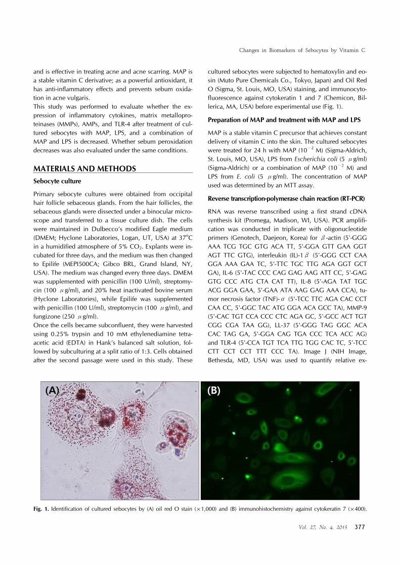
Fig. 1. Identification of cultured sebocytes by (A) oil red O stain (×1,000) and (B) immunohistochemistry against cytokeratin 7 (×400).
and is effective in treating acne and acne scarring. MAP is a stable vitamin C derivative; as a powerful antioxidant, it has anti-inflammatory effects and prevents sebum oxida-tion in acne vulgaris. This study was performed to evaluate whether the ex-pression of inflammatory cytokines, matrix metallopro-teinases (MMPs), AMPs, and TLR-4 after treatment of cul-tured sebocytes with MAP, LPS, and a combination of MAP and LPS is decreased. Whether sebum peroxidation decreases was also evaluated under the same conditions.
MATERIALS AND METHODSSebocyte culture
Primary sebocyte cultures were obtained from occipital hair follicle sebaceous glands. From the hair follicles, the sebaceous glands were dissected under a binocular micro-scope and transferred to a tissue culture dish. The cells were maintained in Dulbecco’s modified Eagle medium (DMEM; Hyclone Laboratories, Logan, UT, USA) at 37oC in a humidified atmosphere of 5% CO2. Explants were in-cubated for three days, and the medium was then changed to Epilife (MEPI500CA; Gibco BRL, Grand Island, NY, USA). The medium was changed every three days. DMEM was supplemented with penicillin (100 U/ml), streptomy-cin (100 μg/ml), and 20% heat inactivated bovine serum (Hyclone Laboratories), while Epilife was supplemented with penicillin (100 U/ml), streptomycin (100 μg/ml), and fungizone (250 μg/ml).Once the cells became subconfluent, they were harvested using 0.25% trypsin and 10 mM ethylenediamine tetra-acetic acid (EDTA) in Hank’s balanced salt solution, fol-lowed by subculturing at a split ratio of 1:3. Cells obtained after the second passage were used in this study. These
cultured sebocytes were subjected to hematoxylin and eo-sin (Muto Pure Chemicals Co., Tokyo, Japan) and Oil Red O (Sigma, St. Louis, MO, USA) staining, and immunocyto-fluorescence against cytokeratin 1 and 7 (Chemicon, Bil-lerica, MA, USA) before experimental use (Fig. 1).
Preparation of MAP and treatment with MAP and LPS
MAP is a stable vitamin C precursor that achieves constant delivery of vitamin C into the skin. The cultured sebocytes were treated for 24 h with MAP (10−2 M) (Sigma-Aldrich, St. Louis, MO, USA), LPS from Escherichia coli (5 μg/ml) (Sigma-Aldrich) or a combination of MAP (10−2 M) and LPS from E. coli (5 μg/ml). The concentration of MAP used was determined by an MTT assay.
Reverse transcription-polymerase chain reaction (RT-PCR)
RNA was reverse transcribed using a first strand cDNA synthesis kit (Promega, Madison, WI, USA). PCR amplifi-cation was conducted in triplicate with oligonucleotide primers (Genotech, Daejeon, Korea) for β-actin (5'-GGG AAA TCG TGC GTG ACA TT, 5'-GGA GTT GAA GGT AGT TTC GTG), interleukin (IL)-1β (5'-GGG CCT CAA GGA AAA GAA TC, 5'-TTC TGC TTG AGA GGT GCT GA), IL-6 (5'-TAC CCC CAG GAG AAG ATT CC, 5'-GAG GTG CCC ATG CTA CAT TT), IL-8 (5'-AGA TAT TGC ACG GGA GAA, 5'-GAA ATA AAG GAG AAA CCA), tu-mor necrosis factor (TNF)-α (5'-TCC TTC AGA CAC CCT CAA CC, 5'-GGC TAC ATG GGA ACA GCC TA), MMP-9 (5'-CAC TGT CCA CCC CTC AGA GC, 5'-GCC ACT TGT CGG CGA TAA GG), LL-37 (5'-GGG TAG GGC ACA CAC TAG GA, 5'-GGA CAG TGA CCC TCA ACC AG) and TLR-4 (5'-CCA TGT TCA TTG TGG CAC TC, 5'-TCC CTT CCT CCT TTT CCC TA). Image J (NIH Image, Bethesda, MD, USA) was used to quantify relative ex-

WJ Lee, et al
378 Ann Dermatol
pression levels. mRNA levels were normalized to β-actin and represented as relative ratios. ANOVA was used for the statistical analysis of all data. A probability value of less than 0.05 was considered statistically significant.
1) Total RNA extraction
TRIzol reagent (Invitrogen, Grand Island, NY, USA) with a modified acid phenol method was used to isolate total RNA from the cells.
2) RT-PCR
cDNA was synthesized from 5 μg total RNA using a cDNA synthesis kit containing ImProm-II reverse tran-scriptase and random primers (Promega). GoTaq Flexi DNA Polymerase was used for PCR amplification. LL-37 amplifications were performed for 28 cycles at annealing temperature of 61oC. TLR-4 amplifications were performed for 35 cycles at annealing temperature of 56oC. All other amplifications were performed for 35 cycles under the fol-lowing conditions: 95oC for 1 min, 56oC for 1 min, and 72oC for 1 min, with the exception of IL-1β, which was performed for 25 cycles under the same program.
Enzyme-linked immunosorbent assay (ELISA)
IL-1β, IL-6, IL-8, TNF-α, and MMP-9 (R&D Systems, Shanghai, China) protein expression was assessed by ELISA according to the manufacturer’s instructions. Briefly, 50 μl of the sample was added to each well in triplicate. Next, 200 μl of prepared streptavidin-horseradish peroxidase and 200 μl of premixed 3,3',5,5'-tetramethylbenzidine substrate solution were added to each well in that order. The plates were developed in the dark at room temper-ature for 30 min, and the reaction was stopped by adding 50 μl of stop solution to each well. Finally, absorbance was measured on a VERSA maxmicroplate reader (Molecular Devices, Sunnyvale, CA, USA). ANOVA was used for statistical analysis of the data. A probability value of less than 0.05 was considered statistically significant.
Western blot analysis for LL-37 and TLR-4
Cultured sebocytes before and after treatment with MAP, LPS, and a combination of MAP and LPS were lysed in a lysis buffer (25 mM Tris-HCl [pH 7.2], 150 mM KCl, 0.1% sodium dodecyl sulfate, 1% Triton X-100, 5 mM EDTA [pH 8.0], 2 mM phenylmethylsulfonyl fluoride) supplemented with protease inhibitor cocktail. Equal protein amounts of total cell lysates were resolved by sodium dodecyl sul-fate-polyacrylamide gel electrophoresis, transferred to a polyvinylidene difluoride membrane, and blotted with an-tibodies against LL-37 (Abcam, Cambridge, UK) and TLR-4 (Abnova, Jhongli City, Taiwan), followed by enhanced
chemiluminescence and autoradiography. This procedure was performed in triplicate. ANOVA was used for stat-istical analysis of the data. A probability value of less than 0.05 was considered statistically significant.
Quantification of lipid peroxidation
BioVision lipid peroxidation assay kit (Biovision, Milpitas, CA, USA) was used according to the manufacturer’s in-structions for the sensitive detection of malondialdehyde assays. Sebocytes (1×10⁶) treated with MAP, LPS, or a combination of MAP and LPS were homogenized on ice in 300 μl of MDA Lysis Buffer with 3 μl butylated hy-droxytoluene (100×) and then centrifuged (13,000g, 10 min) to remove the insoluble material. The 200 μl super-natant from each homogenized sample and standard was placed into a microcentrifuge tube and 600 μl of thio-barbituric acid solution was added. The mixture was in-cubated at 95oC for 60 min and then cooled in ice bath. For the standard curve, 0, 2, 4, 6, 8, and 10 μl of the 2 mM MDA Standard were added into separate micro-centrifuge tubes. Then, 200 μl of each homogenized sam-ple and standard were placed into a 96-well microplate. Absorbance was read at 532 nm.
RESULTSMAP inhibited the increased expression of inflammatory cytokines in cultured sebocytes after treatment with LPS
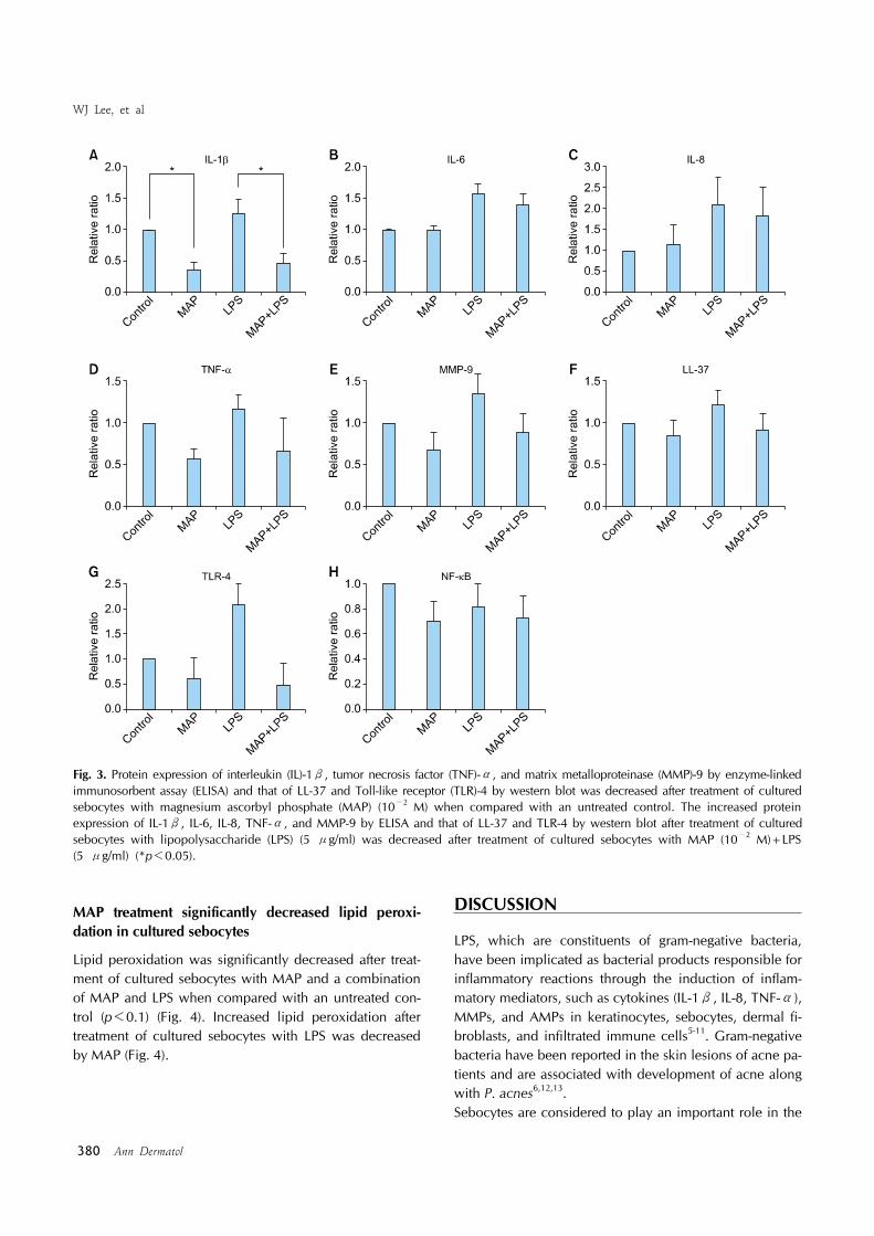
Inflammatory cytokines such as IL-1β, IL-6, IL-8 and TNF-α are produced in unstressed cultured sebocytes. Their expression is influenced by many factors. In this study, LPS upregulated the gene and protein expression of the inflammatory cytokines in cultured sebocytes (Fig. 2, 3). MAP treatment of cultured sebocytes induced a significant decrease in IL-1β gene and protein expression when compared with the untreated control (p<0.05) (Fig. 2, 3). The LPS-induced increase in IL-1β gene and protein ex-pression decreased significantly after treatment with MAP (p<0.05) (Fig. 2, 3). The LPS-induced increase in IL-6 pro-tein expression in cultured sebocytes was inhibited by treatment with MAP (Fig. 3). The increased IL-8 gene ex-pression after treatment with LPS was significantly de-creased by treatment with MAP (p<0.05) (Fig. 2). In addi-tion, increase in TNF-α gene and protein expression after treatment with LPS was inhibited by treatment with MAP (Fig. 2, 3).
MAP decreased TLR-4 expression in cultured sebocytes
Binding of LPS to TLR-4 may lead to increased expression of inflammatory cytokines. The treatment of cultured sebo-cytes with MAP decreased TLR-4 gene and protein ex-

Changes in Biomarkers of Sebocytes by Vitamin C
Vol. 27, No. 4, 2015 379
Fig. 2. Gene expression of interleukin (IL)-1β, IL-8, tumor necrosis factor (TNF)-α, matrix metalloproteinase (MMP)-9, LL-37, and Toll-like receptor (TLR)-4 was decreased after treatment of cultured sebocytes with magnesium ascorbyl phosphate (MAP) (10−2 M) when compared with an untreated control. The increased gene expression of IL-1β, IL-8, TNF-α, MMP-9, LL-37, and TLR-4 after treatment of cultured sebocytes with lipopolysaccharide (LPS) (5 μg/ml) was decreased after treatment of cultured sebocytes with MAP (10−2 M)+LPS (5 μg/ml) (*p<0.05).
pression when compared with an untreated control (Fig. 2, 3). In addition, an increase in TLR-4 gene and protein expression after treatment with LPS was decreased by treatment with MAP (Fig. 2, 3).
MAP decreased the expression of MMPs in cultured sebocytes
The treatment of cultured sebocytes with MAP showed a decrease in the gene and protein expression of MMP-9 when compared with untreated control (Fig. 2, 3). The in-creased gene and protein expression of MMP-9 in cultured sebocytes after treatment with LPS was decreased by treat-
ment with MAP (Fig. 2, 3).
MAP decreased the expression of AMPs in cultured sebocytes
MAP treatment of cultured sebocytes decreased LL-37 gene and protein expression when compared with the un-treated control (Fig. 2, 3). Increased LL-37 gene and pro-tein expression in cultured sebocytes after treatment with LPS was decreased by treatment with MAP (Fig. 2, 3).
WJ Lee, et al
380 Ann Dermatol
Fig. 3. Protein expression of interleukin (IL)-1β, tumor necrosis factor (TNF)-α, and matrix metalloproteinase (MMP)-9 by enzyme-linked immunosorbent assay (ELISA) and that of LL-37 and Toll-like receptor (TLR)-4 by western blot was decreased after treatment of cultured sebocytes with magnesium ascorbyl phosphate (MAP) (10−2 M) when compared with an untreated control. The increased protein expression of IL-1β, IL-6, IL-8, TNF-α, and MMP-9 by ELISA and that of LL-37 and TLR-4 by western blot after treatment of cultured sebocytes with lipopolysaccharide (LPS) (5 μg/ml) was decreased after treatment of cultured sebocytes with MAP (10−2 M)+LPS (5 μg/ml) (*p<0.05).
MAP treatment significantly decreased lipid peroxi-dation in cultured sebocytes
Lipid peroxidation was significantly decreased after treat-ment of cultured sebocytes with MAP and a combination of MAP and LPS when compared with an untreated con-trol (p<0.1) (Fig. 4). Increased lipid peroxidation after treatment of cultured sebocytes with LPS was decreased by MAP (Fig. 4).
DISCUSSION
LPS, which are constituents of gram-negative bacteria, have been implicated as bacterial products responsible for inflammatory reactions through the induction of inflam-matory mediators, such as cytokines (IL-1β, IL-8, TNF-α), MMPs, and AMPs in keratinocytes, sebocytes, dermal fi-broblasts, and infiltrated immune cells5-11. Gram-negative bacteria have been reported in the skin lesions of acne pa-tients and are associated with development of acne along with P. acnes6,12,13.Sebocytes are considered to play an important role in the

Changes in Biomarkers of Sebocytes by Vitamin C
Vol. 27, No. 4, 2015 381
Fig. 4. Lipid peroxidation was significantly decreased after treatment with magnesium ascorbyl phosphate (MAP) (10−2 M) and MAP (10−2 M)+lipopolysaccharide (LPS) (5 μg/ml) and increased after treatment with LPS (5 μg/ml) when compared with an untreated control (*p<0.1).
pathogenesis of inflammatory acne through not only se-bum production and changes in sebum composition but also the production of inflammatory mediators. Sebocytes respond to microbial products such as LPS through the ac-tivation of TLRs, especially TLR-4, and produce various cy-tokines that may evoke an immune response in inflammatory acne. In addition, sebocytes can induce prostaglandins, MMPs, and AMPs.Pharmacologically active vitamin C has antioxidant and anti-inflammatory effects. However, vitamin C is very un-stable under normal environmental conditions. MAP is a stable vitamin C compound that can attenuate the pro-duction of inflammatory mediators. It has not been re-ported whether MAP influences the inhibition of inflam-matory mediators induced by LPS in cultured sebocytes. Therefore, we assessed the effect of MAP on the responses of sebocytes treated with LPS. In this study, MAP inhibited the production of IL-1β, IL-8, and TNF-α in cultured se-bocytes after treatment with LPS. IL-1β is considered an important marker of inflammation in acne. IL-8 is known to act as a neutrophilic chemotactic factor in inflammation. TNF-α induces the production of cytokines, chemokines, and reactive oxygen species in keratinocytes through the activation of transcription factor NF-κB14. Inflammatory reactions in acne have been reported to include extracellular matrix remodeling mediated by MMPs6,15-17. MMP isoforms have been reported in sebum, derived from sebocytes16. Furthermore, sebocytes were found to spontaneously express TLR-4. Expression of MMP-9 increased following treatment of cultured sebo-cytes with LPS, which was subsequently decreased by MAP treatment. Cathelicidin is also detected in cultured
human sebocytes, and its expression levels are upregu-lated in the presence of P. acnes9. The LPS-induced in-creased expression of LL-37 in cultured sebocytes was de-creased by MAP. Similarly, the increased expression of TLR-4 with LPS treatment was decreased by MAP. The TLR-4 pathway may be inhibited by MAP. LPS-induced stimulation of the TLR-4 pathway and subsequent increase in expression of inflammatory cytokines, MMPs, and AMPs in cultured se-bocytes warrant further evaluation of TLR-4-related tran-scription factors.Akitomo et al.18 reported that treatment of cultured sebo-cytes with LPS facilitates the peroxidation of sebum lipids. Vitamin C has antioxidative properties. MAP is effective for the prevention of sebum peroxidation on the skin be-cause of its own antioxidative activity. In this study, in-creased sebum peroxidation after treatment of cultured se-bocytes with LPS was decreased by MAP.In conclusion, this study showed that MAP as vitamin C may be an effective anti-inflammatory and antioxidant fac-tor in cultured sebocytes. Thus, we propose that vitamin C should be considered as a supplementary material to treat inflammatory acne.
ACKNOWLEDGMENT
This research was supported by Basic Science Research Program through the National Research Foundation of Korea funded by the Ministry of Education, Science and Technology (2012R1A1A2007017); grant of Amore-Pacific Corporation, 2014.
REFERENCES
1. Winston MH, Shalita AR. Acne vulgaris. Pathogenesis and treatment. Pediatr Clin North Am 1991;38:889-903.
2. Zouboulis CC. Acne and sebaceous gland function. Clin Dermatol 2004;22:360-366.
3. Zouboulis CC, Adjaye J, Akamatsu H, Moe-Behrens G, Niemann C. Human skin stem cells and the ageing process. Exp Gerontol 2008;43:986-997.
4. Park YK, Chung WS, Lee H, Jung SW. Whitening effect of cosmetics containing magnesium l-ascorbyl-2-phosphate (VC-PMG, vitamin C derivatives) assessed by colorimeter. Ann Dermatol 2002;14:63-70.
5. Alestas T, Ganceviciene R, Fimmel S, Müller-Decker K, Zouboulis CC. Enzymes involved in the biosynthesis of leukotriene B4 and prostaglandin E2 are active in sebaceous glands. J Mol Med (Berl) 2006;84:75-87.
6. Corbel M, Theret N, Caulet-Maugendre S, Germain N, Lagente V, Clement B, et al. Repeated endotoxin exposure induces interstitial fibrosis associated with enhanced gela-tinase (MMP-2 and MMP-9) activity. Inflamm Res 2001;50:

WJ Lee, et al
382 Ann Dermatol
129-135.7. Iwata C, Akimoto N, Sato T, Morokuma Y, Ito A. Aug-
mentation of lipogenesis by 15-deoxy-Delta12,14-prostag-landin J2 in hamster sebaceous glands: identification of cytochrome P-450-mediated 15-deoxy-Delta12,14-prostag-landin J2 production. J Invest Dermatol 2005;125:865-872.
8. Kim J. Review of the innate immune response in acne vulgaris: activation of Toll-like receptor 2 in acne triggers inflammatory cytokine responses. Dermatology 2005;211: 193-198.
9. Nagy I, Pivarcsi A, Kis K, Koreck A, Bodai L, McDowell A, et al. Propionibacterium acnes and lipopolysaccharide induce the expression of antimicrobial peptides and proin-flammatory cytokines/chemokines in human sebocytes. Mi-crobes Infect 2006;8:2195-2205.
10. Oeff MK, Seltmann H, Hiroi N, Nastos A, Makrantonaki E, Bornstein SR, et al. Differential regulation of Toll-like re-ceptor and CD14 pathways by retinoids and corticosteroids in human sebocytes. Dermatology 2006;213:266.
11. Triantafilou M, Triantafilou K. The dynamics of LPS recog-nition: complex orchestration of multiple receptors. J En-dotoxin Res 2005;11:5-11.
12. Bojar RA, Holland KT. Acne and Propionibacterium acnes.
Clin Dermatol 2004;22:375-379.13. Böni R, Nehrhoff B. Treatment of gram-negative folliculitis
in patients with acne. Am J Clin Dermatol 2003;4:273-276.14. Zhou BR, Zhang JA, Zhang Q, Permatasari F, Xu Y, Wu D,
et al. Palmitic acid induces production of proinflammatory cytokines interleukin-6, interleukin-1β, and tumor necrosis factor-α via a NF-κB-dependent mechanism in HaCaT kera-tinocytes. Mediators Inflamm 2013;2013:530429.
15. Neely AN, Clendening CE, Gardner J, Greenhalgh DG, Warden GD. Gelatinase activity in keloids and hypertrophic scars. Wound Repair Regen 1999;7:166-171.
16. Papakonstantinou E, Aletras AJ, Glass E, Tsogas P, Diony-ssopoulos A, Adjaye J, et al. Matrix metalloproteinases of epithelial origin in facial sebum of patients with acne and their regulation by isotretinoin. J Invest Dermatol 2005;125: 673-684.
17. Tanriverdi-Akhisaroglu S, Menderes A, Oktay G. Matrix me-talloproteinase-2 and -9 activities in human keloids, hyper-trophic and atrophic scars: a pilot study. Cell Biochem Funct 2009;27:81-87.
18. Akitomo Y, Akamatsu H, Okano Y, Masaki H, Horio T. Effects of UV irradiation on the sebaceous gland and sebum secretion in hamsters. J Dermatol Sci 2003;31:151-159.