mads on the move - core.ac.uk · pdf filepeople in cars with bumper stickers have a higher...
TRANSCRIPT


Propositions
1. Intercellular movement needs to be considered in the context of the specific location and
developmental stage within the plant.
[This thesis, Wu et al. (2003) Development]
2. The observation that some MADS domain transcription factors move and others do not,
indicates that the movement has a biological relevant function. [This thesis]
3. In confocal laser scanning microscopy good controls are essential to avoid the mistake of
regarding fluorescent artefacts as interesting results.
4. Insect-directed guiding patterns in flowers and inflorescences created by UV reflection
and absorption might interfere with confocal laser scanning microscopy of these tissues.
5. Depictions of fish with air bubbles coming out of their mouths to indicate their presence in
water are in most cases wrong.
6. If chewing gum improves cognitive abilities, ruminants such as cattle, goats and sheep
must be philosophers.
[Scholey et al. (2009) Physiology & Behavior, Smith (2009) Nutritional Neuroscience]
7. People in cars with bumper stickers have a higher tendency to show aggressive road
behaviour, so watch out for cars that say “Peace”.
[Szlemko et al. (2008) Journal of Applied Social Psychology]
8. Growing older is the process of becoming less naive and more cynical.
Propositions belonging to the thesis, entitled
“MADS on the move: a study on MADS domain protein function and movement during floral
development in Arabidopsis thaliana”.
Susanna L. Urbanus
Wageningen, 19 May 2010

MADS on the move A study on MADS domain protein function and movement
during floral development in Arabidopsis thaliana
Susanna Leonora Urbanus

Thesis Committee Thesis supervisor Prof. dr. ir. G.C. Angenent Personal chair at the Laboratory of Molecular Biology Wageningen University Thesis co-supervisor Dr. ir. R.G.H. Immink Senior scientist, Business unit Bioscience Plant Research International, Wageningen Other members Prof. dr. M.M. Kater, University of Milan, Italy Dr. ir. M.J. Ketelaar, Wageningen University Prof. dr. R.E. Koes, VU University Amsterdam Prof. dr. S.C. de Vries, Wageningen University This research was conducted under the auspices of the Graduate School of Experimental Plant Sciences

MADS on the move A study on MADS domain protein function and movement
during floral development in Arabidopsis thaliana
Susanna Leonora Urbanus
Thesis submitted in fulfilment of the requirements for the degree of doctor
at Wageningen University by the authority of the Rector Magnificus
Prof. dr. M.J. Kropff, in the presence of the
Thesis Committee appointed by the Academic Board to be defended in public
on Wednesday 19 May 2010 at 1.30 p.m. in the Aula

Susanna L. Urbanus MADS on the move: a study on MADS domain protein function and movement during floral development in Arabidopsis thaliana 138 pages Thesis, Wageningen University, Wageningen, NL (2010) With references, with summaries in Dutch and English ISBN 978-90-8585-624-5

You don’t need eyes to see,
you need vision
[Faithless | Reverence]


Contents
Chapter 1 General introduction 9
Chapter 2 Tagging of MADS domain proteins for chromatin immunoprecipitation 19
Chapter 3 In planta localisation patterns of MADS domain proteins during floral 37
development in Arabidopsis thaliana
Chapter 4 Investigating intercellular protein transport 61
Chapter 5 Intercellular transport of epidermis-expressed MADS domain 75
transcription factors and their effect on plant morphology and
floral transition
Chapter 6 About SEP3, black holes and UFO 101
Chapter 7 Concluding remarks and perspectives 117
Summary 126 Nederlandse samenvatting 128 Editors’ choice in Science 131 Acknowledgements 132 Curriculum vitae 135 EPS education statement 136


Chapter 1 General introduction

Chapter 1
Flowers and MADS domain transcription factors Evolution has graced our world with plants that bear their reproductive organs in a spectacular array of diverse and beautiful flowers. Although the flowering plant Arabidopsis thaliana has only small and rather humble flowers, they have been a major contributor in our understanding of floral development. Indeed, Arabidopsis thaliana has been in the scientific spotlight of the plant biology field ever since it was recognized that this small weed could be a good model plant under laboratory conditions [1]. This is, among others, due to its small size, rapid generation time, efficient self-pollination, prolific seed production, and the relative easy manipulation of its genetic material. Arabidopsis thaliana flowers consist of four sepals, four petals, six stamens, and two carpels that form the pistil (Figure 1). The stamens are the male reproductive organs and carry pollen inside the anther. The pistil, which is the female reproductive organ, holds the ovules that will develop into seeds after fertilization by the pollen. All these floral organs are developed from a source of proliferating pluripotent cells, called the floral meristem [2]. The MADS domain transcription factor family plays an important role during floral development, among others by specifying the different floral organ identities through distinct combinations of MADS domain transcription factor types. These combinations are illustrated in the extended ‘ABC’ model, where A+E-type MADS domain transcription factors together specify sepal identity, A+B+E-type proteins together specify petal identity, B+C+E-type proteins together specify stamen identity, C+E-type proteins together specify carpel identity, and finally C+D+E-type proteins together specify ovule identity (Figure 1) [3-9].
MADS domain transcription factors can initiate developmental programs through the regulation of downstream target genes [10]. Besides regulating the expression of these target genes, MADS domain transcription factors are also able to regulate their own expression via autoregulatory loops [11-14]. It is thought that MADS domain transcription factors need to be in a dimeric form, either homo- or heterodimeric, to be able to enter the nucleus where they perform their regulatory function [15-17]. The binding of MADS domain proteins to the regulatory DNA sequences of target genes is mediated by the N-terminal located conserved MADS domain, and occurs either in the form of a dimer or in a multimeric fashion, for instance in the tetrameric form that was proposed in the ‘quartet’ model [10, 12, 18-20]. The E-type SEPALLATA (SEP) MADS domain transcription factors that play an important role in all floral organs (Figure 1), seem to function as ‘glue’ proteins that facilitate the formation and functioning of these multimeric MADS domain protein complexes [5, 7, 14, 20-22].
That MADS domain transcription factors can be transported between cells was shown in Antirrhinum majus by the two B-type proteins DEFICIENS (DEF) and GLOBOSA (GLO) that can move outwards to the epidermal cell layer in floral meristems [23]. In contrast, the respective Arabidopsis thaliana B-type orthologues APETALA3 (AP3) and PISTILLATA (PI) do not appear to have the same ability for outward intercellular transport in the floral meristem, nor are these proteins able to travel inwards [24]. Also the A-type protein APETALA1 (AP1) does not travel inwards in floral meristems [25, 26]. There are however
10

General introduction
Figure 1. Arabidopsis thaliana flower and the extended ‘ABC’ model. On the left, a photograph of an Arabidopsis thaliana flower indicating the green sepals (s), the white petals (p), the stamens (st) that carry pollen in the yellow anthers, and the pistil (pi) that is formed from two carpels and contains ovules. On the right, a representation of the extended ‘ABC’ model that illustrates the distinct combinations of MADS domain transcription factor types that are needed for the specification of the sepal, petal, stamen, carpel and ovule identities.
Figure 2. Schematic representation of a plasmodesma spanning the cell wall between adjacent cells. Indicated are the neck region, the central cavity, the endoplasmic reticulum (ER), the cytoplasmic sleeve (CS), the desmotubule (D), the central rod (CR), the plasma membrane (PM), and the spoke-like connections (SP) between the desmotubule and the plasma membrane that may control the aperture of the plasmodesma. Blue and red circles represent plasmodesma-specific proteins. Reprinted with permission [27]
11

Chapter 1
suggestions that certain MADS domain transcription factors have non-cell-autonomous functions, i.e. functions beyond the cell in which the protein is expressed, such as the C-type protein AGAMOUS (AG) in floral meristem integrity [28, 29] and FRUITFULL (FUL) in pistil development [30]. Intercellular transport of transcription factors In plants, intercellular transport of macromolecules, such as transcription factors, is thought to be mediated by dynamic channels that connect the cytoplasm of neighbouring cells [27, 31, 32]. These symplastic connections, called plasmodesmata, consist of a plasma membrane-lined channel through the cell wall that is filled with a central desmotubule derived from the endoplasmic reticulum (ER) and a cytoplasmic sleeve (Figure 2). The cytoplasmic sleeve is the major conduit for macromolecular transport and contains components that regulate plasmodesmal structure and transport. Based on how they are created, plasmodesmata can be classified into two types [33]. Primary plasmodesmata are formed during cell divisions when cytoplasmic strands with ER tubules become enclosed in newly created cell walls. These primary plasmodesmata are initially simple channels, but they can be modified into more complex, branched structures later on. Secondary plasmodesmata, on the other hand, are actively created across existing cell walls and are often complex, branched structures.
Plasmodesmata play a crucial role in developmental processes in a plant through their control over intercellular transport of developmental signals and their ability to join cells on a supracellular level into symplastic domains [34-39]. These symplastic domains, which are partially or completely isolated from intercellular communication with surrounding tissues, make it possible that different developmental processes proceed next to each other in relative isolation. The diffusion of macromolecules through plasmodesmata is controlled by the aperture of the plasmodesmal channel. This aperture appears to reduce in size with increasing levels of tissue differentiation, allowing increasingly smaller sized macromolecules for intercellular transport. However, besides passive transport there is also the possibility of active transport through the plasmodesmata. Especially viral movement proteins are known for their ability to actively enlarge the plasmodesmal channel and thereby help the spread of viral infections throughout the plant [40]. There are also some known plant-specific proteins that have a similar ability to enlarge the plasmodesmal channel, such as the KNOTTED-LIKE HOMEOBOX (KNOX) transcription factors that are involved in the establishment and maintenance of plant meristems [41, 42]. Transport through plasmodesmata can also be reversibly blocked to create boundaries between symplastic domains or in response to abiotic or biotic stress [43-47]. This is achieved through callose deposition in the neck region of the plasmodesmal channel.
12

General introduction
Looking inside the living plant Although genetic screens and in vitro and in vivo studies on protein-protein and protein-DNA interactions provide important information on how MADS domain transcription factors are able to function, understanding of the behaviour of MADS domain transcription factors within their own context in planta is still limited. Since the discovery of the GREEN FLUORESCENT PROTEIN (GFP) from the jellyfish Aequorea Victoria and the subsequent improvement and development of similar fluorescent markers with other spectra and physical characteristics, it has become possible to observe cellular structures and processes in living cells by fluorescence microscopy [48-53]. Especially in combination with Confocal Laser Scanning Microscopy (CLSM), where thick tissues can be optically sectioned and later reconstructed into 3-D images, the visualization of processes in intact plant tissues has become possible. In this thesis we investigated the behaviour of fluorescently-tagged MADS domain proteins during Arabidopsis thaliana floral development, and explored the importance of intercellular transport for MADS domain transcription factor functioning.
In Chapter 2, we describe different methods of tagging the MADS domain proteins AG, SEP3 and FUL for chromatin immunoprecipitation, chromatin affinity purification and in planta imaging. This research demonstrates that tagging of MADS domain proteins frequently results in loss-of-function phenotypes in the plant, especially when the MADS box genes are under the control of the constitutive CaMV35S promoter. Plants that express tagged MADS box genes from genomic fragments that include all or most of the regulatory elements, and therefore mimic the natural expression pattern as much as possible, show lower levels of loss-of-function phenotypes and are also more useful to investigate biologically relevant behaviour of the MADS domain proteins.
In Chapter 3, the spatio-temporal localisation patterns of GFP-tagged MADS domain proteins AG, AP1, SEP3 and FUL during floral development by CLSM are reported. These analyses show that there are several cases of MADS domain protein presence in specific tissues where no mRNA could be detected. This could indicate that there is intercellular transport of MADS domain proteins in meristematic tissues during floral development. The implications of the observed behaviour of the different MADS domain proteins for MADS domain protein functioning are discussed.
In Chapters 4 and 5 we describe the different methods that were used to investigate whether MADS domain proteins are indeed able to transport between cells during floral development. Attempts to investigate intercellular MADS domain protein transport with microinjection techniques and by using the photoconvertible fluorescent mEosFP-tag are discussed. In plants that specifically overexpress GFP-tagged MADS domain proteins AG, AP3, PI, or SEP3 in the epidermis, all tested proteins were able to move within the epidermal cell layer, while only AG could also move from the epidermis to the subepidermis. In these plants we analyzed the effects of epidermal MADS domain protein expression on the plant phenotype and also the ability of both epidermis-expressed GFP-tagged AG and AP3 to complement their corresponding mutant backgrounds.
In Chapter 6, we explore the mechanisms behind the previously observed behaviour of
13

Chapter 1
GFP-tagged SEP3 during petal and stamen development (Chapter 3). Just prior to the initiation of petal and stamen primordia GFP-tagged SEP3 proteins change their subcellular localisation from predominantly nuclear to more cytoplasmic, and at later stages GFP-tagged SEP3 protein seems to disappear in the middle of the primordia without the loss of SEP3 mRNA expression. These two processes could be regulated at a post-transcriptional level by mechanisms that are discussed. Additionally, we investigated the possible involvement of the F-box protein UNUSUAL FLORAL ORGANS (UFO) that regulates petal and stamen development.
Finally, in Chapter 7 the results of this thesis are discussed and placed into a broader context. Special attention is given to the ability of GFP-tagged AG to travel between cells and the behaviour of GFP-tagged SEP3 during petal and stamen development.
14

General introduction
References 1. Pang PP, Meyerowitz EM: Arabidopsis thaliana - A model system for plant molecular-
biology. Bio-Technology 1987, 5(11):1177-1181. 2. Carles CC, Fletcher JC: Shoot apical meristem maintenance: the art of a dynamic
balance. Trends in Plant Science 2003, 8(8):394-401. 3. Coen ES, Meyerowitz EM: The war of the whorls - Genetic interactions controlling flower
development. Nature 1991, 353(6339):31-37. 4. Colombo L, Franken J, Koetje E, Vanwent J, Dons HJM, Angenent GC, Vantunen AJ: The
Petunia MADS box gene FBP11 determines ovule identity. Plant Cell 1995, 7(11):1859-1868.
5. Pelaz S, Ditta GS, Baumann E, Wisman E, Yanofsky MF: B and C floral organ identity functions require SEPALLATA MADS-box genes. Nature 2000, 405(6783):200-203.
6. Pinyopich A, Ditta GS, Savidge B, Liljegren SJ, Baumann E, Wisman E, Yanofsky MF: Assessing the redundancy of MADS-box genes during carpel and ovule development. Nature 2003, 424(6944):85-88.
7. Ditta G, Pinyopich A, Robles P, Pelaz S, Yanofsky MF: The SEP4 gene of Arabidopsis thaliana functions in floral organ and meristem identity. Current Biology 2004, 14(21):1935-1940.
8. Ferrario S, Immink RG, Angenent GC: Conservation and diversity in flower land. Current Opinion in Plant Biology 2004, 7(1):84-91.
9. Favaro R, Pinyopich A, Battaglia R, Kooiker M, Borghi L, Ditta G, Yanofsky MF, Kater MM, Colombo L: MADS-box protein complexes control carpel and ovule development in Arabidopsis. Plant Cell 2003, 15(11):2603-2611.
10. Riechmann JL, Meyerowitz EM: MADS domain proteins in plant development. Biological Chemistry 1997, 378(10):1079-1101.
11. Jack T, Fox GL, Meyerowitz EM: Arabidopsis homeotic gene APETALA3 ectopic expression - transcriptional and posttranscriptional regulation determine floral organ identity. Cell 1994, 76(4):703-716.
12. Schwarz-Sommer Z, Hue I, Huijser P, Flor PJ, Hansen R, Tetens F, Lonnig WE, Saedler H, Sommer H: Characterization of the Antirrhinum floral homeotic MADS-Box gene DEFICIENS - Evidence for DNA-binding and autoregulation of its persistent expression throughout flower development. Embo Journal 1992, 11(1):251-263.
13. Gomez-Mena C, de Folter S, Costa MMR, Angenent GC, Sablowski R: Transcriptional program controlled by the floral homeotic gene AGAMOUS during early organogenesis. Development 2005, 132(3):429-438.
14. Kaufmann K, Muino JM, Jauregui R, Airoldi CA, Smaczniak C, Krajewski P, Angenent GC: Target Genes of the MADS Transcription Factor SEPALLATA3: Integration of Developmental and Hormonal Pathways in the Arabidopsis Flower. Plos Biology 2009, 7(4):854-875.
15. McGonigle B, Bouhidel K, Irish VF: Nuclear localization of the Arabidopsis APETALA3 and PISTILLATA homeotic gene products depends on their simultaneous expression. Genes & Development 1996, 10(14):1812-1821.
16. Immink RG, Gadella TW, Jr., Ferrario S, Busscher M, Angenent GC: Analysis of MADS box protein-protein interactions in living plant cells. Proc Natl Acad Sci U S A 2002, 99(4):2416-2421.
15

Chapter 1
17. Bemer M, Wolters-Arts M, Grossniklaus U, Angenent GC: The MADS domain protein DIANA acts together with AGAMOUS-LIKE80 to specify the central cell in Arabidopsis ovules. Plant Cell 2008, 20(8):2088-2101.
18. Egea-Cortines M, Saedler H, Sommer H: Ternary complex formation between the MADS-box proteins SQUAMOSA, DEFICIENS and GLOBOSA is involved in the control of floral architecture in Antirrhinum majus. Embo Journal 1999, 18(19):5370-5379.
19. Honma T, Goto K: Complexes of MADS-box proteins are sufficient to convert leaves into floral organs. Nature 2001, 409(6819):525-529.
20. Theissen G, Saedler H: Plant biology - Floral quartets. Nature 2001, 409(6819):469-471. 21. Immink RGH, Tonaco IAN, de Folter S, Shchennikova A, van Dijk ADJ, Busscher-Lange J,
Borst JW, Angenent GC: SEPALLATA3: the 'glue' for MADS box transcription factor complex formation. Genome Biology 2009, 10(2).
22. Melzer R, Theissen G: Reconstitution of floral quartets in vitro involving class B and class E floral homeotic proteins. Nucleic Acids Research 2009, 37(8):2723-2736.
23. Perbal MC, Haughn G, Saedler H, Schwarz-Sommer Z: Non-cell-autonomous function of the Antirrhinum floral homeotic proteins DEFICIENS and GLOBOSA is exerted by their polar cell-to-cell trafficking. Development 1996, 122(11):3433-3441.
24. Jenik PD, Irish VF: The Arabidopsis floral homeotic gene APETALA3 differentially regulates intercellular signaling required for petal and stamen development. Development 2001, 128(1):13-23.
25. Sessions A, Yanofsky MF, Weigel D: Cell-cell signaling and movement by the floral transcription factors LEAFY and APETALA1. Science 2000, 289(5480):779-781.
26. Wu XL, Dinneny JR, Crawford KM, Rhee Y, Citovsky V, Zambryski PC, Weigel D: Modes of intercellular transcription factor movement in the Arabidopsis apex. Development 2003, 130(16):3735-3745.
27. Zambryski P: Plasmodesmata. Current Biology 2008, 18(8):R324-R325. 28. Sieburth LE, Drews GN, Meyerowitz EM: Non-autonomy of AGAMOUS function in flower
development: use of a Cre/loxP method for mosaic analysis in Arabidopsis. Development 1998, 125(21):4303-4312.
29. Jenik PD, Irish VF: Regulation of cell proliferation patterns by homeotic genes during Arabidopsis floral development. Development 2000, 127(6):1267-1276.
30. Gu Q, Ferrandiz C, Yanofsky MF, Martienssen R: The FRUITFULL MADS-box gene mediates cell differentiation during Arabidopsis fruit development. Development 1998, 125(8):1509-1517.
31. Maule AJ: Plasmodesmata: structure, function and biogenesis. Current Opinion in Plant Biology 2008, 11(6):680-686.
32. Lucas WJ, Ham LK, Kim JY: Plasmodesmata - bridging the gap between neighboring plant cells. Trends in Cell Biology 2009, 19(10):495-503.
33. Ehlers K, Kollmann R: Primary and secondary plasmodesmata: structure, origin, and functioning. Protoplasma 2001, 216(1-2):1-30.
34. Kim I, Kobayashi K, Cho E, Zambryski PC: Subdomains for transport via plasmodesmata corresponding to the apical-basal axis are established during Arabidopsis embryogenesis. Proceedings of the National Academy of Sciences of the United States of America 2005, 102(33):11945-11950.
35. Kim I, Hempel FD, Sha K, Pfluger J, Zambryski PC: Identification of a developmental transition in plasmodesmatal function during embryogenesis in Arabidopsis thaliana. Development 2002, 129(5):1261-1272.
16

General introduction
36. Gisel A, Barella S, Hempel FD, Zambryski PC: Temporal and spatial regulation of symplastic trafficking during development in Arabidopsis thaliana apices. Development 1999, 126(9):1879-1889.
37. Rinne PLH, van der Schoot C: Symplasmic fields in the tunica of the shoot apical meristem coordinate morphogenetic events. Development 1998, 125(8):1477-1485.
38. Duckett CM, Oparka KJ, Prior DAM, Dolan L, Roberts K: Dye-coupling in the root epidermis of Arabidopsis is progressively reduced during development. Development 1994, 120(11):3247-3255.
39. Kim I, Cho E, Crawford K, Hempel FD, Zambryski PC: Cell-to-cell movement of GFP during embryogenesis and early seedling development in Arabidopsis. Proceedings of the National Academy of Sciences of the United States of America 2005, 102(6):2227-2231.
40. Wolf S, Deom CM, R.N. B, Lucas WJ: Movement protein of tobacco mosaic virus modifies plasmodesmatal size exclusion limit. Science 1989, 246(4928):377-379.
41. Lucas WJ, Bouchepillon S, Jackson DP, Nguyen L, Baker L, Ding B, Hake S: Selective trafficking of KNOTTED1 homeodomain protein and its messenger-RNA through plasmodesmata. Science 1995, 270(5244):1980-1983.
42. Kim JY, Yuan Z, Jackson D: Developmental regulation and significance of KNOX protein trafficking in Arabidopsis. Development 2003, 130(18):4351-4362.
43. Rinne PLH, van der Schoot C: Plasmodesmata at the crossroads between development, dormancy, and defense. Canadian Journal of Botany-Revue Canadienne De Botanique 2003, 81(12):1182-1197.
44. Benitez-Alfonso Y, Cilia M, Roman AS, Thomas C, Maule A, Hearn S, Jackson D: Control of Arabidopsis meristem development by thioredoxin-dependent regulation of intercellular transport. Proceedings of the National Academy of Sciences of the United States of America 2009, 106(9):3615-3620.
45. Simpson C, Thomas C, Findlay K, Bayer E, Maule AJ: An Arabidopsis GPI-Anchor Plasmodesmal Neck Protein with Callose Binding Activity and Potential to Regulate Cell-to-Cell Trafficking. Plant Cell 2009, 21(2):581-594.
46. Radford JE, Vesk M, Overall RL: Callose deposition at plasmodesmata. Protoplasma 1998, 201(1-2):30-37.
47. Radford JE, White RG: Effects of tissue-preparation-induced callose synthesis on estimates of plasmodesma size exclusion limits. Protoplasma 2001, 216(1-2):47-55.
48. Chalfie M, Tu Y, Euskirchen G, Ward WW, Prasher DC: GREEN FLUORESCENT PROTEIN as a marker for gene-expression. Science 1994, 263(5148):802-805.
49. Sheen J, Hwang SB, Niwa Y, Kobayashi H, Galbraith DW: Green fluorescent protein as a new vital marker in plant-cells. Plant Journal 1995, 8(5):777-784.
50. Haseloff J, Dorman, E.-L., Brand, A.H.: Live imaging with green fluorescent protein, vol. 122: Humana Press; 1999.
51. Tsien RY: Building and breeding molecules to spy on cells and tumors. FEBS Lett 2005, 579(4):927-932.
52. Chapman S, Oparka KJ, Roberts AG: New tools for in vivo fluorescence tagging. Current Opinion in Plant Biology 2005, 8(6):565-573.
53. Shimomura O, Johnson FH, Saiga Y: Extraction, purification and properties of AEQOURIN, a bioluminescent protein from luminous hydromedusan, Aequorea. Journal of Cellular and Comparative Physiology 1962, 59(3):223-239.
17


Chapter 2 Tagging of MADS domain proteins for
chromatin immunoprecipitation
Stefan de Folter
Susan L Urbanus
Lisette GC van Zuijlen
Kerstin Kaufmann
Gerco C Angenent
Published in BMC Plant Biology, 7:47 (2007)

Chapter 2
Abstract Background: Most transcription factors fulfil their role in complexes and regulate their target genes upon binding to DNA motifs located in upstream regions or introns. To date, knowledge about transcription factor target genes and their corresponding transcription factor binding sites are still very limited. Two related methods that allow in vivo identification of transcription factor binding sites are chromatin immunoprecipitation (ChIP) and chromatin affinity purification (ChAP). For ChAP, the protein of interest is tagged with a peptide or protein, which can be used for affinity purification of the protein-DNA complex and hence, the identification of the target gene. Results: Here, we present the results of experiments aiming at the development of a generic tagging approach for the Arabidopsis MADS domain proteins AGAMOUS, SEPALLATA3, and FRUITFULL. For this, Arabidopsis wild type plants were transformed with constructs containing a MADS-box gene fused to either a double Strep-tag® II-FLAG-tag, a triple HA-tag, or an eGFP-tag, all under the control of the constitutive double 35S Cauliflower Mosaic Virus (CaMV) promoter. Strikingly, in all cases, the number of transformants with loss-of-function phenotypes was much larger than those with an overexpression phenotype. Using endogenous promoters in stead of the 35S CaMV resulted in a dramatic reduction in the frequency of loss-of-function phenotypes. Furthermore, pleiotropic defects occasionally caused by an overexpression strategy can be overcome by using the native promoter of the gene. Finally, a ChAP result is presented using GFP antibody on plants carrying a genomic fragment of a MADS-box gene fused to GFP. Conclusion: This study revealed that MADS-box proteins are very sensitive to fusions with small peptide tags and GFP tags. Furthermore, for the expression of chimeric versions of MADS-box genes it is favourable to use the entire genomic region in frame to the tag of choice. Interestingly, though unexpected, it appears that the use of chimeric versions of MADS-box genes under the control of the strong 35S CaMV promoter is a very efficient method to obtain dominant-negative mutants, either caused by cosuppression or by alteration of the activity of the recombinant protein. Finally, we were able to demonstrate AGAMOUS binding to one of its targets by ChAP. Background During the last 15 years, many studies have been performed aiming at the understanding of MADS-box gene function in plants using loss- and gain-of-function approaches, which resulted in a wealth of information about their role in development [1, 2]. Far less is known about how they act at the molecular level, how they bind to DNA motifs (cis-elements) and activate down-stream target genes. It has been shown that MADS domain proteins are able to bind to the DNA motif CC(A/T)6GG, the so-called CArG-box (reviewed in [3]). This motif has also been found in promoter sequences of a small number of genes that have been annotated as target genes (e.g. [4-7]). Nevertheless, the exact requirements for this DNA motif to be bound by MADS-box transcription factors in vivo are still unknown. Therefore,
20

Tagging of MADS proteins
methods for the identification of DNA target sites are needed. A powerful method to identify target sites is chromatin immunoprecipitation (ChIP), which
allows purification of in vivo formed complexes of a DNA-binding protein and associated DNA (reviewed in [8]). In short, the method involves the fixation of plant tissue and the isolation of the total protein-DNA mixture, followed by an immunoprecipitation step with an antibody directed against the protein of interest. Next, the DNA can be purified, amplified, and finally identified by sequencing. Alternatively, the amplified DNA can be hybridized to micro arrays containing promoter elements or the entire genome as tiled oligonucleotides (ChIP-chip approach, [9, 10]). The identification of target genes from MADS domain proteins by ChIP has been reported recently [5, 7, 11]. A drawback of ChIP is that for each protein of interest a new specific antibody is required. To overcome this drawback, a protein tagging approach with a general tag could be followed, which we refer to as Chromatin Affinity Purification (ChAP). In this approach, a generic tag is fused to the protein of interest and subsequently used to isolate protein-DNA (or protein-protein) complexes based on affinity purification (reviewed in [12-14]).
In this study we focused on three MADS domain proteins from Arabidopsis, namely AGAMOUS (AG), SEPALLATA3 (SEP3), and FRUITFULL (FUL). AG and SEP3 are both floral organ identity proteins, and based on the ABC model [15], represent C- and E-type proteins, respectively (reviewed in [16]). AG is necessary for the formation of stamens and carpels and is expressed in the inner two floral whorls [17]. SEP3 is expressed in the inner three whorls and is essential for the formation of petals, stamens and carpels in a redundant mode of action with SEP1 and SEP2 [18-21]. FUL has a function in floral meristem identity (early function) and in fruit development (late function) [22-24], and is expressed in the inflorescence meristem, inflorescence stem, cauline leaves, and in developing ovary walls [25]. Here, we report the expression of these three MADS domain proteins in Arabidopsis fused with different tags and the analysis of the phenotypes obtained. Furthermore, the first result obtained with ChAP using a GFP antibody is presented. Results Protein tagging vectors for plant expression Four different binary vectors were used for the tagging approach in plants (Figure 1). The first vector (Figure 1A) contains a double tag, the Strep-tag® II [26], followed by the FLAG-tag [27], located at the C-terminus of the protein of interest. These peptide tags are both very small, each only 8 amino acids long. Two other vectors (Figure 1B and 1C) contain the coding region for eGFP (enhanced GREEN FLUORESCENT PROTEIN, Clonetech) [28, 29], which is either located at the N- or C-terminus of the protein of interest [30]. The fourth vector (Figure 1D) contains a triple HA-tag (hemagglutinin derived) [31], each encoding for a 9 amino acids long peptide. Furthermore, all vectors have a constitutive double 35S CaMV promoter [32, 33] to express the fusion products of AG, SEP3, and FUL in transgenic Arabidopsis plants.
21

Chapter 2
Figure 1. Binary tagging vectors for plant protein expression. (A) C-terminal fusion expression vector with the Strep-tag® II and the FLAG-tag. (B) N-terminal fusion expression vector with eGFP. (C) C-terminal fusion expression vector with eGFP. (D) N-terminal fusion expression vector with a triple HA-tag. All vectors contain the constitutive 35S CaMV promoter with the double enhancer for expression. Phenotypic and expression analyses of Arabidopsis lines expressing chimeric MADS-box versions All constructs were introduced into Arabidopsis wild type plants, ecotype Columbia-0, and the transformants obtained were analyzed for overexpression phenotypes. The results are summarized in Table 1 and Figure 2. The expected overexpression phenotypes for AG are homeotic changes of floral organs, resembling an apetala2-like flower, curly leaves, and early flowering as described by [34]. For ectopic SEP3 expression, curly leaves and early flowering are characteristics to be expected [35], while ectopic expression of FUL results in siliques that fail to shatter, because the dehiscence zone is absent [23, 24].
Overexpression phenotypes were only observed in about 10% of the plants when the eGFP protein was fused either N- or C-terminally (Figure 2B, 2C, and 2J). Surprisingly, many plants containing an eGFP fusion construct revealed a mutant phenotype (Figure 2E, 2F, 2H, 2I, and 2J). Plants with either an overexpression phenotype or a mutant phenotype, obtained with construct pARC276 and pARC277 (Table 1), were analyzed by northern blot hybridization for the expression of the introduced AG or SEP3 transgenes, respectively (Figures 3A and 3B). This revealed a perfect linkage between plants with an overexpression phenotype having a high ectopic gene expression in leaves, while plants with an ag mutant phenotype (pARC276) showed no expression. In stead, the latter plants exhibit a smear in the Northern blot, which is often observed when a gene is cosuppressed [36, 37]. Remarkably, for plants containing the SEP3 fusion construct (pARC277), no loss-of-function
22

Tagging of MADS proteins
phenotypes were observed, though, the Northern blot showed hallmarks of cosuppression, suggesting that silencing of SEP3 may have occurred. Most likely, the paralogs and redundant genes SEP1 and SEP2 are not affected, which explains that no mutant phenotype was obtained. Plants carrying the FUL fusion construct (pARC310) were not molecularly analyzed, but mutant-like plants in a range of severity were observed, which suggest that also cosuppression had occurred. Furthermore, a few overexpression and mutant plants with the AG, SEP3, and FUL fused to eGFP were analyzed for fluorescence (Figures 2K and 2L) and confirmed the same linkage between expression and phenotype.
Plants transformed with constructs containing either the Strep-tag® II-FLAG-tag or the triple HA-tag displayed only a wild-type- or mutant phenotype. Transgenic plants with construct pARC117, containing the double Strep-tag® II-FLAG-tag, were also analyzed by Northern blot for the expression of the FUL fusion product (Figure 3C). Remarkably, in contrast to the eGFP fusion constructs, all plants with a loss-of-function phenotype revealed ectopic FUL expression, which was lacking in plants with a wild-type phenotype. This suggests that this mutant phenotype obtained with the double tag Strep-tag® II-FLAG-tag is caused by a dominant-negative effect and not by a cosuppression mechanism.
Table 1.
Summary of tagged MADS domain proteins in Arabidopsis plants with the observed phenotypes.
Phenotypes (%) Construct Expression cassette Plants (n)
OE LOF WT
pARC117 35S:FUL:StrepII-FLAG:tNOS 21 - 57 43
pARC118 35S:AG:StrepII-FLAG:tNOS 14 - 29 71
pARC276 35S:AG:GFP:t35S 42 12 88 -
pARC277 35S:SEP3:GFP:t35S 60 8 - 92
pARC308 35S:GFP:AG:t35S 54 7 93 -
pARC309 35S:GFP:SEP3:t35S 46 - 100 -
pARC310 35S:GFP:FUL:t35S 49 10 90 -
pARC346 35S:3xHA:AG:tNOS 12 - 50 50
pARC347 35S:3xHA:SEP3:tNOS 15 - - 100
pARC348 35S:3xHA:FUL:tNOS 16 - 38 62
pARC422 gAG:GFP:tNOS 25 - 20 80
pARC423 gSEP3:GFP:tNOS 46 - - 100
pARC424 gFUL:GFP:tNOS 18 - 28 72
n, number of plants; %, percentage of plants; OE, overexpression; LOF, loss-of-function phenotype; WT, wild-type
23
Chapter 2
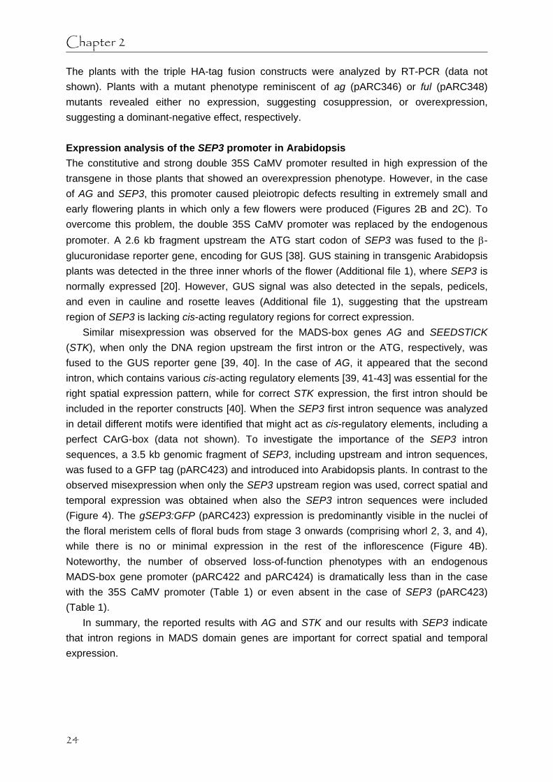
The plants with the triple HA-tag fusion constructs were analyzed by RT-PCR (data not shown). Plants with a mutant phenotype reminiscent of ag (pARC346) or ful (pARC348) mutants revealed either no expression, suggesting cosuppression, or overexpression, suggesting a dominant-negative effect, respectively. Expression analysis of the SEP3 promoter in Arabidopsis The constitutive and strong double 35S CaMV promoter resulted in high expression of the transgene in those plants that showed an overexpression phenotype. However, in the case of AG and SEP3, this promoter caused pleiotropic defects resulting in extremely small and early flowering plants in which only a few flowers were produced (Figures 2B and 2C). To overcome this problem, the double 35S CaMV promoter was replaced by the endogenous promoter. A 2.6 kb fragment upstream the ATG start codon of SEP3 was fused to the β-glucuronidase reporter gene, encoding for GUS [38]. GUS staining in transgenic Arabidopsis plants was detected in the three inner whorls of the flower (Additional file 1), where SEP3 is normally expressed [20]. However, GUS signal was also detected in the sepals, pedicels, and even in cauline and rosette leaves (Additional file 1), suggesting that the upstream region of SEP3 is lacking cis-acting regulatory regions for correct expression.
Similar misexpression was observed for the MADS-box genes AG and SEEDSTICK (STK), when only the DNA region upstream the first intron or the ATG, respectively, was fused to the GUS reporter gene [39, 40]. In the case of AG, it appeared that the second intron, which contains various cis-acting regulatory elements [39, 41-43] was essential for the right spatial expression pattern, while for correct STK expression, the first intron should be included in the reporter constructs [40]. When the SEP3 first intron sequence was analyzed in detail different motifs were identified that might act as cis-regulatory elements, including a perfect CArG-box (data not shown). To investigate the importance of the SEP3 intron sequences, a 3.5 kb genomic fragment of SEP3, including upstream and intron sequences, was fused to a GFP tag (pARC423) and introduced into Arabidopsis plants. In contrast to the observed misexpression when only the SEP3 upstream region was used, correct spatial and temporal expression was obtained when also the SEP3 intron sequences were included (Figure 4). The gSEP3:GFP (pARC423) expression is predominantly visible in the nuclei of the floral meristem cells of floral buds from stage 3 onwards (comprising whorl 2, 3, and 4), while there is no or minimal expression in the rest of the inflorescence (Figure 4B). Noteworthy, the number of observed loss-of-function phenotypes with an endogenous MADS-box gene promoter (pARC422 and pARC424) is dramatically less than in the case with the 35S CaMV promoter (Table 1) or even absent in the case of SEP3 (pARC423) (Table 1).
In summary, the reported results with AG and STK and our results with SEP3 indicate that intron regions in MADS domain genes are important for correct spatial and temporal expression.
24

Tagging of MADS proteins
Figure 2. Phenotypes of transgenic Arabidopsis plants with different tagging constructs. (A) Wild-type Arabidopsis at the rosette stage, (D) at the inflorescence stage, and (G) a close-up of a flower. (B) Line with AG-eGFP fusion construct showing an AG overexpression phenotype (pARC276). (C) Line with SEP3-eGFP fusion construct showing a SEP3 overexpression phenotype (pARC277). Rosette stage images (A-C) were taken from plants grown under the same conditions and were of the same age (bar indicates relative size). (E,H) Line with eGFP-AG fusion construct showing an ag mutant phenotype (pARC308). (F,I) Line with eGFP-SEP3 fusion construct showing a partial sep-like mutant phenotype (pARC309). (J) Siliques of lines with GFP-FUL fusion construct with either a FUL overexpression (FUL), ful mutant (ful) phenotype, or wild-type phenotype (WT) (pARC310). (K) Arabidopsis root tip and (L) open silique with an ovule of a line expressing GFP-FUL fusion construct (pARC310) observed by fluorescence microscopy. dz, dehiscence zone; v, valve; ov, ovule; n, nucleus; ca, carpel wall.
25

Chapter 2
AG protein detection and chromatin affinity purification To investigate whether the ChAP procedure using tags is feasible we used transgenic Arabidopsis plants expressing gAG:GFP (pARC422) as example. Correct spatial and temporal AG expression was observed, predominantly in the nuclei of the floral meristem cells of floral buds from stage 3 onwards (comprising whorl 3 and 4) (Figure 4A).
First, we analyzed the gAG:GFP (pARC422) plants by Western blotting to see whether the chimeric AG protein is detectable with a polyclonal GFP antibody. For this, protein was isolated from nuclei extracts from wild type Arabidopsis (Col-0) plants and compared with extracts from gAG:GFP plants. The Western blot (Figure 4C) shows a specific band of the expected size in the gAG:GFP plants, which was not present in wild type plants.
Finally, a chromatin affinity purification with a GFP antibody was performed on a protein extract derived from gAG:GFP (pARC422) plants. As reported before, AG protein is able to bind to its own intron sequence for autoregulation [7]. This regulatory region was analyzed for enrichment by Real-time PCR, which would demonstrate that the chimeric AG protein is able to bind in vivo to its target sequence. The target DNA sequence (AG second intron) was 10 fold enriched after affinity purification with GFP antibody demonstrating that chimeric AG is indeed able to bind to its regulatory region (Figure 4D). Figure 3. Northern blot analysis of leaf tissue of different Arabidopsis lines containing various tagging constructs. (A) Expression analysis of AG-eGFP (pARC276) lines. (B) Expression analysis of SEP3-eGFP (pARC277) lines. (C) Expression analysis of FUL-Strep-tag® II-FLAG-tag (pARC117) lines, ful-like plants are indicated with ‘m’ and WT-like plants with ‘n’. WT, wild-type; +, line with an overexpression phenotype. Discussion The use of epitope tags can facilitate the isolation of protein-DNA or protein-protein complexes. Here, we report a first attempt of employing a generic tagging approach for the MADS domain proteins AG, SEP3, and FUL. Different tags and a combination of tags were used to produce fusion products expressed in plants. There are two important criteria before further steps can be undertaken to identify target genes by Chromatin Affinity Purification (ChAP). The first basic and most important aspect is to obtain stable expression of the fusion
26

Tagging of MADS proteins
Figure 4. AG and SEP3 expression analysis and chromatin immunoprecipitation (ChIP). Confocal Laser Scanning Microscopical (CLSM) imaging of (A) gAG:GFP (pARC422) and (B) gSEP3:GFP (pARC423) in the inflorescence. Top view (A, B) of an inflorescence with different floral bud stages (indicated by numbers). The GFP expression (green signal) is predominantly localized in the nuclei of floral meristem cells of flower buds from stage 3 onwards (comprising whorl 3 and 4 for AG, and whorl 2, 3, and 4 for SEP3, respectively). Autofluorescence is visible as red signal. (C) Anti-GFP Western blot with material from Arabidopsis WT and gAG:GFP (pARC422) plants. Protein product is detectable in transgenic plants only. Bottom panel shows the Coomassie stained gel serving as loading control. (D) Enrichment of AG target DNA after ChAP with GFP antibody and compared with pre-immune. Quantification of target DNA was done by Real-time PCR using primers corresponding to sequences in the second intron of AG. FM, floral meristem, S, sepal, IM, inflorescence meristem, WT, wild-type.
27

Chapter 2
protein. Secondly, an expressed fusion protein should be biologically active. Both aspects appeared not to be straight forward and appeared to be dependent on the tags used.
The expression experiments in plants using the constitutive and strong 35S CaMV promoter resulted in mutant phenotypes with all constructs, though, in many cases, not the expected overexpression phenotypes. Remarkably, the percentage of loss-of-function phenotypes obtained was very high, even up to 100% in the case of GFP:SEP3 (pARC309). The loss-of-function phenotypes were most likely caused by two phenomena, either by cosuppression in the case of the eGFP fusions, or by a dominant-negative effect in the case of the Strep-tag® II-FLAG-tag fusions. With the triple HA-tag both phenomena could have happened. These different tags have been used in many organisms and with many different proteins (e.g. [13, 31, 44-46]), however it has never been reported that they cause these severe problems related to mRNA expression or activity of a recombinant protein. The high frequency of silencing with the eGFP fusions could be related to the 35S CaMV promoter, causing high expression of the transgene. Expression of MADS-box cDNAs under the control of the 35S CaMV promoter without the GFP tag (e.g. [47]) or expression of GFP tags using endogenous MADS-box gene promoters did not reveal such high percentages of cosuppression plants (Table 1), indicating that the combination of 35S CaMV promoter and the GFP tag may induce silencing. The silencing efficiencies of MADS-box gene expression using the GFP tag in combination with the 35S CaMV promoter appeared to be comparable to using an RNA interference strategy [48]. The only exception on this rule is SEP3:GFP (pARC277), which did not result in any plant with a loss-of-function phenotype. In contrast, all GFP:SEP3 plants show a mutant phenotype. Although an intriguing observation, an explanation is missing. The altered biological activity of the FUL protein fused to short peptide tags, here referred to as ‘dominant-negative’ mode of action, could be caused by either trapping interacting proteins and forming non-functional protein complexes, steric hindrance preventing certain interactions, or altered folding of the protein. However, functionality of a fusion product with an epitope tag has to be analyzed case by case. It depends on the tag used and the effect it may have on the protein of interest. Our results indicate that the activity of MADS-box genes and their products can be dramatically affected by fusions with small peptide tags and GFP tags at both N- and C-termini. This high sensitivity to fusions, however, can also be used as an effective method to obtain high percentages of dominant loss-of-function mutants.
A drawback of an overexpression strategy could be the occurrence of unwanted pleiotropic effects, e.g. early flowering or a reduced number of flowers. Furthermore, overexpression or ectopic expression does not mimic the natural situation. The most elegant solution is to express the genes under their native promoter in a mutant background, which will directly reveal their biological activity and eliminate any competition with the untagged endogenous protein. For the isolation of the native promoter, often DNA sequences upstream the ATG start codon are cloned, although no general rules are available that can predict the promoter region (reviewed in [49]). This approach was followed for the SEP3 promoter, however, it revealed a lack of specificity compared to previously reported in situ hybridization experiments [20]. As described previously for the MADS-box genes AG and
28

Tagging of MADS proteins
STK, intron sequences are important for correct expression [39, 40]. This appears also to be the case for SEP3, because fusion of GFP to a 3.5 kb genomic fragment of SEP3 including upstream and intron sequences revealed correct expression patterns. Finally and most importantly, it appeared possible to perform ChAP using a GFP antibody on plants that carried a genomic AG fragment (including upstream and intron sequences) fused to GFP (pARC422). Conclusion A powerful method to identify target genes is ChIP or related ChAP. ChAP makes use of an epitope tag fused to the protein of interest and this study revealed that the activities of MADS-box proteins are very sensitive to fusions with small peptide and GFP tags. Furthermore, for the expression of chimeric versions of MADS-box genes it is favourable to use the entire genomic region in frame with the tag of choice. Interestingly, though unexpected, it appears that the use of chimeric versions of MADS-box genes under the control of the strong 35S CaMV promoter is a very effective method to obtain loss-of-function mutants, either caused by cosuppression or by alteration of the activity of the recombinant protein. Finally, ChAP is possible with a chimeric MADS-box protein using a GFP antibody. Methods Plant growth Arabidopsis thaliana, ecotype Columbia-0 (Col-0) plants were grown under normal greenhouse or growth chamber conditions (22°C, long day light regime). Construction of binary vectors and plant transformation The vector with the C-terminal double tag Strep-tag® II (WSHPQFEK) and the FLAG-tag (DYKDDDDK) is called pARC113. The double tag is constructed with two forward and three reverse complementary primers resulting in, with Arabidopsis codon usage, 5’-CTCGAGTGGTCTCATCCTCAATTTGAAAAGTCTTCTGATTACAAGGATGATGATGATAAGTAACTCGAG-3’ (nucleotides coding for the tags are underlined). Between the two tags are two serine amino acid residues functioning as linker and after the FLAG-tag a stop codon is introduced. In brief, 1 µl of each primer (100 pmol/µl) were pooled together, incubated for 10 min at 96°C and slowly cooled down to room temperature to create double stranded fragments. The fragments were phosphorylated with 2 µl T4 kinase (10 U/µl) and incubated for 30 min at 37°C. Next, the 69 nucleotides double stranded fragments were isolated from a 12% polyacrylamide gel. Subsequently, the fragment was cloned into an XhoI digested binary pGD121 vector [50], containing a double 35S CaMV promoter (derived from pGD120; [51]). Full length open reading frames for AG (At4g18960; encoding 252 amino acids), SEP3 (At1g24260; encoding 251 amino acids), and FUL (At5g60910; encoding 242 amino acids) were amplified with gene specific primers from the start to the stop codon, clones for C-terminal fusions lack the stop codon, and were subcloned in pGEM-T® Easy (Promega,
29

Chapter 2
Madison, WI) and/or subcloned with the Gateway™ Technology (Invitrogen, Carlsbad, CA). After sequence control, all genes were cloned (in pARC113) and/or recombined (in pARC064, pARC258, and pARC259) in the appropriate vectors to make the fusion constructs.
A 2.6 kb SEP3 region upstream of the ATG was amplified with specific primers (PRO117 5’-CACCGGCGCGCCATCCATCCATCCAAATGGGACC-3’ and PRO118 5’-GAAGCTTTTTC TTTTTCTTTCTCCTCTCCC-3’) and recombined with the Gateway™ Technology in pENTR/D-TOPO (Invitrogen), followed by recombination in the binary vector pBGWFS7 [30], resulting in a transcriptional eGFP-GUS fusion construct (pARC213).
Genomic fragments for AG (6882 bp), SEP3 (3489 bp), and FUL (5298 bp) were amplified with gene specific primers, a forward primer located in the upstream region, PRO433 AG-5’- CACCGATCAAAGACTACACATCAC-3’, PRO407 SEP3-5’- CACCCATACC TTTGTGTCCATCAC-3’, and PRO429 FUL-5’- CACCTCGATCAGAATTTGAGCTG-3’, and a reverse primer in the 3’-region lacking the stop codon for each gene, PRO431 AG-5’-CACTAACTGGAGAGCGGTTTG-3’, PRO408 SEP3-5’- AATAGAGTTGGTGTCATAAGG TAACC-3’, and PRO430 FUL-5’- CTCGTTCGTAGTGGTAGGAC-3’, and recombined in pENTR/D-TOPO. After sequence control, all genomic fragments were recombined in the binary vector pMDC204 [52], resulting in translational GFP6 fusion constructs (pARC422, pARC423, and pARC424, respectively).
Arabidopsis plants were transformed with Agrobacterium tumefaciens strain GV3101 using the floral dip method [53]. RNA gel blot analysis Total RNA was isolated from frozen plant tissue with the RNeasy plant RNA extraction kit (Qiagen). Five micrograms of each RNA sample was denaturated by 1.5 M glyoxal, separated on a 1.2% agarose gel in 15 mM Na-phosphate buffer pH 6.5, checked for equal loading, and followed by blotting onto Hybond-N + membrane (Amersham Biosciences, Piscataway, NJ) in 25 mM Na-phosphate buffer pH 6.5. Probes were labeled with the RadPrime DNA Labeling System (Invitrogen) and blots were hybridized as described by Angenent et al. (1992) [54]. Gene specific probes were amplified by PCR with the following primers: PRO383 AG-5’-GGGTCAATGTCTCCCAAAGA-3’ and PRO384 AG-5’-CTAACTGGAGAGCGGTTTGG-3’, PRO105 SEP3-5’-GTCTAGAATGGGAAGAGGGAGAG TAG-3’ and PRO106 SEP3-5’-CGGATCCAATAGAGTTGGTGTCATAAGGTAACC-3’. The FUL fragment was derived from a pGEM-T® Easy (Promega) clone digested with XbaI-KpnI. GUS assay To detect β-glucuronidase (GUS) activity [38], plant tissue was fixed in 90% ice-cold acetone for 1 h at –20°C, followed by three rinses with 0.1 M Na-phosphate buffer pH 7.0 containing 1 mM potassium ferrocyanide. The three rinse steps in total took 1 h and during the first rinse step vacuum was applied for ~15 min. Finally, the substrate was added to the samples, containing 50 mM Na-phosphate buffer pH 7.0, 1 mM EDTA, 0.1% (v/v) Triton X-100, 1 mM potassium ferrocyanide, and 1 mM X-Gluc (Duchefa, Haarlem, The Netherlands), and
30

Tagging of MADS proteins
vacuum was applied for 5 min, followed by overnight incubation at 37°C in the dark. Chlorophyll was removed by, first, 1 h incubation in 96% ethanol and then transference to 70% ethanol. Microscopy Plant tissue was observed for GFP expression with a Zeiss Axioskop UV-microscope, equipped with filter set 13 (excitation BP 470/20, beamsplitter FT 493, emission BP 505-530). Images were taken with a Leica DFC320 digital camera and an exposure time of 18 seconds was used. Confocal Laser Scanning Microscopical (CLSM) imaging of plant tissue was performed with a Zeiss LSM 510 inverted confocal microscope using a 40x C-Apochromat (NA 1,2 W Korr) lens. The tissue was embedded in the wells of a Silicone Isolator (Grace Bio-Labs, Bend, OR) with 0.8% agar 0.5x MS. GFP was excited with the 488 line of an argon ion laser. The emission of GFP was filtered with a 505-530 nm bandpassfilter, while the red autofluorescence of the plant tissue was filtered with a 650 nm long-pass filter. 3-D projections of the obtained confocal z-stacks were made with the Zeiss LSM Image Browser Version 4. Chromatin Affinity Purification (ChAP) The procedure was performed as previously described [7] with some modifications. Fixed (15–30 min) inflorescence tissue (~0.8 g) was used of transgenic Arabidopsis plants containing construct pARC422 that carries a genomic AG fragment fused with GFP. Chromatin was solubilized on ice with a probe sonicator (MSE, Soniprep 150) by 3 cycles of 15 sec pulses of half maximal power with 30 sec cooling time between pulses. GFP antibody was used for the affinity purification (ab290; Abcam, Cambridge, UK) and for the negative control complete rabbit serum. For pre-clearing and affinity purification Protein A-Agarose beads were used (sc-2001; Santa Cruz Biotechnology, Santa Cruz, CA). After elution of the beads, samples were treated with proteinase K, followed by precipitation. The precipitated DNA was dissolved in 100 µl water, purified with a PCR purification kit (Qiagen, Valencia, CA), and eluted with 30 µl EB (water containing 10 mM Tris, pH 8). Enrichment of the target region was determined using a real-time PCR detection system (MyiQ, Bio-Rad Laboratories, Hercules, CA) by comparing the affinity purified sample (anti-GFP) with the negative control (rabbit serum). The results between the two samples were normalized using sequences of Heat Shock Factor1 (HSF1; At4g17750). The following primers were used, PRO469 AG-5’- TGGTCTGCCTTCTACGATCC-3’ and PRO470 AG-5’-CAACAACCCATTAACACATTGG-3’, PDS1045 HSF1-5’- GCTATCCACAGGTTAGATAAAGGAG-3’ and PDS1046 HSF1-5’- GAGAAAGATTGTGTGAGAATGAAA-3’. Protein isolation and detection Nuclei extraction from 0.5 g of Arabidopsis inflorescences was performed according to the protocol used for ChIP experiments [7]. The nuclei pellet was resuspended in 120 µl 2x SDS sample buffer, incubated on ice and centrifuged at 20800 x g for 10 min at 4°C. The supernatant was boiled for 5 min. Western blotting was performed essentially as described
31

Chapter 2
previously [55]. The GFP antibody (ab290; Abcam) was used in a 1:5000 dilution. Authors’ contributions SdF and GA conceived and designed the experiments. SdF, SU, KK, and LvZ carried out the experiments. SdF and GA drafted the manuscript. All authors read and approved the final manuscript. Acknowledgements We thank François Parcy for providing the Alligator2 vector with the triple HA-tag and Anna Shchennikova for making the genomic AG and FUL entry clones. Furthermore, we thank Nayelli Marsch-Martinez for helpful comments on this manuscript. This work is sponsored by the Netherlands Proteomics Centre (NPC).
Additional file 1. SEP3 expression analysis in transgenic Arabidopsis plants. (A-C) GUS expression patterns of SEP3 promoter GUS fusion (pARC213) in different tissues, (A) inflorescence, (B) silique, and (C) rosette leaf. ov, ovule.
32

Tagging of MADS proteins
References 1. Ng M, Yanofsky MF: Function and evolution of the plant MADS-box gene family. Nat Rev
Genet 2001, 2:186-195. 2. Theissen G, Becker A, Di Rosa A, Kanno A, Kim JT, Munster T, Winter KU, Saedler H: A
short history of MADS-box genes in plants. Plant Mol Biol 2000, 42:115-149. 3. de Folter S, Angenent GC: Trans meets cis in MADS science. Trends Plant Sci 2006,
11:224-231. 4. Sablowski RWM, Meyerowitz EM: A homolog of NO APICAL MERISTEM is an immediate
target of the floral homeotic genes APETALA3/PISTILLATA. Cell 1998, 92:93-103. 5. Wang H, Tang W, Zhu C, Perry SE: A chromatin immunoprecipitation (ChIP) approach to
isolate genes regulated by AGL15, a MADS domain protein that preferentially accumulates in embryos. Plant J 2002, 32:831-843.
6. Ito T, Wellmer F, Yu H, Das P, Ito N, Alves-Ferreira M, Riechmann JL, Meyerowitz EM: The homeotic protein AGAMOUS controls microsporogenesis by regulation of SPOROCYTELESS. Nature 2004, 430:356-360.
7. Gómez-Mena C, de Folter S, Costa MMR, Angenent GC, Sablowski R: Transcriptional program controlled by the floral homeotic gene AGAMOUS during early organogenesis. Development 2005, 132:429-438.
8. Orlando V: Mapping chromosomal proteins in vivo by formaldehyde-crosslinked-chromatin immunoprecipitation. Trends Biochem Sci 2000, 25:99-104.
9. Buck MJ, Lieb JD: ChIP-chip: considerations for the design, analysis, and application of genome-wide chromatin immunoprecipitation experiments. Genomics 2004, 83:349-360.
10. Mockler TC, Ecker JR: Applications of DNA tiling arrays for whole-genome analysis. Genomics 2005, 85:1-15.
11. Zhu C, Perry SE: Control of expression and autoregulation of AGL15, a member of the MADS-box family. Plant J 2005, 41:583-594.
12. Hearn MTW, Acosta D: Applications of novel affinity cassette methods: use of peptide fusion handles for the purification of recombinant proteins. J Mol Recognit 2001, 14:323-369.
13. Lichty JJ, Malecki JL, Agnew HD, Michelson-Horowitz DJ, Tan S: Comparison of affinity tags for protein purification. Protein Expr Purif 2005, 41:98-105.
14. Terpe K: Overview of tag protein fusions: from molecular and biochemical fundamentals to commercial systems. Appl Microbiol Biotechnol 2003, 60:523-533.
15. Coen ES, Meyerowitz EM: The war of the whorls: genetic interactions controlling flower development. Nature 1991, 353:31-37.
16. Ferrario S, Immink RG, Angenent GC: Conservation and diversity in flower land. Curr Opin Plant Biol 2004, 7:84-91.
17. Yanofsky MF, Ma H, Bowman JL, Drews GN, Feldmann KA, Meyerowitz EM: The protein encoded by the Arabidopsis homeotic gene AGAMOUS resembles transcription factors. Nature 1990, 346:35-39.
18. Pelaz S, Ditta GS, Baumann E, Wisman E, Yanofsky MF: B and C floral organ identity functions require SEPALLATA MADS-box genes. Nature 2000, 405:200-203.
19. Honma T, Goto K: Complexes of MADS-box proteins are sufficient to convert leaves into floral organs. Nature 2001, 409:525-529.
20. Mandel MA, Yanofsky MF: The Arabidopsis AGL9 MADS box gene is expressed in young
33
Chapter 2
flower primordia. Sex Plant Reprod 1998, 11:22-28. 21. Pelaz S, Tapia-Lopez R, Alvarez-Buylla ER, Yanofsky MF: Conversion of leaves into petals
in Arabidopsis. Curr Biol 2001, 11:182-184. 22. Ferrandiz C, Gu Q, Martienssen R, Yanofsky MF: Redundant regulation of meristem
identity and plant architecture by FRUITFULL, APETALA1 and CAULIFLOWER. Development 2000, 127:725-734.
23. Gu Q, Ferrandiz C, Yanofsky MF, Martienssen R: The FRUITFULL MADS-box gene mediates cell differentiation during Arabidopsis fruit development. Development 1998, 125:1509-1517.
24. Ferrandiz C, Liljegren SJ, Yanofsky MF: Negative regulation of the SHATTERPROOF genes by FRUITFULL during Arabidopsis fruit development. Science 2000, 289:436-438.
25. Mandel MA, Yanofsky MF: The Arabidopsis AGL8 MADS box gene is expressed in inflorescence meristems and is negatively regulated by APETALA1. Plant Cell 1995, 7:1763-1771.
26. Skerra A, Schmidt TGM: Applications of a peptide ligand for streptavidin: the Strep-tag. Biomol Eng 1999, 16:79-86.
27. Hopp TP, Prickett KS, Price VL, Libby RT, March CJ, Cerretti DP, Urdal DL, Conlon PJ: A short polypeptide marker sequence useful for recombinant protein identification and purification. . Bio/Technology 1988, 6:1204-1210.
28. Chalfie M, Tu Y, Euskirchen G, Ward WW, Prasher DC: Green fluorescent protein as a marker for gene expression. Science 1994, 263:802-805.
29. Chiu W-l, Niwa Y, Zeng W, Hirano T, Kobayashi H, Sheen J: Engineered GFP as a vital reporter in plants. Current Biology 1996, 6:325-330.
30. Karimi M, Inze D, Depicker A: GATEWAY vectors for Agrobacterium-mediated plant transformation. Trends Plant Sci 2002, 7:193-195.
31. Bensmihen S, To A, Lambert G, Kroj T, Giraudat J, Parcy F: Analysis of an activated ABI5 allele using a new selection method for transgenic Arabidopsis seeds. FEBS Lett 2004, 561:127-131.
32. Odell JT, Nagy F, Chua NH: Identification of DNA sequences required for activity of the cauliflower mosaic virus 35S promoter. Nature 1985, 313:810-812.
33. Kay R, Chan A, Daly M, McPherson J: Duplication of CaMV 35S promoter sequences creates a strong enhancer for plant genes Science 1987, 236:1299-1302.
34. Mizukami Y, Ma H: Ectopic expression of the floral homeotic gene AGAMOUS in transgenic Arabidopsis plants alters floral organ identity. Cell 1992, 71:119-131.
35. Pelaz S, Gustafson Brown C, Kohalmi SE, Crosby WL, Yanofsky MF: APETALA1 and SEPALLATA3 interact to promote flower development. Plant J 2001, 26:385-394.
36. Angenent GC, Franken J, Busscher M, Colombo L, van Tunen AJ: Petal and stamen formation in petunia is regulated by the homeotic gene fbp1. Plant J 1993, 4:101-112.
37. Angenent GC, Franken J, Busscher M, Weiss D, van Tunen AJ: Co-suppression of the petunia homeotic gene fbp2 affects the identity of the generative meristem. Plant J 1994, 5:33-44.
38. Jefferson RA, Kavanagh TA, Bevan MW: GUS fusions: beta-glucuronidase as a sensitive and versatile gene fusion marker in higher plants. EMBO J 1987, 6:3901-3907.
39. Sieburth LE, Meyerowitz EM: Molecular dissection of the AGAMOUS control region shows that cis elements for spatial regulation are located intragenically. Plant Cell 1997, 9:355-365.
40. Kooiker M, Airoldi CA, Losa A, Manzotti PS, Finzi L, Kater MM, Colombo L: BASIC
34

Tagging of MADS proteins
PENTACYSTEINE1, a GA binding protein that induces conformational changes in the regulatory region of the homeotic Arabidopsis gene SEEDSTICK. Plant Cell 2005, 17:722-729.
41. Deyholos MK, Sieburth LE: Separable whorl-specific expression and negative regulation by enhancer elements within the AGAMOUS second intron. Plant Cell 2000, 12:1799-1810.
42. Hong RL, Hamaguchi L, Busch MA, Weigel D: Regulatory elements of the floral homeotic gene AGAMOUS identified by phylogenetic footprinting and shadowing. Plant Cell 2003, 15:1296-1309.
43. Busch MA, Bomblies K, Weigel D: Activation of a floral homeotic gene in Arabidopsis. Science 1999, 285:585-587.
44. Witte C-P, Noel L, Gielbert J, Parker J, Romeis T: Rapid one-step protein purification from plant material using the eight-amino acid StrepII epitope. Plant Mol Biol 2004, 55:135-147.
45. Ho Y, Gruhler A, Heilbut A, Bader GD, Moore L, Adams S-L, Millar A, Taylor P, Bennett K, Boutilier K, et al: Systematic identification of protein complexes in Saccharomyces cerevisiae by mass spectrometry. Nature 2002, 415:180-183.
46. Einhauer A, Jungbauer A: The FLAG peptide, a versatile fusion tag for the purification of recombinant proteins. J Biochem Biophys Methods 2001, 49:455-465.
47. Ferrario S, Busscher J, Franken J, Gerats T, Vandenbussche M, Angenent GC, Immink RGH: Ectopic expression of the petunia MADS box gene UNSHAVEN accelerates flowering and confers leaf-like characteristics to floral organs in a dominant-negative manner. Plant Cell 2004, 16:1490-1505.
48. Chuang CF, Meyerowitz EM: Specific and heritable genetic interference by double-stranded RNA in Arabidopsis thaliana. Proc Natl Acad Sci U S A 2000, 97:4985-4990.
49. Rombauts S, Florquin K, Lescot M, Marchal K, Rouze P, van de Peer Y: Computational approaches to identify promoters and cis-regulatory elements in plant genomes. Plant Physiol 2003, 132:1162-1176.
50. de Folter S, Shchennikova AV, Franken J, Busscher M, Baskar R, Grossniklaus U, Angenent GC, Immink RGH: A Bsister MADS-box gene involved in ovule and seed development in petunia and Arabidopsis. Plant J 2006, 47:934-946.
51. Immink RGH, Gadella TWJ, Jr., Ferrario S, Busscher M, Angenent GC: Analysis of MADS box protein-protein interactions in living plant cells. Proc Natl Acad Sci USA 2002, 99:2416-2421.
52. Curtis MD, Grossniklaus U: A gateway cloning vector set for high-throughput functional analysis of genes in planta. Plant Physiol 2003, 133:462-469.
53. Clough SJ, Bent AF: Floral dip: a simplified method for Agrobacterium-mediated transformation of Arabidopsis thaliana. Plant J 1998, 16:735-743.
54. Angenent GC, Busscher M, Franken J, Mol JN, van Tunen AJ: Differential expression of two MADS box genes in wild-type and mutant petunia flowers. Plant Cell 1992, 4:983-993.
55. Lamb RS, Irish VF: Functional divergence within the APETALA3/PISTILLATA floral homeotic gene lineages. Proc Natl Acad Sci U S A 2003, 100:6558-6563.
35


Chapter 3 In planta localisation patterns of MADS domain proteins during floral development
in Arabidopsis thaliana
Susan L Urbanus
Stefan de Folter
Anna V Shchennikova
Kerstin Kaufmann
Richard GH Immink
Gerco C Angenent
Published in BMC Plant Biology, 9:5 (2009)

Chapter 3
Abstract Background: MADS domain transcription factors play important roles in various developmental processes in flowering plants. Members of this family play a prominent role in the transition to flowering and the specification of floral organ identity. Several studies reported mRNA expression patterns of the genes encoding these MADS domain proteins, however, these studies do not provide the necessary information on the temporal and spatial localisation of the proteins. We have made GREEN FLUORESCENT PROTEIN (GFP) translational fusions with the four MADS domain proteins SEPALLATA3, AGAMOUS, FRUITFULL and APETALA1 from the model plant Arabidopsis thaliana and analysed the protein localisation patterns in living plant tissues by confocal laser scanning microscopy (CLSM). Results: We unravelled the protein localisation patterns of the four MADS domain proteins at a cellular and subcellular level in inflorescence and floral meristems, during development of the early flower bud stages, and during further differentiation of the floral organs. The protein localisation patterns revealed a few deviations from known mRNA expression patterns, suggesting a non-cell autonomous action of these factors or alternative control mechanisms. In addition, we observed a change in the subcellular localisation of SEPALLATA3 from a predominantly nuclear localisation to a more cytoplasmic localisation, occurring specifically during petal and stamen development. Furthermore, we show that the down-regulation of the homeodomain transcription factor WUSCHEL in ovular tissues is preceded by the occurrence of both AGAMOUS and SEPALLATA3 proteins, supporting the hypothesis that both proteins together suppress WUSCHEL expression in the ovule. Conclusions: This approach provides a highly detailed in situ map of MADS domain protein presence during early and later stages of floral development. The subcellular localisation of the transcription factors in the cytoplasm, as observed at certain stages during development, points to mechanisms other than transcriptional control. Together this information is essential to understand the role of these proteins in the regulatory processes that drive floral development and leads to new hypotheses. Background Major developmental steps in flowering plants, such as the transition to flowering and floral organ development are, for the most part, controlled by members of the MADS domain family of transcription factors [1]. The action of these transcription factors in defining the identity of the floral organs has been captured in a genetic model, the “ABC” model [2], which was later extended with a “D” and “E” function [3-6]. This model describes how the combinatorial activity of several classes of regulatory genes, most of which encode MIKC-type MADS domain proteins, define the identity of the five different floral organs (sepals, petals, stamen, carpels, and ovules). According to this model, the combination of the class A+E genes specifies the identity of sepals, while the A+B+E genes specify petal identity, the combination of classes B+C+E determines stamen identity, C+E genes together lead to carpel identity,
38

Localisations of MADS proteins
and finally the combination of classes C+D+E is responsible for ovule identity (for review see [7]). Floral organ development in Arabidopsis thaliana is controlled by the following genes: the A-function is represented by the genes APETALA1 (AP1) and APETALA2 (AP2) (not a MADS domain transcription factor); the B-function is controlled by APETALA3 (AP3) and PISTILLATA (PI); AGAMOUS (AG) represents the C-function; the D-function is represented in a redundant manner by SEEDSTICK (STK), SHATTERPROOF1 (SHP1) and SHATTERPROOF2 (SHP2); and the E-function involves the four closely related genes SEPALLATA1 to SEPALLATA4 (SEP1-4). Furthermore, another MADS box gene FRUITFULL (FUL), which is not described in the “ABC” model, is also involved in carpel development. In addition to these functions in floral organ development, some of these genes also have other functions. For instance, both FUL and AP1 are involved in the transition from inflorescence meristem to floral meristem identity, while AG controls the floral meristem determinacy [8-10].
The MADS domain proteins and the “ABC” model are well studied subjects for transcription factor regulation and action. Several studies have shown that at least some MADS domain proteins need to be in a dimeric form, either homo- or heterodimeric, before they can enter the nucleus [11-13]. In the nucleus, the proteins bind to DNA sequences of the target gene with the consensus CC(AT)6GG sequence, also known as the CArG box [14]. Binding to the DNA occurs either in the form of a dimer [14] or in a multimeric fashion [15, 16], for instance in a tetrameric form as proposed in the “quartet” model [17]. Our knowledge about these MADS domain protein interactions has been greatly extended by a study where interaction data obtained from a systematic Yeast Two-Hybrid experiment was combined with large scale microarray co-expression data of the corresponding genes [18]. By considering not only the capacity of proteins to interact with each other, but also the possibility for putative partners to be co-localised in the same tissues and cells, the output is narrowed down to interactions that are likely to be of biological relevance for the plant.
However, microarray studies give only a very broad view on the spatio-temporal expression pattern of genes and do not provide the necessary detail that is needed to demonstrate co-localisation of the encoded proteins. In situ mRNA hybridisation studies and promoter-reporter studies like the ones reported for AG [19], SEP3 [20], AP1 [21], and FUL [22] reveal the expression patterns in more detail, but these might not reflect the protein localisation patterns. In fact, it is difficult to infer the protein localisation pattern from an mRNA expression pattern for two main reasons. First of all, production and degradation rates of mRNA and proteins could be totally different, and secondly, proteins can be transported from an expressing cell to a neighbouring non-expressing cell; all resulting in protein patterns that deviate from the mRNA patterns. It is known from studies on the class B genes DEFICIENS and GLOBOSA from Antirrhinum majus [23] that particular MADS domain transcription factors are able to transfer from one cell to another, where they may have a non-cell-autonomous function. Therefore, to obtain information about the spatio-temporal control of these regulatory proteins, it is essential to study the localisation of the proteins themselves. Additionally, protein localisation studies can be more informative on the functioning of transcription factors by showing the specific subcellular localisation of the
39

Chapter 3
proteins during development. It has been demonstrated that some MADS domain transcription factors are localised in the cytoplasm when an interaction partner is absent and only become functional when they enter the nucleus after dimerisation [11-13, 24]. Preferably, one would like to obtain a three-dimensional map of protein localisations with cellular resolution and information about the dynamics of proteins during plant development. The discovery of GREEN FLUORESCENT PROTEIN (GFP) and similar fluorescent proteins and their use as visual tags for proteins, in combination with Confocal Laser Scanning Microscopy (CLSM) has made this visualisation of fluorescently tagged proteins in living plant tissue possible [25-27].
In order to study MADS domain proteins in living tissues with CLSM during floral development, we made C-terminal GFP tagged versions of SEP3, AG, FUL, and AP1. Previously it was shown that the fusion of GFP to the C-terminus of the MADS domain protein AP1 does not affect its function, as it is able to complement the ap1 mutant [28]. Furthermore, it is known from studies with AG, STK, and SEP3 that introns can contain important regulatory elements that are required for the correct expression pattern of these MADS box genes [29-31]. For this reason, we made C-terminal GFP tagged versions of SEP3, AG, FUL, and AP1 using genomic fragments [29]. Here we describe the protein localisation patterns of gSEP3:GFP, gAG:GFP, gFUL:GFP and gAP1:GFP on a cellular and subcellular level in the inflorescence meristem and at various stages of floral meristem and organ formation. This detailed study reveals discrepancies between the previously reported mRNA expression patterns and the protein localisations, and sheds new light on the functioning of the MADS domain proteins in floral organ patterning and formation. Results and Discussion In this study we analysed the spatio-temporal protein localisation patterns of C-terminally GFP tagged genomic clones of MADS domain proteins SEP3, AG, FUL and AP1 during floral development, hereafter referred to as SEP3:GFP, AG:GFP, FUL:GFP, and AP1:GFP. The generated constructs were transformed to Arabidopsis thaliana wild type Col-0 plants, and at least four GFP-expressing stable primary transformants per construct were analysed for their protein localisation. These were found to be very similar in localisation patterns, although some differences in expression levels were observed. These differences in expression levels may be due to differences in transgene copy numbers, but they may also be caused by positional effects of the insertion of the transgene. Three constructs, namely AG:GFP, FUL:GFP and AP1:GFP, were also introduced into their respective mutant lines. These complementation experiments showed that C-terminal GFP tagged MADS domain proteins are functional, as the AG:GFP, FUL:GFP and AP1:GFP proteins can rescue the mutant phenotypes of ag, ful and ap1 mutants, respectively. As the single sep3 mutant shows very subtle phenotypic alterations due to the redundancy of SEP3 with SEP1 and SEP2 [4], the SEP3:GFP construct was only transformed into the wild type background. The spatio-temporal protein localisation patterns described here are from representative lines homozygous for the transgene in the Col-0 wild type background.
40

Localisations of MADS proteins
SEP3 localisation in inflorescence meristem and early flower bud stages In the inflorescence meristem, SEP3:GFP plants had a very low but definite signal in the epidermal layer that was located mostly in the nucleus (Figures 1A-C). During the initiation of the floral primordia and flower bud stages 1 and 2 this epidermal localisation pattern remained. However, stage 2 flower buds also showed a few cells in the subepidermal and inner cell layers in the centre of the floral meristem that started to have a much higher level of SEP3:GFP signal, also located mostly in the nucleus (Figure 1C). During the development towards stage 3 flower buds this signal spread out to encompass the whole dome of the floral meristem from which the second, third and fourth whorl will develop at later stages (Figure 1D). This increasing SEP3:GFP localisation fits with the reported mRNA expression pattern [20], where SEP3 expression starts in late stage 2 flower buds and is largely confined to the three inner whorls of the flower primordium. However, the weak, but distinct epidermal presence of SEP3:GFP in the inflorescence meristem and stage 1 flower buds has not been reported before. This signal could originate from the epidermis itself, but it cannot be excluded that it is the result of epidermal transport from nearby, high expressing tissues, such as the floral meristem in stage 2/3 flower buds. We also observed signal in the epidermal layer of sepal primordia. During the development of the sepals, the SEP3:GFP signal became weaker at the abaxial side of the sepals, while the signal remained at the adaxial side (Figure 1I). In agreement with this, low level SEP3 expression is occasionally detected on the adaxial side of sepals at later stages [20], and it was shown that SEP1-SEP4 are involved in specifying adaxial sepal surface identity [6].
Interestingly, the subcellular localisation of the SEP3:GFP protein changed dramatically from stage 3 onwards. Just before the petal and stamen initiation, SEP3:GFP proteins in the future second and third whorl became both cytoplasmically and nuclear localised. At the same time, the proteins in the innermost part of the floral meristem clearly remained nuclear localised (Figures 1E and F). This cytoplasmic SEP3:GFP signal in the future second and third whorl could be due to higher expression levels of the gene, resulting in a temporary accumulation of SEP3:GFP proteins in the cytoplasm waiting for transportation to the nucleus. Another option could be that the appearance of new interaction partners in this region results in the cytoplasmic localisation of SEP3:GFP. For instance, it is known that the expression of AP3, which determines petal and stamen identity together with PI and SEP3, also starts at stage 3 and is restricted to the same area where whorl 2 and 3 will develop [32]. It would be interesting to investigate if AP3 is also located predominantly in the cytoplasm at this point in development. Another possibility could be that higher cell division rates in the area of the future second and third whorl which cause the petal and stamen primordia to arise, result in increased unloading of previously nuclear localised proteins into the cytoplasm [33]. The smaller cell sizes in the area of the future second and third whorl compared to the innermost part of the floral meristem could indicate higher cell division rates (Figure 1E). This unloading of the nuclear localised proteins into the cytoplasm during cell division may also allow the proteins to meet new partners and form new complexes in the cytoplasm, for instance with AP3 and PI. In stage 5 flower buds, the initiating petal and stamen primordia revealed an epidermal layer with mostly nuclear SEP3:GFP, while the
41

Chapter 3
Figure 1. Confocal microscopic analysis of SEP3:GFP localisation in inflorescence meristem and early flower bud stages. (A) Overview of an inflorescence with the inflorescence meristem and early flower bud stages 1 to 5 indicated. SEP3:GFP protein is detected as green signal and cell membranes are stained with the red dye FM4-64. (B) Detail of an inflorescence meristem and a stage 2 flower bud. (C) Section through tissue in (B) showing the SEP3:GFP signal in the epidermis and the beginning SEP3:GFP signal in the centre of the stage 2 flower bud. (D) Detail of a stage 4 flower bud showing the highest SEP3:GFP signal in the entire floral meristem and only in the epidermis of the four sepals. (E) Section through the stage 4 flower bud in (D) showing both cytoplasmic and nuclear localisation of SEP3:GFP in the future second and third whorl, and only nuclear localisation in the innermost part of the floral meristem. (F) More basal section through the stage 4 flower bud in (D) showing again both cytoplasmic and nuclear localisation of SEP3:GFP in the future second and third whorl. (G) Detail of a stage 5 flower bud showing the initiating petal and stamen primordia. (H) Section through the stage 5 flower bud in (G) illustrating that the SEP3:GFP signal in whorl 4 is mostly nuclear, while the reduced signal in the initiating petal and stamen primordia is both cytoplasmically and nuclear localised. (I) More basal section through the stage 5 flower bud in (G) showing the SEP3:GFP signal at the adaxial and abaxial sides of a sepal. (1-5) flower bud stages; (ab) abaxial; (ad) adaxial; (FM) floral meristem; (IM)
42

Localisations of MADS proteins
inflorescence meristem; (i) innermost part floral meristem; (L1) epidermal cell layer; (L3) inner cell layers; (p) petal; (s) sepal; (st) stamen; (w4) whorl 4. Scale bars of (A) 50 µm and of (B-I) 25 µm. subepidermal and inner cell layers showed a lower SEP3:GFP signal, both cytoplasmically and nuclear localised (Figures 1G-I). Apparently, SEP3 is less needed in the inner layers of the emerging petals and stamen than in the epidermis. In the mean time the SEP3:GFP proteins in the fourth whorl remained nuclear localised (Figure 1H). AG localisation in inflorescence meristem and early flower bud stages The AG:GFP signal appeared in a cluster of subepidermal and inner layer cells in very early stage 3 flower buds, at the time when the first sepal primordium started to arise. During the development of stage 3 flower buds the AG:GFP localisation enlarged to encompass the part of the floral meristem from which the third and fourth whorl will develop, and this pattern remained in later stages (Figures 2A, B, and D). This corresponds well to the observed AG mRNA expression pattern that starts in the floral meristem of stage 3 flower buds and continues in whorl 3 and 4 in later stages [34, 35]. During all the early stages of flower bud development the AG:GFP protein seemed to be primarily localised in the nucleus, but a substantial part of the signal was also localised in the cytoplasm (Figure 2C). In a stage 5 flower bud, AG:GFP was present throughout all cell layers of the developing stamen primordia and in the region of whorl 4 (Figure 2D). FUL localisation in inflorescence meristem and early flower bud stages In the inflorescence meristem, FUL:GFP plants exhibited a very high fluorescence signal throughout all cell layers (Figure 3A). This signal was mostly located in the nucleus, but also in the cytoplasm (Figure 3B). As soon as the flower bud primordia were initiated the FUL:GFP signal started to reduce in the subepidermal and inner cell layers (Figure 3B), while in the centre of the emerging flower buds some cells maintained the high level of FUL:GFP signal. During the development of the sepals, the basal part of the sepal had FUL:GFP proteins in both epidermal and subepidermal layer, whereas the apical part of the sepal had only epidermal FUL:GFP. This FUL:GFP signal was maintained at the abaxial side of the sepal, whereas the adaxial side and the abaxial side at the tip showed a reduction in signal (Figures 3C and D). In stage 3, 4 and 5 flower buds the floral meristem had the highest amount of FUL:GFP protein in the innermost part of the floral meristem, while in the area of the future second and third whorl, the amount gradually reduced. However, in these initiating petal and stamen primordia the presence of FUL:GFP in the epidermis remained the longest (Figures 3C and D).
FUL mRNA is expressed in two distinct phases: in the inflorescence meristem and in the centre of the floral meristem from stage 3 flower bud onwards, but not in the intermediate flower bud stages 1 and 2 [22, 36]. Therefore, the FUL:GFP presence that was observed in flower bud stages 1 and 2 (Figures 3A and B) might be due to FUL:GFP protein that remained from previous stages. The renewed FUL gene expression in stage 3 flower buds in the centre of the floral meristem corresponds with the increased FUL:GFP protein
43

Chapter 3
Figure 2. Confocal microscopic analysis of AG:GFP localisation in inflorescence meristem and early flower bud stages. (A) Overview of an inflorescence with the inflorescence meristem and early flower bud stages 2 to 6 indicated. AG:GFP protein is detected as green signal and cell membranes are stained with the red dye FM4-64. (B) Detail of a stage 4 flower bud showing AG:GFP signal in the future third and fourth whorl. (C) Section through the stage 4 flower bud in (B) showing that AG:GFP is located in both the cytoplasm and the nuclei. (D) Detail of the stage 5 flower bud in (A) showing AG:GFP signal in the developing stamen and in whorl 4. (2-6) flower bud stages; (FM) floral meristem; (IM) inflorescence meristem; (p) petal; (s) sepal; (st) stamen; (w4) whorl 4. Scale bar of (A) 50 µm and scale bars of (B –D) 25 µm.
44

Localisations of MADS proteins
Figure 3. Confocal microscopic analysis of FUL:GFP localisation in inflorescence meristem and early flower bud stages. (A) Overview of an inflorescence with the inflorescence meristem and early flower bud stages 2 to 7 indicated. FUL:GFP protein is detected as green signal and cell membranes are stained with the red dye FM4-64. (B) Section through an inflorescence meristem showing high FUL:GFP signal in all layers, while stage 1 and 2 flower bud primordia have reduced signal. (C) Detail of a stage 3 flower bud showing the highest FUL:GFP signal in the floral meristem and only epidermal signal in the four sepals. (D) Detail of a stage 5 flower bud showing the highest FUL:GFP signal in whorl 4 and reducing signal in the petal and stamen primordia. (2-7) flower bud stages; (FM) floral meristem; (IM) inflorescence meristem; (p) petal; (s) sepal; (st) stamen; (w4) whorl 4. Scale bar of (A) 50 µm and scale bars of (B –D) 25 µm.
45

Chapter 3
accumulation in the centre of the floral meristem from stage 3 onwards (Figure 3C). In summary, throughout the development from inflorescence meristem to floral organs it seems that the tissues that need to remain undifferentiated, such as the inflorescence and floral meristem, have FUL:GFP protein in all cell layers. Differentiating tissues however, like the developing floral organs, seem to loose the subepidermal and inner cell layer FUL:GFP signal and only retain the signal in the epidermis. AP1 localisation in inflorescence meristem and early flower bud stages The AP1:GFP signal was first detected in a few cells of the epidermal and internal cell layers of the emerging flower bud primordium (Figures 4A and B). From flower bud stages 1 to 3 the AP1 fusion protein was found throughout all cell layers and was located predominantly in the nucleus (Figure 4C). In flower bud stages 4 and 5, the AP1:GFP protein in the sepals was most abundant in the apical tips (Figures 4A and D). The signal diminished in the third and fourth whorl at this stage, while the signal remained in the second whorl where the petal primordia would emerge. However, a few epidermal cells in the centre of the fourth whorl still had a low AP1:GFP signal, perhaps representing the last meristematic cells in the differentiating floral meristem (Figure 4D). This localisation pattern corresponds with the reported AP1 mRNA expression [21, 37], where AP1 expression starts in stage 1 flower bud primordia and increases during the development of the flower bud until stage 3. At the end of stage 3, the AP1 expression starts to reduce in the centre of the floral meristem as a result of the negative regulation of AG protein present there [37] (Figures 2A and B). However, as our AG:GFP and AP1:GFP localisation studies revealed, there is a clear time-lag between the termination of AP1 mRNA expression in early stage 3 flower buds, and the reduction in time of the AP1:GFP protein starting in late stage 3 flower buds (Figure 4C). The same time-lag is seen for the reduction of the FUL:GFP signal starting in stage 1 flower buds (Figure 3B), where FUL mRNA expression is suppressed due to the presence of AP1 protein [36].This negative regulation of AP1 on FUL expression is probably also apparent in the developing sepals, where eventually the FUL:GFP protein presence is highest in the basal parts of the sepals (Figure 3D), while AP1:GFP protein is more abundant in the apical tips of the sepals (Figure 4D). SEP3, FUL and AP1 localisation during petal development The function of the MADS domain proteins in the floral transition and in the determination of floral organ identities is well established, however, less is known about putative later functions of MADS domain proteins in differentiating floral organs. Recently, a report showed that AG has a late function in stamen development [38], and it was already known that FUL has a late function in pistil and fruit development [22, 39]. In view of this and the fact that MADS domain proteins were still present in differentiating floral organs, we studied the SEP3:GFP, FUL:GFP and AP1:GFP localisations during the development of the petal in more detail.
At stage 5, we observed that SEP3:GFP signal was predominantly located in the epidermis of the emerging petal primordium (Figures 1G and 1I), while AP1:GFP signal was
46

Localisations of MADS proteins
Figure 4. Confocal microscopic analysis of AP1:GFP localisation in inflorescence meristem and early flower bud stages. (A) Overview of an inflorescence with the inflorescence meristem and early flower bud stages 1 to 6 indicated. AP1:GFP protein is detected as green signal in a red autofluorescent background. (B) Detail of the inflorescence meristem in (A) showing that the AP1:GFP signal starts in a few cells in stage 1 flower buds and spreads to all cell layers in flower bud stage 2 and 3. The asterisk denotes AP1:GFP signal in nuclei of the adjacent stage 2 flower bud projecting through the overlying inflorescence meristem tissue. (C) Detail of a late stage 3 flower bud showing AP1:GFP signal in all cell layers of the four sepals and the floral meristem. (D) Detail of a stage 4 flower bud showing the highest AP1:GFP signal in the tips of the four sepals and lower signal at positions where the petals will emerge. The signal in whorl 3 and whorl 4 is reducing, although the signal does remain in whorl 4. (1-6) flower bud stages; (FM) floral meristem; (IM) inflorescence meristem; (p) petal; (s) sepal; (w4) whorl 4. Scale bar of (A) 50 µm and scale bars of (B–D) 25 µm.
47

Chapter 3
present throughout all cell layers (Figure 4D). FUL:GFP protein was hardly present at this stage of petal development (Figure 3D). Around stage 9, when petals start to increase rapidly in size [40], high AP1:GFP signal was detected throughout all cell layers. This signal slowly reduced in time and was almost abolished in a stage 12 petal (data not shown). This is in agreement with the AP1 mRNA expression pattern reported for petals [21, 37]. Remarkably, FUL:GFP protein was observed in the centre of the claw around stage 10 (data not shown), where it might be involved in the vascular development of the petal [22]. The SEP3:GFP signal in a stage 9 petal was higher in the adaxial epidermis of the petal than the abaxial epidermis (Figure 5B). This difference in adaxial and abaxial patterning of SEP3:GFP fusion protein remained until stage 12 petals, although the signal gradually reduced. In a stage 11 petal the SEP3:GFP signal on the adaxial side was strongest in the blade and in the middle of the claw (Figure 5A), while at the abaxial side the edges of the blade and the middle of the claw had the strongest SEP3:GFP signal (Figure 5C). The asymmetric accumulation of SEP3:GFP protein in the petal epidermis is in contrast with the uniform SEP3 mRNA expression reported for petals [20], but it does resemble the epidermal SEP3:GFP localisation pattern that we observed in sepals (Figure 1I), suggesting that SEP3 could play a role in the adaxial/abaxial patterning of both organs. FUL, SEP3 and AG localisation during pistil development It is well-known that FUL has a late function in valve differentiation in the developing pistil and in fruit elongation [22, 39]. In contrast, much less is known about putative late functions of SEP3 and AG during pistil development. Therefore, we studied the localisation patterns of FUL:GFP, SEP3:GFP and AG:GFP in the developing pistil until flower bud stage 12 (stage after [40]), when style, valves, valve margins, and replum are being formed. Note that in stage 12 pistil tissue the confocal microscope laser could not reliably penetrate beyond five cell layers.
We observed that FUL:GFP is predominantly located in the two valves and the replum and, to a much lesser extent, in the valve margins and the basal half of the style (Figure 6A). In the valves FUL:GFP was present in the first five cell layers, whereas we detected signal in the replum and the style only in the first two layers, and in the valve margins only in the first layer (Figure 6F). In a younger gynoecium (stage 10) this FUL:GFP localisation pattern was already visible, although with lower intensity, but in this stage the future replum had only relatively high signal in the basal part (data not shown). The presence of FUL in the epidermal and subepidermal layers of the replum and valve margins has not been reported before in either developing pistils or fruits [9, 22, 36]. Nevertheless, ful-1 mutant fruits do not only have defects in the valve tissue but also fail to dehisce, indicating defects in the replum and valve margins [9, 22]. Also, the presence of the FUL:GFP protein in the replum does not transform the cells into valve cells, whereas this was reported happening when constitutively expressing FUL in the entire gynoecium [41]. This suggests that either the replum identity is already established prior to or around stage 10 before the FUL:GFP expression is basal-apically up-regulated in the replum, or that for the conversion of replum to valve cells FUL protein needs to be present in all cell layers of the replum. The suggestion that FUL could be
48

Localisations of MADS proteins
Figure 5. Confocal microscopic analysis of SEP:GFP during petal development. (A) SEP3:GFP signal at the adaxial side of a stage 11 petal. Nuclei with SEP3:GFP proteins are visible as bright green spots against the green autofluorescent background of the petal. (B) Cross section through a petal of approximately stage 10 showing higher SEP3:GFP signal in the adaxial epidermis than the abaxial epidermis. Cell membranes are stained with the red dye FM4-64. (C) SEP3:GFP signal at the abaxial side of a stage 11 petal. The images in (A) and (B) are each composed of two separate, overlapping projections. (ab) abaxial side; (ad) adaxial side; (b) blade of petal; (c) claw of petal. Scale bars of (A) and (C) 50 µm and scale bar of (B) 15 µm. intercellularly transported from the valve tissue to the replum [22] is supported by our localisation data. On the other hand, most studies on FUL expression focused more specifically on fruit development (after stage 12), it could therefore be that FUL expression in the epidermal and subepidermal layers of the replum and valve margins in the developing pistil (until stage 12) has been overlooked.
SEP3:GFP fusion protein is most abundant in the replum and the valve margins, while the valves and the style showed a much lower signal (Figures 6B and D). In the replum and the valve margins SEP3:GFP was located in the first two cell layers, whereas the style and the valves had only signal in the epidermal layer at this stage of pistil development (Figure 6F). However, in a younger gynoecium (stage 10) the same localisation pattern with lower intensity was found, but at least three cell layers deep in the future style and two cell layers deep in the future valve margins, the future replum, and the future valves (data not shown). Therefore, it seems that during the development of the gynoecium SEP3:GFP becomes restricted to the epidermal and subepidermal layers.
49

Chapter 3
Figure 6. Confocal microscopic analysis of FUL:GFP, SEP3:GFP and AG:GFP localisation in stage 12 pistils. (A) FUL:GFP localisation in a stage 12 pistil, with the highest signal in the valves and the replum, and lower signal in the valve margins and the basal half of the style. The image is composed of three separate, overlapping projections. Nuclei with FUL:GFP proteins are visible as bright green spots against the green autofluorescent background of the pistil. (B) SEP3:GFP localisation in the apical part of a stage 12 pistil with the highest signal in the valve margins and replum, and lower signal in the style and valves. (C) AG:GFP signal in all cell layers of the apical part of a stage 12 pistil. (D) SEP3:GFP localisation in the middle part of a stage 12 pistil with the highest signal in the valve margins and replum, and lower signal in the valves. (E) AG:GFP signal in all cell layers of the middle part of a stage 12 pistil. (F) Schematic cross sections of the FUL:GFP, SEP3:GFP and AG:GFP localisations in the ovary wall. (r) replum; (s) stigma; (st) style; (v) valve; (vm) valve margin. All scale bars 50 µm.
50
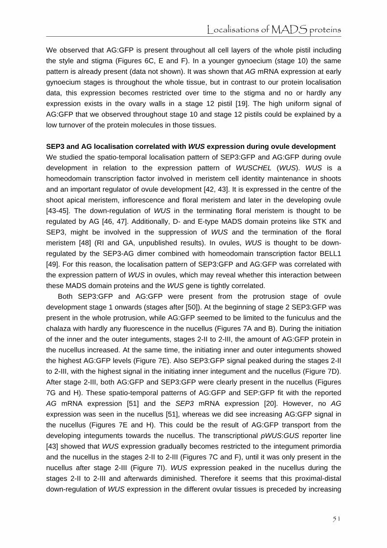
Localisations of MADS proteins
We observed that AG:GFP is present throughout all cell layers of the whole pistil including the style and stigma (Figures 6C, E and F). In a younger gynoecium (stage 10) the same pattern is already present (data not shown). It was shown that AG mRNA expression at early gynoecium stages is throughout the whole tissue, but in contrast to our protein localisation data, this expression becomes restricted over time to the stigma and no or hardly any expression exists in the ovary walls in a stage 12 pistil [19]. The high uniform signal of AG:GFP that we observed throughout stage 10 and stage 12 pistils could be explained by a low turnover of the protein molecules in those tissues. SEP3 and AG localisation correlated with WUS expression during ovule development We studied the spatio-temporal localisation pattern of SEP3:GFP and AG:GFP during ovule development in relation to the expression pattern of WUSCHEL (WUS). WUS is a homeodomain transcription factor involved in meristem cell identity maintenance in shoots and an important regulator of ovule development [42, 43]. It is expressed in the centre of the shoot apical meristem, inflorescence and floral meristem and later in the developing ovule [43-45]. The down-regulation of WUS in the terminating floral meristem is thought to be regulated by AG [46, 47]. Additionally, D- and E-type MADS domain proteins like STK and SEP3, might be involved in the suppression of WUS and the termination of the floral meristem [48] (RI and GA, unpublished results). In ovules, WUS is thought to be down-regulated by the SEP3-AG dimer combined with homeodomain transcription factor BELL1 [49]. For this reason, the localisation pattern of SEP3:GFP and AG:GFP was correlated with the expression pattern of WUS in ovules, which may reveal whether this interaction between these MADS domain proteins and the WUS gene is tightly correlated.
Both SEP3:GFP and AG:GFP were present from the protrusion stage of ovule development stage 1 onwards (stages after [50]). At the beginning of stage 2 SEP3:GFP was present in the whole protrusion, while AG:GFP seemed to be limited to the funiculus and the chalaza with hardly any fluorescence in the nucellus (Figures 7A and B). During the initiation of the inner and the outer integuments, stages 2-II to 2-III, the amount of AG:GFP protein in the nucellus increased. At the same time, the initiating inner and outer integuments showed the highest AG:GFP levels (Figure 7E). Also SEP3:GFP signal peaked during the stages 2-II to 2-III, with the highest signal in the initiating inner integument and the nucellus (Figure 7D). After stage 2-III, both AG:GFP and SEP3:GFP were clearly present in the nucellus (Figures 7G and H). These spatio-temporal patterns of AG:GFP and SEP:GFP fit with the reported AG mRNA expression [51] and the SEP3 mRNA expression [20]. However, no AG expression was seen in the nucellus [51], whereas we did see increasing AG:GFP signal in the nucellus (Figures 7E and H). This could be the result of AG:GFP transport from the developing integuments towards the nucellus. The transcriptional pWUS:GUS reporter line [43] showed that WUS expression gradually becomes restricted to the integument primordia and the nucellus in the stages 2-II to 2-III (Figures 7C and F), until it was only present in the nucellus after stage 2-III (Figure 7I). WUS expression peaked in the nucellus during the stages 2-II to 2-III and afterwards diminished. Therefore it seems that this proximal-distal down-regulation of WUS expression in the different ovular tissues is preceded by increasing
51

Chapter 3
proximal-distal AG:GFP and SEP3:GFP signals and only occurs in the tissues where both SEP3:GFP and AG:GFP were present, supporting the hypothesis that SEP3 and AG are together involved in the repression of WUS expression [49]. Figure 7. Localisation of SEP3:GFP, AG:GFP and expression of pWUS:GUS during ovule development. Confocal microscopic analysis of SEP3:GFP localisation (A, D, and G) and AG:GFP localisation (B, E, and H) during ovule development. GFP tagged MADS domain proteins are detected as green signal and cell membranes are stained with the red dye FM4-64. DIC microscopy of pWUS:GUS expression in developing ovules (C, F, and I) where GUS activity is detected as a blue colour. (A) SEP3:GFP signal in the nucellus, chalaza, and funiculus of an ovule at stage 2-II. (B) AG:GFP signal in the chalaza and funiculus of an ovule at stage 2-II. (C) pWUS:GUS expression in an ovule at stage 2-II in an increasing basal-to-apical gradient. (D) High SEP3:GFP signal specifically in the nucellus, the inner integument, and the funiculus in an ovule at stage 2-III. (E) AG:GFP signal in an ovule at stage 2-III, with beginning signal in the nucellus and the highest signal in the developing inner and outer integuments. (F) pWUS:GUS expression in the nucellus and the two integuments of an ovule at stage 2-III in an increasing basal-to-apical gradient. (G) SEP3:GFP signal in an ovule at stage 2-IV with the highest signal in the nucellus. (H) AG:GFP signal in the nucellus, the two integuments, and the funiculus of an ovule at stage 2-IV. (I) pWUS:GUS expression only in the nucellus in an ovule at stage 2-IV. (c) chalaza; (f) funiculus; (ii) inner integument; (oi) outer integument; (n) nucellus. All scale bars 10 μm.
52

Localisations of MADS proteins
Conclusions The results described here show that in some cases a discrepancy exists between the previously reported mRNA expression patterns and our protein localisations. Therefore, mRNA expression patterns alone are not sufficient to form hypotheses about gene function and they need to be supported by protein localisation data. For instance, in a recent paper [52] it was suggested that FUL, unlike AP1, cannot be directly involved in the regulation of flowering time genes (e.g. SUPPRESSOR OF OVEREXPRESSION OF CONSTANS1), simply because it is not expressed in the early flower bud stages. Our localisation data, however, shows that FUL:GFP is present in the early flower bud stages and therefore could have the same function as AP1 in regulating these flowering time genes. This is also in agreement with the hypothesis that both AP1 and FUL act as hubs between the flower induction protein network and the floral organ protein network [18], and previous genetic studies that showed redundancy between AP1 and FUL during the floral transition [9]. Other examples, such as the asymmetric localisation of SEP3 in the epidermis of both sepals and petals and its possible role in the adaxial/abaxial patterning of these organs, the presence of FUL protein in the replum of developing pistils, or the presence of AG in the nucellus of the developing ovule during the down-regulation of WUS were also not apparent from the reported mRNA patterns. These cases demonstrate the importance of studying protein patterns and protein levels for a better understanding of transcription factor functioning. Some caution is needed however, as we analysed the behaviour of tagged proteins, and we cannot exclude that the localisation patterns of these fusion proteins differ from those of the native proteins. It is possible that the increased size of the fusion protein could have an effect on the transport abilities of the protein, or that the presence of the tag could interfere with the ability to form (multimeric) protein complexes. Furthermore, the presence of the tag could change the stability of the protein, which could lead to a different localisation pattern. We also have to realise that the transgenes are inserted on other positions in the genome than the endogenous genes, which may cause some positional effects on expression pattern and level. Nevertheless, because of the capacity of the AG, FUL and AP1 fusion proteins to complement their respective mutants and the lack of ectopic expression phenotypes in the transgenic lines during floral development, it is very likely that the localisations of the fusion proteins mimic the patterns of the endogenous proteins.
Discrepancies between mRNA and protein patterns may suggest non-cell autonomous action of the proteins by intercellular transport. This has been shown for a number of transcription factors, such as KNOTTED-1 in maize [53], and SHORTROOT [54] and CAPRICE [55] in Arabidopsis. Also MADS domain proteins may move from one cell to another, as has been reported for the Antirrhinum majus MADS domain proteins DEFICIENS and GLOBOSA [23]. In our study there are a few examples where the presence of MADS domain proteins could be due to intercellular transport. For instance, the low level, but distinct presence of SEP3 in the epidermis of the inflorescence meristem and stage 1 flower buds could be due to epidermal transport from high expressing stage 2/3 flower buds. Another example is the increasing AG presence in the nucellus in the developing ovule,
53

Chapter 3
where the high expressing integuments might be the source of the AG proteins in the nucellus. Also, as previously suggested [22], the FUL proteins in the replum in developing pistils could originate from the high expressing valve cells.
Subcellular localisations of proteins can provide clues on the regulation and functioning of transcription factors. The subcellular SEP3:GFP localisation in the cytoplasm in the area of the floral meristem that will become whorl 2 and 3 is an example of a localisation that is not expected based on the nature of transcription factors. The cytoplasmic localisation could indicate that the SEP3:GFP protein in the cytoplasm is not in a dimeric form, and therefore cannot be transported to the nucleus [11-13, 24]. Alternatively, it could also be that post-translational modifications cause the cytoplasmic retention of the SEP3:GFP protein, possibly facilitating intercellular transport [56] or breakdown of the protein [57]. Recently, it has also been shown that the MADS domain protein MPF2 from Physalis floridana is located in the cytoplasm and imported into the nucleus upon hormone treatment with cytokinin [58]. This shows that non-nuclear localisation is an intriguing mechanism for the regulation of transcription factor functioning.
Summarising, our analysis with GFP tagged proteins expressed under the control of the endogenous promoter revealed the spatio-temporal dynamics of the MADS domain proteins in various tissues of the living plant, leading to a deeper understanding of the behaviour of these MADS domain proteins and allowing the formation of new hypotheses about their function and regulation during early and later stages of floral development. Methods Arabidopsis thaliana plants All plants were grown at 22 ºC in growth chambers under a long-day light regime (16 h light/8 h dark).
The construction of transgenic lines expressing gAG:GFP, gFUL:GFP and gSEP3:GFP was previously described [29]. The AG genomic clone has a promoter region of approximately 2.6 kb upstream from the translational start, the FUL genomic clone has a promoter region of approximately 2 kb and the SEP3 clone has a promoter region of approximately 1.5 kb. To make the translational gAP1:GFP fusion construct, a genomic clone fragment of AP1 (6616 bp) was amplified with the following two gene specific primers: the forward primer PDS298 (5’-GGGGACAAGTTTGTACAAAAAAGCAGGCTGTTTAACATCCA AGATTTGTTTTACATAATCGTTAC-3’) located 2992 bp upstream from the translational start, and the reverse primer PDS297 (5’-GGGGACCACTTTGTACAAGAAAGCTGG GTCTGCGGCGAAGCAGCCAAGGTT-3’) lacking the stop codon at the 3’ end of the coding sequence. The amplified product was inserted into the pENTR/D-TOPO vector (Invitrogen) and, after sequence controls, recombined into the binary vector pMDC107 [59]. Arabidopsis plants were transformed with Agrobacterium tumefaciens strain GV3101 using the floral dip method [60]. Transformed seeds were selected on MS agar plates with 10 µg/ml hygromycin, and stable transgenic lines were maintained afterwards. Furthermore, a transcriptional pWUSCHEL:GUS line [43] was used to analyse the expression pattern of WUSCHEL in
54

Localisations of MADS proteins
developing ovules. Complementation experiments Stable transgenic plant lines expressing gAG:GFP and gFUL:GFP in wild type Col-0 background were crossed with the SALK_014999 ag T-DNA insertion mutant line and the ful-1 mutant [22] respectively, while the gAP1:GFP construct was directly transformed into the SALK_056708 ap1 T-DNA insertion mutant line. In the progeny, the presence of the wild type allele, the mutant allele, and the GFP tagged MADS box gene construct was determined by PCR and CLSM. For AG the following primer pairs were used: AG wild type allele, forward primer PRO182 (5’-GGATCCATGGCGTACCAATCGGAGCT-3’) annealing immediately after the START codon and reverse primer PDS1985 (5’- CATTTCCTTCAGCCTATATTACC-3’) located in the 3’ UTR 18 bp downstream of the STOP codon; ag T-DNA mutant allele, forward primer PRO433 (5’-CACCGATCAAAGACTACACATCAC-3’) located in the 5’ UTR 2634 bp upstream of the START codon and reverse primer PDS404 (5’- TGGTTCACGTAGTGGGCCATCG-3’) located on the left border of the T-DNA. The presence of gAG:GFP was determined by CLSM in the inflorescences. For FUL, the following primer pairs were used: FUL wild type allele, forward primer PDS1024 (5’-CTTACGTCACTGTAGACTCACG- 3’) located in the 5’ UTR 201 bp upstream of the START codon and reverse primer PDS1023 (5’-AAAGAGTGAGATAGTTCTACTCG-3’) in the 3’ UTR 16 bp downstream of the STOP codon; ful-1 mutant allele, forward primer PDS1025 (5’-TTCATCCCTTTTTCAGGGTTGTC-3’) corresponding with the inserted DsE element and reverse primer PDS1023; and gFUL:GFP, forward primer PDS920 (5’-ATCACTTACGTCACTGTAGACTCACG-3’) in the 5’ UTR 204 bp upstream and reverse primer PDS914 (5’-CATCATGTTTGTATAGTTCATCCATGCC-3’) 5 bp upstream of the STOP codon of mGFP6. For AP1, the following primer pairs were used: AP1 wild type allele, forward primer PDS912 (5’-AAAACTTTAGGGCCGTAGTGAAGTGAAC-3’) 385 bp downstream of the START codon and reverse primer PDS1105 (5’-ATTGGATGAAAAGAGCCTAGCCAC-3’) in the 3’ UTR 89 bp downstream of the STOP codon (which will give no product for the ap1 T-DNA mutant allele); gAP1:GFP, forward primer PDS912 and reverse primer PDS915 (5’-GACCAGGGTTGGCCATGGAACAGG-3’) 183 bp downstream of the START codon of mGFP6. Confocal laser scanning microscopy To observe the localisation of the GFP tagged proteins in living plant tissue, inflorescence material was dissected until the relevant meristems and flower buds became visible. After stage 5 flower buds, when sepals started to enclose the floral meristem, it became difficult to visualise the underlying developing floral organs without dissecting the flower buds. The tissues were embedded as previously described [29]. The dye FM4-64 (Molecular Probes, Leiden, The Netherlands) was used as a red counter stain for cell membranes and added at a concentration of 5 µM to the embedding mixture of 0.8% agar, 0.5x MS. The incubation time of the sample in the embedding mixture with FM4-64 was at least 20 minutes. Confocal Laser Scanning Microscopy (CLSM) of the living plant tissue was performed with a Zeiss
55

Chapter 3
LSM 510 inverted confocal microscope using a 40 x C-Apochromat (NA 1.2 W korr) lens. Both GFP and the FM4-64 dye were excited with the 488 nm line of an Argon ion laser. The GFP emission was filtered with a 505-530 nm band pass filter, while the FM4-64 dye emission and red autofluorescence was filtered with a 650 nm long pass filter. The optical slices in the confocal z-stacks were made as a sum of 4 scans and were median filtered afterwards. Three-dimensional projections of the obtained confocal z-stacks were made with the Zeiss LSM Image Browser version 4 and adjusted with Adobe Photoshop version 5.0. GUS assay To analyse the expression pattern of WUSCHEL in developing ovules, inflorescences of the pWUS:GUS line were fixed and a β-glucuronidase (GUS) assay was performed overnight at 37 °C as previously described [61] (modified from [62]). After GUS detection and chlorophyll removal, the inflorescences were kept in Hoyer’s solution (7.5 g Arabic gum, 100 g chloral hydrate, 5 ml glycerol, and 60 ml water). Whole siliques of one inflorescence were put under a cover slip and observed with a Nikon Optiphot microscope. Bright field images of the ovules were taken with a Leica DFC320 digital camera using a 40x Plan DIC objective. Authors' contributions For the MADS domain protein localisations SdF, AV and KK created the GFP-tagged MADS box constructs, and SU selected the transgenic plant lines and analysed the protein localisation patterns with CLSM. For the WUS expression pattern in ovules RI performed the GUS assay on inflorescences of pWUS:GUS plants and SU imaged the ovules. SU wrote the manuscript and RI and GA critically revised it. All authors read and approved the final manuscript. Acknowledgements We are grateful to Thomas Laux for providing pWUS:GUS seeds. This work was in part financially supported by the Dutch Ministry of Agriculture, Nature management and Food quality and the Centre for BioSystems Genomics (CBSG).
56

Localisations of MADS proteins
References 1. Riechmann JL, Meyerowitz EM: MADS domain proteins in plant development. Biological
Chemistry 1997, 378(10):1079-1101. 2. Coen ES, Meyerowitz EM: The war of the whorls - Genetic interactions controlling flower
development. Nature 1991, 353(6339):31-37. 3. Colombo L, Franken J, Koetje E, Vanwent J, Dons HJM, Angenent GC, Vantunen AJ: The
Petunia MADS box gene FBP11 determines ovule identity. Plant Cell 1995, 7(11):1859-1868.
4. Pelaz S, Ditta GS, Baumann E, Wisman E, Yanofsky MF: B and C floral organ identity functions require SEPALLATA MADS-box genes. Nature 2000, 405(6783):200-203.
5. Pinyopich A, Ditta GS, Savidge B, Liljegren SJ, Baumann E, Wisman E, Yanofsky MF: Assessing the redundancy of MADS-box genes during carpel and ovule development. Nature 2003, 424(6944):85-88.
6. Ditta G, Pinyopich A, Robles P, Pelaz S, Yanofsky MF: The SEP4 gene of Arabidopsis thaliana functions in floral organ and meristem identity. Current Biology 2004, 14(21):1935-1940.
7. Ferrario S, Immink RG, Angenent GC: Conservation and diversity in flower land. Current Opinion in Plant Biology 2004, 7(1):84-91.
8. Mizukami Y, Ma H: Separation of AG function in floral meristem determinacy from that in reproductive organ identity by expressing antisense AG RNA. Plant Molecular Biology 1995, 28(5):767-784.
9. Ferrandiz C, Gu Q, Martienssen R, Yanofsky MF: Redundant regulation of meristem identity and plant architecture by FRUITFULL, APETALA1 and CAULIFLOWER. Development 2000, 127(4):725-734.
10. Mandel MA, Yanofsky MF: A gene triggering flower formation in Arabidopsis. Nature 1995, 377(6549):522-524.
11. McGonigle B, Bouhidel K, Irish VF: Nuclear localization of the Arabidopsis APETALA3 and PISTILLATA homeotic gene products depends on their simultaneous expression. Genes & Development 1996, 10(14):1812-1821.
12. Immink RG, Gadella TW, Jr., Ferrario S, Busscher M, Angenent GC: Analysis of MADS box protein-protein interactions in living plant cells. Proc Natl Acad Sci U S A 2002, 99(4):2416-2421.
13. Bemer M, Wolters-Arts M, Grossniklaus U, Angenent GC: The MADS domain protein DIANA acts together with AGAMOUS-LIKE80 to specify the central cell in Arabidopsis ovules. Plant Cell 2008, 20(8):2088-2101.
14. Schwarz-Sommer Z, Hue I, Huijser P, Flor PJ, Hansen R, Tetens F, Lonnig WE, Saedler H, Sommer H: Characterization of the Antirrhinum floral homeotic MADS-Box gene DEFICIENS - Evidence for DNA-binding and autoregulation of its persistent expression throughout flower development. Embo Journal 1992, 11(1):251-263.
15. Egea-Cortines M, Saedler H, Sommer H: Ternary complex formation between the MADS-box proteins SQUAMOSA, DEFICIENS and GLOBOSA is involved in the control of floral architecture in Antirrhinum majus. Embo Journal 1999, 18(19):5370-5379.
16. Honma T, Goto K: Complexes of MADS-box proteins are sufficient to convert leaves into floral organs. Nature 2001, 409(6819):525-529.
17. Theissen G, Saedler H: Plant biology - Floral quartets. Nature 2001, 409(6819):469-471.
57

Chapter 3
18. de Folter S, Immink RGH, Kieffer M, Parenicova L, Henz SR, Weigel D, Busscher M, Kooiker M, Colombo L, Kater MM et al: Comprehensive interaction map of the Arabidopsis MADS box transcription factors. Plant Cell 2005, 17(5):1424-1433.
19. Bowman JL, Drews GN, Meyerowitz EM: Expression of the Arabidopsis floral homeotic gene AGAMOUS is restricted to specific cell-types late in flower development. Plant Cell 1991, 3(8):749-758.
20. Mandel MA, Yanofsky MF: The Arabidopsis AGL9 MADS box gene is expressed in young flower primordia. Sexual Plant Reproduction 1998, 11(1):22-28.
21. Mandel MA, Gustafsonbrown C, Savidge B, Yanofsky MF: Molecular characterization of the Arabidopsis floral homeotic gene APETALA1. Nature 1992, 360(6401):273-277.
22. Gu Q, Ferrandiz C, Yanofsky MF, Martienssen R: The FRUITFULL MADS-box gene mediates cell differentiation during Arabidopsis fruit development. Development 1998, 125(8):1509-1517.
23. Perbal MC, Haughn G, Saedler H, Schwarz-Sommer Z: Non-cell-autonomous function of the Antirrhinum floral homeotic proteins DEFICIENS and GLOBOSA is exerted by their polar cell-to-cell trafficking. Development 1996, 122(11):3433-3441.
24. Ferrario S, Busscher J, Franken J, Gerats T, Vandenbussche M, Angenent GC, Immink RGH: Ectopic expression of the petunia MADS box gene UNSHAVEN accelerates flowering and confers leaf-like characteristics to floral organs in a dominant-negative manner. Plant Cell 2004, 16(6):1490-1505.
25. Haseloff J, Dorman, E.-L., Brand, A.H.: Live imaging with green fluorescent protein, vol. 122: Humana Press; 1999.
26. Sheen J, Hwang SB, Niwa Y, Kobayashi H, Galbraith DW: Green fluorescent protein as a new vital marker in plant-cells. Plant Journal 1995, 8(5):777-784.
27. Shu XK, Shaner NC, Yarbrough CA, Tsien RY, Remington SJ: Novel chromophores and buried charges control color in mFruits. Biochemistry 2006, 45(32):9639-9647.
28. Wu XL, Dinneny JR, Crawford KM, Rhee Y, Citovsky V, Zambryski PC, Weigel D: Modes of intercellular transcription factor movement in the Arabidopsis apex. Development 2003, 130(16):3735-3745.
29. de Folter S, Urbanus SL, van Zuijlen LG, Kaufmann K, Angenent GC: Tagging of MADS domain proteins for chromatin immunoprecipitation. BMC Plant Biol 2007, 7(1):47.
30. Sieburth LE, Meyerowitz EM: Molecular dissection of the AGAMOUS control region shows that cis elements for spatial regulation are located intragenically. Plant Cell 1997, 9(3):355-365.
31. Kooiker M, Airoldi CA, Losa A, Manzotti PS, Finzi L, Kater MM, Colombo L: BASIC PENTACYSTEINE1, a GA binding protein that induces conformational changes in the regulatory region of the homeotic arabidopsis gene SEEDSTICK. Plant Cell 2005, 17(3):722-729.
32. Jack T, Brockman LL, Meyerowitz EM: The homeotic gene APETALA3 of Arabidopsis thaliana encodes a MADS box and is expressed in petals and stamens. Cell 1992, 68(4):683-697.
33. Chytilova E, Macas J, Galbraith DW: Green fluorescent protein targeted to the nucleus, a transgenic phenotype useful for studies in plant biology. Annals of Botany 1999, 83(6):645-654.
34. Yanofsky MF, Ma H, Bowman JL, Drews GN, Feldmann KA, Meyerowitz EM: The protein encoded by the Arabidopsis homeotic gene AGAMOUS resembles transcription factors. Nature 1990, 346(6279):35-39.
58

Localisations of MADS proteins
35. Drews GN, Bowman JL, Meyerowitz EM: Negative regulation of the Arabidopsis homeotic gene AGAMOUS by the APETALA2 product. Cell 1991, 65(6):991-1002.
36. Mandel MA, Yanofsky MF: The Arabidopsis AGL8 MADS box gene is expressed in inflorescence meristems and is negatively regulated by APETALA1. Plant Cell 1995, 7(11):1763-1771.
37. Gustafson-Brown C, Savidge B, Yanofsky MF: Regulation of the Arabidopsis floral homeotic gene APETALA1. Cell 1994, 76(1):131-143.
38. Ito T, Ng KH, Lim TS, Yu H, Meyerowitz EM: The homeotic protein AGAMOUS controls late stamen development by regulating a jasmonate biosynthetic gene in Arabidopsis. Plant Cell 2007, 19(11):3516-3529.
39. Liljegren SJ, Roeder AHK, Kempin SA, Gremski K, Ostergaard L, Guimil S, Reyes DK, Yanofsky MF: Control of fruit patterning in Arabidopsis by INDEHISCENT. Cell 2004, 116(6):843-853.
40. Smyth DR, Bowman JL, Meyerowitz EM: Early flower development in Arabidopsis. Plant Cell 1990, 2(8):755-767.
41. Ferrandiz C, Liljegren SJ, Yanofsky MF: Negative regulation of the SHATTERPROOF genes by FRUITFULL during Arabidopsis fruit development. Science 2000, 289(5478):436-438.
42. Laux T, Mayer KFX, Berger J, Jurgens G: The WUSCHEL gene is required for shoot and floral meristem integrity in Arabidopsis. Development 1996, 122(1):87-96.
43. Gross-Hardt R, Lenhard M, Laux T: WUSCHEL signaling functions in interregional communication during Arabidopsis ovule development. Genes & Development 2002, 16(9):1129-1138.
44. Mayer KFX, Schoof H, Haecker A, Lenhard M, Jurgens G, Laux T: Role of WUSCHEL in regulating stem cell fate in the Arabidopsis shoot meristem. Cell 1998, 95(6):805-815.
45. Schoof H, Lenhard M, Haecker A, Mayer KFX, Jurgens G, Laux T: The stem cell population of Arabidopsis shoot meristems is maintained by a regulatory loop between the CLAVATA and WUSCHEL genes. Cell 2000, 100(6):635-644.
46. Lenhard M, Bohnert A, Jurgens G, Laux T: Termination of stem cell maintenance in Arabidopsis floral meristems by interactions between WUSCHEL and AGAMOUS. Cell 2001, 105(6):805-814.
47. Lohmann JU, Hong RL, Hobe M, Busch MA, Parcy F, Simon R, Weigel D: A molecular link between stem cell regulation and floral patterning in Arabidopsis. Cell 2001, 105(6):793-803.
48. Ferrario S, Shchennikova AV, Franken J, Immink RGH, Angenent GC: Control of floral meristem determinacy in petunia by MADS-box transcription factors. Plant Physiology 2006, 140(3):890-898.
49. Brambilla V, Battaglia R, Colombo M, Masiero S, Bencivenga S, Kater MM, Colombo L: Genetic and molecular interactions between BELL1 and MADS box factors support ovule development in Arabidopsis. Plant Cell 2007, 19(8):2544-2556.
50. Schneitz K, Hulskamp M, Pruitt RE: Wild-type ovule development in Arabidopsis thaliana - A light-microscope study of cleared whole-mount tissue. Plant Journal 1995, 7(5):731-749.
51. Reiser L, Modrusan Z, Margossian L, Samach A, Ohad N, Haughn GW, Fischer RL: The BELL1 gene encodes a homeodomain protein involved in pattern-formation in the Arabidopsis ovule primordium. Cell 1995, 83(5):735-742.
52. Liu C, Zhou J, Bracha-Drori K, Yalovsky S, Ito T, Yu H: Specification of Arabidopsis floral
59

Chapter 3
meristem identity by repression of flowering time genes. Development 2007, 134(10):1901-1910.
53. Jackson D: Double labeling of KNOTTED1 mRNA and protein reveals multiple potential sites of protein trafficking in the shoot apex. Plant Physiology 2002, 129(4):1423-1429.
54. Nakajima K, Sena G, Nawy T, Benfey PN: Intercellular movement of the putative transcription factor SHR in root patterning. Nature 2001, 413(6853):307-311.
55. Wada T, Kurata T, Tominaga R, Koshino-Kimura Y, Tachibana T, Goto K, Marks MD, Shimura Y, Okada K: Role of a positive regulator of root hair development, CAPRICE, in Arabidopsis root epidermal cell differentiation. Development 2002, 129(23):5409-5419.
56. Lucas WJ, Lee JY: Plant cell biology - Plasmodesmata as a supracellular control network in plants. Nature Reviews Molecular Cell Biology 2004, 5(9):712-726.
57. Smalle J, Vierstra RD: The ubiquitin 26S proteasome proteolytic pathway. Annual Review of Plant Biology 2004, 55:555-590.
58. He CY, Saedler H: Hormonal control of the inflated calyx syndrome, a morphological novelty, in Physalis. Plant Journal 2007, 49(5):935-946.
59. Curtis MD, Grossniklaus U: A gateway cloning vector set for high-throughput functional analysis of genes in planta. Plant Physiology 2003, 133(2):462-469.
60. Clough SJ, Bent AF: Floral dip: a simplified method for Agrobacterium-mediated transformation of Arabidopsis thaliana. Plant Journal 1998, 16(6):735-743.
61. de Folter S, Shchennikova AV, Franken J, Busscher M, Baskar R, Grossniklaus U, Angenent GC, Immink RGH: A B-sister MADS-box gene involved in ovule and seed development in petunia and Arabidopsis. Plant Journal 2006, 47(6):934-946.
62. Bade J, van Grinsven E, Custers J, Hoekstra S, Ponstein A: T-DNA tagging in Brassica napus as an efficient tool for the isolation of new promoters for selectable marker genes. Plant Molecular Biology 2003, 52(1):53-68.
60

Chapter 4 Investigating intercellular protein transport
Susan L Urbanus
Jan W Vos
Martijn A Fiers
QD (Peter) Dinh
Richard GH Immink
Gerco C Angenent

Chapter 4
Abstract Comparisons between mRNA patterns of MADS box genes and their protein localisations indicate that during floral development in Arabidopsis thaliana intercellular transport of MADS domain proteins probably occurs. To support this hypothesis we investigated the intercellular trafficking ability of MADS domain proteins with more direct methods. We attempted to set up microinjection systems in Tradescantia virginiana stamen hairs and Arabidopsis thaliana inflorescences in which transiently expressed MADS domain proteins with a fluorescent tag could be tested for their ability to actively transport between cells. In a different line of investigation we created two different sets of stable transgenic Arabidopsis thaliana plants that express fluorescently-tagged MADS domain proteins in inflorescence tissue. In one set of transgenic plants expressing mEosFP-tagged MADS domain proteins under the control of their own promoter, we attempted to follow the intercellular transport of these proteins in their native environment by using a photoconversion technique. In another set of transgenic plants expressing GFP-tagged MADS domain proteins specifically in the epidermal cell layer, we successfully demonstrated both active and passive intercellular transport of MADS domain proteins by combining confocal laser scanning microscopy with a photobleaching technique. Introduction In the previous chapter the localisations of selected GFP-tagged Arabidopsis thaliana MADS domain proteins are described and compared to their known mRNA expression patterns. This comparison shows that there are several instances during floral development where MADS domain proteins may be transported between cells. We tried to test with more direct methods if MADS domain proteins indeed have the ability to traffic between cells. Generally it is thought that intercellular transport of proteins is mediated through dynamic channels, called plasmodesmata, which connect the cytoplasm of neighbouring cells [1]. The connectivity between cells depends on the size exclusion limit (SEL) of the plasmodesmata, which is a measure for the maximum size of macromolecules that can freely diffuse through the plasmodesmata. Previously it was shown that less differentiated tissues, such as meristems, have a higher SEL than more differentiated tissues [2-4]. Besides a passive flow of macromolecules between cells, there is also the possibility of active transport of large macromolecules by temporarily enlarging the SEL of plasmodesmata to allow passage [5, 6]. With the methods described in this chapter we tried to determine if MADS domain proteins have any ability for active or passive intercellular transport in inflorescences during floral development.
62

Investigating intercellular transport
Microinjection experiments Two different types of microinjection techniques were used to transiently express fluorescently-tagged MADS domain proteins and fluorescent dyes in plant cells, and to subsequently follow their possible transport to non-injected cells with confocal laser scanning microscopy (CLSM). For our first microinjection experiments we made use of a pressure-microinjection system with living stamen hairs from Tradescantia virginiana, used frequently in cell division research [7]. Microinjection experiments in stamen hairs from Tradescantia virginiana and Setcreasea purpurea have been used before in research on plasmodesmata, because the spread of fluorescently-tagged macromolecules is easily followed in the single file of large stamen hair cells [8, 9]. We attempted to introduce binary vectors into the stamen hair cells, containing sequences that allow the constitutive expression of fluorescently-tagged MADS domain proteins from Arabidopsis thaliana or Petunia hybrida involved in stamen development. Control dyes, such as fluorescein isothiocyanate (FITC)-dextran, Rhodamine 123 and Lucifer Yellow, were used to visualize if the microinjections in the cytoplasm were successful (Figure 1). The aim of the experiment was to compare the possible transport of the fluorescently-tagged MADS domain proteins to the passive transport of fluorescent dyes of different molecular weights, and in that way determine if MADS domain proteins are able to actively transport through the plasmodesmata of the stamen hair cells. However, since we were not able to detect fluorescently-tagged MADS domain proteins in the injected stamen hair cells or in adjacent cells within two or three days after the injection, we discontinued this line of investigation. We also tried to perform microinjections on the relatively small Arabidopsis thaliana inflorescence meristem and floral meristems. Since Arabidopsis thaliana meristems consist of very compact cells, pressurized microinjections would probably be too destructive for the cells. Instead we used an iontophoretic microinjection technique where the injection load is introduced into the cell by a small current. To our knowledge there are no reports on microinjections in Arabidopsis thaliana inflorescences, although there are reports on iontophoretic microinjections in the bigger shoot apical meristems of Betula pubescens and Sinapis alba [10, 11]. The aim of the experiment was similar to the previous microinjections, but with the difference that binary vectors would be injected into cells that normally express the endogenous untagged genes. We were able to perform successful injections with fluorescent dyes in both inflorescence and floral meristems, which demonstrated that there are open connections between the meristematic cells (Figure 2). Unfortunately, we did not perform injections with binary vectors in Arabidopsis thaliana inflorescences due to time limitations. Specific ectopic expression in transgenic plants Another method of introducing proteins into specific tissues is by stably expressing them in a plant under the control of a tissue-specific promoter. We created transgenic Arabidopsis thaliana plants that express selected GFP-tagged MADS box genes under the control of an epidermis-specific promoter, and determined the presence of the GFP-tagged proteins in
63

Chapter 4
Figure 1. FITC-dextran microinjection into a Tradescantia virginiana stamen hair cell. Example of a pressure microinjection with fluorescein isothiocyanate (FITC)-labeled dextran (10 kDa) in a Tradescantia virginiana stamen hair cell. The green fluorescent dye is visible in the tip of the needle (n), the cytoplasm (c) and the cytoplasmic strands (s), but not in the nucleus (nu), the vacuole (v), or the neighbouring cells (left and right). Scale bar is 10 µm.
both epidermal and non-epidermal tissues by CLSM. Although this is not a method that directly visualizes protein transport, it can demonstrate that proteins have moved out from the cell layers where they were expressed, if leaky expression and intercellular transport of mRNA can be excluded. With this transgenic approach we demonstrated that one particular MADS domain protein, AGAMOUS, can move inwards from the epidermal cell layer to the subepidermal cell layer in a controlled manner in floral meristems. This result and others are further described in Chapter 5. Photobleaching experiments To be able to directly follow the movement of proteins between cells, we made use of the phenomenon that fluorophores can irreversibly lose their fluorescent ability when they are photochemically altered by high intensity laser light. This photobleaching allows the tracking of fluorescently-tagged protein movement by monitoring the recovery of fluorescence in a photobleached area, commonly called Fluorescence Recovery After Photobleaching (FRAP). Alternatively, photobleaching can be used to track the movement of photobleached fluorescently-tagged proteins by monitoring the decrease in fluorescence in non-bleached areas, known as Fluorescence Loss In Photobleaching (FLIP) [12, 13]. These FRAP and FLIP methods are generally used to quantitatively investigate intracellular movement of molecules and to show continuity or transport between subcellular compartments in the cell.
64

Investigating intercellular transport
Figure 2. Rhodamine-6G microinjection into an Arabidopsis thaliana floral meristem. Confocal microscopy time series of an iontophoretic microinjection with the red fluorescent dye Rhodamine-6G (0.48 kDa) in an Arabidopsis thaliana floral meristem stage 5. The floral meristem (fm), the two visible sepals (s) and the needle (n) are indicated. The sepal on the right side (*) already contains some Rhodamine-6G dye from a previous microinjection in the epidermis. (A) The floral bud before microinjection. (B) The floral bud after insertion of the needle in the center of the floral meristem in the subepidermal layer, (C) after 10 seconds of iontophoresis and (D) after 20 seconds of iontophoresis. (E-H) Spread of the Rhodamine-6G dye in the floral meristem after 5 minutes (E), 10 minutes (F), 20 minutes (G), and 40 minutes (H). Scale bar is 20 µm.
However, we used photobleaching to investigate the intercellular movement of proteins by removing the fluorescence from a few selected epidermal cells in floral meristems of transgenic Arabidopsis thaliana plants that specifically express GFP-tagged MADS domain proteins in the epidermis. The two-dimensional aspect of the fluorescent epidermal cell layer of the floral meristem greatly simplified the monitoring of the fluorescent recovery in the bleached area by CLSM. With this photobleaching technique we demonstrated that several MADS domain proteins can diffuse through the epidermal cell layer, presumable in a passive manner. This technique and the results are further described in Chapter 5.
65

Chapter 4
Photoconversion experiments With the fairly new photoconvertible fluorescent tags it is possible to visualize the behaviour of a selected subset of tagged proteins within a total pool of tagged proteins, simply by changing the fluorescent colour of the tag [14]. The photoconvertible tag monomeric EosFP (mEosFP) can be changed from green to red fluorescence by irreversibly changing the structure of the fluorophore with laser light of around 400 nm [15]. To study the intercellular movement of MADS domain proteins in their own environment, we attempted to create transgenic Arabidopsis thaliana plants that express selected mEosFP-tagged MADS box genes under control of their own promoter, similar to the DNA constructs used in Chapter 2. Unfortunately we encountered two separate problems in this endeavour. First of all, it was very difficult to obtain enough stable primary transformants without mutant phenotypes and with good amounts of mEosFP fluorescence. It seemed that there was a high rate of transgene silencing in these plants, however transformation in a mutant background for gene silencing (sgs2-1) did not improve the number of stable transformants (Table 1). Another, more prominent problem was the inadvertent photoconversion of mEosFP-tagged proteins in non-selected regions in the inflorescences of stable transgenic plants. There seemed to be extensive scattering of the photoconverting 405 nm laser light far away from the selected photoconversion area, since we observed unwanted photoconversion of the mEosFP-tagged proteins in remote areas (Figures 3 and 4). Changing the embedding medium for the samples from agar to water did not change this scattering phenomenon. It is known that cells and their subcellular components can extensively scatter laser light [16], and it is therefore very likely that the easily diffracted short wavelength laser light is scattered by the curved and dense inflorescence tissue. As a result of this inadvertent photoconversion of non-selected areas, reliable investigation of intercellular movement of mEosFP-tagged proteins in Arabidopsis thaliana inflorescences was not possible. Discussion Comparisons of mRNA patterns and correlated protein localisations in plant tissues can reveal discrepancies between mRNA presence and protein presence in cells. These discrepancies can indicate intercellular transport of the protein if the protein is present in cells where the mRNA is not. However, to proof this intercellular protein transport other methods are needed, preferable methods that can show both intercellular transport of proteins from expressing cells to non-expressing cells and intercellular transport between expressing cells. The proposed microinjection experiments in wild type Arabidopsis thaliana inflorescence tissue would have been a relatively quick, transient method to test the transport abilities of fluorescently-tagged MADS domain proteins in their native environment. However, where other studies report protein expression after introducing constructs into tissues by way of microinjection [17, 18] or microbombardment [19, 20], we were not able to detect fluorescently-tagged proteins after injecting binary constructs into Tradescantia virginiana stamen hair cells in different phases of cell division. Other researchers circumvented this
66

Investigating intercellular transport
Figure 3. Photoconversion of AP1:mEosFP with 405 nm laser light at 100% power. Confocal microscopy of mEosFP-tagged MADS domain protein APETALA1 (AP1) in a wild type Arabidopsis thaliana inflorescence before and after photoconversion of the mEosFP tag. The nuclei filled with the unchanged AP1:mEosFP are visible as bright green dots, while the green autofluorescence is visible as small green dots. Nuclei filled with the photoconverted red version of AP1:mEosFP are visible as bright white dots, while the preconversion scans show the presence of red autofluorescence in white. AP1 proteins are not present in the inflorescence meristem (im) and start to appear from floral bud stage 1 onwards. In a stage 5 floral bud especially the tips of the sepals (s) show high levels of AP1:mEosFP. Photoconversion was performed in a maximal zoomed-in target area (indicated) in the stage 2 floral bud by scanning one z-section 30 times with 405 nm laser light at 100% laser power. Localisations of green mEosFP-tagged AP1 proteins before (A) and after photoconversion (C); showing overall reduced green fluorescence in the targeted floral bud after photoconversion. Localisations of red mEosFP-tagged AP1 proteins before (B) and after photoconversion (D); demonstrating that not only the targeted floral bud was photoconverted, but also the adjacent floral buds. Scale bar is 25 µm.
67

Chapter 4
Figure 4. Photoconversion of AP1:mEosFP with 405 nm laser light at 25% power. Confocal microscopy of mEosFP-tagged MADS domain protein APETALA1 (AP1) in a wild type Arabidopsis thaliana inflorescence before and after photoconversion of the mEosFP tag. The nuclei filled with the unchanged AP1:mEosFP are visible as bright green dots, while the green autofluorescence is visible as small green dots. Nuclei filled with the photoconverted red version of AP1:mEosFP are visible as bright white dots, while the preconversion scans show the presence of red autofluorescence in white. AP1 proteins are not present in the inflorescence meristem (im) and start to appear from floral bud stage 1 onwards. In a stage 5 floral bud especially the tips of the sepals (s) show high levels of AP1:mEosFP. Photoconversion was performed in a maximal zoomed-in target area (indicated) in the stage 3 floral bud by scanning one z-section 30 times with 405 nm laser light at 25% laser power. Localisations of green mEosFP-tagged AP1 proteins before (A) and after photoconversion (C); showing reduced green fluorescence in the middle of the targeted floral bud. Localisations of red mEosFP-tagged AP1 proteins before (B) and after photoconversion (D); demonstrating that almost the whole targeted floral bud contains photoconverted AP1:mEosFP proteins. Scale bar is 25 µm.
68

Investigating intercellular transport
problem by injecting purified proteins generated in Escherichia coli cultures [6, 21]. The difficulty of trying to express fluorescently-tagged proteins from injected constructs in combination with time limitations, made us decide to discontinue this line of investigation. An important remark to make on the use of microinjections in protein transport research is that it was shown that microinjections can affect the function of the plasmodesmata, usually by decreasing the SEL of the plasmodesmata [9, 22, 23]. This decrease in SEL is caused by callose deposition in and around the plasmodesmata and is probably a general defensive mechanism against wounding and stress [24]. It is therefore very important that the investigated tissue is manipulated as little as possible. In our experiments with inflorescence tissue from stable transgenic plants it was relatively simple to avoid direct damage to the investigated inflorescence and floral meristems, since only the older flower buds had to be removed from the cut-off inflorescence stem. Although it is a longer process to create transgenic plants, in the end we learned the most about intercellular transport of MADS domain proteins from transgenic plants that stably express fluorescently-tagged versions of these proteins in their native environment. We successfully used a set of transgenic plants that constitutively express fluorescently-tagged MADS domain proteins specifically in the epidermal cell layer, to demonstrate both passive and active transport of these proteins in floral meristems. Unfortunately the plants that promised to be the most informative on protein behaviour, namely the set of transgenic plants that express MADS box genes with the photoconvertible tag mEosFP under the control of their own promoters, were not useful for studying intercellular protein transport. The system for photoconversion of the fluorescence of a subset of proteins in a targeted area lacked the much needed precision to properly study intercellular protein transport over short and medium distances in inflorescence tissue. This precision could be improved upon by using another confocal microscope set-up where it is possible to target areas smaller than a few meristematic cells, since the mEosFP tag and other photoconvertible tags have successfully been used before to study the dynamics of subcellular components [14, 25, 26]. However, the lack of precision could also be a problem inherent to the combination of the short wavelength photoconversion laser light and the curved and dense Arabidopsis thaliana inflorescence tissue. Material and methods Microinjection experiments Microinjection experiments in combination with confocal laser scanning microscopy were performed on Tradescantia virginiana stamen hair cells and Arabidopsis thaliana inflorescences with a Zeiss LSM5 PASCAL Axiovert 200M inverted microscope equipped with a Narishige micromanipulation system. All images were adjusted with Adobe Photoshop version 5.0. The Tradescantia virginiana stamen hair cells were prepared and microinjected with pressure as described previously [7], and imaged with a Plan-Apochromat 63x/ 1.4 Oil DIC lens. We attempted to introduce binary vectors containing GFP- or YFP-tagged MADS box genes under the control of the CaMV35S promoter in a single stamen hair cell. We used the following MADS box genes: Arabidopsis thaliana AGAMOUS (AG) and SEPALLATA3
69

Chapter 4
(SEP3), and Petunia hybrida SEPALLATA-like FLORAL BINDING PROTEIN 5 (FBP5). Control dyes, such as fluorescein isothiocyanate (FITC)-dextran, Rhodamine 123 and Lucifer Yellow, were used to visualize whether the microinjections were successful. Arabidopsis thaliana inflorescences were dissected and prepared in a similar manner as the Tradescantia virginiana stamen hair cells, microinjected with the red dye Rhodamine-6G by way of iontophoresis with a current of 15 nA and imaged with a LD Plan-Neofluar 40x/0.6 Korr lens. Construction of binary vectors for mEosFP-tagging and plant transformation From the pcDNA3 EosFP T158H/V123T vector [15], we amplified the 681 bp monomeric EosFP (mEosFP) fragment by proofreading PCR with the two primers PDS1224 (5’-ATAGGATCCAGTACTATGAGTGCGATTAAGCCAG-3’) and PDS1225 (5’-ATAGGATCC TTATCGTCTGGCATTGTCAG-3’), which introduced a N-terminal BamHI and ScaI restriction sites and also a C-terminal BamHI restriction site. Subsequently this fragment was introduced into the pGD120 vector [27] by BamHI restriction and ligation, creating the vector CZN319. A Gateway box (GW) with reading frame B (Invitrogen) was introduced upstream of the mEosFP fragment by ScaI restriction and ligation, creating the vector CZN320. The expression cassette containing the CaMV35S promoter_GW_mEosFP_NOS terminator from CZN320 was then introduced into the binary vector pGD121 [28] by double digestion with AscI/PacI and ligation, creating the destination vector CZN321. Subsequently, the CaMV35S promoter element was cut out from CZN321 by double digestion with AscI/XbaI followed by self-ligation, creating another destination vector CZN351. This destination vector was used to make the final expression clones by LR reaction with the previously made entry vectors containing the genomic clones of the MADS box genes AGAMOUS (AG), APETALA1 (AP1), FRUITFULL (FUL), and SEPALLATA3 (SEP3) [29, 30]. The expression clones gAG:mEosFP (CZN386), gFUL:mEosFP (CZN387), gSEP3:mEosFP (CZN388), and gAP1:mEosFP (CZN389) were then transformed to wild type Arabidopsis thaliana Columbia-0 (Col-0) plants via Agrobacterium tumefaciens strain GV3101 using the floral dip method [31]. Primary transformants were selected on 0.8% agar 0.5x MS plates with 25 µg/ml kanamycin, and later scored for their level of mEosFP fluorescence. Since most transformants in wild type Col-0 background from two different transformations had severe problems with silencing of the transgene (see Table1), we also used the sgs2-1 (also known as sde1 and rdr6) mutant line for transformation. Sgs2-1 mutant plants are defective in gene silencing, but have no obvious defects in flower development [32, 33]. Also in this mutant background only a few stable transformants with detectable expression levels were obtained (see Table1). Photoconvertible mEosFP Confocal Laser Scanning Microscopy To observe the localisation of the mEosFP-tagged proteins in living inflorescences, the inflorescence material was dissected and embedded as previously described [30]. Confocal laser scanning microscopy of the living plant tissue was performed with a Leica SPE DM5500 upright microscope with a 63x ACS APO (NA 1.15 CORR) lens, using the LAS AF 1.8.2 software. The green version of the photoconvertible mEosFP tag was excited with the 488 nm solid state laser and scanned at a bandwidth of 505-550 nm, while the
70

Investigating intercellular transport
photoconverted red version of the mEosFP tag was excited with the 532 nm solid state laser and scanned at a bandwidth of 575-620 nm. The obtained confocal z-stacks were median filtered, converted to 3-D maximum projections and adjusted with Adobe Photoshop version 5.0. Photoconversion of the mEosFP fluorophore was performed on selected areas by using the maximal square zoom function (approximately 5 to 7 meristematic cells) and repeatedly scanning this zoomed-in area in one z-section with 405 nm laser light. Several laser intensities and photoconversion times were tested in order to obtain optimal photoconversion of the mEosFP-tagged MADS domain proteins. Acknowledgements We are grateful to Jörg Wiedenmann for providing the mEosFP vector.
Table 1. Number of primary transformants after plate selection.
CZN386
gAG:mEosFP
CZN387
gFUL:mEosFP
CZN388
gSEP3:mEosFP
CZN389
gAP1:mEosFP
total 2 14 8 21
stable 0 (0%) 5 (36%) 0 (0%) 16 (76%) WT Col-0
background
mEosFP
2 (+/-)
2 (+)
1 (++)
2 (+/-)
4 (+)
8 (++)
2 (+++)
total 4 26 8 13
stable 0 (0%) 12 (46%) 2 (25%) 9 (70%) sgs2-1
background
mEosFP 6 (+)
6 (++)
1 (+/-)
1 (+)
1 (+)
8 (++)
+/- very weak mEosFP fluorescence; + weak mEosFP fluorescence; ++ mEosFP fluorescence;
+++ good mEosFP fluorescence
71

Chapter 4
References 1. Lucas WJ, Ham LK, Kim JY: Plasmodesmata - bridging the gap between neighboring
plant cells. Trends in Cell Biology 2009, 19(10):495-503. 2. Duckett CM, Oparka KJ, Prior DAM, Dolan L, Roberts K: Dye-coupling in the root epidermis
of Arabidopsis is progressively reduced during development. Development 1994, 120(11):3247-3255.
3. Gisel A, Barella S, Hempel FD, Zambryski PC: Temporal and spatial regulation of symplastic trafficking during development in Arabidopsis thaliana apices. Development 1999, 126(9):1879-1889.
4. Kim I, Cho E, Crawford K, Hempel FD, Zambryski PC: Cell-to-cell movement of GFP during embryogenesis and early seedling development in Arabidopsis. Proceedings of the National Academy of Sciences of the United States of America 2005, 102(6):2227-2231.
5. Wolf S, Deom CM, R.N. B, Lucas WJ: Movement protein of tobacco mosaic virus modifies plasmodesmatal size exclusion limit. Science 1989, 246(4928):377-379.
6. Lucas WJ, Bouchepillon S, Jackson DP, Nguyen L, Baker L, Ding B, Hake S: Selective Trafficking Of Knotted1 Homeodomain Protein And Its Messenger-RNA Through Plasmodesmata. Science 1995, 270(5244):1980-1983.
7. Vos JW, Valster AH, Hepler PK: Methods for studying cell division in higher plants. In: Methods in Cell Biology, vol. 61; 1999: 413-437.
8. Tucker EB, Tucker JE: Cell-to-cell diffusion selectivity in staminal hairs of Setcreasea-purpurea. Protoplasma 1993, 174(1-2):36-44.
9. Radford JE, White RG: Effects of tissue-preparation-induced callose synthesis on estimates of plasmodesma size exclusion limits. Protoplasma 2001, 216(1-2):47-55.
10. Rinne PLH, Kaikuranta PM, van der Schoot C: The shoot apical meristem restores its symplasmic organization during chilling-induced release from dormancy. Plant Journal 2001, 26(3):249-264.
11. Ormenese S, Havelange A, Bernier G, van der Schoot C: The shoot apical meristem of Sinapis alba L. expands its central symplasmic field during the floral transition. Planta 2002, 215(1):67-78.
12. White J, Stelzer E: Photobleaching GFP reveals protein dynamics inside live cells. Trends in Cell Biology 1999, 9(2):61-65.
13. Goodwin JS, Kenworthy AK: Photobleaching approaches to investigate diffusional mobility and trafficking of Ras in living cells. Methods 2005, 37(2):154-164.
14. Chapman S, Oparka KJ, Roberts AG: New tools for in vivo fluorescence tagging. Current Opinion in Plant Biology 2005, 8(6):565-573.
15. Wiedenmann J, Ivanchenko S, Oswald F, Schmitt F, Rocker C, Salih A, Spindler KD, Nienhaus GU: EosFP, a fluorescent marker protein with UV-inducible green-to-red fluorescence conversion. Proceedings of the National Academy of Sciences of the United States of America 2004, 101(45):15905-15910.
16. Dobrucki JW, Feret D, Noatynska A: Scattering of exciting light by live cells in fluorescence Confocal imaging: Phototoxic effects and relevance for FRAP studies. Biophysical Journal 2007, 93(5):1778-1786.
17. Brucker G, Zeidler M, Kohchi T, Hartmann E, Lamparter T: Microinjection of heme oxygenase genes rescues phytochrome-chromophore-deficient mutants of the moss Ceratodon purpureus. Planta 2000, 210(4):529-535.
72

Investigating intercellular transport
18. Wymer CL, Fernandez-Abalos JM, Doonan JH: Microinjection reveals cell-to-cell movement of green fluorescent protein in cells of maize coleoptiles. Planta 2001, 212(5-6):692-695.
19. Kim JY, Yuan ZA, Cilia M, Khalfan-Jagani Z, Jackson D: Intercellular trafficking of a KNOTTED1 green fluorescent protein fusion in the leaf and shoot meristem of Arabidopsis. Proceedings of the National Academy of Sciences of the United States of America 2002, 99(6):4103-4108.
20. Marzin S, Mihaly R, Pauk J, Schweizer P: A transient assay system for the assessment of cell-autonomous gene function in dehydration-stressed barley. Journal of Experimental Botany 2008, 59(12):3359-3369.
21. Bouyer D, Geier F, Kragler F, Schnittger A, Pesch M, Wester K, Balkunde R, Timmer J, Fleck C, Hulskamp M: Two-dimensional patterning by a trapping/depletion mechanism: The role of TTG1 and GL3 in Arabidopsis trichome formation. Plos Biology 2008, 6(6):1166-1177.
22. Storms MMH, van der Schoot C, Prins M, Kormelink R, van Lent JWM, Goldbach RW: A comparison of two methods of microinjection for assessing altered plasmodesmal gating in tissues expressing viral movement proteins. Plant Journal 1998, 13(1):131-140.
23. Holdaway-Clarke TL, Walker NA, Hepler PK, Overall RL: Physiological elevations in cytoplasmic free calcium by cold or ion injection result in transient closure of higher plant plasmodesmata. Planta 2000, 210(2):329-335.
24. Radford JE, Vesk M, Overall RL: Callose deposition at plasmodesmata. Protoplasma 1998, 201(1-2):30-37.
25. Sinclair AM, Trobacher CP, Mathur N, Greenwood JS, Mathur J: Peroxule extension over ER-defined paths constitutes a rapid subcellular response to hydroxyl stress. Plant Journal 2009, 59(2):231-242.
26. Dhonukshe P, Aniento F, Hwang I, Robinson DG, Mravec J, Stierhof YD, Friml J: Clathrin-mediated constitutive endocytosis of PIN auxin efflux carriers in Arabidopsis. Current Biology 2007, 17(6):520-527.
27. Immink RG, Gadella TW, Jr., Ferrario S, Busscher M, Angenent GC: Analysis of MADS box protein-protein interactions in living plant cells. Proc Natl Acad Sci U S A 2002, 99(4):2416-2421.
28. de Folter S, Shchennikova AV, Franken J, Busscher M, Baskar R, Grossniklaus U, Angenent GC, Immink RGH: A B-sister MADS-box gene involved in ovule and seed development in petunia and Arabidopsis. Plant Journal 2006, 47(6):934-946.
29. Urbanus SL, de Folter S, Shchennikova AV, Kaufmann K, Immink RGH, Angenent GC: In planta localisation patterns of MADS domain proteins during floral development in Arabidopsis thaliana. BMC Plant Biology 2009, 9.
30. de Folter S, Urbanus SL, van Zuijlen LG, Kaufmann K, Angenent GC: Tagging of MADS domain proteins for chromatin immunoprecipitation. BMC Plant Biology 2007, 7.
31. Clough SJ, Bent AF: Floral dip: a simplified method for Agrobacterium-mediated transformation of Arabidopsis thaliana. Plant Journal 1998, 16(6):735-743.
32. Elmayan T, Balzergue S, Beon F, Bourdon V, Daubremet J, Guenet Y, Mourrain P, Palauqui J-C, Vernhettes S, Vialle T et al: Arabidopsis mutants impaired in cosuppression. Plant Cell 1998, 10(10):1747-1758.
33. Peragine A, Yoshikawa M, Wu G, Albrecht HL, Poethig RS: SGS3 and SGS2/SDE1/RDR6 are required for juvenile development and the production of trans-acting siRNAs in Arabidopsis. Genes & Development 2004, 18(19):2368-2379.
73


Chapter 5 Intercellular transport of epidermis-expressed
MADS domain transcription factors and their effect on plant morphology
and floral transition
Susan L Urbanus
Adriana P Martinelli
QD (Peter) Dinh
Lilian CB Aizza
Marcelo C Dornelas
Gerco C Angenent
Richard GH Immink
Manuscript accepted by The Plant Journal

Chapter 5
Summary During the lifetime of an angiosperm plant various important processes, such as floral transition, floral organ identity specification and floral determinacy, are controlled by members of the MADS domain transcription factor family. To investigate the possible non-cell-autonomous function of MADS domain proteins, we expressed GFP-tagged clones of AGAMOUS (AG), APETALA3 (AP3), PISTILLATA (PI) and SEPALLATA3 (SEP3) under the control of the MERISTEMLAYER1 promoter in Arabidopsis thaliana plants. Morphological analyses revealed that epidermal overexpression was sufficient for homeotic changes in floral organs, but that it did not result in early flowering or terminal flower phenotypes that are associated with constitutive overexpression of these proteins. Localisations of the tagged proteins in these plants were analyzed with confocal laser scanning microscopy in leaf tissue, inflorescence meristems and floral meristems. We demonstrated that only AG is able to move via secondary plasmodesmata from the epidermal cell layer to the subepidermal cell layer in the floral meristem and to a lesser extent in the inflorescence meristem. To study the homeotic effects in more detail, the capacity of trafficking AG to complement the ag mutant phenotype was compared to the capacity of the non-inwards-moving AP3 protein to complement the ap3 mutant phenotype. While epidermal expression of AG gave full complementation, AP3 appeared not to be able to drive all homeotic functions from the epidermis, perhaps reflecting the difference in mobility of these proteins. Introduction For the indefinite growth of above-ground parts, plants depend on a group of proliferating pluripotent cells in the central zone of the shoot apical meristem (SAM) (for reviews see [1-3]). In the surrounding peripheral zone the cells that are displaced from the central zone are recruited for organogenesis and below these zones lies the rib meristem that contributes to stem growth. Additionally, the SAM can be divided into clonal cell layers and as such Arabidopsis thaliana has two tunica cell layers and a corpus. The two tunica layers, the epidermis (L1) and subepidermis (L2), are two sheets of cells with only anticlinal cell divisions that cover the internal corpus cells (L3), which do not have clearly oriented cell divisions. Both the central and peripheral zones consist of cells from all three clonal layers. The A. thaliana SAM produces rosette leaves during the vegetative phase of the plants life cycle, followed by the production of cauline leaves and floral structures after the transition to the reproductive phase. Flowers develop from floral meristems (FM) that arise in the peripheral zone of the reproductive SAM, also called the inflorescence meristem (IM), and from axillary IMs. The structural organization of the FM is similar to that of the SAM, with the difference that FM growth is determinate.
The cells in the SAM and FMs are symplastically connected to each other by intercellular dynamic channels, called plasmodesmata (PD) that allow cell-to-cell transport of macromolecules, such as nutrients, proteins, mRNAs and gene silencing signals [4]. The sister cells in the two tunica layers are mainly connected to each other by primary PD that
76

Transport of MADS proteins
are created during cytokinesis, whereas the secondary PD that connect the different clonal cell layers to each other are actively created across the cell wall [5]. The passive diffusion of macromolecules between cells is controlled by the size exclusion limit (SEL) of the PD. In less differentiated tissues the SEL is larger than in more differentiated tissues, therefore the connectivity between cells is greater in meristems than in differentiated tissues [6-8]. However, transport of proteins larger than the SEL is possible by active enlargement of the symplastic channels. This was first recognized in plant virology research where the spread of viruses in the plant is facilitated by viral movement proteins that actively increase the SEL of PD [9]. The first identified endogenous plant protein that moves between cells by actively increasing the SEL, was the transcription factor KNOTTED1 (KN1) in Zea mays [10]. Nowadays it is known that several transcription factors are able to traffic between cells in the SAM and have non-cell-autonomous functions, such as two other KNOTTED-LIKE HOMEOBOX (KNOX) proteins KNOTTED-LIKE FROM ARABIDOPSIS THALIANA (KNAT1/BP) and SHOOT MERISTEMLESS (STM) [11], the FM identity protein LEAFY (LFY) [12], and the B-type MADS domain proteins GLOBOSA (GLO) and DEFICIENS (DEF) [13].
The family of MADS domain transcription factors plays an important role in floral transition, floral organ identity specification, and floral determinacy. The four different floral organs that are developed from the FM are specified by combinations of different types of MADS domain proteins; A+E-type together specify sepals, A+B+E-type petals, B+C+E-type stamens, C+E-type the carpels that make up the pistil, and the C+D+E-type specify the identity of the ovules inside the pistil [14-19]. The C-type protein AGAMOUS (AG) also plays an important role in floral determinacy by eventually terminating the expression of the homeodomain protein WUSCHEL (WUS) that regulates the proliferation of the pluripotent cells in the central zone [20-23]. In the FM WUS is expressed in the central zone in a few cells located underneath the subepidermis [24], whereas AG protein is present in the centre of the FM in both the tunica layers and the internal cells [25].
As mentioned before, the Antirrhinum majus B-type proteins DEF and GLO are able to traffic between cell layers, but in contrast the respective A. thaliana orthologues APETALA3 (AP3) and PISTILLATA (PI) do not seem to have this capacity [26]. Functional A-type APETALA1 (AP1) proteins also do not seem to have the ability to move from the epidermal layer inwards in the FM [12, 27]. However, it has been suggested that the AG protein might have a non-cell-autonomous role in FM integrity [28-30]. To further investigate the possible non-cell-autonomous abilities of A. thaliana MADS domain proteins, we expressed GFP-tagged clones of AG, AP3, PI and the E-type SEPALLATA3 (SEP3) under the control of the MERISTEMLAYER1 (ML1) promoter in A. thaliana plants. As a control for passive diffusion we used 2xGFP, which has approximately the same molecular weight as a GFP-tagged MADS domain protein. The epidermal overexpression of MADS domain proteins was sufficient to induce homeotic changes in the flowers and morphological changes in the leaves, but it did not affect flowering time or SAM indeterminacy. Subsequently, we analyzed the behaviour of these epidermis-expressed proteins with Confocal Laser Scanning Microscopy (CLSM) in leaf tissue, in IMs, and in FMs. This showed that in the FM all investigated MADS domain proteins were able to move between epidermal cells, but only AG
77

Chapter 5
Figure 1. Effects of epidermal MADS domain protein expression on plant morphology and flowering. (a, e, i) Morphology of a non-transgenic control line showing a rosette (a), an inflorescence (e), and a flower with the sepals, petals, stamens and pistil indicated (i). (b, f, j) Morphology of the double transgenic line pML1::AP3:GFP/pML1::PI:GFP, which is a combination of the two single lines, showing a rosette (b), an inflorescence (f), and side view and top view of a flower with the petaloid sepals and staminoid carpels indicated (j). (c, g, k) Morphology of the transgenic pML1::AG:GFP line showing a rosette with small curled-up leaves (c), an inflorescence (g), and a flower with the first-whorl carpels indicated (k). (d, h, l) Morphology of the transgenic pML1::SEP3:GFP line showing a rosette with small leaves that are curled up at the tip (d), an inflorescence with an increasing level of disorganization in the flowers (h), and a flower with bract, petaloid stamens and chimeric organs indicated (l). (m) Graph showing the flowering time of the transgenic lines pCaMV35S::AG:GFP, pCaMV35S::SEP3:GFP, pML1::AG:GFP, and pML1::SEP3:GFP compared to the non-transgenic control line, as measured by
78
Transport of MADS proteins
the number of rosette leaves. Standard deviation (bars) and significance (a,b,c: p<0.001) are indicated. b, bract; c, carpel; ch, chimeric organ; p, petal; pi, pistil; ps, petaloid sepal; pst, petaloid stamen; s, sepal; sc, staminoid carpel; st, stamen. All scale bars are 0.5 cm. was also able to move inwards from the epidermis to the subepidermis. To demonstrate the functionality of these subepidermal AG proteins we did a complementation assay with the epidermis-expressed AG:GFP in an ag mutant background, which showed that epidermal and subepidermal AG:GFP complements fully. In comparison, complementation of the ap3 mutant with AP3:GFP that was detected only in the epidermal layer showed only partial rescue of floral organ identity. The possible involvement of the non-cell-autonomous activity of AG during floral development is discussed. Results Effect of epidermal overexpression on plant morphology and floral transition To explore the possible non-cell-autonomous functions of MADS domain proteins during floral transition and floral organ identity specification, we constitutively expressed four selected GFP-tagged MADS domain proteins in the epidermal cell layer of A. thaliana plants throughout development. The expression of AP3:GFP and PI:GFP in the epidermis of pML1::AP3:GFP, pML1::PI:GFP plants had no obvious effects on the morphology of the vegetative phase compared to the control line (Figure 1a,b). The double expression of AP3:GFP and PI:GFP in the epidermis of pML1::AP3:GFP/pML1::PI:GFP plants also had no obvious morphological effects (Figure 1b), whereas it was reported previously that AP3/PI overexpression results in early flowering, small plants with curled-up leaves [31]. In the case of pML1::AG:GFP and pML::SEP3:GFP plants, the morphology of the vegetative phase was affected by the expression of epidermal AG:GFP and SEP3:GFP proteins. The rosettes of these plants could be up to six times smaller than the rosettes of the control line and had smaller leaves with curled-up edges (Figure 1c,d), resembling the leaf phenotype of plants constitutively overexpressing AG or SEP3 [32, 33]. As the pML1::AG:GFP and pML::SEP3:GFP plants had an altered vegetative morphology and it is known that constitutive overexpression of AG or SEP3 results in early floral transition [33, 34], we paid special attention to the flowering time of these two lines. Compared to the control line the epidermal overexpressing lines did not flower earlier, while two lines constitutively overexpressing AG:GFP or SEP3:GFP did flower considerably earlier (Figure 1m). The pML1::SEP3:GFP line did on average have a slightly increased flowering time compared to the control line, but also showed a larger variation in flowering time.
In the reproductive phase, the constitutive overexpression of AG or SEP3 is associated with a terminal flower phenotype where the IM is converted into a terminal FM after the production of only a few flowers [32, 33]. In contrast, specific epidermal expression of AG:GFP, SEP3:GFP, AP3:GFP, PI:GFP, or AP3:GFP/PI:GFP did not result in such a terminal flower phenotype (Figure 1e-h). All lines, however, did show homeotic changes in the flowers. In pML1::PI:GFP flowers the first whorl organs changed from green sepals into petaloid sepals with white edges (Figure 1j and Figure S1). These petaloid sepals extended
79

Chapter 5
away from the floral axis at a greater angle than sepals in wild type flowers. These changes are similar to the ones reported for constitutive PI overexpression [31]. In pML1::AP3:GFP flowers the fourth whorl organs changed from two carpels with stigmatic papillae and ovules into more than two staminoid carpels, usually without stigmatic papillae and ovules (Figure 1j and Figure S1). This is only a mild homeotic change compared to the complete homeotic transformation of the fourth whorl carpels into stamens that is seen in constitutive AP3 overexpressing plants [35]. The double transgenic pML1::AP3:GFP/pML1::PI:GFP plants merely combined the mild homeotic changes from the single transgenic plants (Figure 1j), whereas constitutive overexpression of the AP3/PI combination results in flowers with only petals and stamens [31]. In pML1::AG:GFP flowers the first whorl organs changed from sepals into carpels with stigmatic papillae and ovules, while the second whorl petals were either not present or staminoid in nature (Figure 1k), which is similar to the changes reported for constitutive AG overexpression [32]. In pML1::SEP3:GFP inflorescences the first few flowers were normal, although they had an additional bract subtending the flower that varied from a rudimentary organ to a fully developed bract. Later flowers, though, became increasingly more disorganized, both in the specification of the floral organs and in the placement of these organs (Figure 1l). This is very similar to the floral phenotypes reported for the triple mutant of the flowering time genes SUPPRESSOR OF OVEREXPRESSION OF CONSTANS1 (SOC1), SHORT VEGETATIVE PHASE (SVP) and AGAMOUS-LIKE 24 (AGL24) that normally repress ectopic SEP3 expression in a redundant manner to prevent premature floral differentiation [36]. Intercellular transport of MADS domain proteins To correlate the effect of epidermis-expressed GFP-tagged MADS domain proteins with the presence of these proteins in the different tissues and cell layers we analyzed the localisations of the GFP-tagged proteins in rosette leaves, IMs and early stage FMs by CLSM. The control line pML1::GFP:GFP showed both nuclear and cytoplasmic localized 2xGFP protein in all three tissues. However, in the rosette leaf tissue the 2xGFP presence was restricted to the epidermis, while the IM and FM showed an inward decreasing gradient of GFP signal over several cell layers (Figure 2a-c). This demonstrates the ability of the 2xGFP molecule to move between different clonal cell layers in meristematic tissues, as shown before [8, 27]. In pML1::SEP3:GFP plants all tissues showed only epidermal nuclear localized SEP3:GFP protein (Figure 2d-f). In rosette leaf tissue of pML1::AG:GFP plants, the AG:GFP protein is only localized in the nuclei of the epidermal cells (Figure 2g). However, in the IM and FM AG:GFP was visible in both the epidermis and the subepidermis and was predominantly nuclear localized (Figure 2h,i). When the subepidermal GFP signal was calculated as a percentage of the epidermal GFP signal, the FM showed a subepidermal GFP signal of 24.9% ± 5.2 (SD) and the IM only 8.3% ± 2.1 (SD). In contrast, AG:GFP mRNA is expressed only in the epidermis and is not detectable in the subepidermis (Figure 3). This demonstrates the ability of the AG:GFP protein to move from the epidermal cell layer to the subepidermal cell layer via the secondary PD in the FM and to a lesser extent in the IM. Since AG is normally not present in IMs [25], the ability of AG to move from the epidermis
80

Transport of MADS proteins
Figure 2. Protein localisations of 2xGFP, SEP3:GFP and AG:GFP. Confocal microscopy of GFP-tagged proteins in rosette leaf tissue, IM and early flower buds. GFP fluorescence can be seen as bright green signal, while autofluorescence of plastids in the IM and in early flower buds can be seen as small green dots. Autofluorescence of chloroplasts in the leaf tissue is shown in red. Upper panels show the 3-D projections of confocal z-stacks, while the lower panels show a cross section through the confocal z-stack. (a) Cytoplasmic and nuclear localisation of 2xGFP in epidermal cells in rosette leaf tissue in the control line pML1::GFP:GFP. (b, c) Cytoplasmic and nuclear localisation of 2xGFP in an IM (b) and in an early flower bud (c), both with an inward decreasing gradient. (d) Nuclear localisation of SEP3:GFP in epidermal cells in rosette leaf tissue in the pML::SEP3:GFP line. (e, f) Predominant nuclear localisation of SEP3:GFP in epidermal cells in an IM (e) and in an early flower bud (f). (g) Nuclear localisation of AG:GFP in epidermal cells in rosette leaf tissue in the pML::AG:GFP line. (h, i) Predominant nuclear localisation of AG:GFP in both epidermal and subepidermal cells in an IM (h) and in an early flower bud (i). *, speck of dust; 2, stage 2 flower bud; fm, floral meristem: g, guard cell; im, inflorescence meristem; n, nucleus; L2, subepidermal cell layer; s, sepal. Scale bars of (a), (d), and (g) are 10 µm, all other scale bars are 25 µm.
81

Chapter 5
Figure 3. Epidermal expression of AG:GFP mRNA. In situ hybridization on fixed pML1::AG:GFP inflorescence tissue, showing clear expression of AG:GFP mRNA in the epidermis (L1) of the floral bud and not in the subepidermis (L2). The floral meristem (fm) and two sepal primordia (s) are indicated. Scale bar is 25 µm.
to the subepidermis in the IM is probably not of biological relevance. The nuclear presence of SEP3:GFP and AG:GFP in all three tissue types is in agreement with the earlier mentioned changes in the pML1::SEP3:GFP and pML1::AG:GFP plant morphology.
Rosette leaf tissue and IMs of pML1::AP3:GFP plants (Figure 4a,b) and pML1::PI:GFP plants (Figure 4d,e) showed no or only faint cytoplasmic GFP signal in the epidermis. In contrast, pML1::AP3:GFP FMs had a strong GFP signal in the epidermis of the inner three whorls, predominantly nuclear localized, while the first whorl organs had no or very faint AP3:GFP signal (Figure 4c). In the case of pML1::PI:GFP FMs GFP signal was visible in the epidermis of the second and third whorl, predominantly nuclear localized, while the first and fourth whorl had no or very faint PI:GFP signal (Figure 4f). These localisations show that AP3:GFP and PI:GFP are only clearly visible in the tissues where the obligate native heterodimerization partner is present, namely PI in the inner three whorls of the FM and AP3 in the second and third whorl and in a few cells at the base of the first whorl organs, respectively [37-39]. In contrast, when both AP3:GFP and PI:GFP are expressed in the double pML1::AP3:GFP/pML1::PI:GFP plants, all three analyzed tissues have (predominantly) nuclear GFP fluorescence in the epidermis (Figure 4g-i). These results demonstrate that only the AP3/PI heterodimeric form is able to enter the nucleus, as shown earlier [40, 41]. However, the fact that both AP3:GFP and PI:GFP were hardly detectable in the tissues where the partner was not natively present or introduced by us, strongly suggests that the non-heterodimeric forms are not only less stable, but are also rapidly degraded. This could point to a specific mechanism whereby non-heterodimeric AP3 and PI are actively degraded, and by this means leaving only the heterodimeric forms to be transported into the nucleus. The nuclear presence of AP3:GFP or PI:GFP in only parts of the FM (Figure 4c,f) correlates well with the fact that only some floral homeotic changes were found in the pML1::AP3::GFP and pML::PI:GFP plants. Curiously, the double presence of both AP3:GFP and PI:GFP in the epidermis in pML1::AP3:GFP/pML1::PI:GFP plants did not lead to extra morphological changes, such as early flowering, curled-up leaves or flowers consisting of only petals and stamens [31]. This could indicate that only epidermal presence of AP3/PI is not sufficient to bring about these morphological changes. Alternatively it could indicate that, at least for the B-type proteins, the presence of two GFP tags in one heterodimer prevents normal functioning of this particular heterodimer, although it does not seem to affect the heterodimerization or the nuclear transport of this heterodimer.
82

Transport of MADS proteins
Figure 4. Protein localisations of AP3:GFP, PI:GFP and AP3:GFP/PI:GFP. Confocal microscopy of GFP-tagged proteins in rosette leaf tissue, IM and early flower buds. GFP fluorescence can be seen as bright green signal, while autofluorescence of plastids in the IM and in early flower buds can be seen as small green dots. Autofluorescence of chloroplasts in the leaf tissue is shown in red. Upper panels show the 3-D projections of confocal z-stacks, while the lower panels show a cross section through the confocal z-stack. (a) Faint cytoplasmic AP3:GFP fluorescence in epidermal cells in rosette leaf tissue in the pML1::AP3:GFP line. ) No or very faint cytoplasmic AP3:GFP fluorescence in epidermal cells in an IM. (c) Predominant nuclear localisation of AP3:GFP only in those epidermal cells of an early flower bud where its native partner PI is present. (d) Faint cytoplasmic PI:GFP fluorescence in epidermal cells in rosette leaf tissue in the pML1::PI:GFP line. (e) No or very faint cytoplasmic PI:GFP fluorescence in epidermal cells in an IM. (f) Nuclear localisation of PI:GFP only in those epidermal cells of an early flower bud where its native partner AP3 is present. (g) Nuclear localisation of GFP signal in epidermal cells in rosette leaf tissue in double line pML1::AP3:GFP/pML1::PI GFP. (h, i) Predominant nuclear localisation of GFP in epidermal cells in an IM (h) and in epidermal cells in an early flower bud (i). fm, floral meristem; im, inflorescence meristem; s, sepal. Scale bars of (a), (d), and (g) are 10 µm, all other scale bars are 25 µm.
(b
83

Chapter 5
Additionally we assessed the ability of MADS domain proteins to move between cells within the epidermis via primary PD by performing a photobleaching assay on sections of early stage FMs (Figure 5 and Figure S2). The speed and diffusive inward-moving direction of fluorescence recovery in the photobleached area indicates that this fluorescence recovery is largely due to epidermal intercellular transport of the GFP-tagged MADS domain proteins, since de novo protein synthesis is expected to occur simultaneously in all epidermal cells. This demonstrates the ability of MADS domain proteins to traffic between epidermal cells in FM tissue. Complementation assay with epidermis-expressed AG and AP3 To investigate the functional relevance of AG:GFP transport from the epidermis to the subepidermis in the FM, we performed a complementation assay with pML1::AG:GFP in a SALK_014999 ag T-DNA insertion mutant background. We compared this with an assay with pML1::AP3:GFP in an ap3-3 mutant background, where we were not able to detect AP3:GFP transport to subepidermal layers. Since AP3 is a B-type protein involved in petal and stamen formation and AG a C-type protein involved in stamen and pistil formation, the focus of the comparison was on the degree of complementation of the stamens in the third floral whorl.
The control sgs2-1 line flowers were identical to Col-0 wild type flowers (Figure 1a and Figure 6a), whereas the SALK_014999 ag T-DNA insertion mutant flowers developed petals instead of stamens in the third whorl and had an indeterminate FM with repeating whorls of sepals and petals (Figure 6f). The pML1::AG:GFP, ag flower had completely restored stamen and pistil function (Figure 6k) and in addition, the first whorl sepals and second whorl petals of the pML1::AG:GFP, ag flower were changed into carpels and staminoid organs due to ectopic AG overexpression (Figure 1k and Figure 6k,l). The complemented third whorl stamens, which are similar in shape and size to stamens of the control line, consisted of a green filament and a yellow or yellowish anther with two thecae (Figure 6e,m). Of the 16 pML1::AG:GFP, ag plants from the total 59 checked F2 progeny plants, at least two plants produced mature pollen that was released after dehiscence (Figure 6m). Some of this pollen was used to fertilize an emasculated Col-0 wild type plant that successfully produced progeny with the pML1::AG:GFP construct. The complemented pistil, similar in shape and size to pistils of the control line (Figure 6a), consisted of two valves separated by a dehiscent zone and had stigmatic papillae on top of the style (Figure 6n). Inside these complemented pistils normal ovules were present. Younger plants sometimes had flowers with shorter, plumper complemented pistils in which a new flower consisting of staminoid organs and a pistil was formed, demonstrating that there still was some floral indeterminacy in these particular flowers (Figure 6o). Occasional floral indeterminacy also occurs when ag mutant plants are complemented with a wild type AG construct expressed in all three cell layers of the FM [42]. This complementation assay therefore demonstrates that the epidermis-expressed AG:GFP that moves to the subepidermis is sufficient for organ identity specification and outgrowth, which corresponds well with earlier observations that AG needs to be active in the subepidermis for staminoid and carpelloid tissue identity [29]. It also demonstrates that the presence of AG:GFP in both the epidermis and the subepidermis is
84

Transport of MADS proteins
Figure 5. Epidermal intercellular transport of AG:GFP. Recovery of GFP fluorescence after photobleaching as a result of intercellular transport of AG:GFP in the epidermis of an early flower bud. The 3-D projection of the flower bud is shown in artificial heat map colouring, which presents the minimum GFP fluorescence value in light blue and the maximum value in bright red. (a) A pML1::AG:GFP stage 3 flower bud immediately after photobleaching. (b-h) Close-ups of bleached area immediately after photobleaching (b), after 5 minutes (c), after 10 minutes (d), after 15 minutes (e), after 20 minutes (f), after 25 minutes (g), and after 30 minutes (h). A cell in the outer ring of recovery (1) and a cell in the inner region of recovery (2) are indicated with white circles. (i) Colour bars illustrate the GFP fluorescence recovery of the indicated cell 1 and cell 2, as measured by the normalized pixel sum per μm2. Note that the fluorescence recovers first in the outer ring of the bleached area and slightly later in the inner region, demonstrating the inward movement of fluorescent AG:GFP molecules into the bleached area. Scale bar of (a) is 10 μm, all other scale bars are 5 μm.
85
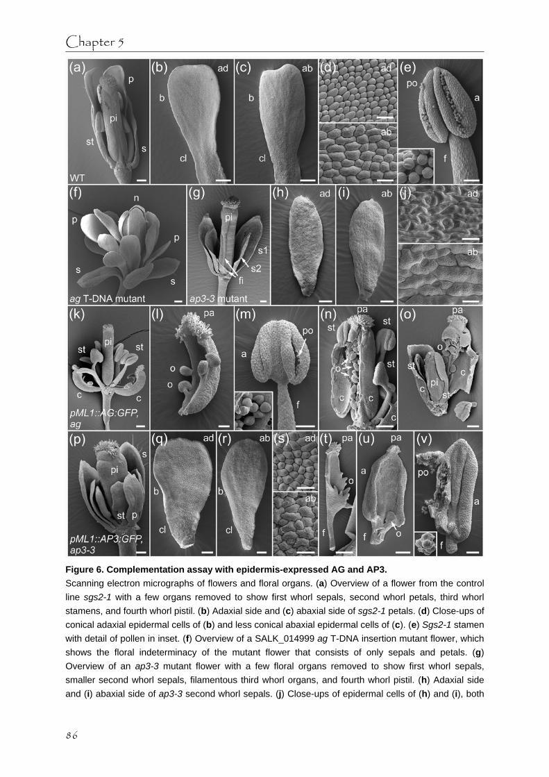
Chapter 5
Figure 6. Complementation assay with epidermis-expressed AG and AP3. Scanning electron micrographs of flowers and floral organs. (a) Overview of a flower from the control line sgs2-1 with a few organs removed to show first whorl sepals, second whorl petals, third whorl stamens, and fourth whorl pistil. (b) Adaxial side and (c) abaxial side of sgs2-1 petals. (d) Close-ups of conical adaxial epidermal cells of (b) and less conical abaxial epidermal cells of (c). (e) Sgs2-1 stamen with detail of pollen in inset. (f) Overview of a SALK_014999 ag T-DNA insertion mutant flower, which shows the floral indeterminacy of the mutant flower that consists of only sepals and petals. (g) Overview of an ap3-3 mutant flower with a few floral organs removed to show first whorl sepals, smaller second whorl sepals, filamentous third whorl organs, and fourth whorl pistil. (h) Adaxial side and (i) abaxial side of ap3-3 second whorl sepals. (j) Close-ups of epidermal cells of (h) and (i), both
86

Transport of MADS proteins
showing irregular shaped, elongated cells interspersed with guard cells. (k) Overview of a pML1::AG:GFP, ag flower with a few organs removed to show first whorl carpels, second whorl staminoid organs, third whorl stamens, and fourth whorl pistil. (l) pML1::AG:GFP, ag first whorl carpel with ovules at basal part of the carpel and papillae at the top. (m) pML1::AG:GFP, ag third whorl stamen with detail of pollen in inset. (n) Opened up pML1::AG:GFP, ag pistil with ovules inside. (o) Opened up pML1::AG:GFP, ag pistil with ovules and new flower with pistil and staminoid organs indicated. (p) Overview of a pML1::AP3:GFP, ap3-3 flower with a few organs removed to show first whorl sepals, second whorl petaloid organs, third whorl staminoid organs, and fourth whorl pistil. (q) Adaxial side and (r) abaxial side of pML1::AP3:GFP, ap3-3 second whorl petaloid organs. (s) Close-up of conical adaxial epidermal cells of (q) and close-up of less conical abaxial epidermal cells interspersed with guard cells of (r). (t) pML1::AP3:GFP, ap3-3 third whorl carpelloid organ on filament with ovular outgrowths at the base and papillae at the top. (u) pML1::AP3:GFP, ap3-3 third whorl carpelloid organ on filament with only minor ovular outgrowth at the base and a few papillae at the top. (v) pML1::AP3:GFP, ap3-3 third whorl stamen with immature pollen locked inside. Detail of immature pollen in inset. a, anther; ab, abaxial; ad, adaxial; b, blade; c, carpel; cl, claw; f, filament; fi, filamentous organ; n, new flower; o, ovule/ovular outgrowth; p, petal/petaloid organ; pa, papillae; pi, pistil; po, pollen; s, sepal; s1, first whorl sepal; s2, second whorl sepal; st, stamen/staminoid organ. Scale bars of (a-c), (f-i), (k), (n-p), (t) are 200 µm, scale bars of (e), (l), (m), (q), (r), (u), (v) are 100 µm, and scale bars of (d), (j), (s) are 20 µm. sufficient for floral determinacy, although it was shown before that AG needs to be active in both the subepidermis and the internal cell layers for floral determinacy [29].
In comparison, the complementation assay with pML1::AP3:GFP in an ap3-3 mutant background showed a strikingly lower degree of complementation. Where the ap3-3 mutant flower had small green sepals in the second whorl and filamentous third whorl organs that were sometimes fused to the fourth whorl pistil (Figure 6g), the pML1::AP3:GFP, ap3-3 flower had second whorl whitish-green petaloid organs, and third whorl carpelloid or staminoid organs that were sometimes fused to the normal pistil (Figure 6p). Compared to petals from the control line (Figure 6b,c), the pML1::AP3:GFP, ap3-3 petaloid organs had a similar shape (Figure 6q,r), but they were smaller in size like ap3-3 second whorl sepals (Figure 6h,i). The epidermal cell identity of pML1::AP3:GFP, ap3-3 petaloid organs was restored, although the abaxial epidermal cells were still interspersed with guard cells as seen in the ap3-3 second whorl sepals (Figure 6d,j,s). Also, the mesophyll cells of the petals still contained chloroplasts as in the ap3-3 second whorl sepals, giving the petaloid organs their whitish-green appearance. The third whorl organs showed a large range of carpelloid/staminoid phenotypes, both between flowers from different plants and between flowers from the same plant (Figure 6t-v). Of the 17 pML1::AP3:GFP, ap3-3 plants from the total 85 checked F2 progeny plants of two independent lines, only three plants showed stamen-like organs consisting of a green filament and a yellowish anther, occasionally with minor ovular outgrowths at the base of the anther (Figure 6p,v). In the thecae of these anthers immature pollen developed (Figure 6v). The other 14 partially complemented plants had either filamentous organs with or without papillae at the top, carpelloid organs on a filament with papillae at the top and ovular outgrowths at the base (Figure 6t), or more anther-like organs on a filament with minor papillar growth at the top and minor ovular
87

Chapter 5
outgrowth at the base (Figure 6u). It therefore seems that epidermal AP3:GFP presence is sufficient only for certain aspects of petal and stamen identity and function, like epidermal cell identity and to some extent the shape of the organs. However, the inner cell layer identity and the outgrowth of the organs are not sufficiently influenced by only epidermal AP3:GFP. This corresponds well with earlier observations that the presence of AP3 in the epidermis of second whorl organs has a strong influence on organ shape, while AP3 presence in the subepidermis influences organ size [26]. Also, it was noted before that in ap3-3 flowers epidermally complemented with either AP3 or the A. majus orthologue DEF, the maturation of pollen, suppression of chlorophyll formation, and elongation of petals and stamen filaments were incomplete [43]. Discussion The family of MADS domain transcription factors controls various important processes in the life of a flowering plant, such as floral transition and floral organ identity specification. To investigate the possibility of non-cell-autonomous function of these transcription factors, we made A. thaliana plants that specifically express GFP-tagged versions of AG, SEP3, AP3, PI, or the combination of AP3 and PI in the epidermal cell layer and analyzed the behaviour of these proteins. Next, the developmental effect of epidermis-expressed MADS domain proteins on plant morphology and flowering time were investigated and related to the localisations and behaviour of these proteins, which lead to insights into the functioning of these MADS domain transcription factors. Intercellular transport The photobleaching experiments we performed demonstrated that all tested epidermis-expressed GFP-tagged MADS domain proteins were able to diffuse through the epidermal cell layer through the primary PD. Whether the intercellular epidermal transport of MADS domain proteins in FMs is more than short-distance cannot be concluded from these experiments. Nevertheless, this intercellular transport of MADS domain proteins within the epidermis could explain previous observations of MADS domain protein localisation in epidermal tissues where their mRNA presence is not reported [25]. Additionally, the observed intercellular transport of AG in the epidermis supports previous suggestions that AG signal can extend laterally in the FM [28, 30]. We also demonstrated that from all the tested epidermis-expressed GFP-tagged MADS domain proteins only AG:GFP was also present in the subepidermal cell layer in FMs and the IM, indicating that AG:GFP proteins are able to move from the epidermis to the subepidermis. Especially in the FM, where AG normally occurs [25, 44, 45], the subepidermal AG:GFP protein amount was a substantial one-fourth of the epidermal protein amount. AG is not the only transcription factor that has the ability to traffic in the SAM, other examples are the MADS domain proteins GLO and DEF [13], the KNOX proteins KN1, KNAT1/BP and STM [10, 11] and the FM identity protein LFY [12]. The transport of AG:GFP via the secondary PD that connect the epidermis and the subepidermis [5] does not seem to be a passive process, as the other GFP-tagged MADS
88

Transport of MADS proteins
domain proteins of similar molecular weight were not present in the subepidermis of the FM or the IM. It is possible that the inward transport of AG is an active process that is controlled by intrinsic properties of AG in combination with the type of secondary PD found in the FM and IM, since there is no inward transport of AG through secondary PD in leaf tissue. Alternatively, the process of AG transport from the epidermis to the subepidermis could be facilitated by a factor or chaperone that is present in the FM and IM and not in leaf tissue [46]. A possible transport facilitator that is indeed specifically expressed in the areas where AG:GFP is able to move between cell layers, is the trafficking KNOX protein STM that accumulates in punctae in the cell wall that are presumably PD [11, 47]. Interestingly, STM is thought to be involved in AG upregulation in the third and fourth whorl and also in carpel development [48, 49]. The AG upregulation would be mediated by heterodimers of STM and the two BELL1-like homeobox (BLH) proteins PENNYWISE (PNY) and POUND-FOOLISH (PNF), which have similar expression domains as STM in the centre of the inflorescence and FM [48, 50-52]. It has been shown before that MADS domain transcription factors can interact with BLH proteins, as BELL1 interacts with AG/SEP3 heterodimers to promote ovule development [53]. Perhaps STM, possibly together with PNY and PNF, interacts with AG to facilitate both AG intercellular transport and AG upregulation in the FM centre. Floral transition and SAM indeterminacy Both the epidermal overexpression experiments with AG:GFP, SEP3:GFP, AP3:GFP, PI:GFP and the complementation assays with epidermis-expressed AG:GFP and AP3:GFP showed that the GFP-tagged versions of these transcription factors are active. Furthermore, the fact that full complementation was obtained upon expression of C-terminally GFP-tagged version of the MADS domain proteins AG, AP1 and FRUITFULL (FUL) in their respective mutants, provides evidence that GFP does not interfere with the functioning of these MADS domain proteins [25]. Nevertheless, we cannot exclude the possibility that the fluorescent tag might hinder the functioning of particular MADS protein complexes, as might be the case for the double GFP-tagged AP3/PI heterodimer. The floral homeotic changes observed in plants with epidermis-expressed AG:GFP, SEP3:GFP, AP3:GFP and PI:GFP are similar to the changes seen in plants that constitutively overexpress these proteins [31, 32, 35, 36]. Also, the altered leaf morphology of plants overexpressing epidermal AG:GFP and SEP3:GFP is similar to that of constitutive overexpressing plants [32, 33]. Unlike constitutive overexpression, specific epidermal expression of AG:GFP, SEP3:GFP, AP3:GFP, or PI:GFP does not lead to early flowering plants or terminal flower phenotypes [32, 33]. This demonstrates that epidermal presence of MADS domain proteins, or in the case of AG:GFP both epidermal and subepidermal presence, does not affect floral transition. Does AG have a non-cell-autonomous function? Is it possible that AG requires non-cell-autonomous abilities to fulfil its two functions of floral organ identity specification and floral determinacy? The results from this study demonstrated that in the FM AG is able to move between epidermal cells and is also able to move inwards from the epidermal layer to the subepidermal layer. In addition, the complementation assay
89

Chapter 5
with epidermis-expressed AG:GFP demonstrated that this construct can fully complement the ag mutant, suggesting that the trafficking AG proteins are functional in both floral organ identity specification and floral determinacy. Earlier it was proposed that AG might be able to move outward from the subepidermis to the epidermis during floral development [29] and we demonstrated that AG is able to move inwards from the epidermis to the subepidermis, so perhaps AG protein can freely move in both directions between the epidermis and the subepidermis. It might be that the ability of AG to traffic between cells and between cell layers is essential for the spread of the AG expression domain over all the three clonal layers in the FM [25, 44]. AG expression is thought to be initiated by the combination of WUS, LFY and possibly also SEP3 in a few inner layer cells in the central zone, called the organizing centre [21, 24, 36]. As soon as AG expression has been initiated, it can become a self-perpetuating signal due to the AG positive feed-back loop [54]. Protein localisations demonstrated that AG presence is first visible in a few scattered cells in the subepidermal and inner cell layers in the central zone in early stage 3 flower buds, while later stage 3 flower buds show AG presence in all the three cell layers in the presumptive third and fourth whorl [25]. It is therefore conceivable that the AG expression domain spreads outward from the organizing centre to the subepidermal and epidermal layers through the action of a few AG proteins that traffic a short distance across cell layers and subsequently initiate AG expression in those cell layers. In line with this model, enhancement of AG expression in the centre of the floral meristem by expression of this gene from the CLAVATA3 (CLV3) promoter results in lateral expansion of AG expression [30]. Our results strongly suggest that the so called principle of ‘regulation by tuning’ for C-type gene expression [30, 55] is mediated by an AG autoregulatory loop in A. thaliana involving short distance AG protein transport. Additionally, the ability of AG to traffic inwards in the FM could be required for the AG-induced termination of the WUS expression in the organizing centre of the FM, which results in the consumption of the last meristematic cells for the proper development of the pistil and ovules [2, 21, 23]. Especially for the floral determinacy function of AG it seems that a high amount of AG proteins needs to be present in the subepidermis and the internal cells to be able to eventually repress WUS expression in the organizing centre underneath the subepidermis [20, 21, 29]. The ability of AG proteins to travel inwards in the FM could provide a failsafe mechanism, on top of the transcriptional regulation, to ensure that enough AG molecules are eventually present in the organizing centre to repress WUS expression and to develop a fully functional flower. Experimental procedures Plant materials and growth conditions All A. thaliana plant lines were grown at 22ºC in growth chambers under a long-day light regime (16 h light/8 h dark). For the transformation of all constructs the sgs2-1 mutant line in Columbia-0 (Col-0) background was used. The sgs2-1 (also known as sde1 and rdr6) mutant line is defective in gene silencing, but has no obvious defects in flower development [56, 57]. This mutant background was chosen because of problems with transgene silencing in the
90

Transport of MADS proteins
Col-0 wildtype background [58]. A double transgenic line expressing both pML1::AP3:GFP and pML1::PI:GFP constructs was obtained by crossing the single transgenic lines. For the complementation experiments the pML1::AG:GFP and pML1::AP3:GFP transgenic lines were crossed into the SALK_014999 ag T-DNA insertion mutant line and the ap3-3 mutant line [38], respectively. Construction of binary vectors and plant transformation A 3388 bp promoter fragment from the MERISTEM LAYER 1 (ML1) gene was generated from Col-0 gDNA by proofreading PCR with primers PDS1209 and PDS1210 (see Table S1 in supplementary material for sequences). The same fragment was shown to be sufficient to give specific epidermal expression in both vegetative and reproductive tissues [59]. Next, the pK7FWG2 gateway destination vector [60] was modified by removing the CaMV35S promoter element with a HindIII/SpeI double digestion and replacing this fragment with the amplified ML1 promoter fragment. Gateway entry clones containing cDNA clones of AG, AP3, GFP, PI, SEP3 lacking their STOP codon, were recombined into the new destination vector. The resulting six expression clones pML1::AG:GFP (CZN301), pML1::AP3:GFP (CZN303), pML1::GFP:GFP (CZN304), pML1::PI:GFP (CZN305) and pML1::SEP3:GFP (CZN306) were introduced into Agrobacterium tumefaciens strain GV3101 and transformed to sgs2-1 mutant plants by floral dip method [61]. Additionally, two constructs pCaMV35S::AG:GFP (pARC276) and pCaMV35S::SEP3:GFP (pARC277) previously made [58], were also transformed into the sgs2-1 mutant line. Confocal Laser Scanning Microscopy The presence of the GFP-tagged proteins was determined in three different living tissues, namely rosette leaves, IMs and stage 3-5 floral buds (stages after [62]). In order to observe the localisation in rosette leaves, tips of young leaves were put between glass slides in 0.5 x MS and scanned. To observe the localisation of the GFP-tagged proteins in inflorescences, the inflorescence material was dissected and embedded as previously described [58]. Confocal Laser Scanning Microscopy (CLSM) of the living plant tissue was performed with a Leica SPE DM5500 upright microscope with a 63x ACS APO (NA 1.15 CORR) lens, using the LAS AF 1.8.2 software (Leica, http://www.leica-microsystems.com). Further details about the imaging and photobleaching experiments are provided in the supporting information. In situ hybridization Inflorescences from pML1::AG:GFP plants were fixed and sections were hybridized with sense and antisense probes for the GFP gene. A detailed protocol is provided in the supporting information. Plant morphology and microscopy The plant morphology of all the transgenic lines was analyzed, with specific attention to the flower phenotypes. Bright field images of the mutant and rescued flower phenotypes were taken with a Leica DFC320 digital camera mounted on a Zeiss Stemi SV8 binocular (Carl
91

Chapter 5
Zeiss, http://www.zeiss.com) with IM500 software. For scanning electron microscopy (SEM), inflorescences were collected and fixed overnight at 4oC in aqueous 4% paraformaldehyde. Samples were dehydrated on an ethanol series to 70% ethanol and stored at 4oC until needed. Samples were further dehydrated through absolute ethanol and critical point dried through CO2 in a Balzers CPD 020 (Baltec, http://www.bal-tec.com). Dried samples were dissected under a dissecting microscope before being mounted on metallic stubs with adhesive tape, and were further dissected when essential. Samples were then sputter-coated with colloidal gold and observed at 10-20 kV under a LEO 435 VP scanning electron microscope (Carl Zeiss) and digital micrographs were obtained. Flowering time measurement Plants of pML1::AG:GFP and pML1::SEP3:GFP transgenic lines were grown together with sgs2-1 control plants on soil, while pCaMV35S::AG:GFP and pCaMV35S::SEP3:GFP primary transformants were grown on 0.5 x MS 0.8% agar plates containing 25 μg/ml kanamycin. As a measure of flowering time the number of rosette leaves was counted at the time of bolting. The presence of the constructs in the plants was determined by examining the flower phenotype and the presence of GFP fluorescence in the leaves. In total 25 pML::AG:GFP plants, 23 pML::SEP3:GFP plants, 46 sgs2-1 control plants, 10 pCaMV35S::AG:GFP plants and 7 pCaMV35S::SEP3:GFP plants were analyzed. This data was explored with a one-way ANOVA and Tukey HSD post hoc test in the statistical program SPSS version 11.5 (SPSS, http://www.spss.com). Complementation analysis The presence of the wild type allele and the mutant allele was determined by PCR in the F2 progeny of the two crosses, pML1::AG:GFP x SALK_014999 ag T-DNA insertion mutant line and pML1::AP3:GFP x ap3-3 mutant line, while the presence of the GFP-tagged MADS box gene construct was determined by CLSM. For the AG wild type allele primers PRO182 and PDS1985 were used and for the ag T-DNA mutant allele primers PRO433 and PDS404 (see Table S1). For the AP3 wild type allele primers PDS2027 and PDS2029 were used and for the ap3-3 mutant allele primers PDS2028 and PDS2029. Acknowledgements APM, LCBA and MCD are financed by CAPES and CNPq, Brazil. GA and RI are financed by CBSG. We acknowledge NAP/MEPA-ESALQ, University of São Paulo, for use of the electron microscopy facilities.
92

Transport of MADS proteins
Supporting information Figure S1. Floral phenotypes of pML1::AP3:GFP and pML1::PI:GFP plants. (a) A pML1::AP3:GFP flower showing an unfused pistil with more than two staminoid carpels. (b) A pML1::PI:GFP flower showing petaloid sepals that extend at a great angle from the floral axis.
Figure S2. Epidermal intercellular transport of SEP3:GFP and AP3:GFP/PI:GFP. Recovery of GFP fluorescence after photobleaching as a result of intercellular transport of GFP-tagged MADS domain proteins in the epidermis of early flower buds. The 3-D projections of the flower buds are shown in artificial heat map colouring, which presents the minimum GFP fluorescence value in light blue and the maximum value in red. (a) A pML1::SEP3:GFP stage 3 flower bud immediately after photobleaching. (b-g) Close-ups of bleached area immediately after photobleaching (b), after 5
93

Chapter 5
minutes (c), after 10 minutes (d), after 15 minutes (e), after 20 minutes (f), and after 25 minutes (g). A cell in the outer ring of recovery (1) and a cell in the inner region of recovery (2) are indicated with white circles. (h) Colour bars illustrate the GFP fluorescence recovery of the indicated cell 1 and cell 2, as measured by the normalized pixel sum per μm2. Note that the fluorescence recovers first in the outer ring of the bleached area and slightly later in the inner region, demonstrating the inward movement of fluorescent SEP3:GFP molecules into the bleached area. (i) A pML1::AP3:GFP/pML1::PI:GFP stage 3 flower bud immediately after photobleaching. (j-o) Close-ups of bleached area immediately after photobleaching (j), after 5 minutes (k), after 10 minutes (l), after 15 minutes (m), after 20 minutes (n), and after 25 minutes (o). (p) Colour bars illustrate the GFP fluorescence recovery of the indicated cell 1 and cell 2, as measured by the normalized pixel sum per μm2. Note that the fluorescence recovers first in the outer ring of the bleached area and slightly later in the inner region, demonstrating the inward movement of fluorescent AP3/PI molecules into the bleached area. Scale bars of (a) and (i) are 30 µm, all other scale bars are 5 µm. Appendix S1. Detailed experimental procedures for confocal laser scanning microscopy and photobleaching assay GFP was excited with the 488 nm solid state laser. The GFP emission was scanned at a bandwidth of 505-530 nm, while the red autofluorescence of the chloroplasts in leaf tissue was detected at a bandwidth of 600-700 nm. All scans for the leaf tissue and also all scans for the IMs and FMs were made with the same confocal settings, which sometimes resulted in some oversaturated images like the scans for the 2x GFP control plants. The obtained confocal z-stacks were median-filtered and converted to 3-D maximum projections or cross section images. All images were adjusted with Adobe Photoshop version 5.0 (Adobe, http://www.adobe.com).
To quantify the GFP signal ratio between the epidermal and subepidermal cell layers in the centre of the IM and FM in cross sections, the pixel sum per μm2 in the GFP channel was determined in two regions of interest (ROI) created around the epidermal nuclei and the subepidermal nuclei. For both the IM and the FM, the ratios were calculated in two perpendicular cross sections through the centre of the meristem in four different samples. The cross sections used for this quantification had a minimal amount of saturated pixels (<3%). Oversaturated scans were occasionally made to verify the absence of GFP fluorescence in the subepidermal and inner cell layers.
Photobleaching experiments were performed on early flower buds by using the maximal square zoom function and repeatedly photobleaching this zoomed-in area in one z-section with 488 nm laser light at 100% power for five minutes. The fluorescence recovery in the photobleached area was monitored by scanning the whole flower bud immediately after the photobleaching and every five minutes afterwards, until a maximum of thirty minutes. The obtained confocal z-stacks were median-filtered and converted to 3-D maximum projections and adjusted with Adobe Photoshop version 5.0. An artificial heat map colouring was used for easy visualization of signal intensity changes over time. For two cells in each experiment the recovery of the GFP fluorescence in time was quantified in two circular ROIs in the 3-D projection as the normalized pixel sum per μm2, and visualized in a colour-coded bar.
94

Transport of MADS proteins
Appendix S2. Detailed experimental procedure for in situ hybridization Inflorescences from pML1::AG:GFP plants were collected and fixed in aqueous 4% paraformaldehyde with 0.25% glutaraldehyde for two hours at room temperature. After fixation the samples were dehydrated through an ethanol/xylene series and finally embedded in Paraplast Plus (Sigma-Aldrich, http://www.sigmaaldrich.com). Sections of 5-6 µm were obtained with a rotary microtome, placed on 3-aminopropyltrietoxysilane-covered slides (Pierce, http://www.piercenet.com) and baked for 16 hours at 50°C. Paraplast was removed from the sections by three consecutive five minute washes in xylene. A pre-hybridization treatment with proteinase-K (1 µg/ml in 0.05 M Tris-HCl, pH7.5) for five minutes at 37ºC was followed by three washes with diethyl pyrocarbonate (DEPC)-treated water. The sections were hybridized with sense and antisense probes for the GFP gene. The digoxigenin (DIG-UTP)-labelled probes were made according to the instructions of the manufacturer (Roche, http://www.roche.com), with the use of a 372 bp PCR product template made from the pML1::AG:GFP construct with primers GFF1 and GFR1 (see Table S1). After overnight hybridization at 42ºC, the sections were washed four times to remove excess probe. The hybridization signal was detected with an anti-DIG antibody diluted 1:1000 in 0.01 M Tris-HCl, 0.15 M NaCl, 1% Blocking Reagent (Roche) and visualized after reaction with a commercial solution of nitro-blue tetrazolium chloride (NBT) and 5-bromo-4-chloro-3´indolyphosphate p-toluidine (BCIP) plus 1 mM levamisole (Pierce). Bright field images were taken with a Zeiss Axioscope (Carl Zeiss, http://www.zeiss.com), and adjusted with Adobe Photoshop version 5.0.
Table S1. Sequences of primers used in experiments.
Primer Sequence 5’- 3’ Annealing site
GFF1 TGAATTTAGGTGACACTATAGTGCTCAGGTAGTGGTTGTCG SP6 promoter
157 bp downstream of START codon of GFP
GFR1 GCTCTAATACGACTCACTATAGGGCACATGAAGCAGCACGACTT T7 promoter
529 bp downstream of START codon of GFP
PRO182 GGATCCATGGCGTACCAATCGGAGCT Immediately after START codon of AGAMOUS
PRO433 CACCGATCAAAGACTACACATCAC 2634 bp upstream of START codon of AGAMOUS
PDS404 TGGTTCACGTAGTGGGCCATCG On left border of T-DNA
PDS1209 TTAAAGCTTATCAAAGAAAAAACAAGAAC HindIII 4983 bp upstream of START codon of MERISTEMLAYER1
PDS1210 AAAACTAGTTAACCGGTGGATTCAGGGAGTTTCTTTA SpeI 1596 bp upstream of START codon of MERISTEMLAYER1
PDS1985 CATTTCCTTCAGCCTATATTACC In 3’ UTR of AGAMOUS 18 bp downstream of STOP codon
PDS2027 AGAACCAGACAAACAGACAAG In first exon of APETALA3 32 bp downstream of START codon
PDS2028 AGAACCAGACAAACAGATAAG In the first exon of apetala3-3 32 bp downstream of START codon
PDS2029 GCACCAGCAAACCTTTTAGC In the 3’ UTR of APETALA3 6 bp downstream of STOP codon
95

Chapter 5
References 1. Carles CC, Fletcher JC: Shoot apical meristem maintenance: the art of a dynamic
balance. Trends in Plant Science 2003, 8(8):394-401. 2. Sablowski R: Flowering and determinacy in Arabidopsis. Journal of Experimental Botany
2007, 58(5):899-907. 3. Kwiatkowska D: Flowering and apical meristem growth dynamics. Journal of Experimental
Botany 2008, 59(2):187-201. 4. Zambryski P: Plasmodesmata. Current Biology 2008, 18(8):R324-R325. 5. Ehlers K, Kollmann R: Primary and secondary plasmodesmata: structure, origin, and
functioning. Protoplasma 2001, 216(1-2):1-30. 6. Duckett CM, Oparka KJ, Prior DAM, Dolan L, Roberts K: Dye-coupling in the root epidermis
of Arabidopsis is progressively reduced during development. Development 1994, 120(11):3247-3255.
7. Gisel A, Barella S, Hempel FD, Zambryski PC: Temporal and spatial regulation of symplastic trafficking during development in Arabidopsis thaliana apices. Development 1999, 126(9):1879-1889.
8. Kim I, Cho E, Crawford K, Hempel FD, Zambryski PC: Cell-to-cell movement of GFP during embryogenesis and early seedling development in Arabidopsis. Proceedings of the National Academy of Sciences of the United States of America 2005, 102(6):2227-2231.
9. Wolf S, Deom CM, R.N. B, Lucas WJ: Movement protein of tobacco mosaic virus modifies plasmodesmatal size exclusion limit. Science 1989, 246(4928):377-379.
10. Lucas WJ, Bouchepillon S, Jackson DP, Nguyen L, Baker L, Ding B, Hake S: Selective trafficking of KNOTTED1 homeodomain protein and its messenger-RNA through plasmodesmata. Science 1995, 270(5244):1980-1983.
11. Kim JY, Yuan Z, Jackson D: Developmental regulation and significance of KNOX protein trafficking in Arabidopsis. Development 2003, 130(18):4351-4362.
12. Sessions A, Yanofsky MF, Weigel D: Cell-cell signaling and movement by the floral transcription factors LEAFY and APETALA1. Science 2000, 289(5480):779-781.
13. Perbal MC, Haughn G, Saedler H, Schwarz-Sommer Z: Non-cell-autonomous function of the Antirrhinum floral homeotic proteins DEFICIENS and GLOBOSA is exerted by their polar cell-to-cell trafficking. Development 1996, 122(11):3433-3441.
14. Coen ES, Meyerowitz EM: The war of the whorls - Genetic interactions controlling flower development. Nature 1991, 353(6339):31-37.
15. Colombo L, Franken J, Koetje E, Vanwent J, Dons HJM, Angenent GC, Vantunen AJ: The Petunia MADS box gene FBP11 determines ovule identity. Plant Cell 1995, 7(11):1859-1868.
16. Pelaz S, Ditta GS, Baumann E, Wisman E, Yanofsky MF: B and C floral organ identity functions require SEPALLATA MADS-box genes. Nature 2000, 405(6783):200-203.
17. Pinyopich A, Ditta GS, Savidge B, Liljegren SJ, Baumann E, Wisman E, Yanofsky MF: Assessing the redundancy of MADS-box genes during carpel and ovule development. Nature 2003, 424(6944):85-88.
18. Ditta G, Pinyopich A, Robles P, Pelaz S, Yanofsky MF: The SEP4 gene of Arabidopsis thaliana functions in floral organ and meristem identity. Current Biology 2004, 14(21):1935-1940.
19. Ferrario S, Immink RG, Angenent GC: Conservation and diversity in flower land. Current
96

Transport of MADS proteins
Opinion in Plant Biology 2004, 7(1):84-91. 20. Mizukami Y, Ma H: Separation of AG function in floral meristem determinacy from that in
reproductive organ identity by expressing antisense AG RNA. Plant Molecular Biology 1995, 28(5):767-784.
21. Lohmann JU, Hong RL, Hobe M, Busch MA, Parcy F, Simon R, Weigel D: A molecular link between stem cell regulation and floral patterning in Arabidopsis. Cell 2001, 105(6):793-803.
22. Sun B, Xu YF, Ng KH, Ito T: A timing mechanism for stem cell maintenance and differentiation in the Arabidopsis floral meristem. Genes & Development 2009, 23(15):1791-1804.
23. Lenhard M, Bohnert A, Jurgens G, Laux T: Termination of stem cell maintenance in Arabidopsis floral meristems by interactions between WUSCHEL and AGAMOUS. Cell 2001, 105(6):805-814.
24. Mayer KFX, Schoof H, Haecker A, Lenhard M, Jurgens G, Laux T: Role of WUSCHEL in regulating stem cell fate in the Arabidopsis shoot meristem. Cell 1998, 95(6):805-815.
25. Urbanus SL, de Folter S, Shchennikova AV, Kaufmann K, Immink RGH, Angenent GC: In planta localisation patterns of MADS domain proteins during floral development in Arabidopsis thaliana. BMC Plant Biology 2009, 9.
26. Jenik PD, Irish VF: The Arabidopsis floral homeotic gene APETALA3 differentially regulates intercellular signaling required for petal and stamen development. Development 2001, 128(1):13-23.
27. Wu XL, Dinneny JR, Crawford KM, Rhee Y, Citovsky V, Zambryski PC, Weigel D: Modes of intercellular transcription factor movement in the Arabidopsis apex. Development 2003, 130(16):3735-3745.
28. Jenik PD, Irish VF: Regulation of cell proliferation patterns by homeotic genes during Arabidopsis floral development. Development 2000, 127(6):1267-1276.
29. Sieburth LE, Drews GN, Meyerowitz EM: Non-autonomy of AGAMOUS function in flower development: use of a Cre/loxP method for mosaic analysis in Arabidopsis. Development 1998, 125(21):4303-4312.
30. Cartolano M, Efremova N, Kuckenberg M, Raman S, Schwarz-Sommer Z: Enhanced AGAMOUS expression in the centre of the Arabidopsis flower causes ectopic expression over its outer expression boundaries. Planta 2009, 230(4):857-862.
31. Krizek BA, Meyerowitz EM: The Arabidopsis homeotic genes APETALA3 and PISTILLATA are sufficient to provide the B class organ identity function. Development 1996, 122(1):11-22.
32. Mizukami Y, Ma H: Ectopic expression of the floral homeotic gene AGAMOUS in transgenic Arabidopsis plants alters floral organ identity. Cell 1992, 71(1):119-131.
33. Honma T, Goto K: Complexes of MADS-box proteins are sufficient to convert leaves into floral organs. Nature 2001, 409(6819):525-529.
34. Mizukami Y, Ma H: Determination of Arabidopsis floral meristem identity by AGAMOUS. Plant Cell 1997, 9(3):393-408.
35. Jack T, Fox GL, Meyerowitz EM: Arabidopsis homeotic gene APETALA3 ectopic expression - transcriptional and posttranscriptional regulation determine floral organ identity. Cell 1994, 76(4):703-716.
36. Liu C, Xi WY, Shen LS, Tan CP, Yu H: Regulation of Floral Patterning by Flowering Time Genes. Developmental Cell 2009, 16(5):711-722.
37. Goto K, Meyerowitz EM: Function and regulation of the Arabidopsis floral homeotic gene
97

Chapter 5
PISTILLATA. Genes & Development 1994, 8(13):1548-1560. 38. Jack T, Brockman LL, Meyerowitz EM: The homeotic gene APETALA3 of Arabidopsis
thaliana encodes a MADS box and is expressed in petals and stamens. Cell 1992, 68(4):683-697.
39. Weigel D, Meyerowitz EM: Activation of floral homeotic genes in Arabidopsis. Science 1993, 261(5129):1723-1726.
40. Immink RGH, Tonaco IAN, de Folter S, Shchennikova A, van Dijk ADJ, Busscher-Lange J, Borst JW, Angenent GC: SEPALLATA3: the 'glue' for MADS box transcription factor complex formation. Genome Biology 2009, 10(2).
41. McGonigle B, Bouhidel K, Irish VF: Nuclear localization of the Arabidopsis APETALA3 and PISTILLATA homeotic gene products depends on their simultaneous expression. Genes & Development 1996, 10(14):1812-1821.
42. Sieburth LE, Running MP, Meyerowitz EM: Genetic separation of 3rd and 4th whorl functions of AGAMOUS. Plant Cell 1995, 7(8):1249-1258.
43. Efremova N, Perbal MC, Yephremov A, Hofmann WA, Saedler H, Schwarz-Sommer Z: Epidermal control of floral organ identity by class B homeotic genes in Antirrhinum and Arabidopsis. Development 2001, 128(14):2661-2671.
44. Drews GN, Bowman JL, Meyerowitz EM: Negative regulation of the Arabidopsis homeotic gene AGAMOUS by the APETALA2 product. Cell 1991, 65(6):991-1002.
45. Yanofsky MF, Ma H, Bowman JL, Drews GN, Feldmann KA, Meyerowitz EM: The protein encoded by the Arabidopsis homeotic gene AGAMOUS resembles transcription factors. Nature 1990, 346(6279):35-39.
46. Lucas WJ: Plant viral movement proteins: Agents for cell-to-cell trafficking of viral genomes. Virology 2006, 344(1):169-184.
47. Long JA, Moan EI, Medford JI, Barton MK: A member of the KNOTTED class of homeodomain proteins encoded by the STM gene of Arabidopsis. Nature 1996, 379(6560):66-69.
48. Yu LF, Patibanda V, Smith HMS: A novel role of BELL1-like homeobox genes, PENNYWISE and POUND-FOOLISH, in floral patterning. Planta 2009, 229(3):693-707.
49. Scofield S, Dewitte W, Murray JAH: The KNOX gene SHOOT MERISTEMLESS is required for the development of reproductive meristematic tissues in Arabidopsis. Plant Journal 2007, 50(5):767-781.
50. Kanrar S, Onguka O, Smith HMS: Arabidopsis inflorescence architecture requires the activities of KNOX-BELL homeodomain heterodimers. Planta 2006, 224(5):1163-1173.
51. Smith HMS, Hake S: The interaction of two homeobox genes, BREVIPEDICELLUS and PENNYWISE, regulates internode patterning in the Arabidopsis inflorescence. Plant Cell 2003, 15(8):1717-1727.
52. Smith HMS, Campbell BC, Hake S: Competence to respond to floral inductive signals requires the homeobox genes PENNYWISE and POUND-FOOLISH. Current Biology 2004, 14(9):812-817.
53. Brambilla V, Battaglia R, Colombo M, Masiero S, Bencivenga S, Kater MM, Colombo L: Genetic and molecular interactions between BELL1 and MADS box factors support ovule development in Arabidopsis. Plant Cell 2007, 19(8):2544-2556.
54. Gomez-Mena C, de Folter S, Costa MMR, Angenent GC, Sablowski R: Transcriptional program controlled by the floral homeotic gene AGAMOUS during early organogenesis. Development 2005, 132(3):429-438.
55. Cartolano M, Castillo R, Efremova N, Kuckenberg M, Zethof J, Gerats T, Schwarz-Sommer Z,
98

Transport of MADS proteins
Vandenbussche M: A conserved microRNA module exerts homeotic control over Petunia hybrida and Antirrhinum majus floral organ identity. Nature Genetics 2007, 39(7):901-905.
56. Elmayan T, Balzergue S, Beon F, Bourdon V, Daubremet J, Guenet Y, Mourrain P, Palauqui J-C, Vernhettes S, Vialle T et al: Arabidopsis mutants impaired in cosuppression. Plant Cell 1998, 10(10):1747-1758.
57. Peragine A, Yoshikawa M, Wu G, Albrecht HL, Poethig RS: SGS3 and SGS2/SDE1/RDR6 are required for juvenile development and the production of trans-acting siRNAs in Arabidopsis. Genes & Development 2004, 18(19):2368-2379.
58. de Folter S, Urbanus SL, van Zuijlen LG, Kaufmann K, Angenent GC: Tagging of MADS domain proteins for chromatin immunoprecipitation. BMC Plant Biology 2007, 7.
59. Sessions A, Weigel D, Yanofsky MF: The Arabidopsis thaliana MERISTEM LAYER 1 promoter specifies epidermal expression in meristems and young primordia. Plant Journal 1999, 20(2):259-263.
60. Karimi M, Inze D, Depicker A: GATEWAY vectors for Agrobacterium-mediated plant transformation. Trends in Plant Science 2002, 7(5):193-195.
61. Clough SJ, Bent AF: Floral dip: a simplified method for Agrobacterium-mediated transformation of Arabidopsis thaliana. Plant Journal 1998, 16(6):735-743.
62. Smyth DR, Bowman JL, Meyerowitz EM: Early flower development in Arabidopsis. Plant Cell 1990, 2(8):755-767.
99


Chapter 6 About SEP3, black holes and UFO
Susan L Urbanus
QD (Peter) Dinh
Zijing (Tina) Liu
Gerco C Angenent
Richard GH Immink

Chapter 6
Abstract In Arabidopsis thaliana, the E-type SEPALLATA MADS domain transcription factors play an important role in the development of the four floral organs, sepals, petals, stamens and carpels. The spatio-temporal localisation pattern of GFP-tagged SEPALLATA3 (SEP3) shows that prior to the initiation of the petal and stamen primordia in the second and third whorl, the SEP3:GFP protein presence suddenly changes from predominantly nuclear localized to more cytoplasmically localized. During further development of the petal and stamen primordia the SEP3:GFP presence in subepidermal and inner cell layers reduces greatly, while the SEP3 presence in the epidermal cell layer remains. We named this last phenomenon SEP3:GFP ‘black holes’. The SEP3 mRNA expression in the second and third whorl tissues during these flower bud stages has previously been described as uniform, which suggests that the SEP3:GFP black holes are caused by post-transcriptional processes. We tried to determine if the SEP3:GFP black holes are due to protein degradation, but could not proof the involvement of the 26s proteasome. We also investigated whether the second and third whorl expressed F-box protein UNUSUAL FLORAL ORGANS (UFO), thought to be a subunit of an SCF E3 ubiquitin ligase complex, is involved in the sudden change in SEP3:GFP subcellular localisation and the formation of SEP3:GFP black holes. However, SEP3:GFP localisations in different genetic backgrounds did not reveal an involvement of UFO in these two processes. Introduction In Arabidopsis thaliana flowers the E-type MADS domain transcription factors play an important role in the development of the four different floral organ types from the floral meristem [1-3]. SEPALLATA3 (SEP3) and the other SEPALLATA proteins seem to function as ‘glue’ proteins that facilitate the formation and functioning of the different multimeric MADS domain protein complexes that are essential for the identity specification of the first-whorl sepals, the second-whorl petals, the third-whorl stamens and the fourth-whorl carpels [4-8]. The spatio-temporal localisation pattern of a GFP-tagged genomic SEP3 clone shows that prior to the initiation of the petal and stamen primordia in the second and third whorl, SEP3:GFP protein presence suddenly changes from predominantly nuclear localized to more cytoplasmically localized. During further development of the petal and stamen primordia the SEP3:GFP presence in subepidermal and inner cell layers reduces greatly, while the SEP3 presence in the epidermal cell layer remains [9]. Because of the lack of SEP3:GFP presence in the centre of these developing organs, we named the last phenomenon SEP3:GFP ‘black holes’. Interestingly, the SEP3 mRNA expression is described as uniform during the early development of petals and stamens [1], which could indicate that these SEP3:GFP black holes are the result of post-transcriptional processes.
Simultaneously with the change to a more cytoplasmic SEP3:GFP localisation in specific regions of stage 3 flower buds [10], the overlapping expression of the two B-type MADS domain transcription factor genes APETALA3 (AP3) and PISTILLATA (PI) starts in these
102
SEP3, black holes and UFO
same regions [11-13]. According to the ‘ABC’ model the heterodimeric complex of these two B-type proteins in combination with an E-type protein and an A- or C-type protein is required for petal and stamen development, respectively [3, 7, 14, 15]. It is known that AP3 expression is regulated by UNUSUAL FLORAL ORGANS (UFO), which is also expressed in the presumptive second and third whorl [16-19]. UFO is an F-box protein that is thought to be a subunit of an SCF E3 ubiquitin ligase complex that modifies target proteins by ubiquitination [20-23]. In these SCF complexes it is the F-box protein that provides the specificity for the target proteins. Since poly-ubiquitination commonly marks proteins for degradation by the 26s proteasome [24], the attractive theory was formed that UFO could be involved in the degradation of a repressor of LEAFY (LFY), the transcriptional activator of AP3 [21]. Later it was confirmed that proteasome activity is indeed required for LFY activity, but it was also shown that UFO physically interacts with LFY and may function as a transcriptional co-factor [19, 25]. Additionally, mono-ubiquitination is thought to affect the function of target proteins without proteasome degradation, for instance by modifying the activity or the subcellular localisation [26].
In this chapter we describe the different experiments that were performed to discover whether the sudden change in subcellular SEP3:GFP localisation and the formation of SEP3:GFP black holes are indeed regulated at a post-transcriptional level and whether the F-box protein UFO is involved in these two processes. Although targeted SEP3 protein degradation seems to be the most likely cause of the SEP3:GFP black holes in developing petal and stamen primordia, we could not proof SEP3 protein degradation by the 26s proteasome. Furthermore, SEP3:GFP localisations in different genetic backgrounds did not give clear indications that UFO affects the subcellular localisation of SEP3:GFP protein or that UFO is involved in the formation of SEP3:GFP black holes. Results SEP3 presence during petal and stamen development The spatio-temporal localisation pattern of a GFP-tagged genomic SEP3 clone shows that SEP3:GFP protein in the inflorescence meristem and the first two flower bud stages is predominantly nuclear localized [9]. However, from flower bud stage 3 onwards SEP3:GFP protein suddenly becomes more cytoplasmically localized in the presumptive second and third whorl, while the SEP3:GFP protein in the centre of the floral meristem and in the epidermis of the first whorl sepal primordia remains predominantly nuclear localized. During the initiation of the petal and stamen primordia the cytoplasmically and nuclear localized SEP3:GFP starts to fade in the subepidermal and inner cell layers, until SEP3:GFP protein is mostly found in the epidermal cell layer in stage 5 flower buds (Figure 1A,C,E). We named this absence of SEP3:GFP fluorescence in the middle of the developing petal and stamen primordia ‘black holes’. In comparison, the spatio-temporal localisation pattern of a GFP-tagged genomic AGAMOUS (AG) clone, expressed in the floral meristem at the position of the future third and fourth whorl, shows a more uniform AG:GFP presence in all three cell layers throughout development (Figure 1B,D,F) [9]. The confocal microscopic scans that
103

Chapter 6
104
Figure 1. Different behaviour of SEP3 and AG during petal and stamen development. Simultaneous localisations of SEP3:GFP and AG:TagRFP in a stage 5 flower bud from a gSEP3:GFP, gAG:TagRFP plant. (A) Green channel 3-D projection showing SEP3:GFP presence in all three cell

SEP3, black holes and UFO
layers in the three inner whorls and also in the epidermal cell layer of the first-whorl sepal primordia. The nuclei filled with SEP3:GFP protein are visible as big white dots, while the autofluorescence of the plastids are visible as smaller dots. (B) Red channel 3-D projection showing AG:TagRFP presence only in the developing third-whorl stamen primordia and the fourth whorl. The nuclei filled with AG:TagRFP protein are visible as big white dots, while the epidermal cell layer of the sepal primordia shows some autofluorescent background signal. (C, D) Optical sections showing that in stamen primordia SEP3:GFP protein is both cytoplasmically and nuclear localized and more restricted to the epidermal cell layer (C), while AG:GFP protein is more nuclear localized and more uniformly present in all cell layers in the stamen primordia (D). Note that the stamen primordium in the upper right corner is already developing a so-called SEP3:GFP black hole. (E, F) Optical sections showing a SEP3:GFP black hole in the stamen primordia and a developing black hole in the petal primordia (E), while AG:GFP protein is more uniformly present in all cell layers in the stamen primordia (F). (*) black hole; (4) whorl 4; (p) petal primordium; (s) sepal primordium; (st) stamen primordium. Scale bars are 25 µm. show AG:GFP fluorescence in the same regions where SEP3:GFP black holes are observed, demonstrate that the SEP3:GFP black holes are specific for SEP3:GFP and not artefacts of confocal laser scanning microscopy through thick tissues. Unlike SEP3:GFP protein, AG:GFP protein is predominantly nuclear localized throughout development [9]. However, in initiating stamen primordia both AG:GFP protein and SEP3:GFP protein are more cytoplasmically localized in the same cells (Figure 1C-F). This could be the result of cell divisions in these initiating primordia, since nuclear localized MADS domain proteins show a more uniform distribution throughout the whole cell during the cell division process (Figure 2). Post-transcriptional regulation versus transcriptional regulation The SEP3 mRNA expression pattern during the early stages of petal and stamen development has been described as uniform [1], whereas the SEP3:GFP protein localisation pattern shows greatly reduced SEP3:GFP presence in the subepidermal and inner cell layers of initiating petal and stamen primordia in the so-called black holes (Figure 1A,C,E) [9]. This suggests that the observed SEP3:GFP black holes are the result of post-transcriptional processes, such as protein modifications that alter the behaviour of the protein or protein degradation.
To determine whether the observed SEP3:GFP behaviour during petal and stamen development is indeed regulated at post-transcriptional level and not simply due to a reduction in gene expression, we analyzed the localisation pattern of SEP3:GFP protein during second and third whorl development in plants constitutively overexpressing a SEP3:GFP cDNA clone. Since the spatio-temporal localisation pattern of genomic AG:GFP does not show the formation of AG:GFP black holes (Figure 1B,D,F) [9], we used plants constitutively overexpressing an AG:GFP cDNA clone as control plants. While the early-flowering pCaMV35S::SEP3:GFP plants commonly have only one incomplete terminal flower, the early-flowering pCaMV35S::AG:GFP plants usually produce a few more flowers that consist of carpelloid sepals, stamens or staminoid organs, and a pistil [27]. In pCaMV35S::SEP3:GFP flower buds we observed a clear reduction in GFP fluorescence in
105

Chapter 6
Figure 2. Altered subcellular SEP3:GFP localisation during cell division. A 3-D close-up of epidermal cells expressing SEP3:GFP proteins in an inflorescence meristem. Most cells have predominantly nuclear localized SEP3:GFP protein, but cells that are in the process of cell division (arrows) show a more uniform distribution of the SEP3:GFP signal over the whole cell. Scale bar is 5 µm.
Figure 3. Black holes in plants constitutively overexpressing SEP3:GFP or AG:GFP. Confocal microscopic analyses of the presence of so-called black holes in floral meristems with constitutive overexpression of either SEP3:GFP or AG:GFP. (A) 3-D projection of a terminal pCaMV35S::SEP3:GFP floral meristem of approximately stage 5, showing black holes in three initiating (presumably) third-whorl primordia. (B) 3-D projection of a pCaMV35S::AG:GFP stage 5 floral meristem, showing black holes in four initiating (presumably) third-whorl primordia. (C) Longitudinal section through the pCaMV35S::SEP3:GFP floral meristem as indicated with a dashed line in (A), showing the presence of two black holes on either side of whorl 4. (D) Longitudinal section through the pCaMV35S::AG:GFP floral meristem as indicated with a dashed line in (B), showing the presence of two black holes on either side of whorl 4. (*) black hole; (4) whorl 4; (lp) dissected leaf primordium; (s) sepal primordium. All scale bars 25 µm.
106

SEP3, black holes and UFO
the second and third whorl primordia (Figure 3A,C), very similar to the genomic SEP3:GFP black holes that were found (Figure 1A,C,E) [9]. Surprisingly, the pCaMV35S::AG:GFP flower buds showed a similar reduction of GFP fluorescence in second and third whorl primordia (Figure 3B,D), which was unexpected based on the observed behaviour of genomic AG:GFP during flower development (Figure 1B,D,F) [9]. This suggests that both SEP3:GFP and AG:GFP proteins are post-transcriptionally regulated in these developing second and third whorl organs, for instance by targeted protein degradation. However, it was shown before that the CaMV35S promoter is not very active in petals until stage 8 flower buds and also drives lower levels of expression in stage 5 and 6 stamens [28]. Taking this information together with the unexpected absence of AG:GFP in the second and third whorl tissues, it seems more likely that the SEP3:GFP and AG:GFP black holes seen in these constitutive overexpressing plants are at least partly due to lack of strong activity of the CaMV35S promoter in these tissues at these developmental stages. Proteasome inhibitors do not affect the black holes We tried to determine if the SEP3:GFP and AG:GFP black holes are created by 26s proteasome mediated degradation of the GFP-tagged proteins by applying proteasome inhibitors to the flower buds. Three different proteasome inhibitors were tested at several concentrations and incubation times, namely the untagged proteasome inhibitor MG132 and two fluorescently-tagged epoxomicin analogs MVB003 and MV151 [29]. In all treated pCaMV35S::SEP3:GFP, pCaMV35S::AG:GFP and also gSEP3:GFP flower buds we still observed black holes in initiating second or third whorl primordia (results not shown). However, this assay does not only depend on the effectiveness of the proteasome inhibitors, but also on the ability of the sample tissue to produce enough de novo synthesized GFP-tagged proteins to detectably fill up the black holes. Does UFO regulate the presence of SEP3? We investigated whether the second and third whorl expressed F-box protein UFO, which controls petal and stamen development together with the transcription factor LFY, is involved in the observed change in SEP3:GFP subcellular localisation and the formation of SEP3:GFP black holes during petal and stamen initiation and outgrowth. Given the fact that UFO seems to be a subunit of an SCF E3 ubiquitin ligase complex [20-22], it is possible that SEP3 is a target for post-translational modification by this SCF E3 ubiquitin ligase complex. Mono-ubiquitination could alter the behaviour of the SEP3 protein, while poly-ubiquitination could target SEP3 proteins for degradation by the 26s proteasome [23, 26].
We first determined the localisation of UFO protein in inflorescence tissues from transgenic plants that express a fluorescently-tagged genomic UFO construct under the control of its own promoter. These plants showed that UFO:TagRFP protein was both nuclear and cytoplasmically localized and present in the inflorescence meristem and in the presumptive second and third whorl of flower bud stage 2/3 onwards (Figure 4). At later developmental stages the UFO signal reduced in the stamen primordia and became more restricted to the petal primordia (Figure 4D,E). This UFO localisation pattern corresponds
107
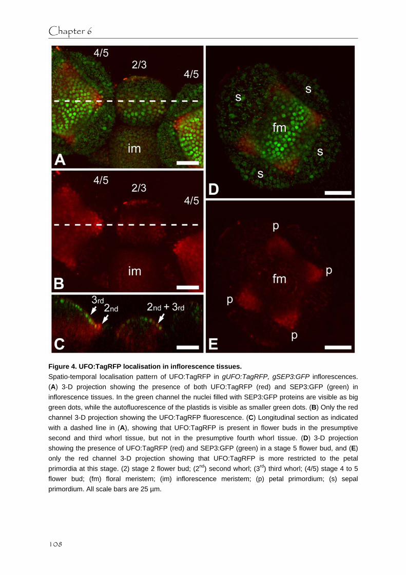
Chapter 6
Figure 4. UFO:TagRFP localisation in inflorescence tissues. Spatio-temporal localisation pattern of UFO:TagRFP in gUFO:TagRFP, gSEP3:GFP inflorescences. (A) 3-D projection showing the presence of both UFO:TagRFP (red) and SEP3:GFP (green) in inflorescence tissues. In the green channel the nuclei filled with SEP3:GFP proteins are visible as big green dots, while the autofluorescence of the plastids is visible as smaller green dots. (B) Only the red channel 3-D projection showing the UFO:TagRFP fluorescence. (C) Longitudinal section as indicated with a dashed line in (A), showing that UFO:TagRFP is present in flower buds in the presumptive second and third whorl tissue, but not in the presumptive fourth whorl tissue. (D) 3-D projection showing the presence of UFO:TagRFP (red) and SEP3:GFP (green) in a stage 5 flower bud, and (E) only the red channel 3-D projection showing that UFO:TagRFP is more restricted to the petal primordia at this stage. (2) stage 2 flower bud; (2nd) second whorl; (3rd) third whorl; (4/5) stage 4 to 5 flower bud; (fm) floral meristem; (im) inflorescence meristem; (p) petal primordium; (s) sepal primordium. All scale bars are 25 µm.
108

SEP3, black holes and UFO
Figure 5. SEP3:GFP localisation in ufo mutant background. Localisation of SEP3:GFP in a gSEP3:GFP, ufo-2 flower bud of approximately stage 5. SEP3:GFP protein in the nuclei is visible as big green dots, while the red dye FM4-64 stains the cell membranes. (A) 3-D projection of the flower bud showing strong SEP3:GFP signal in all three cell layers in the floral meristem (fm) and in the epidermis of the sepal primordia (s). (B-D) Progressive optical sections through the flower bud in (A) from top to bottom, indicating clusters of cells where SEP3:GFP is almost absent (arrows). Scale bar is 25 µm. with the previously reported mRNA pattern, although the withdrawal of UFO:TagRFP protein to the petal primordia seemed to occur a little bit later in development than the withdrawal of the UFO mRNA to the petal primordia [18]. These localisations demonstrate that UFO protein is indeed present in the second and third whorl tissues where more cytoplasmic SEP3:GFP is detected and during the formation of SEP3:GFP black holes in the developing petals and stamens.
109

Chapter 6
To determine if the absence of UFO has an effect on the subcellular localisation of SEP3:GFP or the formation of the SEP3:GFP black holes, we analyzed the presence and subcellular localisation of SEP3:GFP in a ufo-2 mutant background. Ufo-2 flowers have impaired petal and stamen development and commonly have a variety of second and third whorl organs with mixed identities or homeotic conversions, such as sepals, petaloid organs, petals, filamentous organs, staminoid organs, stamens and carpelloid organs [17]. Additionally, the fourth whorl gynoecium often consists of more than two carpels. The localisation pattern of SEP3:GFP in the ufo-2 inflorescence meristem and the early flower bud stages was similar to the localisation pattern of SEP3:GFP in a wild type background (Figure 1A,C,E) [9]. However, in ufo-2 flower buds of approximately stage 4/5 there was less cytoplasmic SEP3:GFP visible in the second and third whorl cell layers (Figure 5). In ufo-2 flower buds of approximately stage 5, when petal and stamen primordia are visible in wild type flowers, there were often no distinct bulges of second and third whorl organ primordia visible in the floral meristem. However, at these positions in the floral meristem we did observe clusters of cells with less SEP3:GFP signal (Figure 5B-D), resembling the black hole phenotype described for SEP3:GFP during wild type flower development. This suggests that UFO is not involved in the formation of SEP3:GFP black holes during second and third whorl development. However, UFO could be involved in the change in subcellular localisation of SEP3:GFP, since SEP3:GFP protein in the ufo-2 mutant plants seemed to be less cytoplasmically localized in the second and third whorl than in wild type plants. To study this in more detail we analyzed the spatio-temporal localisation pattern of SEP3:GFP in a pCaMV35S::UFO background. pCaMV35S::UFO flowers develop supernumerary petals and stamens at the expense of sepals and carpels [18]. No obvious difference was found between the spatio-temporal localisation pattern of SEP3:GFP in a pCaMV35S::UFO background (Figure 6) and in a wild type background (Figure 1A,C,E) [9]. This indicates that UFO does not influence the subcellular localisation of SEP3:GFP protein during petal and stamen development. Discussion In Arabidopsis thaliana the E-type SEPALLATA MADS domain transcription factors play an important role as ‘glue’ proteins in the different multimeric MADS domain protein complexes that specify the identity of the four different floral organs, namely sepals, petals, stamens and carpels [4-8]. During the initiation of petal and stamen primordia, the spatio-temporal localisation pattern of a GFP-tagged genomic SEP3 clone shows an unexpected SEP3:GFP pattern when compared to the previously described SEP3 mRNA pattern [1, 9]. In contrast to the uniform SEP3 mRNA expression in early stages of petal and stamen development, SEP3:GFP protein becomes restricted to the epidermal cell layer of initiating petal and stamen primordia and disappears in the subepidermal and inner cell layers. We named this phenomenon SEP3:GFP ‘black holes’. Prior to the development of these SEP3:GFP black holes, the subcellular localisation of SEP3:GFP in cells of the presumptive second and third whorl changes suddenly from predominantly nuclear to more cytoplasmically localized and
110

SEP3, black holes and UFO
Figure 6. SEP3:GFP localisation in constitutive UFO overexpressing plants. Spatio-temporal localisation pattern of SEP3:GFP in gSEP3:GFP, pCaMV35S::UFO plants. The nuclei filled with SEP3:GFP proteins are visible as big green dots, while the autofluorescence of the plastids is visible as smaller green dots. (A) 3-D projections of a stage 4 flower bud and (B) a stage 5 flower bud with initiating petal and stamen primordia. (C) Optical section through the stage 4 flower bud from (A), demonstrating that in third whorl cell layers the SEP3:GFP protein is more cytoplasmically localized than in fourth whorl cell layers. (D) Optical section through the stage 5 flower bud from (B), showing the presence of SEP3:GFP black holes in the developing stamen primordia. (*) black hole; (3) whorl 3; (4) whorl 4; (fm) floral meristem; (p) petal primordium; (s) sepal primordium; (st) stamen primordium. All scale bars are 25 µm.
111

Chapter 6
remains that way throughout later developmental stages. Assuming that the spatio-temporal localisation pattern of SEP3:GFP is a good reflection of the localisation pattern of endogenous SEP3, the black holes would suggest that after primordium initiation SEP3 is required mostly in the epidermis of the developing organs and less in the subepidermal and inner cell layers. The predominant SEP3:GFP localisation in the epidermis of older petals, stamens, but also the gynoecium and sepals, is in agreement with the hypothesis that SEP3 is mainly active in the epidermal cell layer during floral organ outgrowth and differentiation [9] (unpublished data).
The fact that SEP3 mRNA expression is uniform during early petal and stamen development [1] while gSEP3:GFP and also pCaMV35S::SEP3:GFP plants show SEP3:GFP black holes, suggests that the SEP3:GFP black holes are caused by post-transcriptional processes. However, in the case of the pCaMV35S::SEP3:GFP plants it cannot be excluded that the black holes appear due to the lack of strong expression from the CaMV35S promoter in particular stages of petal and stamen development [28]. Since we have no indication that SEP3 presence is controlled through post-transcriptional inhibition by miRNAs [30, 31], we focused on post-translational processes that could cause the SEP3:GFP black holes. One explanation for the formation of the SEP3:GFP black holes could be the targeted degradation of SEP3 proteins in the subepidermal and inner cell layers in the second and third whorl tissues by the 26s proteasome from flower bud stage 4 onwards. Unfortunately, we were not able to show involvement of the 26s proteasome in the formation of the black holes in the experiments where we treated inflorescences with proteasome inhibitors. An alternative explanation for the formation of SEP3:GFP black holes could be that the sudden change to more cytoplasmically localized SEP3:GFP proteins from flower bud stage 3 onwards makes the SEP3:GFP proteins available for intercellular transport towards the epidermal cell layer [32], where they accumulate.
One known regulator of petal and stamen development that could potentially alter the behaviour of a protein through ubiquitin modification and mark proteins for degradation by the 26s proteasome, is the F-box protein UFO that is thought to be a subunit of an SCF E3 ubiquitin ligase complex [20-22]. We therefore investigated whether the sudden change in SEP3:GFP subcellular localisation and the formation of SEP3:GFP black holes during petal and stamen development is due to UFO-mediated modifications of SEP3 proteins. The spatio-temporal localisation pattern of a TagRFP-tagged genomic UFO clone demonstrated that UFO protein is indeed present in flower buds from stage 2/3 onwards in the presumptive second and third whorl tissues from which the petals and stamen will develop. However, UFO does not seem to be required for the formation of SEP3:GFP black holes, since SEP3:GFP localisations in a ufo-2 mutant background still showed clusters of cells with less SEP3:GFP in the positions where second and third whorl organs would eventually arise. On the other hand, there was less cytoplasmically localized SEP3:GFP in these ufo-2 mutant plants, which could indicate that UFO does play a role in the subcellular localisation of SEP3. However, the localisations of SEP3:GFP in a pCaMV35S::UFO background did not reveal overall increased cytoplasmic SEP3:GFP localisation, which would be expected if UFO indeed regulates the cytoplasmic localisation of SEP3 proteins. Therefore, our experiments
112

SEP3, black holes and UFO
did not provide evidence for a direct role of UFO in the sudden change in SEP3:GFP subcellular localisation and the formation of SEP3:GFP black holes during petal and stamen development. Material and methods Plant materials and growth conditions All Arabidopsis thaliana plant lines were grown at 22 ºC in growth chambers under a long-day light regime (16 h light/8 h dark). The previously described gSEP3:GFP (pARC423) plant line [27] was crossed into the ufo-2 mutant line N6294 [17, 18]. Homozygous ufo-2 mutant plants carrying the gSEP3:GFP construct were selected from the F2 progeny based on the presence of ufo-2 mutant phenotypes and the presence of GFP fluorescence. The gUFO:TagRFP construct (CZN695) and the gAG:TagRFP construct (CZN690) were transformed into Columbia-0 (Col-0) wild type plants and gSEP3:GFP expressing plants. The pCaMV35S::UFO (CZN684) construct was transformed into the Col-0 wild type plant line and the gSEP3:GFP plant line. Additionally, we used two plant lines expressing pCaMV35S::AG:GFP (pARC276) and pCaMV35S::SEP3:GFP (pARC277) in sgs2-1 mutant background that were previously described in chapter 5. Construction of binary vectors and plant transformation To generate a TagRFP-tagged genomic clone of UFO, a 6152 bp genomic fragment was made from Col-0 gDNA by proofreading PCR with the primer PDS1274 (5’-TCAAGGATGTTACCGGAGAG-3’) 4826 bp upstream of the START codon and the primer PDS1269 (5’-ACAGACTCCAGGAAATGGAAG-3’) immediately upstream of the STOP codon. After adding a T/A overhang, this genomic fragment was introduced into the pCR8/GW/TOPO TA vector by using the pCR8/GW/TOPO TA Cloning kit (Invitrogen), after which the right orientation of the genomic UFO insert was ensured. An LR reaction between this gUFO entry clone and the destination vector CZN652 containing the expression cassette GW_TagRFP _NOS terminator, created the expression clone gUFO:TagRFP (CZN695). A similar TagRFP-tagged genomic clone of AG (CZN690) was created by LR reaction between a gAG entry clone (pARC353) and the same destination vector CZN652. These two expression clones were introduced into Agrobacterium tumefaciens strain GV3101 and transformed to Col-0 wild type plants and gSEP3:GFP plants by floral dip method [33].
An untagged overexpressor UFO construct was created by LR reaction between the UFO cDNA clone CZN300 (pENTR221-AT1G30950 from ABRC DNA Stock Center) and the destination vector pARC959 (pB7WG2,0) [34]. The resulting expression clone pCaMV35S::UFO (CZN684) was introduced into Agrobacterium tumefaciens strain GV3101 and transformed to Col-0 wild type plants and gSEP3:GFP plants by floral dip method. Confocal laser scanning microscopy To observe the localisation of the GFP-tagged protein in rosette leaves, inflorescence meristems and floral meristems, plant tissues were dissected and embedded as previously
113

Chapter 6
described [9, 27]. Confocal Laser Scanning Microscopy (CLSM) of the living plant tissue was performed with a Leica SPE DM5500 upright microscope with a 63x ACS APO (NA 1.15 CORR) lens, using the LAS AF 1.8.2 software. The fluorescent tag GFP was excited with the 488 nm solid state laser and the emission scanned at a bandwidth of 505-530 nm. The red dye FM4-64 (Molecular Probes) was excited with the 488 nm solid state laser and the emission scanned at a bandwidth of 600-700 nm. The fluorescent tag TagRFP was excited with the 532 nm solid state laser and the emission scanned at a bandwidth of 560-650 nm. The obtained confocal z-stacks were median-filtered and converted to 3-D maximum projections or cross section images. All images were adjusted with Adobe Photoshop version 5.0. Proteasome inhibitor experiments Inflorescences from gSEP3:GFP plants, pCaMV35S::SEP3:GFP and pCaMV35S::AG:GFP plants were treated with three proteasome inhibitors, namely MG132 and the two BODIPY-labelled epoxomicin analogs MVB003 and MV151 [29]. After initial experimentation with different incubation methods, the preferred method was to submerge dissected inflorescences in different proteasome inhibitor solutions in multiwell plates. The inflorescences were prepared by removing the older flower buds until the relevant flower buds and inflorescence meristem were visible, while a long enough stem was kept for easy handling of the samples. After incubation in the proteasome inhibitor solutions the inflorescence samples were further prepared for imaging as previously described [27]. The three proteasome inhibitor solutions were made in tap water or 0.5 x MS, and ranged in concentration from 0.5 µM to 500 µM. To reduce the surface tension of the solutions and aid the infiltration of the proteasome inhibitors into the inflorescence samples the surfactants Triton X-100, Tween-20 and Tween-80 were also sometimes added to the proteasome inhibitor solutions at an end concentration of 0.03%. Inflorescences in the same solution without proteasome inhibitor were used as control samples. The incubation times ranged from 30 minutes to six hours. Acknowledgements We gratefully acknowledge Martijn Verdoes from Leiden University for providing the BODIPY labelled proteasome probes MVB003 and MV151.
114

SEP3, black holes and UFO
References 1. Mandel MA, Yanofsky MF: The Arabidopsis AGL9 MADS box gene is expressed in young
flower primordia. Sexual Plant Reproduction 1998, 11(1):22-28. 2. Ditta G, Pinyopich A, Robles P, Pelaz S, Yanofsky MF: The SEP4 gene of Arabidopsis
thaliana functions in floral organ and meristem identity. Current Biology 2004, 14(21):1935-1940.
3. Pelaz S, Ditta GS, Baumann E, Wisman E, Yanofsky MF: B and C floral organ identity functions require SEPALLATA MADS-box genes. Nature 2000, 405(6783):200-203.
4. Immink RGH, Tonaco IAN, de Folter S, Shchennikova A, van Dijk ADJ, Busscher-Lange J, Borst JW, Angenent GC: SEPALLATA3: the 'glue' for MADS box transcription factor complex formation. Genome Biology 2009, 10(2).
5. Kaufmann K, Muino JM, Jauregui R, Airoldi CA, Smaczniak C, Krajewski P, Angenent GC: Target Genes of the MADS Transcription Factor SEPALLATA3: Integration of Developmental and Hormonal Pathways in the Arabidopsis Flower. Plos Biology 2009, 7(4):854-875.
6. Melzer R, Theissen G: Reconstitution of floral quartets in vitro involving class B and class E floral homeotic proteins. Nucleic Acids Research 2009, 37(8):2723-2736.
7. Theissen G, Saedler H: Plant biology - Floral quartets. Nature 2001, 409(6819):469-471. 8. de Folter S, Immink RGH, Kieffer M, Parenicova L, Henz SR, Weigel D, Busscher M, Kooiker
M, Colombo L, Kater MM et al: Comprehensive interaction map of the Arabidopsis MADS box transcription factors. Plant Cell 2005, 17(5):1424-1433.
9. Urbanus SL, de Folter S, Shchennikova AV, Kaufmann K, Immink RGH, Angenent GC: In planta localisation patterns of MADS domain proteins during floral development in Arabidopsis thaliana. BMC Plant Biology 2009, 9.
10. Smyth DR, Bowman JL, Meyerowitz EM: Early flower development in Arabidopsis. Plant Cell 1990, 2(8):755-767.
11. Jack T, Brockman LL, Meyerowitz EM: The homeotic gene APETALA3 of Arabidopsis thaliana encodes a MADS box and is expressed in petals and stamens. Cell 1992, 68(4):683-697.
12. Goto K, Meyerowitz EM: Function and regulation of the Arabidopsis floral homeotic gene PISTILLATA. Genes & Development 1994, 8(13):1548-1560.
13. Weigel D, Meyerowitz EM: Activation of floral homeotic genes in Arabidopsis. Science 1993, 261(5129):1723-1726.
14. Coen ES, Meyerowitz EM: The war of the whorls - Genetic interactions controlling flower development. Nature 1991, 353(6339):31-37.
15. Gutierrez-Cortines ME, Davies B: Beyond the ABCs: ternary complex formation in the control of floral organ identity. Trends in Plant Science 2000, 5(11):471-476.
16. Ingram GC, Goodrich J, Wilkinson MD, Simon R, Haughn GW, Coen ES: Parallels between UNUSUAL FLORAL ORGANS and FIMBRIATA, genes controlling flower development in Arabidopsis and Antirrhinum. Plant Cell 1995, 7(9):1501-1510.
17. Levin JZ, Meyerowitz EM: UFO - an Arabidopsis gene involved in both floral meristem and floral organ development. Plant Cell 1995, 7(5):529-548.
18. Lee I, Wolfe DS, Nilsson O, Weigel D: A LEAFY co-regulator encoded by UNUSUAL FLORAL ORGANS. Current Biology 1997, 7(2):95-104.
19. Chae E, Tan QKG, Hill TA, Irish VF: An Arabidopsis F-box protein acts as a
115

Chapter 6
transcriptional co-factor to regulate floral development. Development 2008, 135(7):1235-1245.
20. Samach A, Klenz JE, Kohalmi SE, Risseeuw E, Haughn GW, Crosby WL: The UNUSUAL FLORAL ORGANS gene of Arabidopsis thaliana is an F-box protein required for normal patterning and growth in the floral meristem. Plant Journal 1999, 20(4):433-445.
21. Zhao DZ, Yu QL, Chen M, Ma H: The ASK1 gene regulates B function gene expression in cooperation with UFO and LEAFY in Arabidopsis. Development 2001, 128(14):2735-2746.
22. Ni WM, Xie DX, Hobbie L, Feng BM, Zhao DZ, Akkara J, Ma H: Regulation of flower development in Arabidopsis by SCF complexes. Plant Physiology 2004, 134(4):1574-1585.
23. Callis J, Vierstra RD: Protein degradation in signaling. Current Opinion in Plant Biology 2000, 3(5):381-386.
24. Sullivan JA, Shirasu K, Deng XW: The diverse roles of ubiquitin and the 26S proteasome in the life of plants. Nature Reviews Genetics 2003, 4(12):948-958.
25. Souer E, Rebocho AB, Bliek M, Kusters E, de Bruin RAM, Koes R: Patterning of inflorescences and flowers by the F-box protein DOUBLE TOP and the LEAFY homolog ABERRANT LEAF AND FLOWER of petunia. Plant Cell 2008, 20(8):2033-2048.
26. Hicke L: Protein regulation by monoubiquitin. Nature Reviews Molecular Cell Biology 2001, 2(3):195-201.
27. de Folter S, Urbanus SL, van Zuijlen LG, Kaufmann K, Angenent GC: Tagging of MADS domain proteins for chromatin immunoprecipitation. BMC Plant Biology 2007, 7.
28. Jenik PD, Irish VF: Regulation of cell proliferation patterns by homeotic genes during Arabidopsis floral development. Development 2000, 127(6):1267-1276.
29. Verdoes M, Florea BI, Menendez-Benito V, Maynard CJ, Witte MD, Van der Linden WA, Van den Nieuwendijk A, Hofmann T, Berkers CR, van Leeuwen FWB et al: A fluorescent broad-spectrum proteasome inhibitor for labeling proteasomes in vitro and in vivo. Chemistry & Biology 2006, 13(11):1217-1226.
30. Voinnet O: Origin, Biogenesis, and Activity of Plant MicroRNAs. Cell 2009, 136(4):669-687.
31. Ehrenreich IM, Purugganan MD: Sequence variation of microRNAs and their binding sites in Arabidopsis. Plant Physiology 2008, 146(4):1974-1982.
32. Crawford KM, Zambryski PC: Subcellular localization determines the availability of non-targeted proteins to plasmodesmatal transport. Current Biology 2000, 10(17):1032-1040.
33. Clough SJ, Bent AF: Floral dip: a simplified method for Agrobacterium-mediated transformation of Arabidopsis thaliana. Plant Journal 1998, 16(6):735-743.
34. Karimi M, Inze D, Depicker A: GATEWAY vectors for Agrobacterium-mediated plant transformation. Trends in Plant Science 2002, 7(5):193-195.
116

Chapter 7 Concluding remarks and perspectives

Chapter 7
MADS domain transcription factors The MADS domain transcription factor family plays an important regulatory role in the development of angiosperm flowers. Although previous studies report the mRNA expression patterns of most of the MADS box genes involved in Arabidopsis thaliana floral development, often the more important information on the proteins encoded by these MADS box genes is missing. Protein localisation patterns can differ substantially from mRNA expression patterns, due to post-transcriptional and post-translational processes such as translational inhibition by miRNAs, protein degradation, and intercellular protein transport [1-3]. Intercellular transport of proteins and other macromolecules is thought to be mediated by dynamic channels that connect neighbouring cells, called plasmodesmata [4, 5]. Only recently it has been realized that transport of non-cell-autonomous proteins through plasmodesmata is a highly dynamic process that can play an important regulatory role in developmental processes. The only two known intercellularly transported MADS domain proteins are the B-type proteins DEFICIENS (DEF) and GLOBOSA (GLO) in Antirrhinum majus that are able to move towards the epidermis in the floral meristem [6]. Intriguingly, their respective Arabidopsis thaliana orthologues APETALA3 (AP3) and PISTILLATA (PI) do not seem to have the same ability [7]. Moreover, AP3, PI and also the A-type protein APETALA1 (AP1) seem to lack the ability for inward movement in floral meristems [7-9]. Nevertheless, it has been suggested that certain MADS domain transcription factors have non-cell-autonomous functions, such as the C-type protein AGAMOUS (AG) in floral meristem integrity [10-12] and FRUITFULL (FUL) in pistil development [13]. Tagging of MADS domain proteins Since the discovery of the GREEN FLUORESCENT PROTEIN (GFP) and the subsequent development of similar fluorescent tags, it has become possible to observe the subcellular localisation and behaviour of fluorescently-tagged proteins in living tissues with confocal laser scanning microscopy [14-16]. In this thesis we show that fluorescent tagging of MADS domain proteins for in planta imaging should be used with some caution. Adding a fluorescent tag or a small peptide tag to MADS domain proteins easily leads to transgene silencing, which often results in specific loss-of-function mutant phenotypes. This is not only the case with constructs overexpressing tagged MADS box gene versions, but also with tagged MADS box gene constructs that are expressed under the control of the endogenous regulatory elements by using full genomic clones [17-20]. In the case of GFP-tagged MADS box gene constructs this silencing problem could be circumvented by using plants with an sgs2-1 mutant background that are defective in gene silencing [21, 22]. However, with the photoconvertible mEosFP-tagged MADS box gene constructs even the sgs2-1 mutant background did not help in the recovery of stable primary transformants.
Of course it should be taken into account that the presence of a tag can interfere with the functioning of the protein. We demonstrated that C-terminal GFP-tagged MADS domain proteins in stable transgenic plants are functional and able to complement their respective
118

Concluding remarks and perspectives
mutant backgrounds, but we cannot exclude that the GFP tag interferes with certain functions or in specific complexes. Especially if several MADS domain proteins in a multimeric complex are tagged, it is possible that this will hinder the functioning or even the formation of the complex. For instance, we provide some evidence that the presence of both GFP-tagged AP3 and GFP-tagged PI in one complex might make this complex non-functional. Nevertheless, when the tag is thoughtfully placed and stable transgenic plants are obtained, in planta imaging of fluorescently-tagged proteins by confocal laser scanning microscopy is a valuable tool to investigate protein behaviour during development. Intercellular transport of MADS domain proteins The spatio-temporal localisation patterns of GFP-tagged AG, AP1, FUL, and the E-type protein SEPALLATA3 (SEP3) during floral development demonstrated that there are several tissues, often the epidermal cell layer, where MADS domain proteins could be detected while the available literature describes an absence of mRNA in those tissues. In plants that specifically overexpress GFP-tagged MADS domain proteins in the epidermal cell layer, we were able to demonstrate with a photobleaching technique that AG, SEP3, and the obligate heterodimer AP3/PI are all able to move at least short distances within the epidermal cell layer. This lateral epidermal movement of MADS domain proteins, which seems to be passive diffusion through primary plasmodesmata that connect the epidermal cells, provides an explanation for most of the unexpected MADS domain protein localisations that we found in the spatio-temporal localisation analyses. The amount of passive diffusion of MADS domain proteins that occurs, might be limited by nuclear targeting and sequestration of dimeric MADS domain protein complexes [23-25]. For instance, in roots it was shown that the movement of the GRAS transcription factor SHORT ROOT (SHR) from the stele is limited by nuclear sequestration through the interaction with the GRAS transcription factor SCARECROW (SCR) in the adjacent cell layer [26-28]. According to the extended ‘ABC’ model, the homeotic MADS domain transcription factors are active in clearly defined regions in the floral meristem [29-35]. If MADS domain proteins are allowed to flow freely through the whole epidermis, this could indicate that they lack the capacity to change organ identity from this layer. Supporting this hypothesis is the fact that epidermal-expressed GFP-tagged AP3 is not sufficient for full complementation of the ap3 mutant.
Additionally, we demonstrated that epidermis-expressed GFP-tagged AG is able to move from the epidermis to the subepidermis in the centre of the floral meristem. This observation and the demonstrated lateral movement of AG in the epidermis of the floral meristem provide proof for the previous suggestions that AG acts non-cell-autonomously [10-12]. The transport of AG from the epidermis to the subepidermis through the secondary plasmodesmata that connect these two cell layers seems to be an active process. Perhaps the AG protein possesses a domain that enables it to interact with (secondary) plasmodesmata, while SEP3, AP3 and PI proteins lack this domain. For KNOX transcription factors, which can actively enlarge the aperture of plasmodesmata to allow the passage of large molecules, it was shown that the homeodomain is essential for the interaction with the plasmodesmata [36-39].
119

Chapter 7
Interestingly, the KNOX transcription factor SHOOTMERISTEMLESS (STM), which can actively transport inwards in floral meristems and is involved in AG upregulation in the inner two whorls of the floral meristem, is specifically expressed in the tissues where we observed AG transport [37, 40-42]. It is therefore tempting to speculate that inward AG transport in the floral meristem is mediated by interactions with STM.
Intercellular transport might also play a role in the observed SEP3 behaviour during petal and stamen development. The spatio-temporal localisation pattern of GFP-tagged SEP3 showed that during early stages of petal and stamen development the GFP-tagged SEP3 presence reduced greatly in the subepidermal and inner cell layers, while the mRNA expression in these stages is described as uniform [43]. This seems to imply that SEP3-containing MADS domain protein complexes are needed in all three cell layers only during the initiation of petal and stamen primordia, while epidermal SEP3 presence might suffice for further development and outgrowth. The absence of GFP-tagged SEP3 protein in the subepidermal and inner cell layers in developing primordia, which we named ‘black holes’, could be caused at a post-translational level by targeted SEP3 protein degradation. However, the GFP-tagged SEP3 proteins change their subcellular localisation from predominantly nuclear to more cytoplasmic just prior to the initiation of the petal and stamen primordia. This change to a more cytoplasmic localisation might make the GFP-tagged SEP3 proteins more available for intercellular transport towards the epidermis, where they accumulate [26, 44]. If intercellular transport of SEP3 proteins towards the epidermis is indeed the cause of the black holes, it would indicate that the initiating petal and stamen primordia do not receive SEP3 input from surrounding SEP3-containing tissues. Symplastically isolated primordia can be created by changing the gating properties of the plasmodesmata on the borders of the primordium [4, 45-47]. These symplastic borders could also play a role in limiting the observed lateral transport in the epidermis.
Although intercellular transport of MADS domain proteins in meristems might be only short distanced and often occurs in tissues where the genes themselves are expressed, this does not necessarily imply that this intercellular transport is not biologically relevant. On the contrary, the non-cell-autonomous action of some MADS domain proteins combined with the autoregulatory loops that many MADS proteins have [48-51] could be essential in establishing the observed gene expression patterns during floral development. Additionally, intercellular transport might be needed for the establishment of MADS domain transcription factor gradients that in a concentration-dependent way either initiate different developmental programs as a master regulator, or induce the differentiation of specific tissues as a morphogen [52]. Whether MADS domain transcription factors acquire a different or additional function by going through the process of plasmodesmal transportation is unclear at this moment [53]. Perspectives To complement the performed research on MADS domain protein transport in the floral meristem, it would be good to test whether MADS domain proteins are also able to move
120

Concluding remarks and perspectives
laterally within the subepidermal cell layer or the inner cell layer, and whether they can move from these cell layers towards the epidermal cell layer as was shown for DEF and GLO in Antirrhinum majus [6]. The promoter sequence of WUSCHEL (WUS) or the promoter sequence of CLAVATA1 (CLV1) [54, 55] could for instance be used to specifically express GFP-tagged MADS domain proteins in the centre of the floral meristem in the subepidermal and inner cell layers. This approach might also demonstrate whether there is a limit to the lateral transport in these cell layers, because it is conceivable that developing floral organ primordia represent symplastic domains that are (partially) isolated from MADS domain protein input from surrounding tissues. To investigate the possible role of intercellular SEP3 transport in developing petal and stamen primordia it might be useful to express fluorescently-tagged SEP3 proteins specifically in these developing petal and stamen primordia, for instance under the control of the UNUSUAL FLORAL ORGANS (UFO) promoter [56, 57]. To demonstrate SEP3 transport towards the epidermis, a GFP tag could be used in combination with a photobleaching technique, or photoconvertible fluorescent tags could be used. However, to make this approach successful there should be sufficient precision in the confocal microscopic set-up to target only the desired cell layers for photobleaching or photoconversion, respectively. Additionally, for AG it might be worthwhile to investigate whether active AG transport between cell layers in the floral meristem is indeed mediated by interactions with the KNOX transcription factor STM, by determining the degree of AG transport between cell layers under varying amounts of STM and associated proteins.
In conclusion, the work performed in this project shows the importance of studying transcription factor localisation and dynamics at the protein level in living tissues. Furthermore, the fluorescent tag GFP has highlighted various aspects of MADS domain protein functioning that we currently do not fully understand. The challenge for the near future will be to determine the biological relevance of the observed MADS domain protein behaviour and movement.
121

Chapter 7
References 1. Voinnet O: Origin, Biogenesis, and Activity of Plant MicroRNAs. Cell 2009, 136(4):669-
687. 2. Kurata T, Okada K, Wada T: Intercellular movement of transcription factors. Current
Opinion in Plant Biology 2005, 8(6):600-605. 3. Vierstra RD: The ubiquitin-26S proteasome system at the nexus of plant biology. Nature
Reviews Molecular Cell Biology 2009, 10(6):385-397. 4. Lucas WJ, Ham LK, Kim JY: Plasmodesmata - bridging the gap between neighboring
plant cells. Trends in Cell Biology 2009, 19(10):495-503. 5. Zambryski P: Plasmodesmata. Current Biology 2008, 18(8):R324-R325. 6. Perbal MC, Haughn G, Saedler H, Schwarz-Sommer Z: Non-cell-autonomous function of
the Antirrhinum floral homeotic proteins DEFICIENS and GLOBOSA is exerted by their polar cell-to-cell trafficking. Development 1996, 122(11):3433-3441.
7. Jenik PD, Irish VF: The Arabidopsis floral homeotic gene APETALA3 differentially regulates intercellular signaling required for petal and stamen development. Development 2001, 128(1):13-23.
8. Sessions A, Yanofsky MF, Weigel D: Cell-cell signaling and movement by the floral transcription factors LEAFY and APETALA1. Science 2000, 289(5480):779-781.
9. Wu XL, Dinneny JR, Crawford KM, Rhee Y, Citovsky V, Zambryski PC, Weigel D: Modes of intercellular transcription factor movement in the Arabidopsis apex. Development 2003, 130(16):3735-3745.
10. Sieburth LE, Drews GN, Meyerowitz EM: Non-autonomy of AGAMOUS function in flower development: use of a Cre/loxP method for mosaic analysis in Arabidopsis. Development 1998, 125(21):4303-4312.
11. Jenik PD, Irish VF: Regulation of cell proliferation patterns by homeotic genes during Arabidopsis floral development. Development 2000, 127(6):1267-1276.
12. Cartolano M, Efremova N, Kuckenberg M, Raman S, Schwarz-Sommer Z: Enhanced AGAMOUS expression in the centre of the Arabidopsis flower causes ectopic expression over its outer expression boundaries. Planta 2009, 230(4):857-862.
13. Gu Q, Ferrandiz C, Yanofsky MF, Martienssen R: The FRUITFULL MADS-box gene mediates cell differentiation during Arabidopsis fruit development. Development 1998, 125(8):1509-1517.
14. Haseloff J, Dorman, E.-L., Brand, A.H.: Live imaging with green fluorescent protein, vol. 122: Humana Press; 1999.
15. Sheen J, Hwang SB, Niwa Y, Kobayashi H, Galbraith DW: Green fluorescent protein as a new vital marker in plant-cells. Plant Journal 1995, 8(5):777-784.
16. Chapman S, Oparka KJ, Roberts AG: New tools for in vivo fluorescence tagging. Current Opinion in Plant Biology 2005, 8(6):565-573.
17. Sieburth LE, Meyerowitz EM: Molecular dissection of the AGAMOUS control region shows that cis elements for spatial regulation are located intragenically. Plant Cell 1997, 9(3):355-365.
18. Deyholos MK, Sieburth LE: Separable whorl-specific expression and negative regulation by enhancer elements within the AGAMOUS second intron. Plant Cell 2000, 12(10):1799-1810.
19. Kooiker M, Airoldi CA, Losa A, Manzotti PS, Finzi L, Kater MM, Colombo L: BASIC
122

Concluding remarks and perspectives
PENTACYSTEINE1, a GA binding protein that induces conformational changes in the regulatory region of the homeotic arabidopsis gene SEEDSTICK. Plant Cell 2005, 17(3):722-729.
20. de Folter S, Urbanus SL, van Zuijlen LG, Kaufmann K, Angenent GC: Tagging of MADS domain proteins for chromatin immunoprecipitation. BMC Plant Biology 2007, 7.
21. Elmayan T, Balzergue S, Beon F, Bourdon V, Daubremet J, Guenet Y, Mourrain P, Palauqui J-C, Vernhettes S, Vialle T et al: Arabidopsis mutants impaired in cosuppression. Plant Cell 1998, 10(10):1747-1758.
22. Peragine A, Yoshikawa M, Wu G, Albrecht HL, Poethig RS: SGS3 and SGS2/SDE1/RDR6 are required for juvenile development and the production of trans-acting siRNAs in Arabidopsis. Genes & Development 2004, 18(19):2368-2379.
23. McGonigle B, Bouhidel K, Irish VF: Nuclear localization of the Arabidopsis APETALA3 and PISTILLATA homeotic gene products depends on their simultaneous expression. Genes & Development 1996, 10(14):1812-1821.
24. Immink RG, Gadella TW, Jr., Ferrario S, Busscher M, Angenent GC: Analysis of MADS box protein-protein interactions in living plant cells. Proc Natl Acad Sci U S A 2002, 99(4):2416-2421.
25. Bemer M, Wolters-Arts M, Grossniklaus U, Angenent GC: The MADS domain protein DIANA acts together with AGAMOUS-LIKE80 to specify the central cell in Arabidopsis ovules. Plant Cell 2008, 20(8):2088-2101.
26. Cui HC, Levesque MP, Vernoux T, Jung JW, Paquette AJ, Gallagher KL, Wang JY, Blilou I, Scheres B, Benfey PN: An evolutionarily conserved mechanism delimiting SHR movement defines a single layer of endodermis in plants. Science 2007, 316(5823):421-425.
27. Heidstra R, Welch D, Scheres B: Mosaic analyses using marked activation and deletion clones dissect Arabidopsis SCARECROW action in asymmetric cell division. Genes & Development 2004, 18(16):1964-1969.
28. Sena G, Jung JW, Benfey PN: A broad competence to respond to SHORT ROOT revealed by tissue-specific ectopic expression. Development 2004, 131(12):2817-2826.
29. Coen ES, Meyerowitz EM: The war of the whorls - Genetic interactions controlling flower development. Nature 1991, 353(6339):31-37.
30. Colombo L, Franken J, Koetje E, Vanwent J, Dons HJM, Angenent GC, Vantunen AJ: The Petunia MADS box gene FBP11 determines ovule identity. Plant Cell 1995, 7(11):1859-1868.
31. Pelaz S, Ditta GS, Baumann E, Wisman E, Yanofsky MF: B and C floral organ identity functions require SEPALLATA MADS-box genes. Nature 2000, 405(6783):200-203.
32. Pinyopich A, Ditta GS, Savidge B, Liljegren SJ, Baumann E, Wisman E, Yanofsky MF: Assessing the redundancy of MADS-box genes during carpel and ovule development. Nature 2003, 424(6944):85-88.
33. Ditta G, Pinyopich A, Robles P, Pelaz S, Yanofsky MF: The SEP4 gene of Arabidopsis thaliana functions in floral organ and meristem identity. Current Biology 2004, 14(21):1935-1940.
34. Ferrario S, Immink RG, Angenent GC: Conservation and diversity in flower land. Current Opinion in Plant Biology 2004, 7(1):84-91.
35. Favaro R, Pinyopich A, Battaglia R, Kooiker M, Borghi L, Ditta G, Yanofsky MF, Kater MM, Colombo L: MADS-box protein complexes control carpel and ovule development in Arabidopsis. Plant Cell 2003, 15(11):2603-2611.
123

Chapter 7
36. Lucas WJ, Bouchepillon S, Jackson DP, Nguyen L, Baker L, Ding B, Hake S: Selective trafficking of KNOTTED1 homeodomain protein and its messenger-RNA through plasmodesmata. Science 1995, 270(5244):1980-1983.
37. Kim JY, Yuan Z, Jackson D: Developmental regulation and significance of KNOX protein trafficking in Arabidopsis. Development 2003, 130(18):4351-4362.
38. Winter N, Kollwig G, Zhang S, Kragler F: MPB2C, a microtubule-associated protein, regulates non-cell-autonomy of the homeodomain protein KNOTTED1. Plant Cell 2007, 19(10):3001-3018.
39. Kim JY, Rim Y, Wang J, Jackson D: A novel cell-to-cell trafficking assay indicates that the KNOX homeodomain is necessary and sufficient for intercellular protein and mRNA trafficking. Genes Dev 2005, 19(7):788-793.
40. Long JA, Moan EI, Medford JI, Barton MK: A member of the KNOTTED class of homeodomain proteins encoded by the STM gene of Arabidopsis. Nature 1996, 379(6560):66-69.
41. Scofield S, Dewitte W, Murray JAH: The KNOX gene SHOOT MERISTEMLESS is required for the development of reproductive meristematic tissues in Arabidopsis. Plant Journal 2007, 50(5):767-781.
42. Yu LF, Patibanda V, Smith HMS: A novel role of BELL1-like homeobox genes, PENNYWISE and POUND-FOOLISH, in floral patterning. Planta 2009, 229(3):693-707.
43. Mandel MA, Yanofsky MF: The Arabidopsis AGL9 MADS box gene is expressed in young flower primordia. Sexual Plant Reproduction 1998, 11(1):22-28.
44. Crawford KM, Zambryski PC: Subcellular localization determines the availability of non-targeted proteins to plasmodesmatal transport. Current Biology 2000, 10(17):1032-1040.
45. Rinne PLH, van der Schoot C: Symplasmic fields in the tunica of the shoot apical meristem coordinate morphogenetic events. Development 1998, 125(8):1477-1485.
46. Gisel A, Barella S, Hempel FD, Zambryski PC: Temporal and spatial regulation of symplastic trafficking during development in Arabidopsis thaliana apices. Development 1999, 126(9):1879-1889.
47. Kim I, Kobayashi K, Cho E, Zambryski PC: Subdomains for transport via plasmodesmata corresponding to the apical-basal axis are established during Arabidopsis embryogenesis. Proceedings of the National Academy of Sciences of the United States of America 2005, 102(33):11945-11950.
48. Jack T, Fox GL, Meyerowitz EM: Arabidopsis homeotic gene APETALA3 ectopic expression - transcriptional and posttranscriptional regulation determine floral organ identity. Cell 1994, 76(4):703-716.
49. Schwarz-Sommer Z, Hue I, Huijser P, Flor PJ, Hansen R, Tetens F, Lonnig WE, Saedler H, Sommer H: Characterization of the Antirrhinum floral homeotic MADS-Box gene DEFICIENS - Evidence for DNA-binding and autoregulation of its persistent expression throughout flower development. Embo Journal 1992, 11(1):251-263.
50. Gomez-Mena C, de Folter S, Costa MMR, Angenent GC, Sablowski R: Transcriptional program controlled by the floral homeotic gene AGAMOUS during early organogenesis. Development 2005, 132(3):429-438.
51. Kaufmann K, Muino JM, Jauregui R, Airoldi CA, Smaczniak C, Krajewski P, Angenent GC: Target Genes of the MADS Transcription Factor SEPALLATA3: Integration of Developmental and Hormonal Pathways in the Arabidopsis Flower. Plos Biology 2009, 7(4):854-875.
52. Benkova E, Ivanchenko MG, Friml J, Shishkova S, Dubrovsky JG: A morphogenetic trigger:
124

Concluding remarks and perspectives
is there an emerging concept in plant developmental biology? Trends in Plant Science 2009, 14(4):189-193.
53. Lucas WJ, Lee JY: Plant cell biology - Plasmodesmata as a supracellular control network in plants. Nature Reviews Molecular Cell Biology 2004, 5(9):712-726.
54. Mayer KFX, Schoof H, Haecker A, Lenhard M, Jurgens G, Laux T: Role of WUSCHEL in regulating stem cell fate in the Arabidopsis shoot meristem. Cell 1998, 95(6):805-815.
55. Clark SE, Williams RW, Meyerowitz EM: The CLAVATA1 gene encodes a putative receptor kinase that controls shoot and floral meristem size in Arabidopsis. Cell 1997, 89(4):575-585.
56. Ingram GC, Goodrich J, Wilkinson MD, Simon R, Haughn GW, Coen ES: Parallels between UNUSUAL FLORAL ORGANS and FIMBRIATA, genes controlling flower development in Arabidopsis and Antirrhinum. Plant Cell 1995, 7(9):1501-1510.
57. Lee I, Wolfe DS, Nilsson O, Weigel D: A LEAFY co-regulator encoded by UNUSUAL FLORAL ORGANS. Current Biology 1997, 7(2):95-104.
125

Summary
In this thesis we investigated the behaviour of fluorescently-tagged MADS domain proteins during floral development in the model plant Arabidopsis thaliana, and explored the importance of intercellular transport via plasmodesmata for MADS domain transcription factor functioning. The MADS domain transcription factor family plays an important regulatory role in the development of flowers, among others by establishing the identities of the different floral organs. Although genetic screens and in vitro and in vivo studies on protein-protein and protein-DNA interactions provide important information on how MADS domain transcription factor complexes are able to regulate downstream target genes, understanding of the behaviour of MADS domain transcription factors in planta is still limited. Also, the extent to which intercellular movement of MADS domain transcription factors via plasmodesmata plays a role in developmental processes is poorly understood. Since the discovery of the GREEN FLUORESCENT PROTEIN (GFP) and the subsequent development of similar fluorescent tags, it has become possible to observe the subcellular localisation and behaviour of fluorescently-tagged proteins in living tissues with confocal laser scanning microscopy.
In Chapter 2 of this thesis, different methods of tagging the MADS domain transcription factors AGAMOUS (AG), SEPALLATA3 (SEP3), and FRUITFULL (FUL) for chromatin immunoprecipitation, chromatin affinity purification and in planta imaging are described. This research shows that the addition of a small peptide tag or a fluorescent tag to MADS domain proteins easily leads to transgene silencing and specific loss-of-function mutant phenotypes, especially when the tagged MADS box genes are expressed under the control of the constitutive CaMV35S promoter. Plants that express tagged MADS box genes from genomic fragments that include all or most of the regulatory elements, and therefore mimic the natural expression pattern as much as possible, show lower levels of loss-of-function phenotypes. In addition, these plants are also more useful for investigating biological relevant behaviour of the MADS domain proteins.
In Chapter 3, the spatio-temporal localisation patterns of GFP-tagged MADS domain transcription factors AG, SEP3, FUL and APETALA1 (AP1) during floral development are reported. These analyses demonstrate that there are several tissues, often epidermal cell layers, where MADS domain proteins could be detected, while the available literature describes an absence of mRNA in those tissues. This could indicate that there is intercellular transport of MADS domain proteins in meristematic tissues during floral development. The implications of the observed behaviour of the different MADS domain proteins for MADS domain protein functioning are discussed in this chapter.
In Chapters 4 and 5 we describe the different methods that were used to investigate whether MADS domain proteins are indeed able to transport between cells during floral development. The difficulties that we encountered in our attempts to investigate intercellular MADS domain protein transport with microinjection techniques and by using the photoconvertible fluorescent mEosFP-tag are discussed. In plants that specifically overexpress GFP-tagged MADS domain transcription factors AG, SEP3, APETALA3 (AP3), or PISTILLATA (PI) in the epidermis, we demonstrated with a photobleaching technique that all tested proteins were able to move within the epidermal cell layer. This mechanism of
126

lateral epidermal movement provides an explanation for most of the unexpected MADS domain protein localisations that we found in the spatio-temporal localisation analyses in Chapter 3. Additionally, we demonstrate that epidermis-expressed GFP-tagged AG is able to move from the epidermis to the subepidermis in the centre of the floral meristem, which provides proof for the suggestions that AG acts non-cell-autonomously in the floral meristem. In these plants we also analyzed the effects of epidermal MADS domain protein expression on the plant phenotype. This showed, among others, that epidermis-expressed AG is able to fully complement its own mutant background, while epidermis-expressed AP3 is not.
In Chapter 6, we explore the mechanisms underlying the behaviour of GFP-tagged SEP3 during petal and stamen development that was observed in the spatio-temporal localisation studies described in Chapter 3. Just prior to the initiation of petal and stamen primordia GFP-tagged SEP3 proteins change their subcellular localisation from predominantly nuclear to more cytoplasmic, and at later stages GFP-tagged SEP3 protein seems to disappear in the middle of the primordia without the loss of SEP3 mRNA expression. These two processes could be regulated at a post-transcriptional level by two mechanisms that are discussed, namely 26s proteasome mediated SEP3 protein degradation and epidermal-oriented intercellular transport of SEP3 proteins. Additionally, we demonstrate that there are no clear indications that the observed GFP-tagged SEP3 behaviour is due to the presence of F-box protein UNUSUAL FLORAL ORGANS (UFO), which regulates petal and stamen development.
In Chapter 7, this thesis finishes with some concluding remarks on in planta imaging of MADS domain transcription factors and the possible mechanisms of MADS domain protein movement in the floral meristem. Furthermore, we speculate on the importance of MADS domain protein movement in establishing MADS box gene expression patterns and MADS domain protein gradients, and on the need for symplastically isolated domains for proper floral development.
127

Nederlandse samenvatting
“MADS eiwitten in beweging: een studie naar het functioneren van MADS domein eiwitten tijdens bloemontwikkeling in Arabidopsis thaliana.” Voor dit proefschrift is het gedrag van fluorescent gelabelde MADS domein eiwitten onderzocht tijdens bloemontwikkeling in de model plant Arabidopsis thaliana. Daarbij hebben we ook het belang van intercellulair transport via plasmodesmata voor het functioneren van deze MADS domein transcriptiefactoren verkend. De familie van MADS domein transcriptiefactoren heeft een belangrijke regulerende rol tijdens de ontwikkeling van bloemen, onder andere door de identiteit van de verschillende bloemorganen te bepalen. Genetische onderzoeken en in vitro en in vivo studies op het gebied van eiwit-eiwit en eiwit-DNA interacties hebben veel informatie opgeleverd over hoe MADS domein transcriptiefactor complexen in staat zijn om downstream target genen te reguleren. Echter, over het gedrag van MADS domein transcriptiefactoren in planta en de mate waarin intercellulair transport van transcriptiefactoren via plasmodesmata een rol speelt tijdens ontwikkelingsprocessen is nog maar weinig bekend. Sinds de ontdekking van het fluorescerende eiwit GREEN FLUORESCENT PROTEIN (GFP) en de daaropvolgende ontwikkelingen op het gebied van fluorescente labels, is het mogelijk geworden om het gedrag en de subcellulaire lokalisatie van fluorescent gelabelde eiwitten in levende weefsels te bestuderen met confocale laser scanning microscopie.
In Hoofdstuk 2 van dit proefschrift worden verschillende manieren besproken om de MADS domein transcriptiefactoren AGAMOUS (AG), SEPALLATA3 (SEP3) en FRUITFULL (FUL) te labelen voor chromatine immunoprecipitatie, chromatine affiniteitszuivering en in planta microscopie. Dit onderzoek toont aan dat de toevoeging van een kort peptide label of een fluorescent label aan MADS domein eiwitten makkelijk leidt tot transgen silencing en specifieke loss-of-function mutante fenotypes. Dit is vooral het geval als de gelabelde MADS box genen tot expressie komen onder de controle van de constitutieve CaMV35S promoter. In planten waarbij de gelabelde MADS box genen tot expressie worden gebracht vanaf genomische fragmenten, waardoor de natuurlijke expressiepatronen zoveel mogelijk nagebootst worden, komen deze loss-of-function fenotypes minder vaak voor. Door de nabootsing van de natuurlijke expressiepatronen zijn deze planten ook nuttiger voor het bestuderen van biologisch relevant gedrag van de eiwitten.
In Hoofdstuk 3 worden de lokalisatie patronen in plaats en tijd van de GFP-gelabelde MADS domein transcriptiefactoren AG, SEP3, FUL en APETALA1 (AP1) tijdens de bloemontwikkeling gerapporteerd. Deze analyses laten zien dat in een aantal weefsels, meestal de epidermale cellagen, MADS domein eiwitten aanwezig zijn, terwijl de beschikbare literatuur daar geen mRNA aanwezigheid rapporteert. Dit zou erop kunnen wijzen dat tijdens de bloemontwikkeling in meristematische weefsels intercellulair transport van MADS domein eiwitten plaatsvindt. De implicaties van het waargenomen gedrag van de verschillende MADS domein eiwitten voor hun functioneren worden besproken in dit hoofdstuk.
In Hoofdstuk 4 en 5 beschrijven we de verschillende methoden die gebruikt zijn om te onderzoeken of MADS domein eiwitten inderdaad in staat zijn om tussen cellen te bewegen
128

tijdens de bloemontwikkeling. We bespreken de moeilijkheden die we tegen zijn gekomen bij onze pogingen om intercellulair transport van MADS domein eiwitten te onderzoeken met micro-injectie technieken en door gebruik te maken van het fotoconverteerbare fluorescente mEosFP-label. In planten die de GFP-gelabelde MADS domein transcriptiefactoren AG, SEP3, APETALA3 (AP3) of PISTILLATA (PI) specifiek in de epidermis overproduceren, hebben we met een fotoblekingstechniek gedemonstreerd dat al de geteste eiwitten in staat zijn om in de epidermale cellaag van cel naar cel te bewegen. Dit laterale bewegingsmechanisme geeft een verklaring voor het merendeel van de onverwachte MADS domein eiwit lokalisaties die we gevonden hebben in de gedetaileerde lokalisatie analyses in Hoofdstuk 3. Verder laten we zien dat epidermaal-geproduceerd GFP-gelabeld AG in staat is om van de epidermis naar de subepidermis te bewegen in het midden van het bloemmeristeem. Hiermee is de suggestie dat AG non-cel-autonoom functioneert in het bloemmeristeem voor het eerst van bewijs voorzien. In deze planten hebben we ook de effecten van epidermaal-geproduceerde MADS domain eiwitten op het fenotype van de plant geanalyseerd. Hieruit is onder andere gebleken dat epidermaal-geproduceerd AG in staat is om zijn eigen mutante achtergrond volledig te complementeren, terwijl epidermaal-geproduceerd AP3 dit niet kan.
In Hoofdstuk 6 worden de mechanismen achter het gedrag van GFP-gelabeld SEP3 tijdens bloemblad en meeldraad ontwikkeling onderzocht. Uit het werk beschreven in Hoofdstuk 3 blijkt dat de subcellulaire aanwezigheid van GFP-gelabelde SEP3 eiwitten net voor de initiatie van de bloemblad en meeldraad primordia verandert van hoofdzakelijk kern gelokaliseerd naar meer cytoplasmatisch gelokaliseerd. In latere stadia lijken de GFP-gelabelde SEP3 eiwitten in het midden van de primordia te verdwijnen zonder dat het SEP3 mRNA daar verdwijnt. Deze twee processen zouden op een posttranscriptioneel niveau door twee mechanismen veroorzaakt kunnen worden, namelijk SEP3 eiwitafbraak door het 26s proteasoom en epidermaal-georiënteerd intercellulair transport van SEP3 eiwitten. Deze twee mechanismen worden nader besproken in dit hoofdstuk. Verder laten we zien dat er geen duidelijke aanwijzingen zijn dat het waargenomen gedrag van GFP-gelabeld SEP3 veroorzaakt wordt door de aanwezigheid van het F-box eiwit UNUSUAL FLORAL ORGANS (UFO) dat bloemblad en meeldraad ontwikkeling reguleert.
In Hoofdstuk 7 eindigt dit proefschrift met een aantal concluderende opmerkingen over in planta microscopie en de mogelijke mechanismen van MADS domein eiwit transport in het bloemmeristeem. Verder speculeren we over het belang van MADS domein eiwit transport voor het creëeren van MADS box gen expressiepatronen en MADS domein eiwit gradiënten, als ook over de noodzaak van symplastisch geïsoleerde gebieden voor correcte bloemontwikkeling.
129


131

Acknowledgements
In my life I have had many variations of my name. I started out with the official name Susanna, which was rapidly changed into Susanneke when I was a baby and then shortened to Sanne. At a certain point in my life I foolishly decided to change my name to Susan, because there was this other girl in my class who was also called Sanne. Of course, not long afterwards it turned out there were actually a lot more girls called Susan (or a variation on this spelling). I decided to keep the name, although at home and with old friends I was stilled called Sanne or San. Suddenly people started to call me Suus, which I did not like very much. Several years later, during my PhD time, I had to get used to a nickname that was marginally better than Suus, given to me by a guy with an insatiable appetite for giving everybody a nickname. I tried to fight it, but a lot of people in the lab picked up on this nickname. In the end resistance was futile, so I gave up. Now I am know in the lab as Suzie. Thanks a lot! I would like to thank Gerco Angenent for giving me the opportunity to do my PhD in his Plant Developmental Systems group at Plant Research International in Wageningen. In the beginning of my PhD I was also supervised in the lab by Stefan de Folter and later on Richard Immink became my main supervisor. During this time I learned a lot about being an independent scientist. I want to thank all three of you for the supervision and guidance during these years and I think we got some nice papers out of the research. I would like to thank all the people from the business unit Bioscience for their good advice, help, support and of course the fun and sometimes absurd times in the lab and outside of it. I especially would like to thank all the past and present members of the Plant Developmental Systems group: Andrea, Anneke, Asmini, Camila, Cédric, Cezary, Clarissa, Erica, Froukje, Gerco, Guodong, Huihui, Huong, Isabella, Jan Custers, Jan Kodde, Jacqueline, Jannie, Jennifer, Jeroen van Arkel, Jeroen Peters, John, Kerstin, Kim, Leo, Marco, Marie, Martijn, Merche, Michiel, Mieke, Nayelli, Peter, Priscilla, Richard, Ronny, Rumyana, Ruud, Sela, Silvia, Stefan, Stefano, Steven, Tetty, Tjitske, Tom, Violeta, Wilco, and all the guest workers and students. In the beginning of my PhD I was very glad to get help and advice on some confocal microscopy issues from Jan-Willem Borst and Boudewijn van Veen from the Microspectroscopy Centre in Wageningen, where I did part of my experiments, but also Tijs Ketelaar and Jan Vos from Plant Cell Biology from Wageningen University were very helpful at this point. I also would like to thank Jan Vos for trying some microinjection experiments with me to determine if MADS domain proteins are able to traffic between cells. Later on in my PhD I had assistance on some of my projects from my two students, Q.D. (Peter) Dinh and Zijing (Tina) Liu, and I collaborated with Adriana Martinelli from CENA Universidade de São Paulo and Lilian Aizza and Marcelo Dornelas from Departamento de Biologia Vegetal from Universidade Estadual de Campinas on the pATML1 project. Thank you very much for the hard work, the good times and the sometimes really beautiful results. For the help on my attempts to do immunostainings for SEP3 and in situ’s for pML1::AG:GFP, I would like to
132

thank Mieke Wolters-Arts from the Department of Plant Cell Biology from Radboud University Nijmegen. During the many meetings, conferences and PhD student days I very much enjoyed the company of the people from Plant Research International, Molecular Biology (WU), Biochemistry (WU), Plant Cell Biology (WU), Plant Cell Biology (RU) and Genetics (VU). Additionally, I am very grateful to the LEB fund in Wageningen for the financial support I received for my scientific trips abroad, and to the Max Planck Institut fϋr Ornithologie in Seewiesen for the hospitality during the time I was writing part of my thesis in their guesthouse. Although it was sometimes hard to find the time to visit family and friends during the last five-and-a-half years, I would like to thank you all for the support, advice and good times: Hans & Margriet _ Malene, Claudiu & Lucas _ Ingrid & Jaap _ Hanna _ Jop, Maitso, Damian, Jason, Aiden, Raven & Avalon _ Lien _ Oma _ Theo, Marlies & Miquel _ Marianne & André _ Jeroen & Mariska _ Edith & Jan _ Daan _ Marianne & Ronald _ Stèphanie, Harrie, Noor, Felien & Renée _ Sebastiaan & Anne _ Maartje & Thijs _ Wim & Anneke _ Kee, Pui Yee, Emily, Joyce & Stephany _ Erik & Marit _ Olger-Jan _ Rogier _ Nathalie _ Sidney _ Maarten, Marjolein & Jari _ Yue Xiu. And of course I would like to thank Chiel for going through this with me and for not only being my partner in my personal life, but also my partner in science.
133


Curriculum vitae
Susanna Leonora Urbanus was born on the 25th of December 1978 in Nijmegen, the Netherlands. At VU University in Amsterdam she studied Biology with specialisations in molecular plant biology, plant ecology, and developmental biology. She graduated cum laude in 2004. In this same year she started her part-time PhD in the Plant Developmental Systems group of Prof. Gerco Angenent at Plant Research International in Wageningen, affiliated with the Laboratory of Molecular Biology of Wageningen University. Here she studied MADS domain transcription factor function and movement during floral development in the model plant Arabidopsis thaliana. The results from that study are summarized in this thesis.
135

Susan L. Urbanus19 May 2010Plant Developmental Systems (Bioscience), PRI & Laboratory of Molecular Biology, Wageningen University and Research Centre
date►
Sep 29, 2004► ► ► ►
1.5 credits*
date►
Jun 02, 2005Sep 19, 2006Sep 13, 2007
Oct 01-02, 2008►
Apr 26, 2005May 12, 2006Oct 11, 2007Jan 30, 2009
► Jan 21, 2005Feb 03, 2005Apr 28, 2005
Apr 04-05, 2005Nov 04, 2005
Apr 03-04, 2006Apr 07-08, 2008
Feb 19, 2008►
Nov 04, 2004Sep 26, 2005Oct 24, 2005Apr 13, 2007Nov 26, 2007Apr 14, 2008Jun 26, 2008Sep 11, 2009Feb 02, 2006
2004-2009Dec 21, 2006Feb 28, 2007May 18, 2007Sep 28, 2007
► Nov 26, 2007
► Oct 23-26, 2005
Mar 31-Apr 05, 2007Jul 23-27, 2008
► Oct 23-26, 2005
Feb 10, 2006May 02, 2006Apr 01, 2007May 01, 2007Apr 22, 2008May 28, 2008
Jul 23-27, 2008Oct 02, 2008Jan 30, 2009Apr 14, 2009
► Sep 14, 2007►
22.4 credits*
IAB interviewExcursions
Subtotal Scientific Exposure
1st Leica Confocal User MeetingALW lunteren days 2005Netherlands Proteomic Centre Fall meeting: Protein networks
Montreal, Canada: Poster "In planta localization and dynamics of fluorescently tagged MADS box transcription
Flying seminar Prof Richard Vierstra
Molecular Biology, WU: Inter/intracellular trafficking of MADS box transcription factorsPRI Bioscience thematic meeting: transcription factor trafficking and complex formationSalzburg, Austria: In planta localization and dynamics of MADS box transcription factors
seminar Prof Jan Lohmannseminar Prof Roger Tsien at the MPI for biochemistry in Munich
EPS theme symposia
NWO Lunteren days and other National Platforms2nd Dutch Chromatin Meeting Wageningen
Theme 1 Developmental Biology of Plants, Leiden University
Theme 1 Developmental Biology of Plants, Leiden University
Education Statement of the Graduate SchoolExperimental Plant Sciences
First presentation of your project
Subtotal Start-up Phase
1) Start-up phase
Signal transduction by transcription factors: from synthesis to transcription regulation
Writing a review or book chapterMSc courses
1st joint retreat of the PhD Students in Experimental Plant Sciences in Wageningen
Theme 1 Developmental Biology of Plants, Wageningen University
Grand Opening Netherlands Proteomics Centre Utrecht
Issued to:
Laboratory use of isotopes
Date:Group:
Theme 1 Developmental Biology of Plants, Wageningen University
Writing or rewriting a project proposal
2) Scientific Exposure
PhD student day 2005, Radboud University NijmegenPhD student day 2006, Wageningen UniversityPhD student day 2007, Wageningen University
EPS PhD Student Days
ALW lunteren days 2008
Flying seminar Prof Philip Benfey
EPS Symposium Intracellular SignallingBioscience special seminarspersonal confocal workshop at Leica company in Mannheim
Gargano, Italy: In planta localization of endogenously expressed MADS box transcription factors
ALW lunteren days 2006
Flying seminar Prof Joseph Ecker
Flying seminar Prof Hiroo FukudaFlying seminar Prof Jim Carrington
Progress meeting of Netherlands Proteomics CentreSeminars (series), workshops and symposiaSystems Biology Symposium
Seminar plus Prof. Hiroo FukudaInternational symposia and congressesInternational MADS meeting in Gargnano, Italy8th International Botanical Microscopy Meeting in Salzburg, Austria
personal confocal workshop at Nikon company in BadhoevedorpNVBMB spring symposium organized by Dolf WeijersKNBV Genetics, development and functioning of flowersSeminar plus
19th International Conference on Arabidopsis Research in Montreal, CanadaPresentations
PRI Bioscience thematic meeting: transcription factor localization and movementPRI Bioscience thematic meeting: transcription factor localization and movementWU Biochemistry: Transport and in planta localization of MADS box transcription factors
Wageningen, the Netherlands: MADS transcription factorsLeiden, the Netherlands: Dynamic behaviour of MADS domain proteins in flowersPRI Bioscience thematic meeting: Transcription factor localization and movement

date►
Nov 07-10, 2005Jun 19-21, 2006
► 2004-2009
► 5.1 credits*
date►
Dec 14-15, 2004Dec 16-17, 2004
Oct 13, 2005Oct - Nov, 2006Sep - Nov, 2007May-Jun, 2008Oct 08, 2009Dec 08, 2009
► ►
6.9 credits*
35,9Herewith the Graduate School declares that the PhD candidate has complied with the educational requirements set by theEducational Committee of EPS which comprises of a minimum total of 30 credits
Signalling in plant development and plant defence
Grant writing workshop, Mennen Training & Consultancy
Organisation of PhD students day, course or conference
Time and project management
Career orientation
Scientific publishing workshop
Systems Biology: principle of ~omics data analysis
Subtotal Personal Development
EPS courses or other PhD courses
Skill training courses
PhD scientific writing
4) Personal development
Subtotal In-Depth Studies
Journal clubPlant Developmental Systems Literature DiscussionsIndividual research training
Membership of Board, Committee or PhD council
NWO Talent Class Creative Thinking
3) In-Depth Studies
* A credit represents a normative study load of 28 hours of study
TOTAL NUMBER OF CREDIT POINTS*
Digital scientific artworkAdvanced photoshop

Financial support from Wageningen University for printing this thesis is gratefully acknowledged. Printed by Ipskamp Drukkers, Enschede