macular oct imaging in glaucoma
TRANSCRIPT

Macular OCT Imaging in Glaucoma
Kouros Nouri-Mahdavi, MD, MScAssociate Professor of Ophthalmology
Director, Glaucoma Advanced Imaging LaboratoryStein Eye Institute, UCLA
8th World Glaucoma CongressMarch 27-30, 2019
Melbourne, Australia

Financial Disclosures
• I have the following financial interests or relationships to disclose:– Departmental grant from Research to Prevent Blindness– UCLA Innovation Award 2019– Heidelberg Engineering: Lecture Fees, Grant Support, Hardware and Software Support– Aerie: Ad hoc Advisory Board

2012
2017
Case Review

2012
2017
2012 2017

Why Macula?
Curcio and Allen 1990
Curcio et al. 2011
50% of RGCs
Multilayered GCL

Full macular thickness
FMT
Ganglion cell complexGCC
Ganglion cell/inner plexiform layersGC/IPL
Ganglion cell layerGCL
Macular Outcome Measures

Tan et al. Ophthalmology 2009GCC map
Ganglion Cell Complex (GCC)RTVue/Avanti

Ganglion Cell (GCIPL) Analysis Cirrus HD-OCT
4.8 mm4.
0 m
m
Mwanza et al. IOVS 2011

Posterior Pole Asymmetry Analysis(Spectralis OCT, Full Thickness)
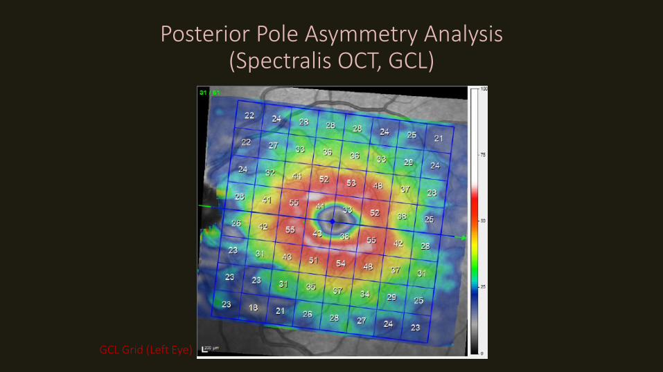
GCL Grid (Left Eye)
Posterior Pole Asymmetry Analysis(Spectralis OCT, GCL)
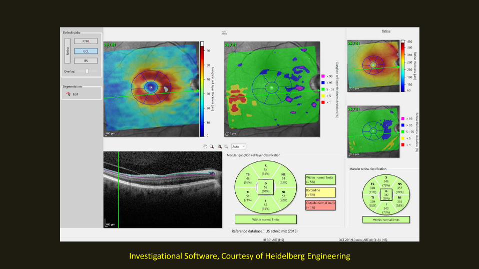
Investigational Software, Courtesy of Heidelberg Engineering
Detection of Progression

Mean baseline MD: -14.3 (±5.5)
Criterion 1 Disc Photos Criterion 2 VFI
Stable Progressed Undetermined P Stable Progressed P
Macular thickness (μm/yr) −0.53 ± 1.44 −4.74 ± 4.40 −2.72 ± 4.75 0.01 −2.22 ± 4.33 −5.12 ± 2.40 0.039
RNFL thickness (μm/yr) −0.33 ± 1.29 −1.19 ± 2.62 −1.21 ± 2.75 0.314 −0.90 ± 2.42 −2.08 ± 2.85 0.459
Glaucoma Progression: RNFL vs. Macular Rates of Progression
Table Reproduced from
Sung KR, Sun JH, Na JH, et al. Progression detection capability of macularthickness in advanced glaucomatous eyes. Ophthalmology 2012; 119:308–313.

Glaucoma Progression: RNFL vs. Full Macular Thickness Event Analysis
Lee et al IOVS 2013 (90% NTG eyes)
average cpRNFLT vs.1 sector in TMT
1 quadrant in cpRNFLT vs. 1 sector in TMT

Add Belghhith here
Advanced glaucoma defined as visual field MD < -21 dB
Belghith et al. IOVS 2016
Improving ImprovingWorse Worse
RNFL GCIPL

16

17

80-year old F with POAG, IOPs <15 on maximal treatment
2011
2015
Follow-up – baseline thickness
Case Review

2012
2015
mRNFLThicknessChange
Humphrey GPA (Glaucoma Progression Analysis)

GCL thickness change over time
Follow-up – baseline thickness
2015
2018

Patterns of Progressive GCIPL Thinning in Glaucoma
Baseline distribution of damage Distribution of progression
Shin JW et al. Ophthalmology 2018
Widening of GCIPL defects: 58% • Deepening of defects: 26%• Newly developed GCIPL defects: 21%

Correspondence of Superpixels and 10-2 Test Locations
3
2
1
Central 18 degrees
Macular superpixels
10-2 test locations

Miraftabi et al. TVST 2016
Within-Session Variability < 3 µm
or GCL
All Macular Outcomes: Very Low Local Within-Session Variability

Miraftabi et al. IOVS 2016
Similar Measurement FloorDynamic range: FMT > GCC > GCIPL > GCL

a C
b
3
2
1
Nouri-Mahdavi et al. ARVO 2017
Within-Eye ModelingCentral 18 degrees
Macular superpixels
10-2 testlocations

Within-Eye Longitudinal Structure-Function Relationships
The ‘Moving Cloud’ Concept

Does Baseline Thickness Affect Rates of Change?
Regress superpixel thickness against time: FMT, GCC, GCIPL, GCL

Prop
ortio
n of
neg
ativ
e ra
tes (
%)
Baseline Thickness, DecilesThinner Thicker
Proportion of Significant Negative Rates vs. Baseline Thickness

GC: The Best Outcome if Outer Retinal Thinning Considered!
Nouri-Mahdavi et al. ARVO 2018

Does Structural Worsening Precede Functional Worsening in Established Glaucoma?
Mohammadzadeh et al.

The Case of Patty B.
• 89 yo F with advanced glaucoma OD• IOPs in low 20s before Trab + MMC• BCVA before surgery: 20/50+2
• BCVA after surgery: 20/80
Mohammadzadeh, Galian, Martinyan, and Nouri-Mahdavi, J Glaucoma 2019


Summary Remarks: Macular OCT Imaging
• Complementary to ONH/RNFL imaging
• Useful for the entire spectrum of glaucoma incl. early glaucoma
• Detection of disease: GCC = GCIPL = GCL
• Excellent reproducibility profile
• Early supporting evidence regarding detection of progression
• GCC: ?optimal outcome measure for monitoring glaucoma at all stages

The Glaucoma Advanced Imaging Laboratory @ Stein

36
Thank You

Detection of Glaucoma
• GCC and GC/IPL > FMT for detection of glaucoma• GCC = GCIPL = GCL

Detection of Glaucoma
• GCC and GC/IPL > FMT for detection of glaucoma• GCC = GCIPL = GCL• Sectoral GC/IPL (or GCC) better than global measures

Detection of Glaucoma
• GCC and GC/IPL > FMT for detection of glaucoma• GCC = GCIPL = GCL• Sectoral GC/IPL (or GCC) better than global measures• RNFL and macular imaging provide complementary information!
– Combining OCT parameters improves performance!

Adopted from Tan et al. Ophthalmology 2009
Perimetric glaucoma Preperimetric glaucoma
65%13% 9% 33%12% 11%
None: 13% None: 45%
RNFL GCC RNFL GCC

68 yr old F with NTG
Case Review