macrophage imbalance (m1 vs. m2) and upregulation of mast ... · pascal jabbour thomas jefferson...
TRANSCRIPT
Thomas Jefferson UniversityJefferson Digital Commons
Department of Neurosurgery Faculty Papers Department of Neurosurgery
1-1-2012
Macrophage imbalance (M1 vs. M2) andupregulation of mast cells in wall of rupturedhuman cerebral aneurysms: preliminary results.David HasanUniversity of Iowa
Nohra ChalouhiThomas Jefferson University, [email protected]
Pascal JabbourThomas Jefferson University, [email protected]
Tomoki HashimotoUniversity of California San Francisco
Let us know how access to this document benefits youFollow this and additional works at: http://jdc.jefferson.edu/neurosurgeryfp
Part of the Neurology Commons, and the Surgery Commons
This Article is brought to you for free and open access by the Jefferson Digital Commons. The Jefferson Digital Commons is a service of ThomasJefferson University's Center for Teaching and Learning (CTL). The Commons is a showcase for Jefferson books and journals, peer-reviewed scholarlypublications, unique historical collections from the University archives, and teaching tools. The Jefferson Digital Commons allows researchers andinterested readers anywhere in the world to learn about and keep up to date with Jefferson scholarship. This article has been accepted for inclusion inDepartment of Neurosurgery Faculty Papers by an authorized administrator of the Jefferson Digital Commons. For more information, please contact:[email protected].
Recommended CitationHasan, David; Chalouhi, Nohra; Jabbour, Pascal; and Hashimoto, Tomoki, "Macrophage imbalance(M1 vs. M2) and upregulation of mast cells in wall of ruptured human cerebral aneurysms:preliminary results." (2012). Department of Neurosurgery Faculty Papers. Paper 25.http://jdc.jefferson.edu/neurosurgeryfp/25
RESEARCH Open Access
Macrophage imbalance (M1 vs. M2) andupregulation of mast cells in wall of rupturedhuman cerebral aneurysms: preliminary resultsDavid Hasan1,4*, Nohra Chalouhi2, Pascal Jabbour2 and Tomoki Hashimoto3
Abstract
Background: M1 and M2 cells are two major subsets of human macrophages that exert opposite effects on theinflammatory response. This study aims to investigate the role of macrophage M1/M2 imbalance and mast cells inthe progression of human cerebral aneurysms to rupture.
Methods: Ten patients with cerebral aneurysms (five ruptured and five unruptured) underwent microsurgicalclipping. During the procedure, a segment of the aneurysm dome was resected and immunostained withmonoclonal antibodies for M1 cells (anti-HLA DR), M2 cells (anti-CD 163), and mast cells (anti-tryptase clone AA). Asegment of the superficial temporal artery (STA) was also removed and immunostained with monoclonal antibodiesfor M1, M2, and mast cells.
Results: All ten aneurysm tissues stained positive for M1, M2, and mast cells. M1 and M2 cells were present inequal proportions in unruptured aneurysms. This contrasted with a marked predominance of M1 over M2 cells inruptured aneurysms (p= 0.045). Mast cells were also prominently upregulated in ruptured aneurysms (p= 0.001).Few M1 and M2 cells were present in STA samples.
Conclusions: M1/M2 macrophages and mast cells are found in human cerebral aneurysms; however, M1 and mastcell expression seems to markedly increase in ruptured aneurysms. These findings suggest that macrophage M1/M2imbalance and upregulation of mast cells may have a role in the progression of cerebral aneurysms to rupture.
Keywords: Aneurysm, Inflammation, Macrophages, M1, M2, Mast cells
BackgroundMonocytes originate from bone marrow-derived pro-genitor cells and do not proliferate in the blood [1-4].Mononuclear phagocytes develop into morphologicallyand functionally distinct cell types in response to the tis-sue microenvironment (e.g., lung alveolar macrophages,Kupffer cells, decidual macrophages) [5,6]. Two majorsubsets of human macrophages can be defined in ath-erosclerotic plaques: CD14highCD16low macrophages,which typically represent 85% to 95% of monocytes, andCD14lowCD16high macrophages, which account for theremaining 5 to 15% [2-4,7]. These macrophages play
opposite roles in inflammation [2-4,7]. A growing bodyof evidence suggests that a polarized macrophage popu-lation can contribute to systemic and neuroinflammatorydiseases [8,9].Macrophages play a critical role in cerebral aneurysm
formation and rupture [10-13]. Macrophage depletionand knockout of monocyte chemotactic protein-1 genein mice reduce the incidence cerebral aneurysm forma-tion [13]. Recent studies also have revealed that mastcells contribute to various vascular diseases through de-granulation and release of cytokines including cerebralaneurysm formation [14,15].We hypothesize that macrophage imbalance and upre-
gulation of mast cells are more pronounced in rupturedcompared to unruptured human cerebral aneurysms.The goal of this study is to provide immunohistologicalevidence of this hypothesis.
* Correspondence: [email protected] of Neurosurgery, Carver College of Medicine, University of Iowa,Iowa City, IA, USA4Department of Neurosurgery, University of Iowa Hospitals and Clinics, 200Hawkins Drive, JCP 1616, Iowa City, IA 52242, USAFull list of author information is available at the end of the article
JOURNAL OF NEUROINFLAMMATION
© 2012 Hasan et al.; licensee BioMed Central Ltd. This is an Open Access article distributed under the terms of the CreativeCommons Attribution License (http://creativecommons.org/licenses/by/2.0), which permits unrestricted use, distribution, andreproduction in any medium, provided the original work is properly cited.
Hasan et al. Journal of Neuroinflammation 2012, 9:222http://www.jneuroinflammation.com/content/9/1/222
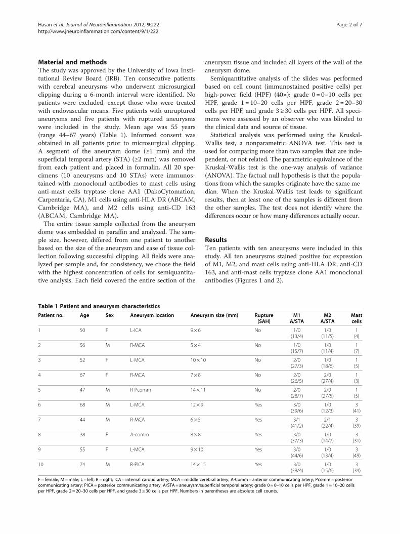
Material and methodsThe study was approved by the University of Iowa Insti-tutional Review Board (IRB). Ten consecutive patientswith cerebral aneurysms who underwent microsurgicalclipping during a 6-month interval were identified. Nopatients were excluded, except those who were treatedwith endovascular means. Five patients with unrupturedaneurysms and five patients with ruptured aneurysmswere included in the study. Mean age was 55 years(range 44–67 years) (Table 1). Informed consent wasobtained in all patients prior to microsurgical clipping.A segment of the aneurysm dome (≥1 mm) and thesuperficial temporal artery (STA) (≥2 mm) was removedfrom each patient and placed in formalin. All 20 spe-cimens (10 aneurysms and 10 STAs) were immunos-tained with monoclonal antibodies to mast cells usinganti-mast cells tryptase clone AA1 (DakoCytomation,Carpentaria, CA), M1 cells using anti-HLA DR (ABCAM,Cambridge MA), and M2 cells using anti-CD 163(ABCAM, Cambridge MA).The entire tissue sample collected from the aneurysm
dome was embedded in paraffin and analyzed. The sam-ple size, however, differed from one patient to anotherbased on the size of the aneurysm and ease of tissue col-lection following successful clipping. All fields were ana-lyzed per sample and, for consistency, we chose the fieldwith the highest concentration of cells for semiquantita-tive analysis. Each field covered the entire section of the
aneurysm tissue and included all layers of the wall of theaneurysm dome.Semiquantitative analysis of the slides was performed
based on cell count (immunostained positive cells) perhigh-power field (HPF) (40×): grade 0 = 0–10 cells perHPF, grade 1 = 10–20 cells per HPF, grade 2 = 20–30cells per HPF, and grade 3 ≥ 30 cells per HPF. All speci-mens were assessed by an observer who was blinded tothe clinical data and source of tissue.Statistical analysis was performed using the Kruskal-
Wallis test, a nonparametric ANOVA test. This test isused for comparing more than two samples that are inde-pendent, or not related. The parametric equivalence of theKruskal-Wallis test is the one-way analysis of variance(ANOVA). The factual null hypothesis is that the popula-tions from which the samples originate have the same me-dian. When the Kruskal-Wallis test leads to significantresults, then at least one of the samples is different fromthe other samples. The test does not identify where thedifferences occur or how many differences actually occur.
ResultsTen patients with ten aneurysms were included in thisstudy. All ten aneurysms stained positive for expressionof M1, M2, and mast cells using anti-HLA DR, anti-CD163, and anti-mast cells tryptase clone AA1 monoclonalantibodies (Figures 1 and 2).
Table 1 Patient and aneurysm characteristics
Patient no. Age Sex Aneurysm location Aneurysm size (mm) Rupture M1 M2 Mast(SAH) A/STA A/STA cells
1 50 F L-ICA 9 × 6 No 1/0 1/0 1(13/4) (11/5) (4)
2 56 M R-MCA 5× 4 No 1/0 1/0 1(15/7) (11/4) (7)
3 52 F L-MCA 10 × 10 No 2/0 1/0 1(27/3) (18/6) (5)
4 67 F R-MCA 7× 8 No 2/0 2/0 1(26/5) (27/4) (3)
5 47 M R-Pcomm 14× 11 No 2/0 2/0 1(28/7) (27/5) (5)
6 68 M L-MCA 12 × 9 Yes 3/0 1/0 3(39/6) (12/3) (41)
7 44 M R-MCA 6× 5 Yes 3/1 2/1 3(41/2) (22/4) (39)
8 38 F A-comm 8× 8 Yes 3/0 1/0 3(37/3) (14/7) (31)
9 55 F L-MCA 9× 10 Yes 3/0 1/0 3(44/6) (13/4) (49)
10 74 M R-PICA 14 × 15 Yes 3/0 1/0 3(38/4) (15/6) (34)
F = female; M=male; L = left; R = right; ICA = internal carotid artery; MCA=middle cerebral artery; A-Comm=anterior communicating artery; Pcomm=posteriorcommunicating artery; PICA = posterior communicating artery; A/STA = aneurysm/superficial temporal artery; grade 0= 0–10 cells per HPF, grade 1 = 10–20 cellsper HPF, grade 2= 20–30 cells per HPF, and grade 3≥ 30 cells per HPF. Numbers in parentheses are absolute cell counts.
Hasan et al. Journal of Neuroinflammation 2012, 9:222 Page 2 of 7http://www.jneuroinflammation.com/content/9/1/222
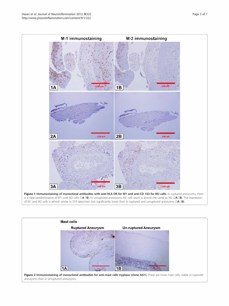
Figure 1 Immunostaining of monoclonal antibodies with anti-HLA DR for M1 and anti-CD 163 for M2 cells. In ruptured aneurysms, thereis a clear predominance of M1 over M2 cells (1A-1B). In unruptured aneurysms, M1 cell count is almost the same as M2 (2A-2B). The expressionof M1 and M2 cells is almost similar in STA specimen, but significantly lower than in ruptured and unruptured aneurysms (3A-3B).
Figure 2 Immunostaining of monoclonal antibodies for anti-mast cells tryptase (clone AA1). There are more mast cells visible in rupturedaneurysms than in unruptured aneurysms.
Hasan et al. Journal of Neuroinflammation 2012, 9:222 Page 3 of 7http://www.jneuroinflammation.com/content/9/1/222
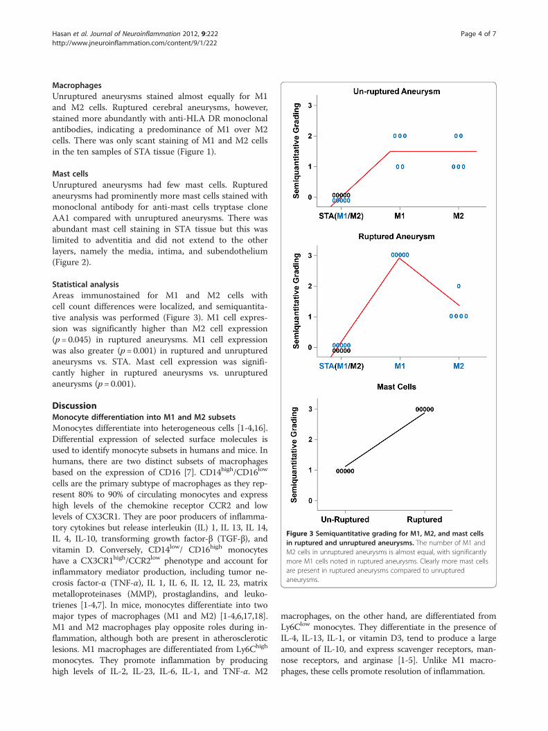
MacrophagesUnruptured aneurysms stained almost equally for M1and M2 cells. Ruptured cerebral aneurysms, however,stained more abundantly with anti-HLA DR monoclonalantibodies, indicating a predominance of M1 over M2cells. There was only scant staining of M1 and M2 cellsin the ten samples of STA tissue (Figure 1).
Mast cellsUnruptured aneurysms had few mast cells. Rupturedaneurysms had prominently more mast cells stained withmonoclonal antibody for anti-mast cells tryptase cloneAA1 compared with unruptured aneurysms. There wasabundant mast cell staining in STA tissue but this waslimited to adventitia and did not extend to the otherlayers, namely the media, intima, and subendothelium(Figure 2).
Statistical analysisAreas immunostained for M1 and M2 cells withcell count differences were localized, and semiquantita-tive analysis was performed (Figure 3). M1 cell expres-sion was significantly higher than M2 cell expression(p= 0.045) in ruptured aneurysms. M1 cell expressionwas also greater (p= 0.001) in ruptured and unrupturedaneurysms vs. STA. Mast cell expression was signifi-cantly higher in ruptured aneurysms vs. unrupturedaneurysms (p= 0.001).
DiscussionMonocyte differentiation into M1 and M2 subsetsMonocytes differentiate into heterogeneous cells [1-4,16].Differential expression of selected surface molecules isused to identify monocyte subsets in humans and mice. Inhumans, there are two distinct subsets of macrophagesbased on the expression of CD16 [7]. CD14high/CD16low
cells are the primary subtype of macrophages as they rep-resent 80% to 90% of circulating monocytes and expresshigh levels of the chemokine receptor CCR2 and lowlevels of CX3CR1. They are poor producers of inflamma-tory cytokines but release interleukin (IL) 1, IL 13, IL 14,IL 4, IL-10, transforming growth factor-β (TGF-β), andvitamin D. Conversely, CD14low/ CD16high monocyteshave a CX3CR1high/CCR2low phenotype and account forinflammatory mediator production, including tumor ne-crosis factor-α (TNF-α), IL 1, IL 6, IL 12, IL 23, matrixmetalloproteinases (MMP), prostaglandins, and leuko-trienes [1-4,7]. In mice, monocytes differentiate into twomajor types of macrophages (M1 and M2) [1-4,6,17,18].M1 and M2 macrophages play opposite roles during in-flammation, although both are present in atheroscleroticlesions. M1 macrophages are differentiated from Ly6Chigh
monocytes. They promote inflammation by producinghigh levels of IL-2, IL-23, IL-6, IL-1, and TNF-α. M2
macrophages, on the other hand, are differentiated fromLy6Clow monocytes. They differentiate in the presence ofIL-4, IL-13, IL-1, or vitamin D3, tend to produce a largeamount of IL-10, and express scavenger receptors, man-nose receptors, and arginase [1-5]. Unlike M1 macro-phages, these cells promote resolution of inflammation.
Figure 3 Semiquantitative grading for M1, M2, and mast cellsin ruptured and unruptured aneurysms. The number of M1 andM2 cells in unruptured aneurysms is almost equal, with significantlymore M1 cells noted in ruptured aneurysms. Clearly more mast cellsare present in ruptured aneurysms compared to unrupturedaneurysms.
Hasan et al. Journal of Neuroinflammation 2012, 9:222 Page 4 of 7http://www.jneuroinflammation.com/content/9/1/222

Recently, there has been a great deal of interest inmacrophage heterogeneity in atherosclerotic lesions,particularly regarding the roles of M1 versus M2 macro-phages. There is also evidence that an imbalance in theratio of M1 to M2 macrophages in advanced atheroscler-osis impairs resolution of inflammation in vitro [1-5].
Macrophages and cerebral aneurysmsMacrophage-depleted and monocyte chemotactic protein-1knockout mice have a reduced incidence of cerebral
aneurysms [13]. Macrophages were observed in the wallof hypertension-induced cerebral aneurysms in rats andwere reported to secrete extracellular matrix-degradingproteolytic enzymes and induce apoptosis of smoothmuscle cells [12]. Additionally, macrophages induce fibro-sis through secretion of TGF-β and promote the release ofreactive oxygen species, TNF-α and IL-1 [10]. Macro-phages are also an important source of MMP 2 and 9,which presumably contribute to the reduction of mechan-ical strength and rupture of aneurysms. MMP-2 isexpressed in most cerebral aneurysms, whereas MMP-9is expressed primarily in aneurysms with atheroscler-otic changes [19,20]. Macrophages are an especiallyimportant source of growth factors and cytokines thatstimulate fibrosis [3].Platelet-derived growth factor, basic fibroblast growth
factor (bFGF), and transforming growth factors alpha(TGF-α) and beta are expressed by immunohistochemis-try in intracranial aneurysms [20-23]. Transcription ofprocollagens I and III is promoted by TGF-β, secreted inlarge part by macrophages [24]. In aneurysms, the pro-gression of smooth muscle cells from a contractile to asynthetic phenotype and the increase in relative numbersof smooth muscle cells (by increased proliferation ofsmooth muscle cells and myointimal hyperplasia) maycompensate for the increased MMP expression and sus-tain aneurysm wall strength [25]. In concordance with thistheory, the aneurysm wall shows, by in situ hybridization,
Figure 4 M1 vs. M2 balance. Schematic diagram illustrating ourhypothesis that a balance between M1 and M2 cells leads to stableaneurysm and that imbalance between M1 and M2 cells withincreased population of M1 cells and upregulation of mast cellspredisposes the aneurysm to rupture.
Figure 5 Polarized monocytes and cytokine production. Schematic diagram demonstrating two populations of monocytes (CD14high
CD16low, CD14low CD16high) and their polarization to two subtypes of macrophages (M1 and M2) with their cell-specific cytokine production.
Hasan et al. Journal of Neuroinflammation 2012, 9:222 Page 5 of 7http://www.jneuroinflammation.com/content/9/1/222

increased synthesis of collagen type III but a slight dimin-ution in expression of collagen type III in immunos-taining, probably indicating faster collagen turnover.Mural cells die during aneurysm progression and theextracellular matrix-synthesizing capability is progres-sively lost, leading to a decrease in tensile wall strengthand an increase in susceptibility of the aneurysm torupture [25].Based on the classification of atherosclerosis by histo-
logical changes, nearly all cerebral aneurysms can beconsidered atherosclerotic [26]. Features of advancedatherosclerosis (including a core of atheromatous debris,a fibrous cap with macrophages and T-cells) are observedin approximately half of intracranial aneurysms [11,26],and myointimal hyperplasia occurs in the other half [25].Thrombosis occurs in some cerebral aneurysms, whichfurther amplifies the ongoing inflammatory reaction andwall degeneration, with loss of tensile strength and ultim-ately aneurysm rupture [24,27-32].
Mast cells and cerebral aneurysmsIshibashi et al. [14] examined the role of mast cells inthe formation of cerebral aneurysms in an experimentalrat model. They found that the number of mast cellswas significantly increased in aneurysm walls. They alsoshowed that using mast cell degranulation inhibitorsattenuated the chronic inflammatory process in theaneurysm wall. This was evident from the decreased nu-clear factor-kappa B activation, macrophage infiltration,and expression of monocyte chemoattractant protein-1,MMP, and interleukin-1beta. They also demonstratedthat the degranulation of mast cells led to increased ex-pression and activation of MMP-2 and -9 and inducednitric oxide synthase in cultured smooth muscle cellsfrom rat intracranial arteries. Their results suggest thatmast cells play a critical role in aneurysm formation inrats through the induction of inflammation. However,they did not address the question of whether mast cellscontribute to the progression of cerebral aneurysms torupture and whether these cells are present in humancerebral aneurysm tissue.
Interpretation of current findingsWe found that M1 and M2 cells were present in equalproportions in unruptured aneurysms with very fewmast cells, which suggests that the pro- and antiinflam-matory activities of M1 and M2 cells, respectively, maybe well balanced in the walls of unruptured aneurysms(Figure 4). However, ruptured aneurysms appear to losethis critical M1/M2 balance, as M1 cell expression wassignificantly more pronounced than M2 cell expressionin ruptured aneurysm walls. Additionally, mast cellswere found to be significantly upregulated in rupturedcerebral aneurysm tissue. Collectively, these data suggest
that a polarized proinflammatory response involving M1macrophages and mast cells may have a role in the cas-cade of events leading to aneurysm rupture (Figures 5and 6). However, the relationship between macrophagesubsets and mast cells remains to be defined.
LimitationsThis study is limited by the small sample size, andgeneralization of the results may not be appropriate. Italso is difficult to determine whether increased expres-sion of M1 vs. M2 and upregulation of mast cells in rup-tured cerebral aneurysms (compared to non-ruptured) isdue to inflammation that occurs following the rupture ofthe aneurysm or whether there was an increase in ex-pression of these molecules that preceded and led torupture of the aneurysm.
ConclusionM1, M2, and mast cells are expressed in the walls ofhuman cerebral aneurysms. However, M1 and mast cellexpression may increase significantly in ruptured aneur-ysms. Our findings suggest that macrophage M1/M2 im-balance and upregulation of mast cells may play a rolein the progression of cerebral aneurysms to rupture.Mast cell activation and M1/M2 macrophage phenotypicmodulation may represent important targets for futuretherapy.
AbbreviationsSTA: Superficial temporal artery; HPF: High-power field; IL: Interleukin; TGF-β: Transforming growth factor-β; TNF-α: Tumor necrosis factor-α; MMP: Matrixmetalloproteinases; TGF-α: Transforming growth factor alpha.
Competing interestsThe authors declare that they have no competing interest.
Authors’ contributionsDH performed all surgical procedures; conceived and designed the study;acquired, analyzed, and interpreted the data; and drafted the manuscript. NChelped in data analysis and interpretation and helped drafting themanuscript. PJ and TH helped design the study, analyzed and interpreted
Figure 6 Mast cell – macrophage interaction. Schematic diagramdemonstrating the interaction of mast cells with macrophages (M1cells) and their cytokine production implicated in aneurysmformation and rupture.
Hasan et al. Journal of Neuroinflammation 2012, 9:222 Page 6 of 7http://www.jneuroinflammation.com/content/9/1/222
the data. All authors have revised the manuscript critically for importantintellectual content and have given final approval of the version to bepublished.
Sources of fundingThis study was supported by NIH grant no. R03NS07922 to DH.
AcknowledgmentsNone
Author details1Department of Neurosurgery, Carver College of Medicine, University of Iowa,Iowa City, IA, USA. 2Department of Neurosurgery, Thomas JeffersonUniversity and Jefferson Hospital for Neuroscience, Philadelphia, PA, USA.3Department of Anesthesia and Perioperative Care, University of CaliforniaSan Francisco, San Francisco, CA, USA. 4Department of Neurosurgery,University of Iowa Hospitals and Clinics, 200 Hawkins Drive, JCP 1616, IowaCity, IA 52242, USA.
Received: 17 July 2012 Accepted: 12 September 2012Published: 21 September 2012
References1. Geissmann F, Jung S, Littman DR: Blood monocytes consist of two
principal subsets with distinct migratory properties. Immunity 2003,19:71–82.
2. Gui T, Shimokado A, Sun Y, Akasaka T, Muragaki Y: Diverse roles ofmacrophages in atherosclerosis: from inflammatory biology tobiomarker discovery. Mediators Inflamm 2012, 2012:693083.
3. Mantovani A, Garlanda C, Locati M: Macrophage diversity and polarizationin atherosclerosis: a question of balance. Arterioscler Thromb Vasc Biol2009, 29:1419–1423.
4. Sica A, Mantovani A: Macrophage plasticity and polarization: in vivoveritas. J Clin Invest 2012, 122:787–795.
5. Mantovani A, Sica A, Locati M: Macrophage polarization comes of age.Immunity 2005, 23:344–346.
6. Mantovani A, Sozzani S, Locati M, Allavena P, Sica A: Macrophagepolarization: tumor-associated macrophages as a paradigm for polarizedM2 mononuclear phagocytes. Trends Immunol 2002, 23:549–555.
7. Passlick B, Flieger D, Ziegler-Heitbrock HW: Identification andcharacterization of a novel monocyte subpopulation in humanperipheral blood. Blood 1989, 74:2527–2534.
8. Kigerl KA, Gensel JC, Ankeny DP, Alexander JK, Donnelly DJ, Popovich PG:Identification of two distinct macrophage subsets with divergent effectscausing either neurotoxicity or regeneration in the injured mouse spinalcord. J Neurosci 2009, 29:13435–13444.
9. Gordon S: Macrophage heterogeneity and tissue lipids. J Clin Invest 2007,117:89–93.
10. Boyle JJ: Macrophage activation in atherosclerosis: pathogenesis andpharmacology of plaque rupture. Curr Vasc Pharmacol 2005, 3:63–68.
11. Chalouhi N, Ali MS, Jabbour PM, Tjoumakaris SI, Gonzalez LF, RosenwasserRH, Koch WJ, Dumont AS: Biology of intracranial aneurysms: role ofinflammation. J Cereb Blood Flow Metab 2012, 32:1659–1676.
12. Jamous MA, Nagahiro S, Kitazato KT, Tamura T, Aziz HA, Shono M, Satoh K:Endothelial injury and inflammatory response induced by hemodynamicchanges preceding intracranial aneurysm formation: experimental studyin rats. J Neurosurg 2007, 107:405–411.
13. Kanematsu Y, Kanematsu M, Kurihara C, Tada Y, Tsou TL, van Rooijen N,Lawton MT, Young WL, Liang EI, Nuki Y, Hashimoto T: Critical roles ofmacrophages in the formation of intracranial aneurysm. Stroke 2011,42:173–178.
14. Ishibashi R, Aoki T, Nishimura M, Hashimoto N, Miyamoto S: Contribution ofmast cells to cerebral aneurysm formation. Curr Neurovasc Res 2010,7:113–124.
15. Xu JM, Shi GP: Emerging role of mast cells and macrophages incardiovascular and metabolic diseases. Endocr Rev 2012, 33:71–108.
16. Auffray C, Sieweke MH, Geissmann F: Blood monocytes: development,heterogeneity, and relationship with dendritic cells. Annu Rev Immunol2009, 27:669–692.
17. Johnson JL, Newby AC: Macrophage heterogeneity in atheroscleroticplaques. Curr Opin Lipidol 2009, 20:370–378.
18. Paulson KE, Zhu SN, Chen M, Nurmohamed S, Jongstra-Bilen J, Cybulsky MI:Resident intimal dendritic cells accumulate lipid and contribute to theinitiation of atherosclerosis. Circ Res 2010, 106:383–390.
19. Bruno G, Todor R, Lewis I, Chyatte D: Vascular extracellular matrixremodeling in cerebral aneurysms. J Neurosurg 1998, 89:431–440.
20. Caird J, Napoli C, Taggart C, Farrell M, Bouchier-Hayes D: Matrixmetalloproteinases 2 and 9 in human atherosclerotic and non-atherosclerotic cerebral aneurysms. Eur J Neurol 2006, 13:1098–1105.
21. Kilic T, Sohrabifar M, Kurtkaya O, Yildirim O, Elmaci I, Gunel M, Pamir MN:Expression of structural proteins and angiogenic factors in normalarterial and unruptured and ruptured aneurysm walls. Neurosurgery 2005,57:997–1007. discussion 1997–1007.
22. Krischek B, Kasuya H, Tajima A, Akagawa H, Sasaki T, Yoneyama T, Ujiie H,Kubo O, Bonin M, Takakura K, et al: Network-based gene expressionanalysis of intracranial aneurysm tissue reveals role of antigenpresenting cells. Neuroscience 2008, 154:1398–1407.
23. Shi C, Awad IA, Jafari N, Lin S, Du P, Hage ZA, Shenkar R, Getch CC, BredelM, Batjer HH, Bendok BR: Genomics of human intracranial aneurysm wall.Stroke 2009, 40:1252–1261.
24. Wynn TA: Cellular and molecular mechanisms of fibrosis. J Pathol 2008,214:199–210.
25. Mimata C, Kitaoka M, Nagahiro S, Iyama K, Hori H, Yoshioka H, Ushio Y:Differential distribution and expressions of collagens in the cerebralaneurysmal wall. Acta Neuropathol 1997, 94:197–206.
26. Kosierkiewicz TA, Factor SM, Dickson DW: Immunocytochemical studies ofatherosclerotic lesions of cerebral berry aneurysms. J Neuropathol ExpNeurol 1994, 53:399–406.
27. Fontaine V, Jacob MP, Houard X, Rossignol P, Plissonnier D, Angles-Cano E,Michel JB: Involvement of the mural thrombus as a site of proteaserelease and activation in human aortic aneurysms. Am J Pathol 2002,161:1701–1710.
28. Fontaine V, Touat Z, Mtairag el M, Vranckx R, Louedec L, Houard X,Andreassian B, Sebbag U, Palombi T, Jacob MP, et al: Role of leukocyteelastase in preventing cellular re-colonization of the mural thrombus.Am J Pathol 2004, 164:2077–2087.
29. Frosen J, Marjamaa J, Myllarniemi M, Abo-Ramadan U, Tulamo R, Niemela M,Hernesniemi J, Jaaskelainen J: Contribution of mural and bone marrow-derived neointimal cells to thrombus organization and wall remodelingin a microsurgical murine saccular aneurysm model. Neurosurgery 2006,58:936–944. discussion 936–944.
30. Frosen J, Piippo A, Paetau A, Kangasniemi M, Niemela M, Hernesniemi J,Jaaskelainen J: Remodeling of saccular cerebral artery aneurysm wall isassociated with rupture: histological analysis of 24 unruptured and 42ruptured cases. Stroke 2004, 35:2287–2293.
31. Peterson JW, Kwun BD, Teramura A, Hackett JD, Morgan JA, Nishizawa S,Bun T, Zervas NT: Immunological reaction against the aging humansubarachnoid erythrocyte. A model for the onset of cerebral vasospasmafter subarachnoid hemorrhage. J Neurosurg 1989, 71:718–726.
32. Wohner N: Role of cellular elements in thrombus formation anddissolution. Cardiovasc Hematol Agents Med Chem 2008, 6:224–228.
doi:10.1186/1742-2094-9-222Cite this article as: Hasan et al.: Macrophage imbalance (M1 vs. M2) andupregulation of mast cells in wall of ruptured human cerebralaneurysms: preliminary results. Journal of Neuroinflammation 2012 9:222.
Submit your next manuscript to BioMed Centraland take full advantage of:
• Convenient online submission
• Thorough peer review
• No space constraints or color figure charges
• Immediate publication on acceptance
• Inclusion in PubMed, CAS, Scopus and Google Scholar
• Research which is freely available for redistribution
Submit your manuscript at www.biomedcentral.com/submit
Hasan et al. Journal of Neuroinflammation 2012, 9:222 Page 7 of 7http://www.jneuroinflammation.com/content/9/1/222