macrophage cyclooxygenase-2 protects against...
TRANSCRIPT
Xin Wang,1,2 Bing Yao,1 Yinqiu Wang,1 Xiaofeng Fan,1 Suwan Wang,1 Aolei Niu,1
Haichun Yang,3 Agnes Fogo,3,4 Ming-Zhi Zhang,1,4,5 and Raymond C. Harris1,4,6
Macrophage Cyclooxygenase-2Protects Against Development ofDiabetic NephropathyDiabetes 2017;66:494–504 | DOI: 10.2337/db16-0773
Diabetic nephropathy (DN) is characterized by increasedmacrophage infiltration, and proinflammatory M1 mac-rophages contribute to development of DN. Previousstudies by us and others have reported that macrophagecyclooxygenase-2 (COX-2) plays a role in polarizationand maintenance of a macrophage tissue-reparative M2phenotype. We examined the effects of macrophageCOX-2 on development of DN in type 1 diabetes. Culturedmacrophages with COX-2 deletion exhibited an M1 phe-notype, as demonstrated by higher inducible nitricoxide synthase and nuclear factor-kB levels but lowerinterleukin-4 receptor-a levels. Compared with corre-sponding wild-type diabetic mice, mice with COX-2 dele-tion in hematopoietic cells (COX-2 knockout bone marrowtransplantation) or macrophages (CD11b-Cre COX2f/f)developed severe DN, as indicated by increased albumin-uria, fibrosis, and renal infiltration of T cells, neutrophils, andmacrophages. Although diabetic kidneys with macrophageCOX-2 deletion had more macrophage infiltration, they hadfewer renal M2 macrophages. Diabetic kidneys with mac-rophage COX-2 deletion also had increased endoplasmicreticulum stress and decreased number of podocytes. Sim-ilar results were found in diabetic mice with macrophagePGE2 receptor subtype 4 deletion. In summary, these stud-ies have demonstrated an important but unexpected rolefor macrophage COX-2/prostaglandin E2/PGE2 receptorsubtype 4 signaling to lessen progression of diabetic kidneydisease, unlike the pathogenic effects of increased COX-2expression in intrinsic renal cells.
In the U.S., .20 million people are currently affected bychronic kidney disease (1). Diabetes is the most commoncause of chronic kidney disease and end-stage renal dis-ease, accounting for about half of all cases. Diabetic ne-phropathy (DN) is characterized by infiltration ofhematopoietic cells, especially macrophages, and there isan association between increased tubulointerstitial in-flammatory cell infiltrate in human diabetic kidneys andloss of renal function (2,3).
The role of macrophages in development of DN is ofparticular interest because they can exhibit distinctlydifferent functional phenotypes, broadly characterized asproinflammatory (M1 or classically activated) and tissue-reparative (M2 or alternatively activated) phenotypes (4).M1 macrophages increase in diabetic kidneys of rodents(5). Furthermore, our previous studies showed that inter-ventions that decrease progression of DN are associatedwith inhibition of renal macrophage infiltration (6–8).
The cyclooxygenase (COX)/prostaglandin system con-tributes to development of DN. COX is the rate-limitingenzyme in metabolizing arachidonic acid to prostaglandinG2 and subsequently to prostaglandin H2, which serves asthe precursor for subsequent metabolism by prostaglan-din and thromboxane synthases. Prostanoid cellular re-sponses are mediated by specific membrane-associatedG-protein–coupled receptors (9–15). Two isoforms ofCOX exist in mammals, constitutive COX-1 and inducibleCOX-2. Previous studies focused on the role of intrinsicrenal cortical COX-2 (macula densa and adjacent cortical
1Division of Nephrology, Department of Medicine, Vanderbilt University School ofMedicine, Nashville, TN2Department of Anesthesiology, Shanghai Cancer Center, Fudan University,Shanghai, China3Department of Pathology, Vanderbilt University School of Medicine, Nashville, TN4Vanderbilt Center for Kidney Disease, Vanderbilt University School of Medicine,Nashville, TN5Department of Cancer Biology, Vanderbilt University School of Medicine, Nash-ville, TN6Nashville Veterans Affairs Hospital, Nashville, TN
Corresponding author: Raymond C. Harris, [email protected], or Ming-ZhiZhang, [email protected].
Received 24 June 2016 and accepted 4 October 2016.
This article contains Supplementary Data online at http://diabetes.diabetesjournals.org/lookup/suppl/doi:10.2337/db16-0773/-/DC1.
© 2017 by the American Diabetes Association. Readers may use this article aslong as the work is properly cited, the use is educational and not for profit, andthe work is not altered. More information is available at http://www.diabetesjournals.org/content/license.
494 Diabetes Volume 66, February 2017
COMPLIC
ATIO
NS
thick ascending limb) in development of DN. Early diabe-tes is characterized by hyperfiltration, and COX-2–derivedprostaglandin E2 (PGE2) contributes to the altered hemo-dynamics. In this regard, selective COX-2 inhibitors havebeen reported to attenuate development of DN (16–18).
In addition to intrinsic kidney cells, COX-2 is alsoexpressed in renal immune cells, particularly renalmonocytes/macrophages. Macrophages express COX-2and are a rich source of prostaglandins, and macrophage-dependent COX-2 expression has been shown to beimportant for macrophage polarization. Previous stud-ies by us and others indicate that COX-2 inhibition ormacrophage deletion of PGE2 receptor subtype 4 (EP4),the major receptor for PGE2 on macrophages, led to de-creased expression of macrophage M2 markers in tumors(19–21). However, the potential role of the macrophageCOX-2/PGE2/EP4 pathway in development of DN hasnot been investigated. Therefore, in the current studies,we determined the role of macrophage COX-2–derivedprostaglandin expression and activity in mediation ofdevelopment of DN.
RESEARCH DESIGN AND METHODS
Animal StudiesAll animal experiments were performed in accordance withthe guidelines of the Institutional Animal Care and UseCommittee of Vanderbilt University. COX-22/2 mice on the129/Bl6 background were originally generated by Dinchuket al. (22). Heterozygous breeding pairs were obtained fromThe Jackson Laboratory (002476; Bar Harbor, ME) andbackcrossed onto 129/svj background for 12 generations(22). EP4
flox/flox mice were generated in Matthew Breyer’slaboratory (23), COX-2flox/flox mice in which exons 6, 7,and 8 of Cox-2 gene are flanked by Lox P sites were orig-inally generated in Dr. Fitzgerald’s laboratory (24), andCD11b-Cre mice with transgene integration in the Y chro-mosome were generated in Dr. Vacher’s laboratory (25), allof which were originally on the C57/Bl6 background andlater backcrossed onto an FVB background for 12 genera-tions. Macrophage COX-2 deletion mice (CD11b-Cre COX-2flox/flox) and macrophage EP4 deletion mice (CD-11b-CreEP4flox/flox) and corresponding wild-type (WT) (COX-2flox/flox
mice and EP4flox/flox mice, respectively) on the C57/Bl6 orFVB backgrounds were used for experiments. Male micereceived daily intraperitoneal injections for 5 consecutivedays of streptozotocin (STZ; 50 mg/kg) that was freshlyprepared in 0.1 mol/L citrate buffer (pH 4.5) (8). The onsetof diabetes was evaluated by measuring fasting blood glu-cose with a B-glucose analyzer (HemoCue, Lake Forest, CA)in conscious mice on saphenous vein samples at noon afterfasting for 6 h initiated at 6:00 A.M. Urinary albumin andcreatinine excretion was determined using Albuwell M kits(Exocell, Philadelphia, PA). Albuminuria is expressed as theurinary albumin-to-creatinine ratio (ACR; mg/mg). Periodicacid-Schiff (PAS)–stained slides were evaluated for glomer-ular injury without knowledge of the identity of variousgroups as described previously (7).
Creation of Chimeric MiceBone marrow transplantation (BMT) was performed aspreviously described (26). Briefly, recipient mice (WTfemale 129/svj mice) were lethally irradiated with 9 Gyusing a cesium g source. Bone marrow cells from donors(male 129/svj WT and COX-22/2 mice) were harvestedfrom femurs and tibias. Recipient mouse received 5 3106 bone marrow cells in 0.2 mL medium through tailvein injection. BMT from males to females made it easierto assess effective engraftment by identification of Ychromosomes in engrafted hematopoietic cells. Fiveweeks after transplantation, blood was sampled to deter-mine chimerism by determination of COX-2 expressionwith PCR.
Cell CultureMale 129/svj WT or COX-22/2 mice were intraperitoneallyinjected with 1 mL sterile thioglycollate (3%; Sigma-Aldrich). Four days later, peritoneal cells including mac-rophages were harvested. Peritoneal macrophages werecultured according to a previous report (27).
Isolation of Kidney Macrophages/Dendritic CellsCD11b-expressing cells in kidney single-cell suspensionswere enriched using mouse CD11b Microbeads and MACS.
AntibodiesRat anti-mouse F4/80 (MCA497R), CD68 (MCA1957),CD11c (MCA1369), CD3 (MCA1477), CD4 (MCA2961),CD8a (MCA2694), and Ly-6G (Gr-1, MCA2387) were pur-chased from AbD Serotec; rabbit anti-human fibronectin(FN; F3648) and mouse anti–a-smooth muscle actin(a-SMA, a marker of myofibroblasts; A5228) were fromSigma-Aldrich; rabbit antimurine collagen type I (600-401-103-01) and collagen type IV (600-401-106-01)were from Rockland Immunochemicals; goat anti-humanconnective tissue growth factor (CTGF; SC-14939) wasfrom Santa Cruz Biotechnology; mouse anti–mannosereceptor (MR or CD206; MAB25341) was from R&DSystems; rabbit anti-human interleukin-4 receptor-a (IL-4Ra; or CD124, NBP1-00884) was from Novus Biologi-cals; rabbit anti–Wilms Tumor Protein (WT1; ab89901),inducible nitric oxide synthase (iNOS; ab3523), and tu-mor necrosis factor-a (ab6671) were from Abcam; andrabbit-anti–nuclear factor-kB p65 (8284) and mouse-anti–C/EBP homologous protein (CHOP; 2895) werefrom Cell Signaling Technology. Rabbit anti-renin antise-rum (1:6,000 dilutions) was a gift from T. Inagami (Van-derbilt University).
RNA Isolation and Quantitative RT-PCRTotal RNA from tissues and cells were isolated usingTRIzol reagents (Invitrogen). Quantitative RT-PCR wasperformed using TaqMan real-time PCR (7900HT; AppliedBiosystems). The Master Mix and all gene probes werealso purchased from Applied Biosystems. The probes usedin the experiments included mouse S18 (Mm02601778),IL-4Ra (Mm01275139), MR (Mm01329362), chitinase3–like protein 3 (Ym-1) (Mm00657889), nephrin (Nphs1;
diabetes.diabetesjournals.org Wang and Associates 495

Mm00497828), podocin (Nphs2, Mm01292252), chemo-kine (C-C motif) ligand 2 (CCL2; MCP-1; Mm00441242),COX-2 (PTGS2; Mm00478374), EP4 (PTGER4; Mm00436053),collagen I (Col1a1; Mm00801666), collagen IV (Col4a1;Mm01210125), a-SMA (acta2; Mm01546133), renin(Mm02342889), angiotensinogen (Mm00599662), ACE(ACE1; Mm00802048), ACE2 (Mm01159003), angio-tensin II type I (AT1a; agtr1a; Mm01166161), AT1b(agtr1b; Mm01701115), AT2 (agtr2; Mm01341373),and Mas (Mas1; Mm00627134).
Immunohistochemistry Staining and QuantitativeImage AnalysisThe animals were anesthetized with Nembutal (70 mg/kg,i.p.; Abbot Laboratories) and given heparin (1,000 units/kg,i.p.) to minimize coagulation. One kidney was removed forimmunoblotting and quantitative RT-PCR, and the animalwas perfused with 3.7% formaldehyde, 10 mmol/L sodiumm-periodate, 40 mmol/L phosphate buffer, and 1% aceticacid through the aortic trunk cannulated by means of theleft ventricle. The fixed kidneys were dehydrated through agraded series of ethanols, embedded in paraffin, sectioned(4 mm), and mounted on glass slides. Immunostaining wascarried out as in previous reports (28). For WT1 staining,antigen retrieval was achieved by boiling in citric acidbuffer (100 mmol/L; pH 6) for 3 3 5 min. For stainingwith mouse monoclonal antibodies (a-SMA and MR), thetissues were blocked and primary and secondary antibodiesdiluted using reagents from the M.O.M. Kit (PK-2200; Vec-tor Laboratories, Burlingame, CA). On the basis of the dis-tinctive density and color of immunostaining in video images,the number, size, and position of stained area were quantifiedby using the BIOQUANT true-color windows system (R&MBiometrics, Nashville, TN). Four representative fields fromeach animal were quantified at3160 magnification, and their
average was used as data from one animal sample. Podocytedensity is expressed as podocytes per glomerulus.
ImmunoblottingCultured cells were lysed, and kidneys were homogenizedwith buffer containing 10 mmol/L Tris-HCl (pH 7.4),50 mmol/L NaCl, 2 mmol/L EGTA, 2 mmol/L EDTA,0.5% Nonidet P-40, 0.1% SDS, 100 mmol/L Na3VO4,100 mmol/L NaF, 0.5% sodium deoxycholate, 10 mmol/Lsodium pyrophosphate, 1 mmol/L phenylmethylsulfonylfluoride, 10 mg/mL aprotinin, and 10 mg/mL leupeptin.The homogenate was centrifuged at 15,000g for 20 minat 4°C. An aliquot of supernatant was taken for proteinmeasurement with a BCA protein assay kit (Thermo FisherScientific, Rockford, IL). Immunoblotting was described in arecent report (29).
StatisticsAll values are presented as means, with error bars repre-senting 6 SEM. Fisher exact test, ANOVA, and Bonferronit tests were used for statistical analysis.
RESULTS
To determine potential effects of COX-2 expression onmonocyte/macrophage phenotype, we isolated and cul-tured peritoneal macrophages fromWT mice and mice withglobal deletion of COX-2 (COX-22/2). As indicated inFig. 1, compared with WT mice, peritoneal macrophagesfrom COX-22/2 mice had increased total expression ofthe proinflammatory, M1 marker iNOS. There was alsoincreased NF-kB expression, indicative of an M1 phenotype.In contrast, there was decreased expression of IL-4Ra, anM2 marker.
As noted in the introductory paragraphs, there isincreasing evidence for an important pathophysiologic
Figure 1—COX-2–deficient macrophages exhibited an M1 phenotype. Peritoneal macrophages from WT and COX-22/2 mice werecultured as described in RESEARCH DESIGN AND METHODS. iNOS (a marker of M1 macrophages) was minimal in WT macrophages but readilydetected in COX-22/2 macrophages. In contrast, levels of IL-4Ra (a marker of M2 macrophages) were markedly lower in COX-22/2
macrophages. NF-kB levels were also significantly higher in COX-22/2 macrophages. **P < 0.01, ***P < 0.001 vs. WT; n = 3.
496 Macrophage COX-2 and Diabetic Nephropathy Diabetes Volume 66, February 2017
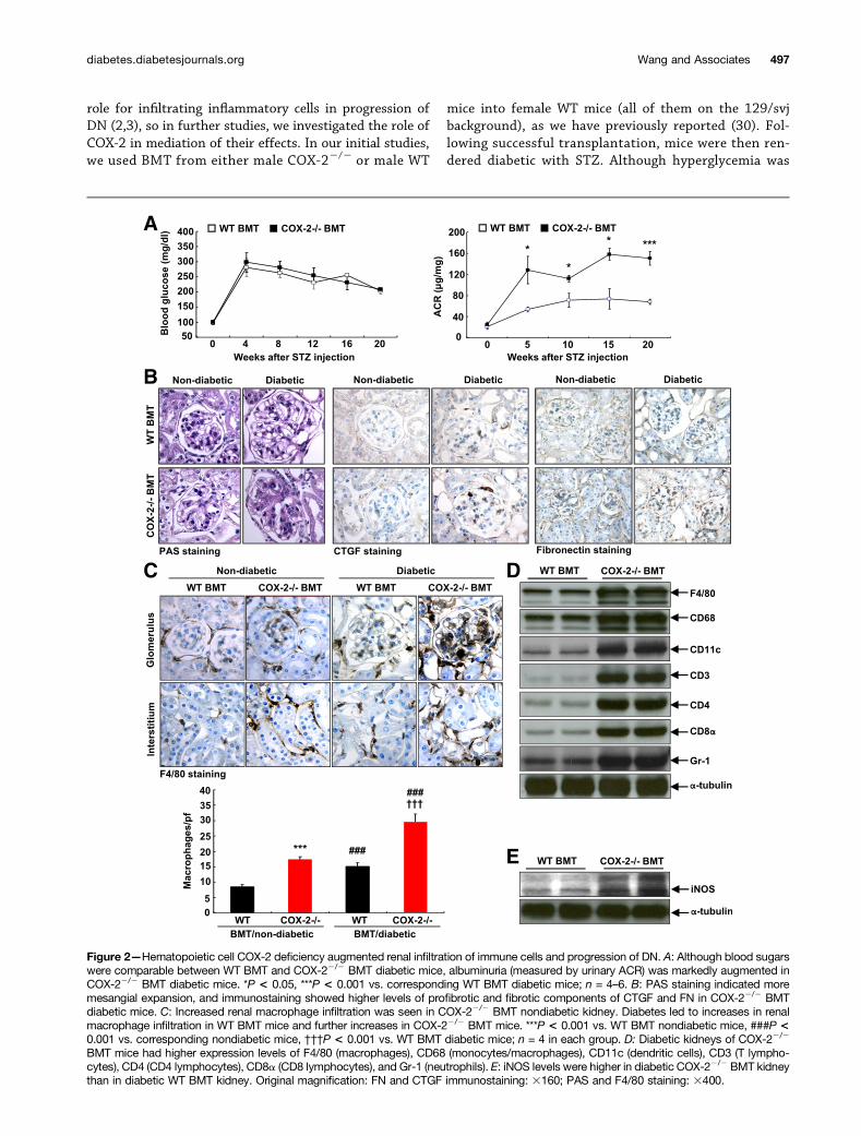
role for infiltrating inflammatory cells in progression ofDN (2,3), so in further studies, we investigated the role ofCOX-2 in mediation of their effects. In our initial studies,we used BMT from either male COX-22/2 or male WT
mice into female WT mice (all of them on the 129/svjbackground), as we have previously reported (30). Fol-lowing successful transplantation, mice were then ren-dered diabetic with STZ. Although hyperglycemia was
Figure 2—Hematopoietic cell COX-2 deficiency augmented renal infiltration of immune cells and progression of DN. A: Although blood sugarswere comparable between WT BMT and COX-22/2 BMT diabetic mice, albuminuria (measured by urinary ACR) was markedly augmented inCOX-22/2 BMT diabetic mice. *P < 0.05, ***P < 0.001 vs. corresponding WT BMT diabetic mice; n = 4–6. B: PAS staining indicated moremesangial expansion, and immunostaining showed higher levels of profibrotic and fibrotic components of CTGF and FN in COX-22/2 BMTdiabetic mice. C: Increased renal macrophage infiltration was seen in COX-22/2 BMT nondiabetic kidney. Diabetes led to increases in renalmacrophage infiltration in WT BMT mice and further increases in COX-22/2 BMT mice. ***P < 0.001 vs. WT BMT nondiabetic mice, ###P <0.001 vs. corresponding nondiabetic mice, †††P < 0.001 vs. WT BMT diabetic mice; n = 4 in each group. D: Diabetic kidneys of COX-22/2
BMT mice had higher expression levels of F4/80 (macrophages), CD68 (monocytes/macrophages), CD11c (dendritic cells), CD3 (T lympho-cytes), CD4 (CD4 lymphocytes), CD8a (CD8 lymphocytes), and Gr-1 (neutrophils). E: iNOS levels were higher in diabetic COX-22/2 BMT kidneythan in diabetic WT BMT kidney. Original magnification: FN and CTGF immunostaining: 3160; PAS and F4/80 staining: 3400.
diabetes.diabetesjournals.org Wang and Associates 497

comparable in the two groups, albuminuria was signifi-cantly increased in the COX-22/2 BMT mice comparedwith the WT BMT mice (Fig. 2A). Nondiabetic kidneysfrom COX-22/2 BMT and WT BMT mice had similarnormal histology and low levels of FN and CTGF, whereas
diabetic kidneys of COX-22/2 BMT mice had increasedmesangial expansion and increased expression of FN andCTGF compared with diabetic kidneys of WT BMT mice(Fig. 2B). There was increased macrophage infiltrationin nondiabetic kidneys of COX-22/2 BMT mice. Although
Figure 3—Macrophage COX-2 deficiency augmented development of DN. A: Urinary ACR was markedly higher in diabetic CD11b-CreCOX2f/f mice than diabetic WT mice at 12 and 18 weeks after initiation of hyperglycemia. **P < 0.01; n = 5–9. B: PAS staining indicatedmore severe glomerulosclerosis in diabetic CD11b-Cre COX2f/f mice. *P < 0.05; n = 5. Original magnification: 3160.
Figure 4—Macrophage COX-2 deficiency led to accelerated podocyte loss in diabetic mice. A: Both nephrin and podocin mRNA levelswere significantly lower in diabetic CD11b-Cre COX2f/f mice than diabetic WT mice at 18 weeks after initiation of hyperglycemia. ***P <0.001; n = 6. B: Podocyte number in each glomerulus (WT1-positive nuclei) was markedly lower in diabetic CD11b-Cre COX2f/f mice thandiabetic wild-type mice. ***P < 0.001; n = 4–7. Original magnification 3400.
498 Macrophage COX-2 and Diabetic Nephropathy Diabetes Volume 66, February 2017
renal macrophage infiltration increased in both diabeticWT BMT and COX-22/2 BMT mice, it was still significantlyhigher in COX-22/2 BMT mice than in WT BMT mice (Fig.2C). The increased renal macrophage infiltration of theCOX-22/2 BMT mice was further confirmed by immunoblot-ting for F4/80 and CD68 (Fig. 2D). Furthermore, there wasincreased expression of the M1 marker iNOS (Fig. 2E). Inaddition to increased macrophage infiltration, kidneys fromCOX-22/2 BMT mice had increased expression of the den-dritic cell marker CD11c, the neutrophil marker Gr-1, and thepan-T cell marker CD3 as well as both CD4 and CD8 (Fig. 2D).
To determine more specifically the role of macrophageCOX-2 in DN, we backcrossed both COX2f/f mice andCD11b-Cre mice to a more kidney injury–prone strain,FVB. In preliminary studies, we found that macrophagesfrom CD11b-Cre COX2f/f mice had essentially no COX-2
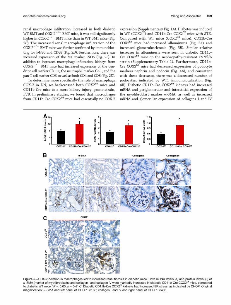
expression (Supplementary Fig. 1A). Diabetes was inducedin WT (COX2f/f) and CD11b-Cre COX2f/f mice with STZ.Compared with WT mice (COX2f/f mice), CD11b-CreCOX2f/f mice had increased albuminuria (Fig. 3A) andincreased glomerulosclerosis (Fig. 3B). Similar relativeincreases in albuminuria were seen in diabetic CD11b-Cre COX2f/f mice on the nephropathy-resistant C57Bl/6strain (Supplementary Table 1). Furthermore, CD11b-Cre COX2f/f mice had decreased expression of podocytemarkers nephrin and podocin (Fig. 4A), and consistentwith these decreases, there was a decreased number ofpodocytes, indicated by WT1 immunolocalization (Fig.4B). Diabetic CD11b-Cre COX2f/f kidneys had increasedmRNA and periglomerular and interstitial expression ofthe myofibroblast marker a-SMA, as well as increasedmRNA and glomerular expression of collagens I and IV
Figure 5—COX-2 deletion in macrophages led to increased renal fibrosis in diabetic mice. Both mRNA levels (A) and protein levels (B) ofa-SMA (marker of myofibroblasts) and collagen I and collagen IV were markedly increased in diabetic CD11b-Cre COX2f/f mice, comparedto diabetic WT mice. *P < 0.05; n = 5–7. C: Diabetic CD11b-Cre COX2f/f kidneys had increased ER stress, as indicated by CHOP. Originalmagnification: a-SMA and left panel of CHOP: 3160; collagen I and IV and right panel of CHOP: 3400.
diabetes.diabetesjournals.org Wang and Associates 499

(Fig. 5A and B). Of note, diabetic CD11b-Cre COX2f/f
kidneys had increased expression of CHOP, a marker ofendoplasmic reticulum (ER) stress, consistent with thedevelopment of more severe DN in the absence of mac-rophage COX-2 expression (Fig. 5C).
Diabetic CD11b-Cre COX2f/f kidneys had increased levelsof the macrophage-attracting chemokine, CCL2 (MCP-1)(Fig. 6A) and increased F4/80-positive macrophage infiltra-tion (Fig. 6B). Furthermore, there were increases in pan-Tcell marker–positive CD3 cells as well as both CD4- andCD8-positive T cells and increased neutrophil infiltrationin CD11b-Cre COX2f/f mice (Supplementary Fig. 2).
Diabetic CD11b-Cre COX2f/f kidneys had decreasedmRNA for the M2 markers CD206 (MR) and Ym-1 (Fig. 7A).Although there were increased F4/80-positive cells in di-abetic CD11b-Cre COX2f/f kidneys, there was a significantdecrease in MR-positive infiltrating cells (Fig. 7B). In addi-tion, macrophages isolated from diabetic CD11b-CreCOX2f/f kidneys had decreased mRNA expression of MRand IL-4Ra (Fig. 7C).
As noted in the introductory paragraphs, there isevidence that macrophage activation of EP4 promotespolarization to an M2 phenotype (19,30). We generatedCD11b-Cre EP4f/f mice on an FVB background and madethem diabetic with STZ. Macrophages from CD11b-CreEP4f/f mice had markedly decreased expression of EP4mRNA (Supplementary Fig. 1). Similar to what we ob-served in diabetic CD11b-Cre COX2f/f mice, macrophage-selective deletion of EP4 led to increased albuminuria(Fig. 8A), increased expression of a-SMA, collagen I, andcollagen IV as well as CHOP, a marker of ER stress (Fig. 8B)
and decreased kidney expression of the M2 markers MR andYm-1 (Fig. 8C). Similar relative increases in albuminuria wereseen in diabetic CD11b-Cre EP4f/f mice on the nephropathy-resistant C57Bl/6 strain (Supplementary Table 1).
Activation of renin-angiotensin system (RAS) plays animportant role in development of DN, and inhibition of RASattenuates kidney injury. We have previously shown thatrenal renin expression and activity is regulated by maculadensa COX-2 (31). Therefore, we first investigated renal re-nin expression with immunohistochemistry. As indicated inSupplementary Fig. 3, renal renin expression was compara-ble between diabetic COX2f/f and CD11b-Cre COX2f/f miceand between diabetic EP4f/f and CD11b-Cre EP4f/f mice.Further analysis showed that renal mRNA levels of the ma-jor components of the RAS including renin, angiotensino-gen, ACE, ACE2, AT1a and AT1b, AT2, and angiotensin 1–7receptor (Mas) were comparable between diabetic COX2f/f
and CD11b-Cre COX2f/f mice (Supplementary Fig. 4).
DISCUSSION
The current studies demonstrate an important role for themacrophage COX-2/PGE2/EP4 axis to mitigate against de-velopment of DN. Both mice with BMT from global COX-2knockout mice and mice with selective deletion of COX-2 inmacrophages had increased structural and functional injuryin response to STZ-induced diabetes. Similar acceleration ofrenal injury was seen in diabetic mice with selective macro-phage deletion of the PGE2 receptor subtype, EP4. Theincreased injury with macrophage COX-2 deletion was ac-companied by increased infiltration of inflammatory cells,macrophages, neutrophils, and T cells. Although there was
Figure 6—COX-2 deletion in macrophages led to increased renal macrophage infiltration in diabetic mice. A: CCL2 (or MCP-1) mRNAlevels were markedly increased in diabetic CD11b-Cre COX2f/f mice. *P < 0.05; n = 5–7. B: Renal macrophage infiltration (F4/80-positivecells) was markedly increased in diabetic CD11b-Cre COX2f/f mice. ***P < 0.001; n = 4. Original magnification: 3250.
500 Macrophage COX-2 and Diabetic Nephropathy Diabetes Volume 66, February 2017

an increase in total numbers of macrophages, the percent-age that expressed M2 markers was significantly decreased.
Previous studies by us and others have demonstratedincreased expression of intrinsic renal COX-2 localizedto the macula densa in both experimental and humandiabetes (16–18,32). Administration of selective COX-2inhibitors decreased diabetes-induced hyperfiltration anddecreased renal injury. In addition to mediating hyperfiltra-tion, our previous studies have demonstrated that maculadensa COX-2 is an important mediator of the RAS so inhi-bition of macula densa COX-2 may also inhibit the intrarenalRAS, which has been implicated in progression of DN (33). Inaddition, we have also previously shown that increased podo-cyte COX-2 expression accelerated diabetic glomerular injurythrough increased prorenin receptor expression (34).
However, in contrast to our previous studies that inhi-bited intrinsic renal COX-2 activity, the current studies indi-cate an opposing, protective role for macrophage-specificCOX-2 activity. COX-2–derived PGE2 acting through theEP4 receptor is important for macrophage polarization toan M2 phenotype (35). We have previously shown thatmacrophage EP4 deletion led to decreased macrophage M2markers in tumors (19). In contrast, EP4 activation inhibitsrelease of the cytokines tumor necrosis factor-a and IL-2from mouse macrophages (36) and inhibits the NLRP3inflammasome in human macrophages (37). EP4 is coupled
to Gs, and receptor activation activates adenylate cyclaseand increases cAMP production, which polarizes macro-phages to an M2 phenotype (38).
In diabetic rats, administration of hemin (a heme-oxygenase inducer) suppressed increased M1 macrophagesand restored decreased M2 macrophages in associationwith decreases in proinflammatory cytokine/chemokine,reduction of extracellular matrix/profibrotic protein, andimprovement of kidney function and histology (5). Simi-larly, pentraxin-3 polarized macrophages to an M2 pheno-type and ameliorated experimental diabetic renal injury(39). In addition, suppression of renal M1 macrophagesby deletion of Toll-like receptors 2 or 4 also protectedagainst development of DN in mice (40,41).
DN is known to be a proinflammatory state. Devarajet al. (40) have shown that peritoneal and kidney macro-phages from mice with STZ diabetes have greater expressionof M1 (Ly6c, IL-6, and CCR2) than M2 markers (CD206 andCD163). Monocytes isolated from patients with both type 1and type 2 diabetes expressed high levels of COX-2 mRNAcompared with minimal levels from those isolated from nor-mal nondiabetic volunteers. In human monocyte THP-1cells, high glucose induced COX-2 mRNA and protein aswell as COX-2–derived PGE2, but had no effect on COX-1expression (42). The kidney macrophages in the dia-betic kidney may be similar to a phenotype described
Figure 7—COX-2 deletion in macrophages led to decreased renal M2 macrophages in diabetic mice. A: The mRNA levels of MR (CD206)and Ym-1, markers of M2 macrophages, were markedly decreased in diabetic CD11b-Cre COX2f/f kidneys. **P< 0.01, ***P< 0.001; n = 5–7. B: The number of renal M2 macrophages (MR-positive cells [arrows]) was significantly lower in diabetic CD11b-Cre COX2f/f mice than indiabetic WT mice. ***P < 0.001; n = 4. Original magnification: 3250. C: The mRNA levels for both MR and IL-4Ra (M2 markers) weremarkedly lower in isolated renal macrophages from CD11b-Cre COX2f/f mice than WT mice at 2 weeks after initiation of hyperglycemia.*P < 0.05; n = 4.
diabetes.diabetesjournals.org Wang and Associates 501

as resolution macrophages. These resolution macrophages donot express markers that characterize them as either classi-cally nor alternatively activated but are a hybrid of bothphenotypes. Of note, they are characterized by increasedCOX-2 expression, and maintenance of this phenotypeis dependent upon macrophage-derived cAMP production.They have been postulated to be dispensable for clear-ing polymorphonuclear neutrophils during self-limitinginflammation but are necessary for postresolution in-nate lymphocyte repopulation and restoring tissue homeo-stasis (43). Further studies will be necessary to characterizethe functions of macrophages in the diabetic kidney.
The role of COX-2 in the development of DN and otherkidney injury is complicated. COX-2 expression increases inboth podocytes and macula densa in diabetic kidney (16–18).Overexpression of COX-2 predisposes to podocyte injury andmacula densa COX-2 contributes to hyperfiltration in earlydiabetes (34). Therefore, systemic COX-2 inhibition has ben-eficial effects because of inhibition of podocyte COX-2 andattenuation of hyperfiltration. The present observations in-dicate that the beneficial effect of macrophage COX-2 is theresult of a specific effect on immune cells. Recently, Nilssonet al. (44) reported that global COX-2 deficiency exacerbatedunilateral ureteral obstruction (UUO)–induced kidney dam-age. Similarly, Kamata et al. (45) reported that inhibition
of COX-2 activity with celecoxib exacerbated developmentof fibrosis in UUO kidneys. However, COX-2 knockdownin macrophages using a chitosan delivery system was report-ed to attenuate UUO-induced kidney damage in associationwith decreases in inflammation, oxidative stress, and apo-ptosis (46). Therefore, the role of COX-2 in kidney injurymay depend on the sources of COX-2, the mechanisms ofrenal injury, the expression and subtype of PGE2 receptors,and timing of COX-2 inhibition.
In summary, these studies have demonstrated an impor-tant but unexpected role for macrophage COX-2 signaling tolessen progression of diabetic kidney disease, unlike the path-ogenic effects of increased COX-2 expression in intrinsicrenal cells. Although increased COX-2 expression in macro-phages is often cited as a characteristic of a proinflammatory,M1 phenotype, these studies indicate that its expression mayactually mitigate against detrimental effects in DN.
Acknowledgments. The authors thank Garret Fitzgerald (University ofPennsylvania, Philadelphia, PA) for providing the COX-2f/f mice.Funding. These studies were supported by National Institutes of Health grantsDK-51265 (to R.C.H. and M.-Z.Z.), DK-62794 (to R.C.H.), DK-95785 (to R.C.H.and M.-Z.Z.), and DK-103067 (to R.C.H., A.F., H.Y., and M.-Z.Z.) and U.S.Department of Veterans Affairs VA Merit Award 00507969 (to R.C.H.).
Figure 8—EP4 deletion in macrophages augmented development of DN. A: Macrophage EP4 deletion augmented albuminuria at 12 weeksafter initiation of hyperglycemia. **P < 0.01; n = 5. B: The levels of a-SMA (marker of myofibroblasts), collagen I, collagen IV, and CHOP(marker of ER stress) were markedly increased in diabetic CD11b-CreEP4f/f mice, compared to diabetic WT mice. Original magnification:a-SMA,3160; and collagen I and IV and CHOP,3400. C: The mRNA levels of MR and Ym-1, markers of M2 macrophages, were markedlydecreased in diabetic CD11b-Cre EP4f/f kidneys. **P < 0.01; n = 5.
502 Macrophage COX-2 and Diabetic Nephropathy Diabetes Volume 66, February 2017
Duality of Interest. No potential conflicts of interest relevant to this articlewere reported.Author Contributions. X.W. researched data and contributed to thediscussion. B.Y., Y.W., X.F., S.W., A.N., and H.Y. researched data. A.F. contributedto the discussion. M.-Z.Z. researched data, contributed to the discussion, wrote themanuscript. R.C.H. contributed to the discussion and wrote the manuscript. M.-Z.Z.and R.C.H. are the guarantors of this work and, as such, had full access to all thedata in the study and take responsibility for the integrity of the data and theaccuracy of the data analysis.
References1. Breyer MD, Susztak K. The next generation of therapeutics for chronic
kidney disease. Nat Rev Drug Discov 2016;15:568–5882. Bohle A, Wehrmann M, Bogenschütz O, Batz C, Müller CA, Müller GA. The
pathogenesis of chronic renal failure in diabetic nephropathy. Investigation of
488 cases of diabetic glomerulosclerosis. Pathol Res Pract 1991;187:251–2593. Woroniecka KI, Park AS, Mohtat D, Thomas DB, Pullman JM, Susztak K.
Transcriptome analysis of human diabetic kidney disease. Diabetes 2011;60:
2354–23694. Rees AJ. Monocyte and macrophage biology: an overview. Semin Nephrol
2010;30:216–2335. Ndisang JF, Jadhav A. Hemin therapy improves kidney function in male
streptozotocin-induced diabetic rats: role of the heme oxygenase/atrial natriuretic
peptide/adiponectin axis. Endocrinology 2014;155:215–2296. Zhang MZ, Wang S, Yang S, et al. Role of blood pressure and the renin-
angiotensin system in development of diabetic nephropathy (DN) in eNOS-/-
db/db mice. Am J Physiol Renal Physiol 2012;302:F433–F4387. Zhang MZ, Wang Y, Paueksakon P, Harris RC. Epidermal growth factor
receptor inhibition slows progression of diabetic nephropathy in association with
a decrease in endoplasmic reticulum stress and an increase in autophagy. Di-
abetes 2014;63:2063–20728. Zhang MZ, Yao B, Yang S, et al. Intrarenal dopamine inhibits progression of
diabetic nephropathy. Diabetes 2012;61:2575–25849. Whorton A, Misono, K, Hollifield, J, Frolich, JC, Inagami, T, Oates, JA.
Prostaglandins and renin release. I. Stimulation of renin release from rabbit renal
cortical slices by PGI2. Prostaglandins 1977;14:1095–110410. Francisco LJ, Osborn JL, and DiBona GF. Prostaglandins in renin release
during sodium deprivation. Am. J. Physiol. 1982;243:261–26811. Linas SL. Role of prostaglandins in renin secretion in the isolated kidney.
Am. J. Physiol. 1984;246:F811–F81812. Ito S, Carretero OA, Abe K, Juncos LA, Yoshinaga K. Macula densa control of
renin release and glomerular hemodynamics. Tohoku J Exp Med 1992;166:27–3913. Ito S, Carretero OA, Abe K, Beierwaltes WH, Yoshinaga K. Effect of pros-
tanoids on renin release from rabbit afferent arterioles with and without macula
densa. Kidney Int 1989;35:1138–114414. Greenberg SG, Lorenz JN, He XR, Schnermann JB, Briggs JP. Effect of
prostaglandin synthesis inhibition on macula densa-stimulated renin secretion.
Am J Physiol 1993;265:F578–F58315. Needleman P, Turk J, Jakschik BA, Morrison AR, and Lefkowith JB. Ara-
chidonic acid metabolism. Ann. Rev. Biochem. 1986;55:69–10216. Cheng HF, Wang CJ, Moeckel GW, Zhang MZ, McKanna JA, Harris RC.
Cyclooxygenase-2 inhibitor blocks expression of mediators of renal injury in a
model of diabetes and hypertension. Kidney Int 2002;62:929–93917. Komers R, Lindsley JN, Oyama TT, Anderson S. Cyclo-oxygenase-2 in-
hibition attenuates the progression of nephropathy in uninephrectomized diabetic
rats. Clin Exp Pharmacol Physiol 2007;34:36–4118. Komers R, Lindsley JN, Oyama TT, et al. Immunohistochemical and func-
tional correlations of renal cyclooxygenase-2 in experimental diabetes. J Clin
Invest 2001;107:889–89819. Chang J, Vacher J, Yao B, et al. Prostaglandin E receptor 4 (EP4) promotes
colonic tumorigenesis. Oncotarget 2015;6:33500–33511
20. Na YR, Yoon YN, Son DI, Seok SH. Cyclooxygenase-2 inhibition blocks M2macrophage differentiation and suppresses metastasis in murine breast cancer
model. PLoS One 2013;8:e6345121. Nakanishi Y, Nakatsuji M, Seno H, et al. COX-2 inhibition alters the phe-notype of tumor-associated macrophages from M2 to M1 in ApcMin/+ mouse
polyps. Carcinogenesis 2011;32:1333–133922. Dinchuk JE, Car BD, Focht RJ, et al. Renal abnormalities and an altered in-
flammatory response in mice lacking cyclooxygenase II. Nature 1995;378:406–40923. Schneider A, Guan Y, Zhang Y, et al. Generation of a conditional allele of themouse prostaglandin EP4 receptor. Genesis 2004;40:7–1424. Wang D, Patel VV, Ricciotti E, et al. Cardiomyocyte cyclooxygenase-2 influ-ences cardiac rhythm and function. Proc Natl Acad Sci U S A 2009;106:7548–755225. Ferron M, Vacher J. Targeted expression of Cre recombinase in macro-
phages and osteoclasts in transgenic mice. Genesis 2005;41:138–14526. Nishida M, Fujinaka H, Matsusaka T, et al. Absence of angiotensin II type 1
receptor in bone marrow-derived cells is detrimental in the evolution of renalfibrosis. J Clin Invest 2002;110:1859–186827. Hoover DL, Nacy CA. Macrophage activation to kill Leishmania tropica:defective intracellular killing of amastigotes by macrophages elicited with sterile
inflammatory agents. J Immunol 1984;132:1487–149328. Zhang MZ, Yao B, Cheng HF, Wang SW, Inagami T, Harris RC. Renal corticalcyclooxygenase 2 expression is differentially regulated by angiotensin II AT(1) and
AT(2) receptors. Proc Natl Acad Sci U S A 2006;103:16045–1605029. Zhang MZ, Wang Y, Yao B, et al. Role of epoxyeicosatrienoic acids (EETs) in
mediation of dopamine’s effects in the kidney. Am J Physiol Renal Physiol 2013;305:F1680–F168630. Zhang MZ, Yao B, Wang Y, et al. Inhibition of cyclooxygenase-2 in hematopoietic
cells results in salt-sensitive hypertension. J Clin Invest 2015;125:4281–429431. Cheng HF, Wang JL, Zhang MZ, et al. Angiotensin II attenuates renal cortical
cyclooxygenase-2 expression. J Clin Invest 1999;103:953–96132. Khan KN, Stanfield KM, Harris RK, Baron DA. Expression of cyclooxygenase-2 in the macula densa of human kidney in hypertension, congestive heart failure,
and diabetic nephropathy. Ren Fail 2001;23:321–33033. Cheng HF, Wang JL, Zhang MZ, Wang SW, McKanna JA, Harris RC. Genetic
deletion of COX-2 prevents increased renin expression in response to ACEinhibition. Am J Physiol Renal Physiol 2001;280:F449–F45634. Cheng H, Wang S, Jo YI, et al. Overexpression of cyclooxygenase-2 pre-
disposes to podocyte injury. J Am Soc Nephrol 2007;18:551–55935. Takayama K, García-Cardena G, Sukhova GK, Comander J, Gimbrone MA Jr,
Libby P. Prostaglandin E2 suppresses chemokine production in human macro-phages through the EP4 receptor. J Biol Chem 2002;277:44147–4415436. Nataraj C, Thomas DW, Tilley SL, et al. Receptors for prostaglandin E(2) that
regulate cellular immune responses in the mouse. J Clin Invest 2001;108:1229–123537. Sokolowska M, Chen LY, Liu Y, et al. Prostaglandin E2 inhibits NLRP3 in-
flammasome activation through EP4 receptor and intracellular cyclic AMP inhuman macrophages. J Immunol 2015;194:5472–548738. Luan B, Yoon YS, Le Lay J, Kaestner KH, Hedrick S, Montminy M. CREBpathway links PGE2 signaling with macrophage polarization. Proc Natl Acad Sci
U S A 2015;112:15642–1564739. Sun H, Tian J, Xian W, Xie T, Yang X. Pentraxin-3 attenuates renal damagein diabetic nephropathy by promoting M2 macrophage differentiation. Inflammation
2015;38:1739–174740. Devaraj S, Tobias P, Kasinath BS, Ramsamooj R, Afify A, Jialal I. Knockout of
toll-like receptor-2 attenuates both the proinflammatory state of diabetes and in-cipient diabetic nephropathy. Arterioscler Thromb Vasc Biol 2011;31:1796–180441. Jialal I, Major AM, Devaraj S. Global Toll-like receptor 4 knockout results in
decreased renal inflammation, fibrosis and podocytopathy. J Diabetes Compli-cations 2014;28:755–76142. Shanmugam N, Gaw Gonzalo IT, Natarajan R. Molecular mechanisms ofhigh glucose-induced cyclooxygenase-2 expression in monocytes. Diabetes
2004;53:795–802
diabetes.diabetesjournals.org Wang and Associates 503

43. Bystrom J, Evans I, Newson J, et al. Resolution-phase macrophages pos-sess a unique inflammatory phenotype that is controlled by cAMP. Blood 2008;112:4117–412744. Nilsson L, Madsen K, Krag S, Frøkiær J, Jensen BL, Nørregaard R. Dis-ruption of cyclooxygenase type 2 exacerbates apoptosis and renal damage duringobstructive nephropathy. Am J Physiol Renal Physiol 2015;309:F1035–F1048
45. Kamata M, Hosono K, Fujita T, Kamata K, and Majima M. Role of cyclo-oxygenase-2 in the development of interstitial fibrosis in kidneys following uni-lateral ureteral obstruction in mice. Biomed Pharmacother. 2015;70:174–18046. Yang C, Nilsson L, Cheema MU, et al. Chitosan/siRNA nanoparticles tar-geting cyclooxygenase type 2 attenuate unilateral ureteral obstruction-inducedkidney injury in mice. Theranostics 2015;5:110–123
504 Macrophage COX-2 and Diabetic Nephropathy Diabetes Volume 66, February 2017