macrophage activation syndrome in a newborn: report of a
TRANSCRIPT

CASE REPORT Open Access
Macrophage activation syndrome in anewborn: report of a case associated withneonatal lupus erythematosus and asummary of the literatureVeerle Heijstek1* , Meelad Habib2, Roel van der Palen3, Remco van Doorn2 and Petra Hissink Muller1
Abstract
Background: Macrophage activation syndrome (MAS) is a life-threatening hyperinflammatory syndrome and iscaused by a severely dysregulated immune response. It has rarely been associated with neonatal lupus.
Case presentation: We present a female neonate with MAS born to a mother who had cutaneous lupuserythematosus with circulating anti-nuclear antibodies (ANA), anti-SSA, anti-SSB and anti-extractable nuclear antigen(anti-ENA) antibodies.Because of neonatal lupus (NLE) with a total atrioventricular block, epicardial pacemaker implantation was requiredon the sixth day of life. Following surgery she developed non-remitting fever and disseminated erythematous skinlesions. A diagnosis of MAS was made based on these symptoms, with hyperferritinemia, elevated transaminases,hypertriglyceridemia, and a skin biopsy that showed hemophagocytosis. Our patient was treated with steroids for 3months with good effect. No relapse has occurred.
Conclusions: MAS is a rare complication of neonatal lupus that may be difficult to diagnose, but needs to betreated promptly. In this article, pathogenesis and overlap of MAS and hemophagocytic lymphohistiocytosis (HLH)has been described.Diagnosis of MAS can be difficult. Different diagnostic criteria are used in both diagnosing MAS and HLH. Validatedcriteria for diagnosis of MAS in other disease than systemic onset JIA have not been validated yet. In NLE,diagnosing MAS is even more difficult, since skin lesions are already common in NLE. We show the potentialadditional value of skin biopsy in diagnosing MAS.
Keywords: Neonatal lupus erythematosus, Complete AV block, Macrophage activation syndrome, Hemophagocyticlymphohistiocytosis
© The Author(s). 2021 Open Access This article is licensed under a Creative Commons Attribution 4.0 International License,which permits use, sharing, adaptation, distribution and reproduction in any medium or format, as long as you giveappropriate credit to the original author(s) and the source, provide a link to the Creative Commons licence, and indicate ifchanges were made. The images or other third party material in this article are included in the article's Creative Commonslicence, unless indicated otherwise in a credit line to the material. If material is not included in the article's Creative Commonslicence and your intended use is not permitted by statutory regulation or exceeds the permitted use, you will need to obtainpermission directly from the copyright holder. To view a copy of this licence, visit http://creativecommons.org/licenses/by/4.0/.The Creative Commons Public Domain Dedication waiver (http://creativecommons.org/publicdomain/zero/1.0/) applies to thedata made available in this article, unless otherwise stated in a credit line to the data.
* Correspondence: [email protected] of Pediatric Rheumatology, Leiden University Medical Center,Leiden, the NetherlandsFull list of author information is available at the end of the article
Heijstek et al. Pediatric Rheumatology (2021) 19:13 https://doi.org/10.1186/s12969-021-00500-w

BackgroundNeonatal lupus erythematosus (NLE) is an auto-immunedisease caused by transplacental transfer of maternalauto-antibodies anti-SSA (anti-Ro) and anti-SSB (anti-La) [1]. The risk of NLE in maternal auto-immune dis-ease with these antibodies is approximately 2 % [1]. Se-verity of disease in the mother is not associated withseverity of disease in the child. Mothers may have Sys-temic Lupus Erythematosus (SLE), subacute cutaneouslupus erythematosus, Sjögren syndrome or may not haveany signs of auto-immune disease.A well-known clinical presentation of NLE is neonatal
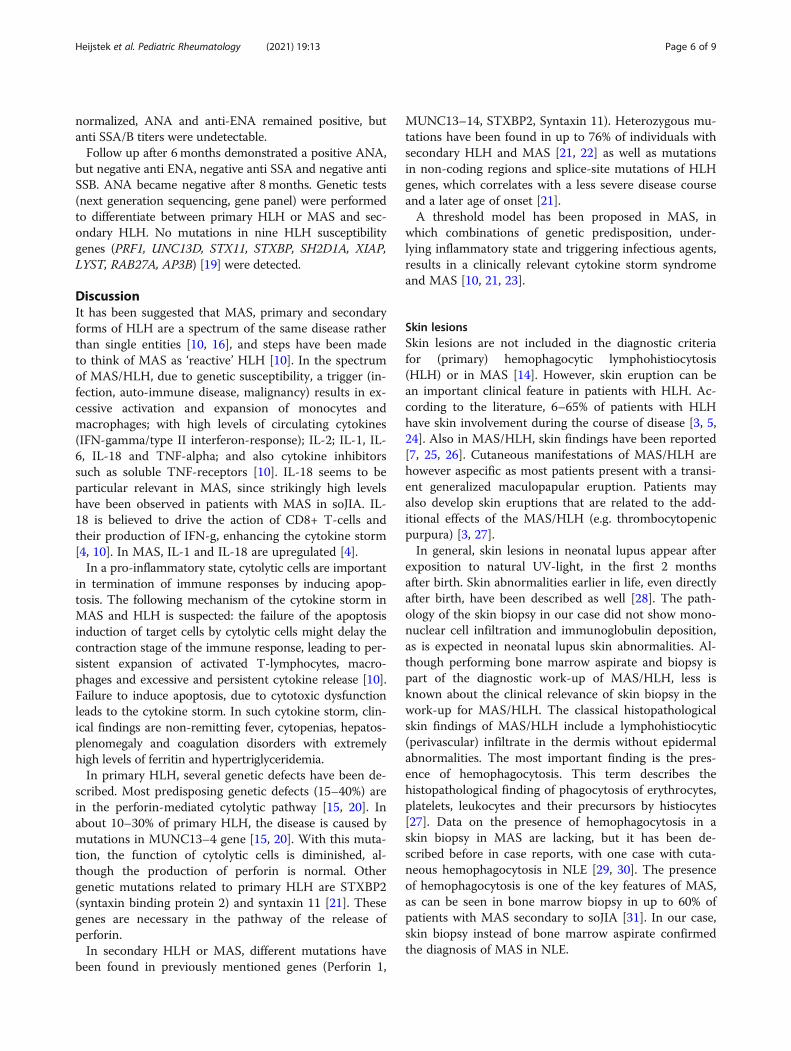
cutaneous lupus and congenital complete heart block;NLE can also present with hepatitis, cytopenias andneurological abnormalities [2]. Macrophage activationsyndrome (MAS) is a life-threatening hyperinflammatorysyndrome and is caused by a severely dysregulated im-mune response. It has rarely been associated with neo-natal lupus. So far, only four patients have been reportedin the literature (Table 1) [6–9].Although MAS is a well-known complication of
systemic-onset juvenile idiopathic arthritis (soJIA) itless frequently occurs associated with other auto-immune diseases such as Kawasaki disease, systemiclupus erythematosus, polyarticular juvenile idiopathicarthritis, juvenile dermatomyositis, anti-phospholipidsyndrome and mixed connective tissue disease [10–13]. MAS is considered to represent a secondary formof hemophagocytic lymphohistiocytosis (HLH). Diag-nosing MAS in diseases other then soJIA is challen-ging, because diagnostic criteria are only validated insoJIA. Skin findings are not included in the diagnosticcriteria for (primary) hemophagocytic lymphohistiocy-tosis (HLH) or in MAS [14]. However, skin eruptioncan be an important clinical finding in patients withHLH.Primary HLH is a group of autosomal recessive im-
mune disorders, all leading to a life-threatening hyperin-flammatory state. It is linked to various genetic defects,mostly affecting the perforin-mediated cytolytic pathway[10, 11]. Perforin is a protein, necessary for inducingapoptosis of target cells (viruses, tumors) [15].It has been suggested that MAS, primary and second-
ary forms of HLH are a spectrum of the same disease ra-ther than single entities [10, 16], and steps have beenmade to think of MAS as ‘reactive’ HLH. Genetic studiesconfirm overlap between these entities.In this case report, we show a case of MAS as a com-
plication of NLE, potentially triggered by infection andmaybe surgery as well [17], without a known HLH-susceptibility gene mutation. Additionally, we present areview of the literature of the pathophysiology of MASand HLH in NLE patients.
CaseA 29-year old, secondary gravida female was referred tothe obstetrics department. She was diagnosed with cuta-neous lupus erythematosus several years before preg-nancy. She had positive anti-nuclear antibodies (ANA),anti-extractable nuclear antigen (anti-ENA), anti-SSA andanti-SSB. Treatment before pregnancy consisted of topicalsteroids. Before pregnancy, she was never treated with sys-temic therapy. During gestation she was asymptomaticwithout medication. Pregnancy was followed carefully atour obstetrics department because of her condition. Astructural ultrasound examination at 20 2/7 weeks of ges-tation showed a fetal bradycardia (50 beats per minute(bpm)), based on a complete atrioventricular (AV) blockin absence of a structural heart disease. No other struc-tural abnormalities were found and no hydrops waspresent. Although controversial and a matter of debate [2,18], mother was treated after diagnosis of the AV blockwith dexamethasone in order to suppress a potential on-going inflammation to prevent fibrotic replacement in theAV node. No improvement of the fetal heart block wasobserved during close follow-up.At 37 2/7 weeks of gestation, a female infant was born,
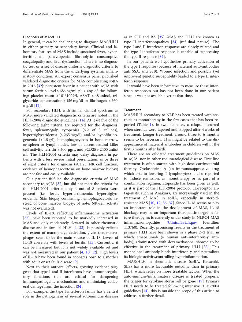
birth weight was 2330 g (p5) and APGAR scores 9/10.Complete AV block was confirmed and there was ajunctional escape rhythm of 50 bpm (Fig. 1a). The childwas admitted to our neonatal intensive care unit forclinical observation. Due to circulatory insufficiencycaused by the total AV block with a junction escaperhythm of 50 bpm, isoprenalin was started. Implantationof an epicardial VVI pacemaker system in abdominalposition (Microny™, St Jude Medical) was inevitable onday 6.Two days later, the patient developed fever and the
pacemaker implantation wound appeared erythematousand inflamed. Blood- and wound cultures acquired be-fore and during antibiotic treatment remained sterile. C-reactive protein (CRP) was maximum 80mg/L on day 9post-birth (ref < 5 mg/L). She was treated for a suspectedwound infection with flucloxacillin and gentamycinintravenously. Furthermore, she was treated for perianalcandidiasis with topical application of miconazole cream.Despite antibiotic therapy, there was an ongoing rednessaround the pacemaker pocket with cloudy, purulent fluiddraining from the wound and fever persisted, one weekafter pacemaker implantation.Surgical wound exploration was performed showing
infiltrates with pus, for which antibiotic treatment wasswitched to ceftazidime. The pacemaker system was leftinside and the wound was left open. In the days after ex-ploration the redness slightly diminished around the ele-vated wound edges with granulation tissue appearing inthe wound bed (Fig. 1b). CRP values remained low; vary-ing between 6 and 13mg/L. In between, fever persisted.
Heijstek et al. Pediatric Rheumatology (2021) 19:13 Page 2 of 9

Table
1Macroph
ageactivationsynd
romein
neon
atallupu
serythe
matosus
-de
scrip
tionof
four
casesin
theliterature
Sex
Antibod
ies
Clin
ical
signs
Maxim
umFe
rritin
Ng/m
l
Aminotransferases
IU/L
Hem
atolog
yTriglycerides
Interleu
kins
Gen
etics
Therap
yOutco
me
Referenc
e
MAntiR
o/SSAAnti
La/SSB
22hafterbirth:
Com
pleteAV-block
Cardiop
ulmon
ary
resuscitatio
n
9769
(32h)
ASA
T1027
ALA
T121
LDH3490
Trom
bope
nia
day24
(90×
10^3)
-SolIL-23230
U/m
lHLH
-ge
nes
negative
Hydrocortison
eRelapseday24th
Hydrocortison
etill3
mon
ths
Severe
psycho
motor
retardation
Suzuki
(2013)
[3]
MAntiR
o/SSA
AntiLa/SSB
Directlyafterbirth:
Skin:ann
ular
plaques
660
ASA
T257
LDH633
Trom
bope
nia
(104
×10^3)
SolIL-22280
U/m
lPred
nisolone
1mg/kg
day8till6
mon
ths
Goo
dShim
ozaw
a(2015)
[4]
FANA
AntiR
o/SSA
10days
afterbirth:
Tachypno
ea,Fever
Hep
atosplen
omeg
aly
Maxim
um2891
Maxim
umday10
ASA
T459
ALA
T463
Ane
mia(12.8
mg/dl)
Trom
bope
nia
lowest12
(day
18)
Day
10:280
mg/dL
IVIGS(1g/kg
2days)
Methylpredn
isolon
epu
lse(HLH
protocol
2004)Relapseday26:
Ciclosporin
6mg/kg.54day’s
steroids
2mon
ths:
good
Park
(2015)
[5]
FANA
AntiENA
AntiR
o/SSA
AntiSSB/La
Fever
Skin
lesion
s(ann
ular
plaques)
Maxim
um4439
Maxim
umASA
T84
ALA
T33
LDH909
Day
5Trom
bocytes
135
Hblowest
7.1mmol/L
day32
3.88
mmol/L
SolIL-
221803
pg/
ml
HLH
-ge
nes
negative
Pred
nisolone
1mg/kg`
Tape
reddo
wnin
6mon
ths
Goo
dAntibod
ies
negativeafter
6mon
ths
2020
(our
case)
Heijstek et al. Pediatric Rheumatology (2021) 19:13 Page 3 of 9

The patient developed a generalized non-pruritic skineruption on the 22nd day of life. Numerous round tooval and partly confluent erythematous papules and pla-ques, located on the forehead and eyebrows, as well ason the trunk and extremities were noted (Fig. 1c). Somepapular lesions had an erosive center. The oral mucosa,palms and soles were not affected. There was no hepa-tosplenomegaly or lymphadenopathy.Persistent fever, despite antibiotic treatment, and the atypical
extensive skin lesions prompted us to think of other diagnosislike MAS. Our differential diagnosis consisted of persistent
infection (systemic candidiasis, CMV, EBV varicella, (perianalskin culture positive for Candida albicans. CMV and EBVboth (maternal) IgG positive, IgM negative)); auto-immune(complex neonatal lupus) or auto-inflammatory disease(MAS, primary HLH) or a primary immunodeficiency.Additional blood tests showed a ferritin level of 4162
mug/L (ref. 10–150 mug/L), a mild anemia (Hb 7.1mmol/L; 114.4 g/L; ref. 8.5–12.5mmol/L), leukocytes andthrombocyte counts remained normal. Fibrinogen was 3.6g/L (ref. 2.1–3.8 g/L), D-dimer 3996 ng/ml; ref. < 500 ng/ml). Furthermore, mildly elevated aminotransferase levels
Fig. 1 a ECG: third degree AV block with junctional escape rhythm (~50 bpm), atrial rate ~115 bpm. b Day 12: Midsternal pacemaker pocketwound 7 days after surgical exploration for infection, pacemaker in situ. c Day 22: Numerous round to oval and partly confluent erythematouspapules and plaques, located on the forehead and eyebrows, as well as on the trunk and extremities were noted. d Pathology of skin biopsy: Adense dermal (perivascular) infiltrate is shown on the left. The red arrow points to a hemophagocytic macrophage. e Resolving skin lesions, fourweeks after start of treatment
Heijstek et al. Pediatric Rheumatology (2021) 19:13 Page 4 of 9
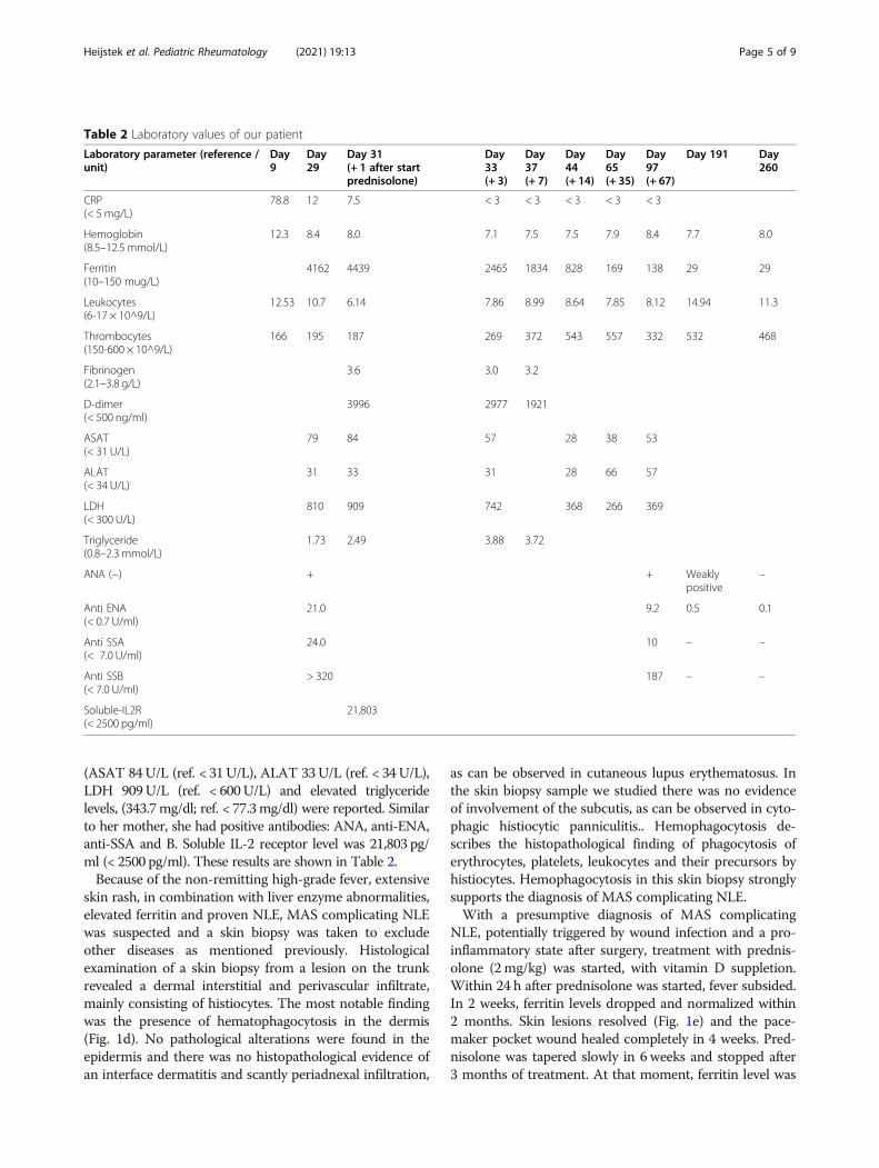
(ASAT 84U/L (ref. < 31U/L), ALAT 33U/L (ref. < 34U/L),LDH 909U/L (ref. < 600U/L) and elevated triglyceridelevels, (343.7mg/dl; ref. < 77.3mg/dl) were reported. Similarto her mother, she had positive antibodies: ANA, anti-ENA,anti-SSA and B. Soluble IL-2 receptor level was 21,803 pg/ml (< 2500 pg/ml). These results are shown in Table 2.Because of the non-remitting high-grade fever, extensive
skin rash, in combination with liver enzyme abnormalities,elevated ferritin and proven NLE, MAS complicating NLEwas suspected and a skin biopsy was taken to excludeother diseases as mentioned previously. Histologicalexamination of a skin biopsy from a lesion on the trunkrevealed a dermal interstitial and perivascular infiltrate,mainly consisting of histiocytes. The most notable findingwas the presence of hematophagocytosis in the dermis(Fig. 1d). No pathological alterations were found in theepidermis and there was no histopathological evidence ofan interface dermatitis and scantly periadnexal infiltration,
as can be observed in cutaneous lupus erythematosus. Inthe skin biopsy sample we studied there was no evidenceof involvement of the subcutis, as can be observed in cyto-phagic histiocytic panniculitis.. Hemophagocytosis de-scribes the histopathological finding of phagocytosis oferythrocytes, platelets, leukocytes and their precursors byhistiocytes. Hemophagocytosis in this skin biopsy stronglysupports the diagnosis of MAS complicating NLE.With a presumptive diagnosis of MAS complicating
NLE, potentially triggered by wound infection and a pro-inflammatory state after surgery, treatment with prednis-olone (2mg/kg) was started, with vitamin D suppletion.Within 24 h after prednisolone was started, fever subsided.In 2 weeks, ferritin levels dropped and normalized within2 months. Skin lesions resolved (Fig. 1e) and the pace-maker pocket wound healed completely in 4 weeks. Pred-nisolone was tapered slowly in 6 weeks and stopped after3 months of treatment. At that moment, ferritin level was
Table 2 Laboratory values of our patient
Laboratory parameter (reference /unit)
Day9
Day29
Day 31(+ 1 after startprednisolone)
Day33(+ 3)
Day37(+ 7)
Day44(+ 14)
Day65(+ 35)
Day97(+ 67)
Day 191 Day260
CRP(< 5mg/L)
78.8 12 7.5 < 3 < 3 < 3 < 3 < 3
Hemoglobin(8.5–12.5 mmol/L)
12.3 8.4 8.0 7.1 7.5 7.5 7.9 8.4 7.7 8.0
Ferritin(10–150 mug/L)
4162 4439 2465 1834 828 169 138 29 29
Leukocytes(6-17 × 10^9/L)
12.53 10.7 6.14 7.86 8.99 8.64 7.85 8.12 14.94 11.3
Thrombocytes(150-600 × 10^9/L)
166 195 187 269 372 543 557 332 532 468
Fibrinogen(2.1–3.8 g/L)
3.6 3.0 3.2
D-dimer(< 500 ng/ml)
3996 2977 1921
ASAT(< 31 U/L)
79 84 57 28 38 53
ALAT(< 34 U/L)
31 33 31 28 66 57
LDH(< 300 U/L)
810 909 742 368 266 369
Triglyceride(0.8–2.3 mmol/L)
1.73 2.49 3.88 3.72
ANA (−) + + Weaklypositive
–
Anti ENA(< 0.7 U/ml)
21.0 9.2 0.5 0.1
Anti SSA(< 7.0 U/ml)
24.0 10 – –
Anti SSB(< 7.0 U/ml)
> 320 187 – –
Soluble-IL2R(< 2500 pg/ml)
21,803
Heijstek et al. Pediatric Rheumatology (2021) 19:13 Page 5 of 9
normalized, ANA and anti-ENA remained positive, butanti SSA/B titers were undetectable.Follow up after 6months demonstrated a positive ANA,
but negative anti ENA, negative anti SSA and negative antiSSB. ANA became negative after 8months. Genetic tests(next generation sequencing, gene panel) were performedto differentiate between primary HLH or MAS and sec-ondary HLH. No mutations in nine HLH susceptibilitygenes (PRF1, UNC13D, STX11, STXBP, SH2D1A, XIAP,LYST, RAB27A, AP3B) [19] were detected.
DiscussionIt has been suggested that MAS, primary and secondaryforms of HLH are a spectrum of the same disease ratherthan single entities [10, 16], and steps have been madeto think of MAS as ‘reactive’ HLH [10]. In the spectrumof MAS/HLH, due to genetic susceptibility, a trigger (in-fection, auto-immune disease, malignancy) results in ex-cessive activation and expansion of monocytes andmacrophages; with high levels of circulating cytokines(IFN-gamma/type II interferon-response); IL-2; IL-1, IL-6, IL-18 and TNF-alpha; and also cytokine inhibitorssuch as soluble TNF-receptors [10]. IL-18 seems to beparticular relevant in MAS, since strikingly high levelshave been observed in patients with MAS in soJIA. IL-18 is believed to drive the action of CD8+ T-cells andtheir production of IFN-g, enhancing the cytokine storm[4, 10]. In MAS, IL-1 and IL-18 are upregulated [4].In a pro-inflammatory state, cytolytic cells are important
in termination of immune responses by inducing apop-tosis. The following mechanism of the cytokine storm inMAS and HLH is suspected: the failure of the apoptosisinduction of target cells by cytolytic cells might delay thecontraction stage of the immune response, leading to per-sistent expansion of activated T-lymphocytes, macro-phages and excessive and persistent cytokine release [10].Failure to induce apoptosis, due to cytotoxic dysfunctionleads to the cytokine storm. In such cytokine storm, clin-ical findings are non-remitting fever, cytopenias, hepatos-plenomegaly and coagulation disorders with extremelyhigh levels of ferritin and hypertriglyceridemia.In primary HLH, several genetic defects have been de-
scribed. Most predisposing genetic defects (15–40%) arein the perforin-mediated cytolytic pathway [15, 20]. Inabout 10–30% of primary HLH, the disease is caused bymutations in MUNC13–4 gene [15, 20]. With this muta-tion, the function of cytolytic cells is diminished, al-though the production of perforin is normal. Othergenetic mutations related to primary HLH are STXBP2(syntaxin binding protein 2) and syntaxin 11 [21]. Thesegenes are necessary in the pathway of the release ofperforin.In secondary HLH or MAS, different mutations have
been found in previously mentioned genes (Perforin 1,
MUNC13–14, STXBP2, Syntaxin 11). Heterozygous mu-tations have been found in up to 76% of individuals withsecondary HLH and MAS [21, 22] as well as mutationsin non-coding regions and splice-site mutations of HLHgenes, which correlates with a less severe disease courseand a later age of onset [21].A threshold model has been proposed in MAS, in
which combinations of genetic predisposition, under-lying inflammatory state and triggering infectious agents,results in a clinically relevant cytokine storm syndromeand MAS [10, 21, 23].
Skin lesionsSkin lesions are not included in the diagnostic criteriafor (primary) hemophagocytic lymphohistiocytosis(HLH) or in MAS [14]. However, skin eruption can bean important clinical feature in patients with HLH. Ac-cording to the literature, 6–65% of patients with HLHhave skin involvement during the course of disease [3, 5,24]. Also in MAS/HLH, skin findings have been reported[7, 25, 26]. Cutaneous manifestations of MAS/HLH arehowever aspecific as most patients present with a transi-ent generalized maculopapular eruption. Patients mayalso develop skin eruptions that are related to the add-itional effects of the MAS/HLH (e.g. thrombocytopenicpurpura) [3, 27].In general, skin lesions in neonatal lupus appear after
exposition to natural UV-light, in the first 2 monthsafter birth. Skin abnormalities earlier in life, even directlyafter birth, have been described as well [28]. The path-ology of the skin biopsy in our case did not show mono-nuclear cell infiltration and immunoglobulin deposition,as is expected in neonatal lupus skin abnormalities. Al-though performing bone marrow aspirate and biopsy ispart of the diagnostic work-up of MAS/HLH, less isknown about the clinical relevance of skin biopsy in thework-up for MAS/HLH. The classical histopathologicalskin findings of MAS/HLH include a lymphohistiocytic(perivascular) infiltrate in the dermis without epidermalabnormalities. The most important finding is the pres-ence of hemophagocytosis. This term describes thehistopathological finding of phagocytosis of erythrocytes,platelets, leukocytes and their precursors by histiocytes[27]. Data on the presence of hemophagocytosis in askin biopsy in MAS are lacking, but it has been de-scribed before in case reports, with one case with cuta-neous hemophagocytosis in NLE [29, 30]. The presenceof hemophagocytosis is one of the key features of MAS,as can be seen in bone marrow biopsy in up to 60% ofpatients with MAS secondary to soJIA [31]. In our case,skin biopsy instead of bone marrow aspirate confirmedthe diagnosis of MAS in NLE.
Heijstek et al. Pediatric Rheumatology (2021) 19:13 Page 6 of 9
Diagnosis of MAS/HLHIn general, it can be challenging to diagnose MAS/HLHin either primary or secondary forms. Clinical and la-boratory features of MAS include sustained fever, hyper-ferritinemia, pancytopenia, fibrinolytic consumptivecoagulopathy and liver dysfunction. There is no diagnos-tic test or a set of disease uniform diagnostic criteria todifferentiate MAS from the underlying systemic inflam-matory condition. An expert consensus panel publishedvalidated diagnostic criteria for MAS complicating soJIAin 2016 [32]: persistent fever in a patient with soJIA withserum ferritin level > 684 ng/ml plus any of the follow-ing: platelet count < 181*10^9/l, ASAT > 48 units/l, tri-glyceride concentration > 156 mg/dl or fibrinogen < 360mg/dl [12].For secondary HLH, with similar clinical spectrum as
MAS, more validated diagnostic criteria are noted in theHLH-2004 diagnostic guidelines [14]. At least five of thefollowing eight criteria are required for the diagnosis:fever, splenomegaly, cytopenias (> 2 of 3 cellines),hypertriglyceridemia (> 265 mg/dl) and/or hypofibrino-genemia (< 1.5 g/l), hemophagocytosis in bone marrowor spleen or lymph nodes, low or absent natural killercell activity, ferritin > 500 μg/L and sCD25 > 2400 units/ml. The HLH-2004 criteria may delay diagnosis in pa-tients with a less severe initial presentation, since threeof eight criteria for diagnosis (sCD25, NK cell function,evidence of hemophagocytosis on bone marrow biopsy)are not fast and easily available.Our patient fulfilled the diagnostic criteria of MAS
secondary to soJIA [32] but did not meet the criteria forthe HLH-2004 criteria: only 4 out of 8 criteria werepresent (i.e. fever, hyperferritinemia, hypertriglyc-eridemia. Skin biopsy confirming hemophagocytosis in-stead of bone marrow biopsy; of note: NK-cell activitywas not evaluated).Levels of IL-18, reflecting inflammasome activation
[33], have been reported to be markedly increased inMAS and only moderately elevated in other rheumaticdisease and in familial HLH [4, 33]. It possibly reflectsthe extent of macrophage activation, given that macro-phages seem to be the main source of IL-18. Levels ofIL-18 correlate with levels of ferritin [33]. Currently, itcan be measured but it is not widely available yet andwas not measured in our patient [4, 10, 12]. High levelsof IL-18 have been found in neonates born to a motherwith adult onset Stills disease [9].Next to their antiviral effects, growing evidence sug-
gests that type I and II interferons have immunoregula-tory functions that are critical for dampeningimmunopathogenic mechanisms and minimizing collat-eral damage from the infection [34].For example, the type I interferon family has a central
role in the pathogenesis of several autoimmune diseases
as in SLE and RA [35]. MAS and HLH are known astype II interferonopathies [34] (ref dual nature). Thetype I and II interferon response are closely related andthe type I interferon response is capable of suppressingthe type II response [34].In our patient; we hypothesize primary activation of
the type I response (because of maternal auto-antibodiesanti SSA, anti SSB). Wound infection and possibly (yetunproven) genetic susceptibility leaded to a type II inter-feron response.It would have been informative to measure these inter-
feron responses but has not been done in our patientsince it was not available yet at that time.
TreatmentMAS/HLH secondary to NLE has been treated with ste-roids as monotherapy in the few cases that has been re-ported (Table 1). In two neonates, a relapse occurredwhen steroids were tapered and stopped after 4 weeks oftreatment. Longer treatment, around three to 6 monthsseems to be necessary. This might be related to the dis-appearance of maternal antibodies in children within thefirst 3 months after birth.There are no validated treatment guidelines on MAS
in soJIA, nor in other rheumatological disease. First-linetreatment is often started with high-dose corticosteroidtherapy. Cyclosporine A (an immunosuppressive drugwhich acts in lowering T-lymphocytes) is also reportedto induce remission, as monotherapy or as part of acombination regimen. Etoposide has been given as well,as it is part of the HLH-2004 protocol. IL-receptor an-tagonists, such as Anakinra, are increasingly used in thetreatment of MAS in soJIA, especially in steroid-resistant MAS [10, 12, 36, 37]. Since IL-18 seems to playan important role in the development of MAS, IL-18blockage may be an important therapeutic target in fu-ture therapy, as is currently under study in NLRC4-MASinflammasomopathies (ClinicalTrials.gov Identifier:113760). Recently, promising results in the treatment ofprimary HLH have been shown in a phase 2–3 trial, inwhich emapalumab (a human anti-interferon-y anti-body); administered with dexamethasone, showed to beeffective in the treatment of primary HLH [38]. Thismonoclonal antibody binds interferon-y and neutralizesits biologic activity,controlling hyperinflammation.MAS/HLH in rheumatic disease (soJIA, Kawasaki,
SLE) has a more favourable outcome than in primaryHLH, which relies on more treatable factors. When theauto-immune/inflammatory disease is treated properly,the trigger for cytokine storm will be gone [19]. PrimaryHLH needs to be treated following intensive HLH-2004guidelines [14], this is outside the scope of this article toaddress in further detail.
Heijstek et al. Pediatric Rheumatology (2021) 19:13 Page 7 of 9

Our patient has been followed for the first 3 years. Shehad a growth restriction; her development is normal.The complete AV block persisted and the implantedpacemaker device is working properly. The trigger forMAS/HLH is gone, autoantibodies have become nega-tive. Although long-term effects of NLE complicated byHLH/MAS are not known, no important sequellae havebeen observed so far.
ConclusionIn children with NLE or in children born from motherswho are SSA- and/or SSB-positive, every condition withpersistent fever, abnormal wound healing, atypical skineruption, should raise awareness of the possible diagno-sis of MAS/HLH complicating NLE.Recent diagnostic criteria of MAS in soJIA seem to be
useful in more rheumatological diseases, for instanceNLE, but have not been validated yet. A lesional skin bi-opsy can be taken to assess the presence of hemophago-cytosis in the dermal compartment. The results of a skinbiopsy may add in the diagnostic work-up and may leadto treatment at an earlier stage.
AbbreviationsANA: Anti-nuclear antibodies; Anti-ENA: Anti-extractable nuclear antigen;Anti-SSA: Anti-Sjögren-syndrome-related antigen A / also called anti-Ro; Anti-SSB: Anti-Sjögren-syndrome-related antigen B / also called anti-La; AVblock: Atrioventricular block; AV node: Atrioventricular node;CMV: Cytomegalovirus; CRP: C-reactive protein; EBV: Epstein-Barr virus;HLH: Hemophagocytic lymphohistiocytosis; JIA: Juvenile idiopathic arthritis;MAS: Macrophage activation syndrome; NLE: Neonatal lupus erythematosus;soJIA: Systemic-onset juvenile idiopathic arthritis; SLE: Systemic lupuserythematosus; LDH: Lactate dehydrogenase
AcknowledgmentsNot applicable.
Authors’ contributionsVH evaluated the patient and wrote the article; MH wrote the part aboutskin findings in neonatal lupus. RvdP analyzed and edited the cardiologicalpart of the article and provided images of the ECG and midsternal woundbed. RvD and PHM were contributors in writing and editing the manuscript.All authors read and approved the final manuscript.
FundingNot applicable.
Availability of data and materialsNot applicable.
Ethics approvalNot applicable.
Consent for publicationSee consent form.
Competing interestsThe authors declare that they have no competing interests.
Author details1Department of Pediatric Rheumatology, Leiden University Medical Center,Leiden, the Netherlands. 2Department of Dermatology, Leiden UniversityMedical Center, Leiden, the Netherlands. 3Division of Pediatric Cardiology,
Department of Pediatrics, Leiden University Medical Center, Leiden, theNetherlands.
Received: 29 July 2020 Accepted: 4 February 2021
References1. Klein-Gitelman MS. Neonatal lupus: what we have learned and current
approaches to care. Curr Rheumatol Rep. 2016;18(9):60.2. Vanoni F, Lava SAG, Fossali EF, Cavalli R, Simonetti GD, Bianchetti MG, et al.
Neonatal systemic lupus erythematosus syndrome: a comprehensive review.Clin Rev Allergy Immunol. 2017;53(3):469–76.
3. Morrell DS, Pepping MA, Scott JP, Esterly NB, Drolet BA. Cutaneousmanifestations of hemophagocytic lymphohistiocytosis. Arch Dermatol.2002;138(9):1208–12.
4. Griffin G, Shenoi S, Hughes GC. Hemophagocytic lymphohistiocytosis: anupdate on pathogenesis, diagnosis, and therapy. Best Pract Res ClinRheumatol. 2020;34(4):101515. https://doi.org/10.1016/j.berh.2020.101515. Epub2020 May 7.
5. Jun HJ, Kim HO, Lee JY, Park YM. Preceding annular skin lesions in a patientwith Hemophagocytic Lymphohistiocytosis. Ann Dermatol. 2015;27(5):608–11.
6. Park JH, Kim SH, Kim HJ, Lee SJ, Jeong DC, Kim SY. Macrophage activationsyndrome in a newborn infant born to a mother with autoimmune disease.J Perinatol. 2015;35(2):158–60.
7. Shimozawa H, Kono Y, Matano M, Suzuki Y, Koike Y, Yada Y, et al. Cytokineprofile in two siblings with neonatal lupus erythematosus. Pediatr Int. 2015;57(6):1211–4.
8. Suzuki Y, Takahashi N, Yada Y, Koike Y, Matano M, Nishimura H, et al.Hemophagocytic lymphohistiocytosis in a newborn infant born to a motherwith Sjogren syndrome antibodies. J Perinatol. 2013;33(7):569–71.
9. Shimizu M, Kizawa T, Kato R, Suzuki T, Yachie A. Macrophage activationsyndrome in neonates born to mothers with adult-onset Still's disease:perinatal effect of maternal IL-18. Clin Immunol. 2019;207:36–9.
10. Grom AA, Horne A, De Benedetti F. Macrophage activation syndrome in theera of biologic therapy. Nat Rev Rheumatol. 2016;12(5):259–68.
11. Sen ES, Steward CG, Ramanan AV. Diagnosing haemophagocytic syndrome.Arch Dis Child. 2017;102(3):279–84.
12. Crayne CB, Albeituni S, Nichols KE, Cron RQ. The immunology ofmacrophage activation syndrome. Front Immunol. 2019;10:119.
13. Lerkvaleekul B, Vilaiyuk S. Macrophage activation syndrome: early diagnosisis key. Open Access Rheumatol. 2018;10:117–28.
14. Henter JI, Horne A, Arico M, Egeler RM, Filipovich AH, Imashuku S, et al.HLH-2004: diagnostic and therapeutic guidelines for hemophagocyticlymphohistiocytosis. Pediatr Blood Cancer. 2007;48(2):124–31.
15. Schulert GS, Grom AA. Pathogenesis of macrophage activationsyndrome and potential for cytokine- directed therapies. Annu RevMed. 2015;66:145–59.
16. Bracaglia C, Prencipe G, De Benedetti F. Macrophage activation syndrome:different mechanisms leading to a one clinical syndrome. PediatrRheumatol Online J. 2017;15(1):5.
17. Dabrowska AM, Slotwinski R. The immune response to surgery andinfection. Cent Eur J Immunol. 2014;39(4):532–7.
18. DeNoble AE, Kuller JA, Rhee EJ. Controversies in the Management ofIsolated Congenital Atrioventricular Block. Obstet Gynecol Surv. 2015;70(8):518–23.
19. Chinn IK, Eckstein OS, Peckham-Gregory EC, Goldberg BR, Forbes LR,Nicholas SK, et al. Genetic and mechanistic diversity in pediatrichemophagocytic lymphohistiocytosis. Blood. 2018;132(1):89–100.
20. Ravelli A, Grom AA, Behrens EM, Cron RQ. Macrophage activation syndromeas part of systemic juvenile idiopathic arthritis: diagnosis, genetics,pathophysiology and treatment. Genes Immun. 2012;13(4):289–98.
21. Zhang M, Behrens EM, Atkinson TP, Shakoory B, Grom AA, Cron RQ. Geneticdefects in cytolysis in macrophage activation syndrome. Curr RheumatolRep. 2014;16(9):439.
22. Kaufman KM, Linghu B, Szustakowski JD, Husami A, Yang F, Zhang K, et al.Whole-exome sequencing reveals overlap between macrophage activationsyndrome in systemic juvenile idiopathic arthritis and familial hemophagocyticlymphohistiocytosis. Arthritis Rheumatol. 2014;66(12):3486–95.
23. Strippoli R, Caiello I, De Benedetti F. Reaching the threshold: a multilayerpathogenesis of macrophage activation syndrome. J Rheumatol. 2013;40(6):761–7.
Heijstek et al. Pediatric Rheumatology (2021) 19:13 Page 8 of 9

24. Bay A, Calka O, Akdeniz N, Oner AF, Kirimi E. Newborn infant withhemophagocytic lymphohistiocytosis and generalized skin eruptions. JDermatol. 2006;33(9):628–31.
25. Shwin KW, Lee CR, Goldbach-Mansky R. Dermatologic manifestations ofmonogenic autoinflammatory diseases. Dermatol Clin. 2017;35(1):21–38.
26. Li X, Qu B, Nie Y, Zhu G, Li W, Mu F. Clinical features of macrophageactivation syndrome in the adult northern Chinese population. Lupus. 2014;23(8):785–92.
27. Santos-Arroyo A, Barrera-Llaurador J, Sanchez JE, Martin-Garcia R, SanchezJL. Role of skin biopsies in the diagnosis of HemophagocyticLymphohistiocytosis. Am J Dermatopathol. 2017;39(7):e86–e9.
28. Cimaz R, Biggioggero M, Catelli L, Muratori S, Cambiaghi S. Ultraviolet lightexposure is not a requirement for the development of cutaneous neonatallupus. Lupus. 2002;11(4):257–60.
29. Chamseddin B, Marks E, Dominguez A, Wysocki C, Vandergriff T. Refractorymacrophage activation syndrome in the setting of adult-onset still diseasewith hemophagocytic lymphohistiocytosis detected on skin biopsy treatedwith canakinumab and tacrolimus. J Cutan Pathol. 2019;46(7):528–31.https://doi.org/10.1111/cup.13466. Epub 2019 Apr 23.
30. Kerl K, Wolf IH, Cerroni L, Wolf P, French LE, Kerl H. Hemophagocytosis incutaneous autoimmune disease. Am J Dermatopathol. 2015;37(7):539–43.
31. Cron RQ, Davi S, Minoia F, Ravelli A. Clinical features and correct diagnosisof macrophage activation syndrome. Expert Rev Clin Immunol. 2015;11(9):1043–53.
32. Ravelli A, Minoia F, Davi S, Horne A, Bovis F, Pistorio A, et al. 2016classification criteria for macrophage activation syndrome complicatingsystemic juvenile idiopathic arthritis: a European league againstrheumatism/American College of Rheumatology/Paediatric rheumatologyinternational trials organisation collaborative initiative. Arthritis Rheumatol.2016;68(3):566–76.
33. Canna SW, Marsh RA. Pediatric hemophagocytic lymphohistiocytosis. Blood.2020;135(16):1332–43.
34. Lee AJ, Ashkar AA. The dual nature of type I and type II interferons. FrontImmunol. 2018;9:2061.
35. El-Sherbiny YM, Psarras A, Md Yusof MY, Hensor EMA, Tooze R, Doody G,et al. A novel two-score system for interferon status segregatesautoimmune diseases and correlates with clinical features. Sci Rep. 2018;8(1):5793.
36. Boom V, Anton J, Lahdenne P, Quartier P, Ravelli A, Wulffraat NM, et al.Evidence-based diagnosis and treatment of macrophage activationsyndrome in systemic juvenile idiopathic arthritis. Pediatr Rheumatol OnlineJ. 2015;13:55.
37. Mehta P, Cron RQ, Hartwell J, Manson JJ, Tattersall RS. Silencing thecytokine storm: the use of intravenous anakinra in haemophagocyticlymphohistiocytosis or macrophage activation syndrome. Lancet Rheumatol.2020.
38. Locatelli F, Jordan MB, Allen C, Cesaro S, Rizzari C, Rao A, et al. Emapalumabin children with primary Hemophagocytic Lymphohistiocytosis. N Engl JMed. 2020;382(19):1811–22.
Publisher’s NoteSpringer Nature remains neutral with regard to jurisdictional claims inpublished maps and institutional affiliations.
Heijstek et al. Pediatric Rheumatology (2021) 19:13 Page 9 of 9