macpherson et al., j drug metab toxicol drug metabolism ... · macpherson et al., j drug metab...
TRANSCRIPT

Volume 4 • Issue 1 • 1000140J Drug Metab ToxicolISSN: 2157-7609 JDMT, an open access journal
Research Article Open Access
Macpherson et al., J Drug Metab Toxicol 2013, 4:1http://dx.doi.org/10.4172/2157-7609.1000140
Research Article Open Access
Drug Metabolism & Toxicology
*Corresponding author: Rainsford KD, Professor, Biomedical Research Centre, Sheffield Hallam University, Howard Street, Sheffield, S1 1WB, UK, Fax: +44-1246-583 815; E-mail: [email protected]
Received January 09, 2013; Accepted January 24, 2013; Published January 28, 2013
Citation: Macpherson D, Best SA, Gedik L, Hewson AT, Rainsford KD, et al. (2013) The Biotransformation and Pharmacokinetics of 14C-Nimesulide in Humans Following a Single Dose Oral Administration. J Drug Metab Toxicol 4: 140. doi:10.4172/2157-7609.1000140
Copyright: © 2013 Macpherson D, et al. This is an open-access article distributed under the terms of the Creative Commons Attribution License, which permits unrestricted use, distribution, and reproduction in any medium, provided the original author and source are credited.
AbstractNimesulide is a preferential cyclo-oxygenase-2 inhibitory non-steroidal anti-inflammatory drug has, infrequently,
been associated with hepatic reactions. To establish the extent of formation of various metabolites (some of which might be hepato-reactive) the whole body metabolism, plasma kinetics and routes of excretion of the radio-labelled drug was undertaken in 4 fasted male volunteers following an oral dose of 100 mg [14C]-nimesulide. Urine, faecal and plasma samples were collected up to 168 h post dose, the total radioactivity and plasma concentrations of nimesulide and its principle metabolite, 4-hydroxynimesulide, were determined. Radio labelled metabolites in these samples was identified by combined liquid chromatography-mass spectrometry.
The mean elimination half-life of total radioactivity in the plasma and whole blood was circa 4.8 h; the ratio whole blood and plasma being circa 0.6 h. The mean elimination half-lives for nimesulide and 4-hydroxynimesulide in plasma were circa 2.5 h and circa 3.9 h, respectively. The drug was rapidly excreted and recoveries were 59-66% in the urine and 33-39% in the faeces at 168 hours.
A total of 16 metabolites were identified including the conjugated metabolites, which exceeds the 5 previously identified. Nimesulide was to be metabolised by 5 pathways involving (a) cleavage of the molecule at the ether linkage (b) reduction of the NO2 group to NH2, and (c) ring hydroxylation followed by conjugation with either glucuronic acid or sulphate. In conclusion, the biotransformation pathway for nimesulide in man has now been comprehensively determined with 92% of the urinary metabolites fully characterised. The identification of some rare metabolites of nimesulide may help in understanding the mechanisms of hepatotoxicity from this drug.
The Biotransformation and Pharmacokinetics of 14C-Nimesulide in Humans Following a Single Dose Oral AdministrationDavid Macpherson1, Stuart A Best1, Layla Gedik1, Alan T Hewson2, Rainsford KD2* and Simona Parisi2
1Department of Metabolism Chemistry, Inveresk Research (now Charles River, Edinburgh), Tranent EH33 2NE, Scotland, UK2Biomedical Research Centre, Sheffield Hallam University, Sheffield, S1 1WB, England, UK
Keywords: Nimesulide; Anti-inflammatory drug; Pharmacokinetics; Mass spectrophotometric analysis
IntroductionNimesulide (2-phenoxy-4-nitro-methanesulphonanilide) is a
non-steroidal anti-inflammatory drug (NSAID) with preferential cyclo-oxygenase-2 inhibitory activity and which is licensed in over 50 countries worldwide for use as an anti-inflammatory, antipyretic and analgesic agent [1]. The pharmacological and clinical features of the agent have been reviewed [1-4] and details about the drug reported in extenso in a monograph [1]. The pharmacokinetics of nimesulide in man have been previously investigated and these studies have been reviewed by Bernareggi [5], Bernareggi and Rainsford [6] and Bjarnason et al. [7]. Nimesulide is a neutral drug (pKa 6.4; solubility in water 5.5-11.4; log POctanol/Water 2.5) is rapidly and completely absorbed from the stomach and small intestine following oral administration of the normal 100-200 mg dose [6]. It is rapidly metabolised and distributed throughout the body (volume of distribution, VZ/F 0.18-0.35 L/Kg) and the parent drug and metabolites are rapidly cleared principally via the kidney (CL/F 31-152 mL/h/kg) depending on dose and duration of oral drug ingestion [6]. Like many other NSAIDs, nimesulide is strongly bound to circulating plasma proteins, principally albumin, and to a lesser extent to α1-acid glycoprotein and lipoproteins, but not to red blood cells [6,7]. The mean plasma elimination half-life ranges from 1.80-4.73 hr, with the decline being mono-exponential [6]. The pharmacokinetics of nimesulide and its principal, 4-hydroxy-metabolite, is markedly affected by severe hepatic impairment and the drug is contraindicated in this state [6]. Age, gender and moderate renal impairment do not markedly affect the pharmacokinetics of this drug [6,7].
The metabolism of nimesulide in humans is thought to be extensive [5,6]. Studies by Carini et al. [8] using liquid-liquid extraction, high
performance liquid chromatography and electron impact mass spectrophotometric analysis of thin-layer chromatographic isolated fractions identified 5 metabolites in the urine of 6 volunteers that received a single dose of 200 mg nimesulide. However, this study accounted for 40% of the drug excreted over 96 hr and was conducted without the advantage of using radio labelled drug [8]. Thus, a substantial number of metabolites including those from minor pathways of metabolism might have been missed. In addition, conjugated metabolites were investigated after non-selective deconjugating procedures involving use of enzyme preparations with both glucuronidase and sulphatase activity. Moreover, the need for a comprehensive understanding of the pathway of nimesulide metabolism in humans is made all the more imperative since a number of studies have implicated certain reactive metabolites as being potentially capable of forming adducts that could be important in the development of liver abnormalities that have been associated with nimesulide [9-19]. The present study was, therefore, conducted to give a more complete characterisation of the metabolic pathways involved in the biotransformation of nimesulide in man, including thorough identification and characterization of the drug conjugates. The identification of several metabolites of nimesulide

Citation: Macpherson D, Best SA, Gedik L, Hewson AT, Rainsford KD, et al. (2013) The Biotransformation and Pharmacokinetics of 14C-Nimesulide in Humans Following a Single Dose Oral Administration. J Drug Metab Toxicol 4: 140. doi:10.4172/2157-7609.1000140
Page 2 of 13
Volume 4 • Issue 1 • 1000140J Drug Metab ToxicolISSN: 2157-7609 JDMT, an open access journal
may give insight into the potential role of these in the aetiology of hepatic reactions associated with this drug [7,20-24]. A brief summary of the results of this study has been included in a review of the pharmacokinetics of nimesulide [7].
MethodsTo enable characterisation of the biodisposition of nimesulide,
the plasma kinetics and the rates and routes of excretion of total radioactivity were investigated following a single oral administration of [14C]-nimesulide to 4 healthy male volunteers at a dose of 100 mg per subject. In addition, nimesulide and its principal metabolite, 4-hydroxynimesulide, were measured in plasma to allow the comparison of their concentrations with total radioactivity. The metabolites present in plasma, urine and faeces were investigated using chromatographic techniques. These samples were first extracted and partially purified prior to analysis by LC-MS/MS.
The male volunteers 30 to 55 years of age and had no clinically important abnormal physical or laboratory findings at the pre-screening examination and underwent screening within 14 days of commencement of the study. They had no history of alcohol or drug abuse (confirmed by Toxilab™ screen), positive test results of tests of HBs-Ag, HCV or HIV Ab, presence of drug allergy or existence of any surgical or medical conditions that might interfere with the pharmacokinetics of the test drug.
On the evening prior the day of dosing they were admitted to the clinic and fasted from 2300 h until approximately 4 h after dosing the next day. Approximately 200 ml of tap water was administered 30 min prior to dosing. Standardised meals were served at regular intervals during the study. Water, fruit juice (excluding grapefruit juice) and decaffeinated drinks were allowed from approximately 4 h post dose. Alcohol was not allowed from 48 h prior to dosing until the end of the study. The study was conducted in accordance with the provisions of the Declaration of Helsinki, 1964, with subsequent amendments approved by a local ethics committee and the exposure of volunteers to radioactive drug approval by the UK Department of Health Administration of Radioactive Substances Advisory Committee (ARSAC).
Chemicals
[14C]-Nimesulide (4-nitro-2-phenoxymethanesulphon [U-14C] anilide) was supplied by Amersham International plc (Buckinghamshire, UK) with a radiochemical purity of 99% and a specific activity of 740 MBq/mmol. The purity of the radiolabelled material was confirmed by TLC as being >97% immediately prior to administration of the dose of labelled drug. The TLC was conducted on glass backed Silica gel60 F254 of 2 mm thickness (Merck, Darmstadt, Germany) using a mobile phase of chloroform:methanol (100:5, v/v) or hexane:ethyl acetate (50:50, v/v). Non-radiolabelled nimesulide (purity 99%), brodimoprim (internal standard) and the reference metabolite standards, M1 (2-(4’-hydroxyphenoxy)-4-nitro-methanesulphonanilide=4-hydroxynumesulide), M2 (2-phenoxy-4-amino- methane sulphonanilide), M3 (2-(4’-hydroxyphenoxy)-4-amino-methanesulphonanilide), M4 (2-phenoxy-4-N-acetylamino-methanesulphonanilide), M5 (2-(4’-hydroxyphenoxy)-4-N-acetylamino-methanesulphonanilide) were supplied by Helsinn Healthcare (Lugano Switzerland). M6 (2’-hydroxy-4’-nitro-methanesulphonanilide), M7 (4’-hydroxy-2’-phenoxy-methanesulphonanilide) and their respective phenolic glucuronides M6-G and M7-G were synthesized as described below.
Quickszint-1™ liquid scintillation fluid was obtained from Zinsser Analytic, Maidenhead, UK. Carbo-Sorb® CO2 absorbing fluid and Permafluor® E+ scintillation fluid were used in conjunction with the Packard Tri-Carb 306 automatic sample oxidiser and were supplied by Canberra Packard Limited, Pangbourne, UK. Spec-Chec™-[14C], used to estimate efficiencies of combustion, was supplied by Canberra Packard Limited, Pangbourne, UK. Acetonitrile and methanol were of HPLC grade and were supplied by Rathburn Chemical Company, UK. Orthoboric acid (AnalaR grade), chloroform (‘HiPerSolv’ grade) and triethylamine (AnalaR grade) were supplied by BDH chemical company, Lutterworth, UK. Potassium hydroxide pellets (‘AR’ grade) and potassium dihydrogen orthophosphate (‘AR’ grade) were supplied by Fisons Chemical Company, Loughborough, UK.
Synthesis of M6
2-Amino-5-nitrophenol (Aldrich) (7.7 g, 50 mmol) was dissolved in pyridine (50 ml), methanesulphonyl chloride (12.6 g, 110 mmol) was added and the mixture was refluxed for 3 h. The mix was then cooled and poured into excess 5 M HCl. The solid product (the N,O-bis sulphonamide) was filtered off, washed with water and then refluxed with 2 M NaOH (150 ml). The reaction was followed by TLC until complete hydrolysis of bis-sulphonamide had occurred (ethyl acetate-petrol, 1:1, Rf of bis-sulphonamide 0.7, Rf of M6 0.5). The solution was then acidified with HCl and the crude M6 was filtered off, washed with water, dried and recrystallised from ethyl acetate-petrol. (Yield 5.4 g, 47%); δH (250 MHz; d6-DMSO) 3.13 (3H, s, CH3), 7.49 (1H, d, J 8.9, ArH), 7.67 (1H, d, J 2.6, ArH), 7.73 (1H, dd, J 8.9, 2.6, ArH), 10.20 (2H, br s, NH+OH).
Synthesis of M7
Nimesulide (1 g), iron powder (2 g) and ammonium chloride (1 g) were added to ethanol (40 ml) and water (20 ml) and the mixture was refluxed for 3 h, cooled and filtered. The filtrate was evaporated to give the crude amine (0.8 g) which was dissolved in 2 M HCl (10 ml) and cooled to 0°C before slowly adding sodium nitrite (0.2 g) dissolved in water (5 ml). The mixture was stirred for 15 mins at ~ 0°C and then added slowly to boiling water (50 ml). After 5 mins, the mixture was cooled and extracted with ethyl acetate. Evaporation of the ethyl acetate gave the crude product which was then chromatographed on silica gel with ethyl acetate-petroleum ether (3:7) to give the product M7. (Yield 0.45 g, 50%); δH (250 MHz; d6-DMSO) 2.90 (3H, s, CH3), 6.19 (1H, d, J 2.3, ArH), 6.49 (1H, dd, J 8.5, 2.4, ArH), 7.13 (4H, m, ArH), 7.42 (2H, m, ArH), 9.00 (1H, s, NH/OH), 9.62 (1H, s, NH/OH).
Synthesis of M6-G
The phenol M6 (160 mg, 0.69 mmol) was suspended in dry dichloromethane (10 ml) and methyl 2,3,4-tri-O-acetyl-1-O-(trichloroacetimidoyl)-α-D-glucopyranuoate (450 mg, 0.82 mmol) was added, followed by 3 drops of boron trifluoride etherate. The mixture was stirred overnight but TLC (ethyl acetate petrol, 1:1) showed only partial reaction had occurred. A further portion of imidate (200 mg) and 3 drops of boron trifluoride etherate were added and the mixture was again stirred overnight. The solvent was removed and the residue was chromatographed on silica gel with dichloromethane-methanol (98:2) to give the methyl ester triacetate of M6-G (310 mg, 82%). This was added to 1 M NaOH (6 ml) and the mixture was left overnight, acidified with 1 M HCl and then extracted with ethyl acetate. The ethyl acetate was evaporated and the residue was chromatographed with dichloromethane-methanol-acetic acid (75:20:5) to give the glucuronide M6-G as an off white solid. (Yield 120 mg, 52%); mass spectrum (FAB) 409 (M++H), 232 (M+-176); δH (250 MHz; CD3OD) 3.10 (3H, s, CH3),

Citation: Macpherson D, Best SA, Gedik L, Hewson AT, Rainsford KD, et al. (2013) The Biotransformation and Pharmacokinetics of 14C-Nimesulide in Humans Following a Single Dose Oral Administration. J Drug Metab Toxicol 4: 140. doi:10.4172/2157-7609.1000140
Page 3 of 13
Volume 4 • Issue 1 • 1000140J Drug Metab ToxicolISSN: 2157-7609 JDMT, an open access journal
3.59 (3H, m, 3 x CHOH), 4.05 (1H, d, J 9.3, OCHCO2H), 5.09 (1H, d, J 7.4, OCHO), 7.75 (1H, d, J 9.0, ArH), 8.02 (1H, dd, J 9.0, 2.5, ArH), 8.10 (1H, d, J 2.5, ArH).
Synthesis of M7-G
Using exactly the same method as described above for the preparation of M6-G, the phenol M7 was converted to the glucuronide M7-G, an off white solid. (Yield 43%); mass spectrum (FAB) 456 (M++H), 279 (M+-176); δH (250 MHz; CD3OD) 2.93 (3H, s, CH3), 3.30-3.52 (3H, m, 3×CHOH), 3.84 (1H, d, J 9.4, OCHCO2H), 4.84 (1H, m, OCHO), 6.55 (1H, d, J 2.6, ArH), 6.82 (1H, dd, J 8.8, 2.6, ArH), 7.15 (3H, m, ArH), 7.40 (3H, m, ArH).
Dose preparation and administration
The specific activity of [14C]-nimesulide supplied by Amersham (740 MBq/mmol) was confirmed during dose formulation and the specific activity of free base [14C]-nimesulide thus calculated to be 0.021 MBq/mg was the total amount of [14C]-nimesulide in the final formulation.
The [14C]-nimesulide was prepared for the dose administration by combining 7.03 mg (16.6 MBq) of [14C]-nimesulide and 795 mg of non-radiolabelled nimesulide in ethyl acetate. The ethyl acetate was removed under N2. The test material was placed in a teflon shaking flask containing grinding balls and placed on a Microdismembrater- II ™(Braun Medical, Melsungen, Germany) to break down the particles so that less than 5% of the particles counted exceeded 5 µm in diameter. The particle size of the nimesulide was using an Aerosizer (Amherst Process Instruments, Tewkesbury, England) and light microscopy.
The target dose was 100 mg and this was formulated as a dry powder in gelatin capsules. The specific activity of the dose was such that each subject received a target radioactive dose of 2.1 MBq (0.5 mSV). This dose complied with the `International Commission on Radiological Protection’ (ICRP) Guidelines (1992) for a Category IIa study (0.1-1 mSV). The actual dose received by each subject was calculated using the weight and specific activity of [14C]-nimesulide in the formulated dose.
Collection of biological samples
Urine samples were collected quantitatively at 0-2, 2-4, 4-6, 6-8, 8-12, 12-24, 24-48, 48-72, 72-96, 96-120, 120-144 and 144-168 h post dose. Faeces were collected over 24 h periods after dose administration up to 168 hours. Venous blood samples (ca 12 ml) were collected from in situ venous cannula or by venepuncture into lithium heparinised tubes at 0.5, 1, 1.5, 2, 3, 4, 6, 8, 10, 12, 24, 48, 72, 96, 120, 144 and 168 h post dose. Aliquots of blood (ca 1 ml) were immediately transferred to a lithium heparinised tube for the determination of radioactivity in whole blood. Plasma was separated from the remaining blood by centrifugation (3000 rpm for 10 min) and divided into 2 tubes. The level of total radioactivity was measured in each sample of urine, faeces, plasma and whole blood collected.
Analysis of samples for total radioactivity
Duplicate aliquots of urine and plasma were taken for the determination of radioactivity. Plasma aliquots were made up to 1 ml with water and mixed with Quickszint-1™ scintillation fluid (10 ml).
Faecal samples were homogenised in water:acetonitrile (50:50) and duplicate sub-samples were weighed into Combustocones™ (Packard Instruments Company Limited) and combusted using a Packard Tri-Carb™ 306 Automatic Sample Oxidiser (Canberra Packard Limited). The resultant 14CO2 generated was collected by absorption in Carbo-
sorb™ CO2 absorbing fluid (8 ml) and Permaflour E+ ™ scintillation fluid (10 ml) added prior to analysis by Liquid Scintillation Analysis. Duplicate aliquots of each blood sample were also combusted using a Packard Tri-Carb™ 306 Automatic Sample Oxidiser. Combustion of standards during faecal and whole blood sample combustion showed that recovery efficiencies were in excess of 97% throughout.
All samples prepared in the scintillant were analysed for 5 min using a Packard 1600 TR Liquid Scintillation Analyser™ (Canberra Packard Limited) with automatic quench correction by external standard method. All biological samples were processed in duplicate. Scintillation vials were allowed to heat and light stabilise prior to analysis. Representative blank sample values were subtracted from sample count rates to give net d.p.m. per sample.
Solid phase extraction of urine samples
Waters 3cc Oasis® cartridges (HLB 3cc, 60 µg) were washed with 2 ml of acetonitrile following by 2 ml of 1% acetic acid in water (v/v). The urine samples were acidified with acetic acid and then applied to a prepared cartridge. The column eluate was collected, the total volume was measured and aliquots taken for LSC. The column was then washed sequentially with 2 ml of each of hexane, ethyl acetate, ether, acetonitrile and methanol. Following each solvent wash the eluate was collected, the total volume was measured and aliquots were taken for LSC to calculate the extraction efficiency and the distribution of radioactivity in the solvent fractions. Each of the fractions were taken to dryness under nitrogen and re-dissolved in acetonitrile:water (1:1, v/v) prior to LC-MS/MS.
Flash column chromatography of urine samples
The methanol fraction from the solid phase extraction procedure was further processed by flash column chromatography. A silica column was prepared in a mobile phase of chloroform:methanol:formic acid (75:25:1, by volume). The methanol fraction was dried and re-dissolved in the mobile phase. The sample was applied to the column and eluted sequentially with chloroform:methanol:formic acid (75:25:1, v/v/v), chloroform:methanol:formic acid (50:50:1, v/v/v), chloroform:methanol:formic acid (25:75:1, v/v/v) and methanol:formic acid (100:1, v/v). Fractions (ca 0.5 ml) were collected and aliquots taken for LSC. Fractions were pooled when they were common to peaks of radioactivity and the pooled fractions were dried under nitrogen then re-dissolved in HPLC mobile phase prior to analysis by LC-MS/MS.
Enzyme hydrolysis of urine samples
A 10 ml sample of pooled urine was dried under nitrogen and the residue re-dissolved in 10 ml of methanol. The sample was centrifuged (3000 rpm for 10 min) and the supernatant decanted. The methanol fraction was dried under nitrogen and the residue re-dissolved in 10 ml of water and aliquots taken for LSC. The overall recovery of radioactivity was 81%. A 1 ml sub-sample was retained for HPLC analysis and the remaining 9 ml was placed in a vial with 1000 units of Escherichia coli-derived β-glucuronidase (Sigma, phosphate buffered to pH 6.8). The sample was placed in a water bath at approximately 37°C for ca 16 h.
Extraction of enzyme hydrolysed urine
Enzyme hydrolysed urine was extracted using 25 ml Isolute™ C18 SPE cartridges that were washed with 5 ml of acetonitrile following by 10 ml of 1% aqueous acetic acid. The hydrolysed urine sample was mixed with 9 ml of 1% aqueous acetic acid and applied to the column. The column eluate was collected, the total volume measured and
Citation: Macpherson D, Best SA, Gedik L, Hewson AT, Rainsford KD, et al. (2013) The Biotransformation and Pharmacokinetics of 14C-Nimesulide in Humans Following a Single Dose Oral Administration. J Drug Metab Toxicol 4: 140. doi:10.4172/2157-7609.1000140
Page 4 of 13
Volume 4 • Issue 1 • 1000140J Drug Metab ToxicolISSN: 2157-7609 JDMT, an open access journal
aliquots taken for LSC. The column was then washed sequentially with 10 mls of each of 1% aqueous acetic acid in water (SPE fraction 1), 1% aqueous acetic acid in water:acetonitrile (95:5, v/v) (SPE fraction 2), 1% aqueous acetic acid in water:acetonitrile (90:10, v/v) (SPE fraction 3), 1% aqueous acetic acid in water:acetonitrile (75:25, v/v) (SPE fraction 4), 1% aqueous acetic acid in water:acetonitrile (50:50, v/v) (SPE fraction 5), and 1% aqueous acetic acid in acetonitrile (SPE fraction 6). The eluates of each fraction were collected, the total volume measured and aliquots were taken for LSC to calculate the extraction efficiency and the distribution of radioactivity in the solvent fractions.
Solvent extraction of faecal samples
Pooled faeces samples (0-48 h from each subject) were macerated in 30 ml of methanol. The sample was centrifuged (3000 rpm for 10 min) and the organic extract was collected. The post extracted solids (PES) were re-extracted similarly with a further 2×30 ml of methanol. The extracts were combined and the total volume recorded. Two aliquots were taken for liquid scintillation counting. Sub-samples were removed from the residual solids for combustion prior to LSC. The PES remaining after methanol extraction was further extracted with methanol:water (80:20, v/v), methanol:water:ammonium hydroxide (80:20:1, v/v/v), methanol:water:acetic acid (80:20:1, v/v/v), 0.1 M HCl, overnight hydrolysis at 40°C and 0.1 M HCl, overnight hydrolysis at 40°C. The total volume of each extract was recorded and aliquots taken for LSC to establish the extractable portion of the radioactivity.
Papain digestion of faecal samples
Pooled faecal samples (10 g) were each mixed with 100 ml of 20 mM sodium phosphate buffer (pH 6.8), containing 1 mM EDTA, 2 mM dithiothreitol and ca 300 µg/ml papain which was used to enable full digestion of faecal proteins. The sample was thoroughly shaken and then incubated at ca 60°C for ca 60 min. At the end of this time the sample was removed from the incubation oven and ca 100 ml of ice-cold methanol was added to terminate the reaction. The post-incubate was then stored in the fridge at ca 4°C for ca 48 h. Upon removal from the fridge the post-incubate was allowed to equilibrate to room temperature prior to being centrifuged at ca 3000 rpm for ca 10 min. The total volume of the extract was recorded and two aliquots were taken for LSC. Sub samples were removed from the final PES for oxidation prior to LSC.
Chromatographic analysis
Metabolites of nimesulide were separated on a Inertsil™ ODS 2 (25 cm×4.6 mm, 5 µm) HPLC column and an HP1050 series HPLC. Gradient elution was used, mobile phase A comprised 50 mM ammonium acetate:acetonitrile (80:20 v/v) and mobile phase B was 100% acetonitrile. Mobile phase A was used for the initial 10 minutes of each run and then a linear gradient over 30 minutes to 75% mobile phase B was used. A flow rate of 1 ml per minute was used and metabolite were detected using a Packard Radiomatic™ Flo-one /Beta, Flow Scintillation Analyser (Model 150TR). Non-radiolabelled reference standard were detected using u.v. The same column and mobile phase were used for LC-MS/MS experiments that were carried out on a Finnigan MAT TSQ 7000 quadrupole mass spectrometer with an electrospray source (Thermoquest™, Paradise, Hemel Hempstead, UK). All spectral acquisitions were made in negative ion mode with a spray voltage of 4.5 kV, capillary temperature of 250°C, sheath gas pressure of 70 psi and an auxiliary gas of 20 units. A Hewlett Packard 1090 (Agilent Technologies, Berkshire, England) was interfaced to the mass spectrometer with the column eluent split with ca 200 μl/min
flow to the mass spectrometer. The remaining eluant was analysed by a Packard Flo-one\Beta Flow Scintillation Analyser, model 150TR (Packard Bioscience Company, Berkshire, UK). The mass spectrometer was programmed to scan over the mass range m/z 100-1000, in one second. Product ion spectra were generated with Argon gas, at a pressure of ca 2 mTorr and a collision offset of 25V.
Method for the determination of nimesulide and 4-hydroxy-nimesulide
Plasma samples (200 µl) were spiked with internal standard (brodimoprim) at a concentration of 1000 ng/ml and then made basic by addition of 500 µl of borate buffer (34.73 g boric acid per litre of deionised water, adjusted to pH 9 with 10 M KOH). Chloroform 8 ml was mixed with each sample by vortexing, the chloroform layer separated by centrifugation (10 min at 960 g) and then transferred to a glass tube. The chloroform fraction was evaporated at 37°C, the residue redissolved in 90 µl of methanol and subsequently mixed with 110 µl of deionised water. These samples were centrifuged prior to injection on to the HPLC.
The HPLC method used an Inertsil™ ODS2, 10×3.2 mm i.d., 5 µm particle size guard column and an Inertsil ODS2, 250×4.6 mm i.d., 5 µm particle size analytical column. The column was maintained at 30°C and a flow rate of 1 ml/min was used. Mobile phase A was acetonitrile:25 mM KH2 PO4 containing 0.1% triethylamine and pH corrected to 7.5 with 10 M K0H 40:60 (v/v) and mobile phase B was acetonitrile:25 mM KH2PO4 containing 0.1% triethylamine and pH corrected to 7.5 with 10 M KOH 70:30 (v/v). A solvent gradient was used to separate nimesulide and 4-hydroxynimesulide. 100% of mobile phase A was pumped for the initial 10 minutes followed by a linear gradient to 100% of mobile phase B over 2 minutes. This was maintained for a further 4 minutes. Analytes were detected using a variable wavelength uv detector set at 230 nm. The retention times of 4-hydroxynimesulide, internal standard and nimesulide were 4.5, 6.6 and 7.4 minutes, respectively. The analytical method was validated for use in human plasma over the concentration range 50 ng/ml to 50 µg/ml for each compound. The accuracy and precision at this level for nimesulide were 97.1% and 7.5%, respectively. The accuracy and precision at this level for 4-hydroxynimesulide were 100.6% and 6.6%, respectively. The extraction procedure was shown to give a quantitative recovery of nimesulide, 4-hydroxynimesulide, and the internal standard.
ResultsThe dose of radiolabelled drug was well tolerated in all of the
volunteers and no clinical observations relating to drug administration were observed.
Plasma concentrations of total radioactivity
Plasma total radioactivity in the blood and plasma following oral administration of [14C]-nimesulide are shown in figure 1. The radioactivity in the urine and faeces as well as total recoveries of radioactivity are shown in figure 2. Plasma concentrations peaked at between 2 and 6 h post dose for each subject with the peak mean concentration of radioactivity occurring 4 h post dose (2.99 ± 1.02 µg equiv/ml). The plasma concentrations decreased slowly to 1.78 ± 0.43 µg equiv/ml at 8 h post dose and further to 0.88 ± 0.33 µg equiv/ml by 12 h post dose. Thereafter, the concentrations continued to decrease and by 48 h post dose were near to the limit of reliable measurement. The mean AUC for total radioactivity for the periods 0-24 h and 0-∞ were calculated to be 29.47 ± 3.97 µg equiv.h/ml and 30.77 ± 4.10 µg equiv.h/

Citation: Macpherson D, Best SA, Gedik L, Hewson AT, Rainsford KD, et al. (2013) The Biotransformation and Pharmacokinetics of 14C-Nimesulide in Humans Following a Single Dose Oral Administration. J Drug Metab Toxicol 4: 140. doi:10.4172/2157-7609.1000140
Page 5 of 13
Volume 4 • Issue 1 • 1000140J Drug Metab ToxicolISSN: 2157-7609 JDMT, an open access journal
ml, respectively. The mean elimination half life of total radioactivity was 4.8 ± 0.35 h.
Plasma Concentrations of Nimesulide
Plasma concentrations of nimesulide following oral administration of [14C]-nimesulide are presented graphically in figure 1. Plasma concentrations were highest between 1.5 and 6 h post dose for each subject, with the peak mean concentration occurring 4 h post dose (1.95 ± 0.67 µg/ml). Concentrations decreased slowly to 0.84 ± 0.43 µg/ml at 8 h post dose and further to 0.30 ± 0.21 µg/ml by 12 h post dose, and by 24 h post dose were below the limit of reliable measurement (0.05 µg/ml).
The mean AUC for nimesulide for the periods 0-24 h and 0-∞
were calculated to be 13.48 ± 2.43 µg.h/ml and 14.67 ± 3.23 µg.h/ml, respectively. The mean elimination half life of nimesulide was 2.5 ± 0.55 h.
Plasma concentrations of 4-Hydroxy-nimesulide
Plasma concentrations of 4-hydroxynimesulide following oral administration of [14C]-nimesulide are presented graphically in figure 1. Plasma concentrations were highest between 4 and 8 h post dose for each subject with the peak mean concentration occurring 4-6 h post dose (0.87 ± 0.41 µg/ml). Concentrations decreased slowly to 0.73 ± 0.10 µg/ml at 8 h post dose and further to 0.57 ± 0.12 µg/ml by 10 h post dose. Thereafter, concentrations continued to decrease and by 48 h post dose were below the limit of reliable measurement (0.05 µg/ml).
3.5
2.5
1.5
3
2
1
0.5
00 5 10
Time (hours)15 20 25 30
Concentration (ug/ml)
Total Radioactivity in whole blood
Total Radioactivity in plasma
Nimesulide
Hydroxy Nimesulide
Combined Nimesulide and Hydroxy Nimesulide
Figure 1: Pharmacokinetics of total radioactivity in whole blood and plasma and nimesulide and 4-hydroxynimesulide in plasma. Radioactivity data is expressed as µg equivalents of 14C-nimesulide per gram.
Time (hours)
Percent of administration dose
100
90
80
70
60
50
40
30
20
10
00 24 48 72 96 120 144 168
Urine
FaecesTotal recovery
Figure 2: Cumulative excretion of radioactivity following the single oral administration of [14C]-nimesulide to male volunteers.
Citation: Macpherson D, Best SA, Gedik L, Hewson AT, Rainsford KD, et al. (2013) The Biotransformation and Pharmacokinetics of 14C-Nimesulide in Humans Following a Single Dose Oral Administration. J Drug Metab Toxicol 4: 140. doi:10.4172/2157-7609.1000140
Page 6 of 13
Volume 4 • Issue 1 • 1000140J Drug Metab ToxicolISSN: 2157-7609 JDMT, an open access journal
The mean AUC for 4-hydroxynimesulide for the periods 0-24 h and 0-∞ were calculated to be 9.12 ± 0.74 µg.h/ml and 10.10 ± 0.86 µg.h/ml, respectively. The mean elimination half life of 4-hydroxynimesulide was 3.9 ± 0.57 h.
Whole blood concentrations and whole blood/plasma ratio of total radioactivity
Whole blood concentrations of total radioactivity following oral administration of [14C]-nimesulide are shown in figure 1. Whole blood concentrations were highest between 1.5 and 6 h post dose. The mean peak of radioactivity (1.72 ± 0.55 µg equiv/g) occurred at 4 h post dose. The mean AUC for total radioactivity in whole blood for the periods 0-24 h and 0-∞ were calculated to be 17.57 and 18.39 µg equiv.h/g, respectively. The mean elimination half life of total radioactivity was 4.8 h. In general, whole blood concentrations paralleled those in plasma but at a lower level. The whole blood:plasma ratio of mean total radioactivity was circa 0.6 at 4 h, ca 0.6 at 8 h, and ca 0.6 at 24 h, which suggests the radiolabelled components were not associated with blood cells.
Excretion of total radioactivity
The cumulative excretion of total radioactivity following oral administration of [14C]-nimesulide is presented graphically in figure 2. The recovery of radioactivity was quantitative in all subjects, ranging from 98.0-99.1% (mean value 98.7%). The major route of excretion was via the urine, accounting for 59-66% (mean 62.5%) of the administered dose. Radioactivity recovered in the faeces accounted for 33-39% (mean value 36.2%). The 48 h faecal sample from Subjects #1% and #2% were combined at analysis in error. The percentage dose of total radioactivity in this combined sample was 64.1%. A value of 32.0% was equally attributed to Subjects #1% and #2% and used in subsequent cumulative excretion calculations. Recovery of radioactivity in the excreta was rapid with 91-95% (mean 93.3%) of the dose recovered in the first 48 h post dose.
Solid phase extraction of urine
When a urine pool was applied to a SPE cartridge, 90.2% of the matrix radioactivity was retained. The radioactivity distributed in hexane, ethyl acetate, ether, acetonitrile and methanol fractions contained 0.0%, 12.9%, 2.1%, 19.9% and 42.6% of the urinary radioactivity, respectively, giving an overall recovery of 77.5% total recoverable radioactivity (TRR). The ethyl acetate, acetonitrile and methanol fractions were analysed by LC-MS. In addition, the methanol extract was further fractionated by flash column chromatography prior to analysis by LC-MS.
Solid phase extraction of enzyme hydrolysed urine
When an enzyme hydrolysed urine sample was applied to a SPE cartridge, 99.5% of the matrix radioactivity was retained. The radioactivity was distributed in fractions as detailed in table 1. The bulk of the radioactivity (87.5%) was eluted in the later fractions (SPE fractions 5a-7a) which contained a greater proportion of acetonitrile. Each of these fractions was analysed by LC-MS.
Sequential solvent extraction of faeces
The methanol extraction of faeces gave an extractability of approximately 53.9% TRR; the further extraction of the residual solids with methanol/water 80:20 v/v gave an additional 1.4% TRR. Altering the pH of the extraction solvent (methanol/water 80:20 v/v) with ammonium hydroxide or acetic acid gave a further 1.3% or 0.9% TRR,
respectively. Overnight hydrolysis (x2) of the residual solids with 0.1 M HCl at 40°C gave a further 8.7% TRR. The total extractability from this sample was 66.2% TRR. Digestion of faecal samples with papain resulted in an overall extractability of 57.6%.
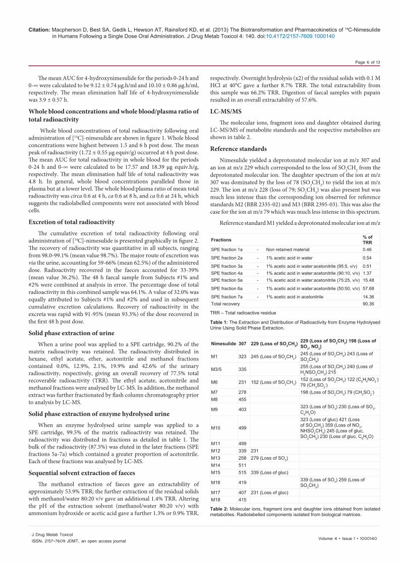
LC-MS/MS
The molecular ions, fragment ions and daughter obtained during LC-MS/MS of metabolite standards and the respective metabolites are shown in table 2.
Reference standards
Nimesulide yielded a deprotonated molecular ion at m/z 307 and an ion at m/z 229 which corresponded to the loss of SO2CH2 from the deprotonated molecular ion. The daughter spectrum of the ion at m/z 307 was dominated by the loss of 78 (SO2CH2) to yield the ion at m/z 229. The ion at m/z 228 (loss of 79; SO2CH3) was also present but was much less intense than the corresponding ion observed for reference standards M2 (BBR 2335-02) and M3 (BBR 2395-03). This was also the case for the ion at m/z 79 which was much less intense in this spectrum.
Reference standard M1 yielded a deprotonated molecular ion at m/z
Fractions % of TRR
SPE fraction 1a - Non retained material 0.46
SPE fraction 2a - 1% acetic acid in water 0.54
SPE fraction 3a - 1% acetic acid in water:acetonitrile (95:5, v/v) 0.51SPE fraction 4a - 1% acetic acid in water:acetonitrile (90:10, v/v) 1.37SPE fraction 5a - 1% acetic acid in water:acetonitrile (75:25, v/v) 15.48
SPE fraction 6a - 1% acetic acid in water:acetonitrile (50:50, v/v) 57.68
SPE fraction 7a - 1% acetic acid in acetonitrile 14.36Total recovery 90.36
TRR – Total radioactive residue
Table 1: The Extraction and Distribution of Radioactivity from Enzyme Hydrolysed Urine Using Solid Phase Extraction.
Nimesulide 307 229 (Loss of SO2CH2)229 (Loss of SO2CH2) 198 (Loss of SO2, NO2)
M1 323 245 (Loss of SO2CH2)245 (Loss of SO2CH2) 243 (Loss of SO2CH4)
M3/5 335 255 (Loss of SO2CH4) 240 (Loss of H2NSO2CH3) 215
M6 231 152 (Loss of SO2CH3)152 (Loss of SO2CH3) 122 (C6H4NO2
-) 79 (CH3SO2
-)M7 278 198 (Loss of SO2CH3) 79 (CH3SO2
-)M8 455
M9 403 323 (Loss of SO3) 230 (Loss of SO3, C6H5O)
M10 499
323 (Loss of gluc) 421 (Loss of SO2CH2) 359 (Loss of NO2, NHSO2CH3) 245 (Loss of gluc, SO2CH2) 230 (Loss of gluc, C6H5O)
M11 499M12 339 231M13 258 279 (Loss of SO3)M14 511M15 515 339 (Loss of gluc)
M16 419 339 (Loss of SO3) 259 (Loss of SO2CH4)
M17 407 231 (Loss of gluc)M18 415
Table 2: Molecular ions, fragment ions and daughter ions obtained from isolated metabolites. Radiolabelled components isolated from biological matrices.

Citation: Macpherson D, Best SA, Gedik L, Hewson AT, Rainsford KD, et al. (2013) The Biotransformation and Pharmacokinetics of 14C-Nimesulide in Humans Following a Single Dose Oral Administration. J Drug Metab Toxicol 4: 140. doi:10.4172/2157-7609.1000140
Page 7 of 13
Volume 4 • Issue 1 • 1000140J Drug Metab ToxicolISSN: 2157-7609 JDMT, an open access journal
of 323 ([M-H]-). The spectrum also contained the ions at m/z 265 and 245. The ion at m/z 265 was not related to M1 (4-hydroxynimesulide), however, the ion at m/z 245 corresponded to the loss of SO2CH2 from the deprotonated molecular ion. The daughter spectrum of the ion at m/z 323 was dominated by the loss of 78 (SO2CH2) to yield the ion at m/z 245. The ion at m/z 243 (loss of 80; SO2, CH4) was also present but was much less intense than the corresponding ion observed for reference standards M2 (BBR 2335-02) and M3 (BBR 2395-03).
Reference standard M2 yielded a deprotonated molecular ion at m/z of 277 ([M-H]-). The daughter spectrum of the ion at m/z 277 contained ions at m/z 79, 197 and 199. The ion at m/z 79 (SO2CH3) was the base peak of this spectrum. The loss of 78 (SO2CH2) and 80 (SO2, CH4) gave the ions at m/z 199 and 197, respectively.
Reference standard M3 yielded a deprotonated molecular ion at m/z of 293 and the daughter spectrum of this ion contained ions at m/z 79, 213 and 215. The loss of 78 (SO2CH2) and 80 (SO2, CH4) gave the ions at m/z 215 and 213, respectively, were significant. The ion at m/z 79 was assigned as SO2CH3.
Reference standard M4 yielded a deprotonated molecular ion at m/z of 319 ([M-H]-). The spectrum also contained an ion at m/z of 239 which corresponded to the loss of SO2 and CH4 from the deprotonated molecular ion. The daughter spectrum of the ion at m/z 319 contained ions at m/z 239 and 241. These ions corresponded to the loss of 80 (SO2, CH4) and 78 (SO2CH2), respectively. The ion at m/z 79 was present but was not as intense.
Reference standard M5 yielded a deprotonated molecular ion at m/z 335 and the daughter spectrum of this ion at m/z 335 showed the loss of 80 (SO2CH4) to yield the ion at m/z 255 and 78 (SO2, CH2) to give m/z 257 The ion at m/z 79 was also present.
Reference standard M6 gave a deprotonated molecular ion at m/z 231 and the fragment ion at 152 produced by the loss of SO2CH3. The daughter spectrum obtained from the base peak at m/z 231 contains an abundant ion at m/z 122 which is produced by the loss of NO from the m/z 152 ion.
The mass spectrum of reference standard M7 contained a deprotonated molecular ion at m/z 278 and a [2M-1]- ion at m/z 557. The daughter spectrum obtained from the base peak at m/z 278 contained ions at m/z 198 (loss of SO2CH3) and m/z 79 (SO2CH3).
Reference standard M6-G yielded a deprotonated molecular ion at m/z 407. A fragment ion was present at m/z 231 which corresponds to the loss of the glucuronide moiety. An ion was present at m/z 815 which corresponds to [2M-1]-.
Reference standard M7-G yielded a deprotonated molecular ion at m/z 454. An ion was present at m/z 909 which corresponds to [2M-1]-.
Identification of metabolites (for structures see figure 4)
Nimesulide was identified in an acetonitrile extract of faeces and in plasma. The molecular weight (308) was confirmed by the [M-1]- ion at m/z 307, the spectrum obtained was consistent with the reference standard. The daughter ion spectrum obtained from nimesulide (m/z 307) in faecal extracts contained the ion at m/z 229 which corresponds to the loss of SO2CH2.
M1 (4-hydroxynimesulide) was identified in urine and plasma. The MW of 324 was confirmed by the [M-1]- ion at m/z 323. The ion at m/z 245 is generated by the loss of SO2CH2 and this was consistent with the reference standard. The daughter ion spectrum produced from M1 was
consistent with the daughter ion spectrum obtained from the reference standard. The principle ions were m/z 245 and 243 which are produced by the loss of SO2CH2 and SO2CH3, respectively. The ion (m/z 323) associated with this metabolite was observed at 2 separate retention times, indicating that isomers are formed with different positions of hydroxylation.
M3 was detected in urine, however, a full scan mass spectrum was not obtained because it co-chromatographed with M5 which was more abundant. The presence of metabolite 3 was confirmed by generating selected ion chromatograms which confirmed the m/z 293 ([M-1]- ion from M3) and the m/z 335 ions ([M-1]- ion from M5). The m/z 293 ion is also present as a contaminating ion in the spectrum obtained for M5.
M5 was detected in deconjugated urine. The Mwr of 336 was confirmed by the ion at m/z 335. The minor ion at m/z 293 is not related to M5 but was produced by the co-elution of M3 which has a molecular weight of 294. The daughter ion mass spectrum obtained for M5 contained a major ion at m/z 255 which corresponded to the loss of SO2CH4 and is consistent with the reference standard.
M6 was identified as a trace metabolite in deconjugated urine. It had a molecular weight of 232 which was confirmed by the ion at m/z 231 in the mass spectrum. The loss of SO2CH4 gave the minor ion at m/z 152 which was consistent with the reference standard. The daughter ion mass spectrum was consistent with the reference standard with major ions at m/z 152, 122 and 79. The ion at m/z 152 is produced by the loss of SO2CH3, the ion at m/z 122 is C6H4NO2 from the m/z 231 ion and the ion at m/z 79 represents the CH3SO2.
M7 was confirmed in deconjugated urine by LC-MS and although the spectrum obtained was weak, a daughter ion spectrum was obtained from the molecular ion at m/z 278. The major daughter ions were m/z 198 and 79, the 198 ion is generated by the loss of SO2CH3. Both the spectra were consistent with the reference standard spectra.
M8 was detected in urine and was identified as a glucuronide conjugate of M7 with a Mwr of 455 which gave the molecular ion at m/z 454, consistent with the reference standard. The occurrence of this metabolite was confirmed by enzyme deconjugation experiments with β-glucuronidase. Following the treatment of urine with β-glucuronidase (sulphatase free) metabolite 7 was identified by LC-MS.
M9 was identified as a sulphate conjugate of M1. The Mwr of 404 was confirmed by the deprotonated molecular ion at m/z 403, the daughter ion spectrum obtained from the 403 contained an ion at m/z 323 which was produced by the loss of SO3.
M10 was identified in urine and was assigned as a glucuronide conjugate of M1. The Mwr of 500 was confirmed by an ion at m/z 499. The daughter ion spectrum obtained from the 499 ion contained an ion at m/z 323 which was produced by the loss of the glucuronide moiety and confirms that this molecule is a glucuronide conjugate of metabolite 1. The ion at m/z 499 was shown to be abundant at 2 main positions in the urinary chromatogram which indicates that 2 positions of hydroxylation are possible. The second position of hydroxylation gave a earlier retention time than that obtained for M10 and this component was labelled M11.
The mass spectrum for M12 was obtained only following enzyme deconjugation with β-glucuronidase which would indicate that this metabolite was not present in the aglycone form. The Mwr of 340 is confirmed by the deprotonated molecular ion at m/z 339. The ion at m/z 231 was consistent with structure.

Citation: Macpherson D, Best SA, Gedik L, Hewson AT, Rainsford KD, et al. (2013) The Biotransformation and Pharmacokinetics of 14C-Nimesulide in Humans Following a Single Dose Oral Administration. J Drug Metab Toxicol 4: 140. doi:10.4172/2157-7609.1000140
Page 8 of 13
Volume 4 • Issue 1 • 1000140J Drug Metab ToxicolISSN: 2157-7609 JDMT, an open access journal
M13 was identified as a sulphate conjugate of M7 with a Mwr of 359. The mass spectrum obtained contained a deprotonated molecular ion at m/z 358 and an ion at m/z 279 produced by the loss of SO3 from the molecule confirming that it is a sulphate conjugate of M7.
M14 was tentatively proposed to be a glucuronide conjugate of M5, with Mwr 512. The mass spectrum obtained was weak but contained an ion at m/z 511 which was proposed to be the deprotonated molecular ion. M5 was a significant peak in urine hydrolysed with beta-glucuronidase.
M15 was identified as the glucuronide conjugate of M12, with Mwr 516. The molecular weight was confirmed by the deprotonated molecular ion at m/z 515, the minor ion at m/z 339 was produced by the loss of the glucuronic acid moiety.
M16 was identified as a sulphate conjugate of M12 and was identified in, with a Mwr 420. The molecular weight was confirmed by the deprotonated molecular ion at m/z 419. The daughter ions produced from the m/z 419 ion confirmed the structure. The ions present (m/z
339 and 259) represent the loss of 80 and 160 which corresponds to SO3 or SO2CH4 and SO3 and SO2CH4.
M17 was identified in urine as a glucuronide conjugate of M6, with Mwr of 408. The molecular weight was confirmed by the deprotonated molecular ion at m/z 407. The loss of glucuronide produces the minor ion at m/z 231. The ions observed were identical with the reference standard.
M18 is proposed to be a sulphate conjugate of M5, with a mwt 416 that consistent with the deprotonated molecular ion is present at m/z 415. However no fragmentation information was present so this assignment is tentative.
Metabolite quantification
The radio-HPLC profile obtained from 0-24 h urine is shown in figures 3a-3e. Peaks were assigned in the chromatogram by monitoring for each of the masses associated with metabolites identified by LC-MS of urinary fractions. 0-24 h urine was found to contain 13 peaks of radioactivity, 10 of these peaks were assigned as identified metabolites
150
140
130
120
110
100
0 10 20 30 40 50
Intensity (mV)
Time (minutes)
M17
M8
M14
2.79
3.49
M11 M
18
M15
Unk
now
n
M16
M1bM1a
M10
Sample Urine Pool 1% 100ul Amount : 1.000
3a
Figure 3a: Radio-HPLC chromatogram of 0-24 h urine.
25. 0
06. 208. 2
01. 3 03. 305. 3 08. 3
02. 484. 4
09. 4 03. 5 07. 5 01. 604. 600. 7
01. 804. 9
16. 905. 01 09. 01
08. 11
02. 41
02. 5106. 51
03. 61 02. 71
09. 81 23. 91
05. 02
09. 32
04. 94
61M
31M09. 41
81M
Intensity (mV)
Time (minutes)
Sample 532U 100ul Amount : 1.000
3b
140
130
120
110
1000 10 20 30 40 50
Figure 3b: HPLC chromatograms of extracts of enzyme hydrolysed urine.

Citation: Macpherson D, Best SA, Gedik L, Hewson AT, Rainsford KD, et al. (2013) The Biotransformation and Pharmacokinetics of 14C-Nimesulide in Humans Following a Single Dose Oral Administration. J Drug Metab Toxicol 4: 140. doi:10.4172/2157-7609.1000140
Page 9 of 13
Volume 4 • Issue 1 • 1000140J Drug Metab ToxicolISSN: 2157-7609 JDMT, an open access journal
(M1, M8, M10, M11, M14, M15, M16, M17 and M18) and a further 2 were assigned as glucuronide conjugates (retention times 2.8 and 3.2 min) because these polar peaks were not present in a sample that had been treated with β-glucuronidase. Table 3 shows the assignment of peaks in urine and each metabolite as a percentage of the dose. Identified metabolites account for 92.4% of the urinary radioactivity and 49.4% of dose (in urine). Unidentified components represented 7.6% of the urinary radioactivity and 4.1% of the dose (in urine).
Figure 3a shows the HPLC profiles of SPE fractions 5a, 6a and 7a which account for 87.52% of the radioactivity from an enzyme hydrolysed sample of 0-24 h urine. The enzyme hydrolysis was conducted with β-glucuronidase from E. coli which does not contain sulphatase activity, as a result the sulphate conjugates M9, M16 and M18 are present in these fractions. Other peaks present are M1, M5, M6, M7 and M12 which are aglycones of M10, M14, M17, M8 and M15, respectively further confirming the assignments made on the
34.0
93.4
93.592.6
95.7
90.9
94.01
90.3193.31
95.3192.41
92.4194.41
97.4186.51 93.51
M5
61M
a1M
b1M
7M
95.6191.7197.61
95.7199.71
92.8194.81
92.91 96.9195.02
90.12 98.1293.22 92.72
92.8296.8294.92
90.23
97.43
97.64
91.15
Intensity (mV)
Time (minutes)
Sample 532U 6E 100ul Amount : 1.000
3c
240
110
180
270
120
150
300
0 10 20 30 40 50
Figure 3c: HPLC chromatograms of extracts of enzyme hydrolysed urine.
Intensity (mV)
Time (minutes)
Sample 532U 7E 100ul Amount : 1.000
3d
160
140
180
200
100
120
0 10 20 30 40 50
03.0105.01 05. 2209. 22
08. 0304. 13
06. 64
b1M
Figure 3d: HPLC chromatograms of extracts of enzyme hydrolysed urine.

Citation: Macpherson D, Best SA, Gedik L, Hewson AT, Rainsford KD, et al. (2013) The Biotransformation and Pharmacokinetics of 14C-Nimesulide in Humans Following a Single Dose Oral Administration. J Drug Metab Toxicol 4: 140. doi:10.4172/2157-7609.1000140
Page 10 of 13
Volume 4 • Issue 1 • 1000140J Drug Metab ToxicolISSN: 2157-7609 JDMT, an open access journal
intact glucuronide conjugates during LC-MS analysis of urine fractions (Table 3).
The radio-HPLC profile (Figure 4) obtained from acetonitrile extract of faeces contained 2 radiolabelled components which were detectable above background. The non polar component which accounted for approximately 50% of the extractable radioactivity was assigned as nimesulide. The polar component was not identified.
The concentrations of radioactivity in plasma were too low for analysis by radio-HPLC, however LC-MS analysis of plasma confirmed the presence of nimesulide and 4-hydroxynimesulide. Nimesulide and M1 were quantified using a validated bioassay and the quantities of these components present would account for virtually the entire radioactivity present in plasma (Figure 1).
DiscussionThis study gives the most comprehensive data on the metabolic
transformation and recovery from the extraction pathways of nimesulide reported to date.
The mean Cmax and Tmax values obtained for nimesulide were 2.35 ± 0.35 µg.ml-1 and 3.88 ± 1.84 hours. These values are similar to the kinetic parameters measured in patients administered 100 mg of nimesulide in tablet or suspension from where the Cmax and Tmax ranges reported have been 2.86-6.50 µg.ml-1 and 1.22-2.75 hours [5]. While the Cmax value obtained in the present study is lower than the published range and the Tmax is later, however the elimination half life (2.49 ± 0.55 hours) and AUC∞ (16.67 ± 3.23 µg/ml.h) are consistent with the published ranges of 1.96-4.73 hours for the elimination half live and 14.65-54.09 µg/ml.h
Intensity (mV)
Time (minutes)
Conc. Faeces extract -100ul inj Amount : 1.000
3e
160
60
80
180
100
120
140
0 10 20 30 40 50
28.3
39.54
Figure 3e: Radio-HPLC chromatogram of acetonitrile extract of faeces.
Fraction Retention Time Metabolite as % Dose Metabolite Names1 2.8 3.11 Unknown glucuronide conjugate2 3.2 4.30 Unknown glucuronide conjugate3 3.7 5.06 Metabolite 17 -Glucuronide of M64 4.3 4.56 Metabolite 14 -Glucuronde of M55 5.8 6.43 Metabolite 8 -Glucuronide of M76 7.8 2.37 Metabolite 11 -Glucronide of M1a7 8.1 2.54 Metabolite 18 -Sulphate of M58 9.5 14.58 Metabolite 10 -Glucuronide of M1b9 10.3 2.32 Metabolite 15 -Glucuronide of M1210 13.2 4.06 Unknown11 20.1 2.80 Metabolite 16 -Sulphate of M1212 23.2 0.97 Metabolite 1a13 24.9 0.41 Metabolite 1b Consistent with Reference Standard M1
Identified % of urinary radioactivity 92.41Unidentified % of urinary radioactivity 7.59
Identified % of dose 49.44Unidentified % of dose 4.06
M3 – Only observed in deconjugated samples as a trace component. The conjugate of M3 was not identified.M9 – Retention time 19 minutes. Trace componentM13 – Retention time at 17 min. Trace component.
Table 3: The Assignment of Peaks in 0-24 h Urine.

Citation: Macpherson D, Best SA, Gedik L, Hewson AT, Rainsford KD, et al. (2013) The Biotransformation and Pharmacokinetics of 14C-Nimesulide in Humans Following a Single Dose Oral Administration. J Drug Metab Toxicol 4: 140. doi:10.4172/2157-7609.1000140
Page 11 of 13
Volume 4 • Issue 1 • 1000140J Drug Metab ToxicolISSN: 2157-7609 JDMT, an open access journal
O
NHSO2CH3
NO2
OH
NHSO2CH3
NO2
O
NHSO2CH3
NHCOCH3
OH
O
NHSO2CH3
NO2
OH
O
NHSO2CH3
OH
Nimesulide MW 308(m/z 307)
M7MW 279(m/z 278)
#
M1MW 324(m/z 323)
0.4% of dose
M10 - Glucuronide of M1MW 500 (m/z 499)
14.6% of dose
M6MW 232(m/z 231)
+
M5MW 336(m/z 335)
#
M9 - Sulphateof M1MW 404 (m/z403)Trace metabolite
M11 - Second position of hydroxylationis possible. This gives a second
peak of MW 500 (m/z 499)2.4% of dose
O
NHSO2CH3
NO2
OH
M12MW 340(m/z 339)
O
NHSO2CH3
NH2
O
NHSO2CH3
NHCOCH3 M4MW 320(m/z 319)
M2mw 278
(m/z 277)
M8 - glucuronide of M7, MW 455( m/z 454)
6.4% of dose
M13 sulphate of M7MW 359 ( m/z 358)
Trace metabolite
M14 Glucuronide of M5MW 512 (m/z 511)
4.6% of dose)
M15 glucuronide of M12MW 516 (m/z 515)
2.3% of dose
M16 sulphate of M12MW420 (m/z 419)
2.8% of dose
M17, Glucuronide of M6MW 408 ( m/z 407)
5.1% of dose
M18 Sulphate of M5MW 416 (m/z 415)
2.5% of dose
O
NHSO2CH3
NH2
M3mw 294
(m/z 293)Trace metabolite in
deconjugated samples
OH
O
NHSO2CH3
NO2
OSO3-
O
NHSO2CH3
NO2
Ogluc
O
NHSO2CH3
NO2
Ogluc
O
NHSO2CH3
NO2
OSO3-
O
NHSO2CH3
NO2
Ogluc
O
NHSO2CH3
NHCOCH3
Ogluc
O
NHSO2CH3
NHCOCH3
OSO3-
M18MW 336(m/z 335)
Ogluc
NHSO2CH3
NO2
O
NHSO2CH3
OSO3-
O
NHSO2CH3
Ogluc
OH
OH
OH
Figure 4: Proposed biotransformation pathway for nimesulide in man.
for AUC∞. This would indicate that in the present study the absorption of nimesulide from the gelatin capsule formulation was slower than that observed previously with other oral formulations [6].
The principal nimesulide metabolite, 4’-hydroxy-nimesulide, was also measured in plasma and the kinetic data are consistent with metabolism of nimesulide to 4’-hydroxy-nimesilde being the major route of elimination for nimesulide. In combination the concentrations of nimesulide and 4’-hydroxy-nimesulide in plasma accounts for virtually all the radioactivity in plasma.
The distribution of radioactivity in excreta was consistent with previous data Bernareggi [5] with urine and faeces containing 62.5% and 36.2% of the administered dose respectively.
A total of 16 metabolites of nimesulide were identified. At least 11 of these have been identified for the first time and these constitute a major proportion (>35-40%) of total drug that was metabolised. The pathways for biotransformation of nimesulide in man have been shown to proceed by 3 principle routes (Figure 4), namely cleavage of the molecule at the ether linkage, reduction of the NO2 group to

Citation: Macpherson D, Best SA, Gedik L, Hewson AT, Rainsford KD, et al. (2013) The Biotransformation and Pharmacokinetics of 14C-Nimesulide in Humans Following a Single Dose Oral Administration. J Drug Metab Toxicol 4: 140. doi:10.4172/2157-7609.1000140
Page 12 of 13
Volume 4 • Issue 1 • 1000140J Drug Metab ToxicolISSN: 2157-7609 JDMT, an open access journal
NH2 and ring hydroxylation. Cleavage of the ether linkage gave M6 which is conjugated with glucuronic acid to give M17. Reduction of the NO2 group to NH2 is proposed to produce the intermediate M2, which is hydroxylated to produce M3. M3 is then acetylated to produce M5. An alternative route is possible for the production of M5 in which M2 is acetylated to the postulated intermediate M4 which is in turn hydroxylated to give M5. The latter pathway is, however, less likely because the acetylation of the NH2 group is thought to occur in the kidney and therefore is a terminal metabolic reaction. M5 is conjugated with sulphate and glucuronide to give M18 and M14, respectively. Ring hydroxylation of nimesulide gives the 4-hydroxy-derivative, M1, conjugation of this molecule with sulphate gives M9. Conjugation of M1 with glucuronic acid gives M10, it is proposed that the principle position of hydroxylation is consistent with the reference standard M1. However, a second position of hydroxylation is proposed to give rise to a second step conjugate of molecular weight 500 (M11). M1 is also hydroxylated in a second position to give M12 which is then conjugated with glucuronide and sulphate to give M15 and M16, respectively.
The metabolites identified during these investigations are in agreement with the work of Carini et al. [8] but these authors only identified 5 metabolites of nimesulide in human urine (M1-M5). Metabolites M1, M2 and M5 were confirmed during the present study but M3 and M4 were not detected. These metabolites were previously reported as being present at very low concentrations. As a consequence they are proposed as intermediates in the biotransformation pathway. Additional phase 1 metabolites (M6, M7 and M12) have been identified which were not previously detected. The structural assignments of M6 and M7 and their glucuronide conjugates were confirmed with authentic reference standards. Carini et al. [8] also reported that the greater portion of an oral administration of nimesulide was excreted as conjugated metabolites. This was based on the extractability of metabolites before and after the enzymatic hydrolysis of urine and this has been confirmed in the present study by isolating conjugated metabolites from urine and analysing these metabolites by LC-MS/MS. This approach confirmed the presence of glucuronide and sulphate conjugates. Beta-glucuronidase from E. coli, which does not have any sulphatase activity, was used to hydrolyse the glucuronide conjugates present in urine. This allowed the confirmation of glucuronide conjugates identified by LC-MS/MS and also allowed the sulphate conjugates to be clearly visible in chromatograms of extracts of hydrolysed urine. Thus, the present experiments enable identification of the biotransformation pathway for nimesulide in man to be extended from 5 metabolites to 16 metabolites and 2 postulated intermediates. Greater than 92.4% of the urinary (0-24) radioactivity was accounted for by characterised metabolites. Thus, the most comprehensive metabolic pathway of nimesulide has now been obtained as a consequence of careful extraction isolation and man spectrokinetic analytical procedures.
From the point of view of likely involvement of the nimesulide metabolites those thought to be involved with the development of hepatotoxicity from the drug include nitroso- and glucuronide metabolites [6,8-14,16-18,22]. Studies by Li et al. [15] indicate that two diimine metabolites of nimesulide may be formed by cytochrome P450 2C19 or 1A2 oxidation of the two 4-amino-metabolites, M3 and the N-de-acetylated form of M5. These postulated diimine reactive metabolites may act in forming adducts in a manner similar to that suggested with diclofenac [25,26] and paracetamol [27]. However, the formation of the N-acetylated derivatives of the 4-amino-metabolites of nimesulide (Figure 4) would be a metabolic means of negating the hepatotoxic impact of these diimine metabolites. The relative proportions of the 4-amino metabolites that are formed
in vivo are small in comparison with the total number and rate of formation of the metabolites of the drug (Table 3). The glucuronide metabolites may also represent another means of forming adducts [11] analogous to that seen with many other NSAIDs [27]. These and other reactive intermediates or metabolites including those with effects on mitochondrial functions and superoxide generation could also be candidates for hepatic injury by nimesulide [9,13,14,17,27,28]. Further quantification of the putative reactive metabolites along with studies on their mode of hepatic reactions may give insight into the relative contributions of these different metabolites in the development of hepatotoxicity form nimesulide.
Acknowledgments
We thank Richard Seabrook of the Biomedical Research Centre of Sheffield Hallam University for expert technical assistance. KDR thanks Dr. Alberto Bernareggi for valuable advice. These studies were funded by grants from Helsinn Healthcare SA (Pambio-Noranco, Lugano, Switzerland).
Declaration
The manuscript was prepared entirely by the authors who are responsible for its content.
References
1. Rainsford KD (ed.) (2005) Nimesulide. Actions and uses. Birkhaüser Basel.
2. Bennett A (2001) Nimesulide: a well-established cyclooxygenase-2 inhibitor with many other pharmacological properties relevant to inflammatory diseases. In: Vane JR and Botting RM (eds). Therapeutic roles of selective COX-2 inhibitors. William Harvey Press, London, 524-540.
3. Rainsford KD; Members of the Consensus Report Group on Nimesulide (2006) Nimesulide -- a multifactorial approach to inflammation and pain: scientific and clinical consensus. Curr Med Res Opin 22: 1161-1170.
4. Mattia C, Ciarcia S, Muhindo A, Coluzzi F (2010) [Nimesulide: 25 years later]. Minerva Med 101: 285-293.
5. Bernareggi A (1998) Clinical pharmacokinetics of nimesulide. Clin Pharmacokinet 35: 247-274.
6. Bernareggi A, Rainsford KD (2005) Pharmacokinetics of nimesulide. In: Rainsford KD (ed.) Nimesulide. Actions and uses. Birkhaüser, Basel, 63-120.
7. Bjarnason I, Bissoli F, Conforti A, Maiden L, Moore N, et al. (2005) Adverse reactions and their mechanisms from nimesulide. In Rainsford KD (ed) Nimesulide. Actions and Uses. Birkhaeuser, Basel. 315-415.
8. Carini M, Aldini G, Stefani R, Marinello C, Facino RM (1998) Mass spectrometric characterization and HPLC determination of the main urinary metabolites of nimesulide in man. J Pharm Biomed Anal 18: 201-211.
9. Boelsterli UA (2002) Nimesulide and hepatic adverse effects: roles of reactive metabolites and host factors. Int J Clin Pract Suppl: 30-36.
10. Boelsterli UA (2002) Mechanisms of NSAID-induced hepatotoxicity: focus on nimesulide. Drug Saf 25: 633-648.
11. Boelsterli UA (2002) Xenobiotic acyl glucuronides and acyl CoA thioesters as protein-reactive metabolites with the potential to cause idiosyncratic drug reactions. Curr Drug Metab 3: 439-450.
12. Rainsford KD, Seabrook RW, Spencer S, Hewson AT (2001) Effects of nimesulide and its metabolites or manufacturing intermediates on the viability and growth of the human hepatoma HepG2 cell line. Life Sci 69: 2965-2973.
13. Tay VK, Wang AS, Leow KY, Ong MM, Wong KP, et al. (2005) Mitochondrial permeability transition as a source of superoxide anion induced by the nitroaromatic drug nimesulide in vitro. Free Radic Biol Med 39: 949-959.
14. Freitas CS, Dorta DJ, Pardo-Andreu GL, Pestana CR, Tudella VG, et al. (2007) 4-hydroxy nimesulide effects on mitochondria and HepG2 cells. A comparison with nimesulide. Eur J Pharmacol 566: 43-49.
15. Li F, Chordia MD, Huang T, Macdonald TL (2009) In vitro nimesulide studies toward understanding idiosyncratic hepatotoxicity: diiminoquinone formation and conjugation. Chem Res Toxicol 22: 72-80.
16. Kale VM, Hsiao CJ, Boelsterli UA (2010) Nimesulide-induced electrophile

Citation: Macpherson D, Best SA, Gedik L, Hewson AT, Rainsford KD, et al. (2013) The Biotransformation and Pharmacokinetics of 14C-Nimesulide in Humans Following a Single Dose Oral Administration. J Drug Metab Toxicol 4: 140. doi:10.4172/2157-7609.1000140
Page 13 of 13
Volume 4 • Issue 1 • 1000140J Drug Metab ToxicolISSN: 2157-7609 JDMT, an open access journal
stress activates Nrf2 in human hepatocytes and mice but is not sufficient to induce hepatotoxicity in Nrf2-deficient mice. Chem Res Toxicol 23: 967-976.
17. Singh BK, Tripathi M, Pandey PK, Kakkar P (2010) Nimesulide aggravates redox imbalance and calcium dependent mitochondrial permeability transition leading to dysfunction in vitro. Toxicology 275: 1-9.
18. Yang M, Chordia MD, Li F, Huang T, Linden J, et al. (2010) Neutrophil- and myeloperoxidase-mediated metabolism of reduced nimesulide: evidence for bioactivation. Chem Res Toxicol 23: 1691-1700.
19. Monteiro JP, Martins AF, Lúcio M, Reis S, Pinheiro TJ, et al. (2011) Nimesulide interaction with membrane model systems: are membrane physical effects involved in nimesulide mitochondrial toxicity? Toxicol In Vitro 25: 1215-1223.
20. Singh BK, Tripathi M, Chaudhari BP, Pandey PK, Kakkar P (2012) Natural terpenes prevent mitochondrial dysfunction, oxidative stress and release of apoptotic proteins during nimesulide-hepatotoxicity in rats. PLoS One 7: e34200.
21. Traversa G, Bianchi C, Da Cas R, Abraha I, Menniti-Ippolito F, et al. (2003) Cohort study of hepatotoxicity associated with nimesulide and other non-steroidal anti-inflammatory drugs. BMJ 327: 18-22.
22. Aithal GP, Day CP (2007) Nonsteroidal anti-inflammatory drug-induced hepatotoxicity. Clin Liver Dis 11: 563-575, vi-vii.
23. Walker SL, Kennedy F, Niamh N, McCormick PA (2008) Nimesulide associated fulminant hepatic failure. Pharmacoepidemiol Drug Saf 17: 1108-1112.
24. Bessone F (2010) Non-steroidal anti-inflammatory drugs: What is the actual risk of liver damage? World J Gastroenterol 16: 5651-5661.
25. Boelsterli UA (2003) Diclofenac-induced liver injury: a paradigm of idiosyncratic drug toxicity. Toxicol Appl Pharmacol 192: 307-322.
26. Lim MS, Lim PL, Gupta R, Boelsterli UA (2006) Critical role of free cytosolic calcium, but not uncoupling, in mitochondrial permeability transition and cell death induced by diclofenac oxidative metabolites in immortalized human hepatocytes. Toxicol Appl Pharmacol 217: 322-331.
27. Walker GS, Atherton J, Bauman J, Kohl C, Lam W, et al. (2007) Determination of degradation pathways and kinetics of acyl glucuronides by NMR spectroscopy. Chem Res Toxicol 20: 876-886.
28. Ong MM, Wang AS, Leow KY, Khoo YM, Boelsterli UA (2006) Nimesulide-induced hepatic mitochondrial injury in heterozygous Sod2(+/-) mice. Free Radic Biol Med 40: 420-429.
Submit your next manuscript and get advantages of OMICS Group submissionsUnique features:
• Userfriendly/feasiblewebsite-translationofyourpaperto50world’sleadinglanguages• AudioVersionofpublishedpaper• Digitalarticlestoshareandexplore
Special features:
• 250OpenAccessJournals• 20,000editorialteam• 21daysrapidreviewprocess• Qualityandquickeditorial,reviewandpublicationprocessing• IndexingatPubMed(partial),Scopus,DOAJ,EBSCO,IndexCopernicusandGoogleScholaretc• SharingOption:SocialNetworkingEnabled• Authors,ReviewersandEditorsrewardedwithonlineScientificCredits• Betterdiscountforyoursubsequentarticles
Submityourmanuscriptat:http://www.editorialmanager.com/pharma
Citation: Macpherson D, Best SA, Gedik L, Hewson AT, Rainsford KD, et al. (2013) The Biotransformation and Pharmacokinetics of 14C-Nimesulide in Humans Following a Single Dose Oral Administration. J Drug Metab Toxicol 4: 140. doi:10.4172/2157-7609.1000140