m-lab 1 orthopedic evaluation of the knee … palpation and special tests ... microsoft powerpoint -...
TRANSCRIPT
Orthopedic Evaluation of the Knee- Palpation and Special Tests
Any knee injuries?
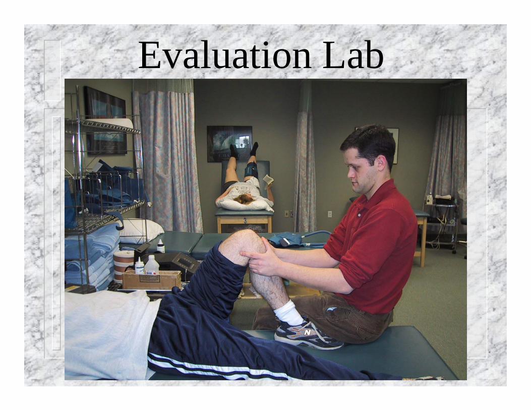
Evaluation Lab
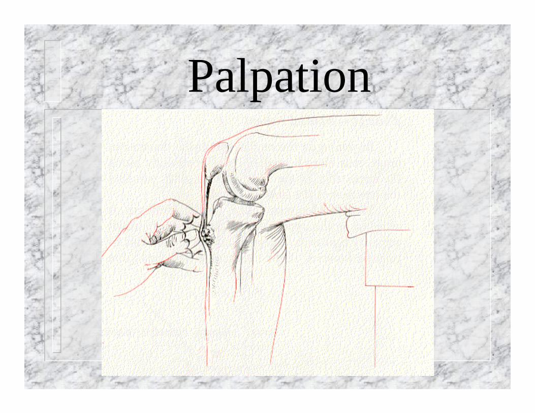
Palpation
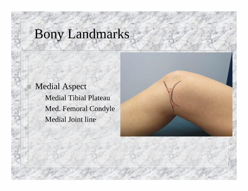
Bony Landmarks
Medial Aspect– Medial Tibial Plateau– Med. Femoral Condyle– Medial Joint line
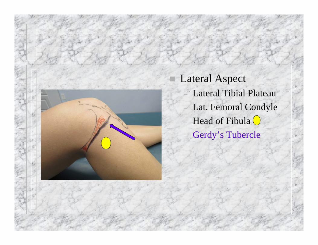
Lateral Aspect– Lateral Tibial Plateau– Lat. Femoral Condyle– Head of Fibula– Gerdy’s Tubercle
Bony Landmarks (cont.)
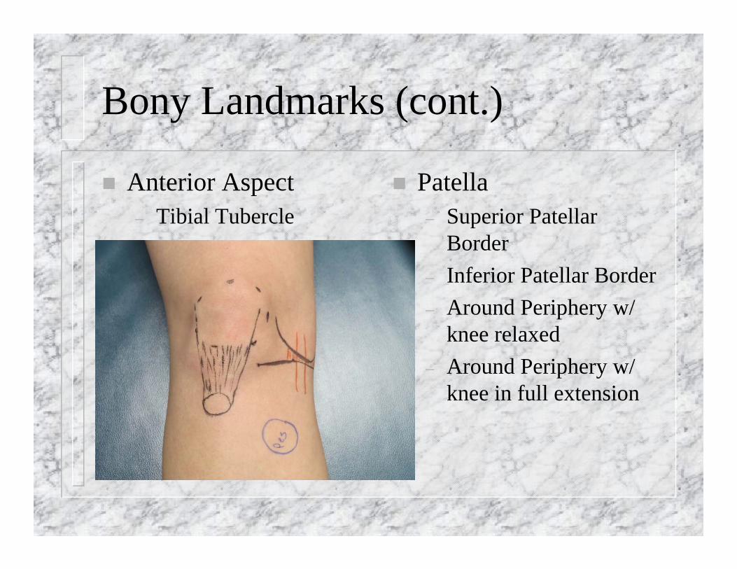
Anterior Aspect– Tibial Tubercle
Patella– Superior Patellar
Border– Inferior Patellar Border– Around Periphery w/
knee relaxed– Around Periphery w/
knee in full extension
Soft-Tissue Palpation
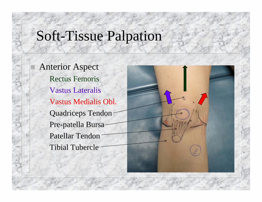
Anterior Aspect– Rectus Femoris– Vastus Lateralis– Vastus Medialis Obl.– Quadriceps Tendon – Pre-patella Bursa– Patellar Tendon– Tibial Tubercle
Medial Soft Tissue
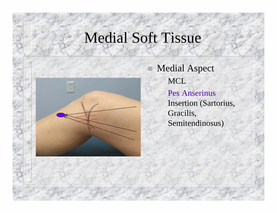
Medial Aspect– MCL– Pes Anserinus
Insertion (Sartorius, Gracilis, Semitendinosus)
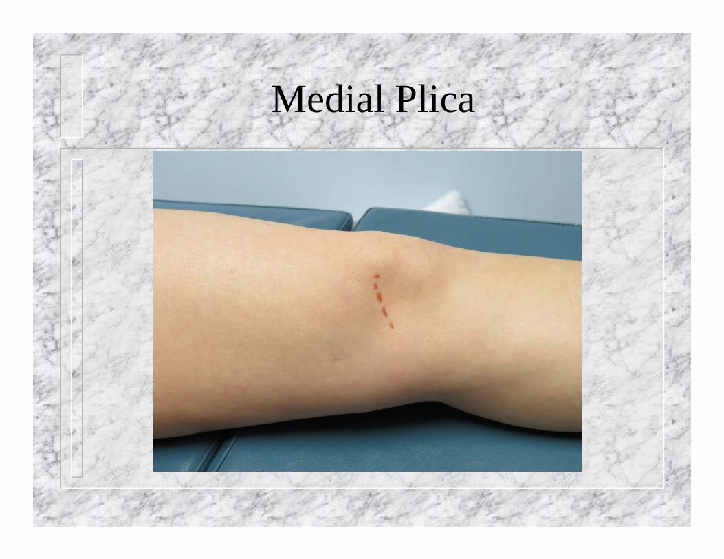
Medial Plica
Soft-Tissue Palpation (cont.)
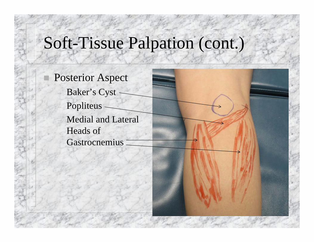
Posterior Aspect– Baker’s Cyst– Popliteus– Medial and Lateral
Heads of Gastrocnemius
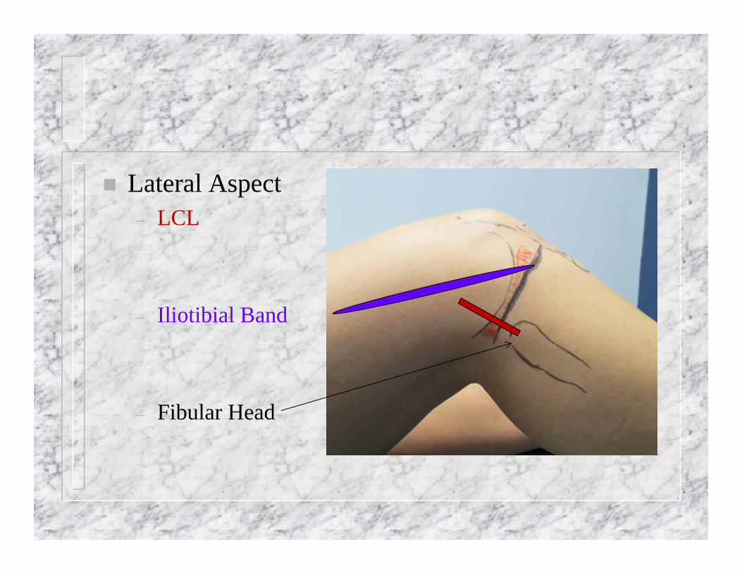
Lateral Aspect– LCL
– Iliotibial Band
– Fibular Head
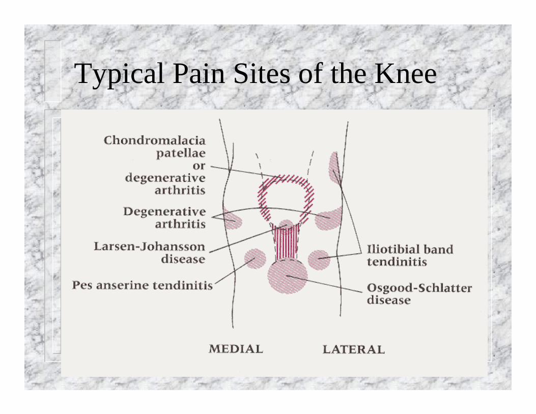
Typical Pain Sites of the Knee
Special Tests
Patella TestsBrush TestBallottementClarke’s TestApprehension
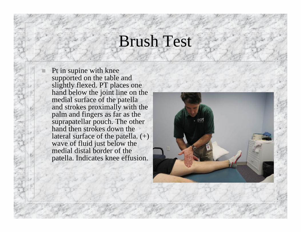
Brush Test Pt in supine with knee
supported on the table and slightly flexed. PT places one hand below the joint line on the medial surface of the patella and strokes proximally with the palm and fingers as far as the suprapatellar pouch. The other hand then strokes down the lateral surface of the patella. (+) wave of fluid just below the medial distal border of the patella. Indicates knee effusion.
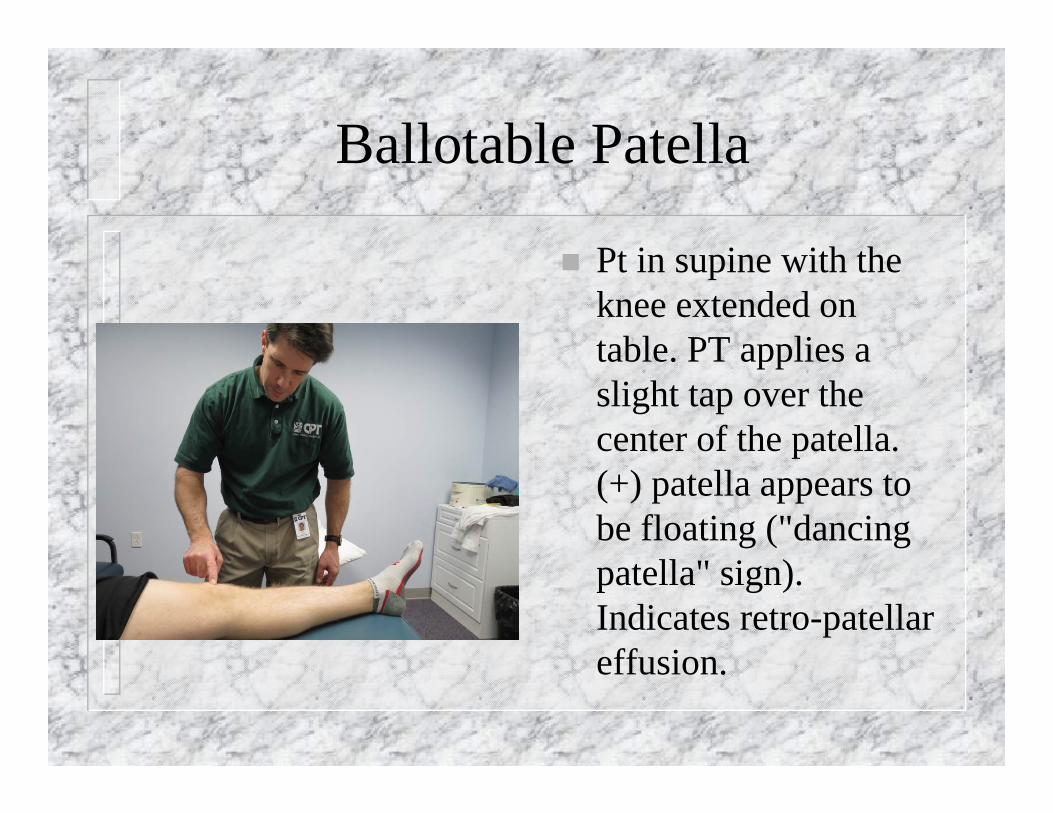
Ballotable Patella
Pt in supine with the knee extended on table. PT applies a slight tap over the center of the patella. (+) patella appears to be floating ("dancing patella" sign). Indicates retro-patellar effusion.
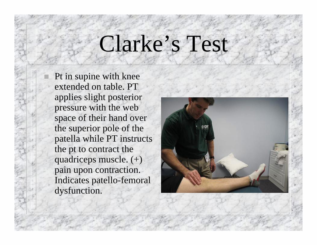
Clarke’s Test Pt in supine with knee
extended on table. PT applies slight posterior pressure with the web space of their hand over the superior pole of the patella while PT instructs the pt to contract the quadriceps muscle. (+) pain upon contraction. Indicates patello-femoral dysfunction.
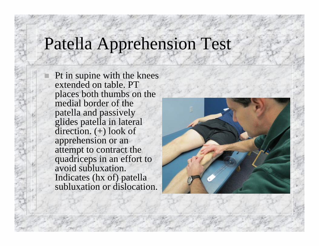
Patella Apprehension Test
Pt in supine with the knees extended on table. PT places both thumbs on the medial border of the patella and passively glides patella in lateral direction. (+) look of apprehension or an attempt to contract the quadriceps in an effort to avoid subluxation. Indicates (hx of) patella subluxation or dislocation.

Ligamentous Tests Valgus stress at 0 degrees = Capsule/MCL Valgus stress at 20-30 degrees =MCL Varus stress at 0 degrees = Capsule/LCL Valgus stress at 20-30 degree = LCL Anterior drawer = ACL Posterior drawer = PCL Lachman test = ACL Modified Lachman = ACL Posterior Sag = PCL Lateral Pivot Shift
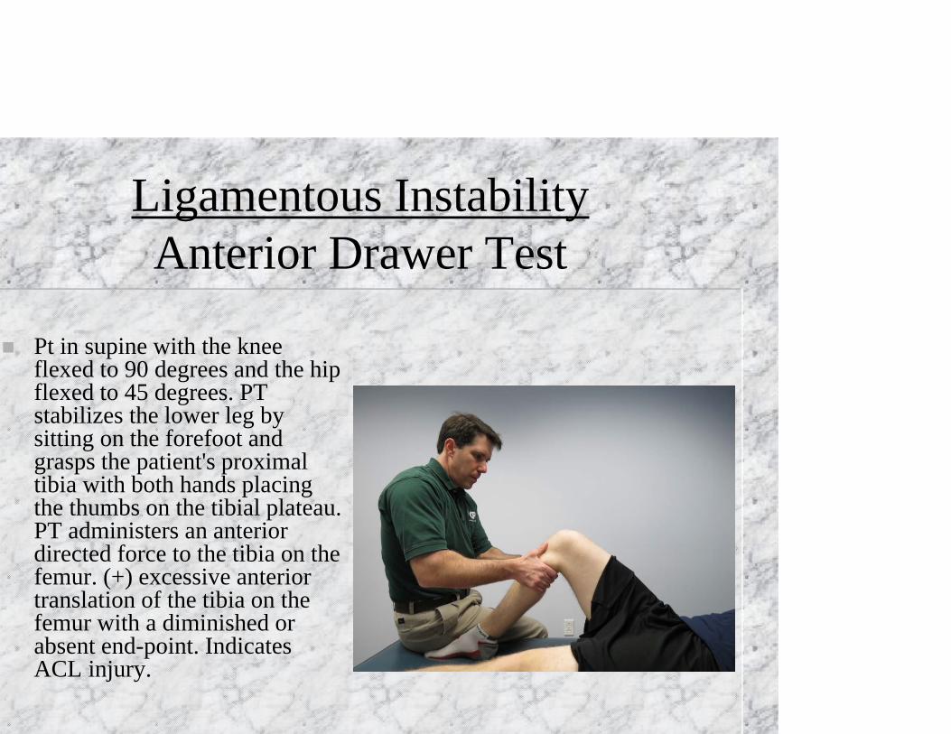
Ligamentous InstabilityAnterior Drawer Test
Pt in supine with the knee flexed to 90 degrees and the hip flexed to 45 degrees. PT stabilizes the lower leg by sitting on the forefoot and grasps the patient's proximal tibia with both hands placing the thumbs on the tibial plateau. PT administers an anterior directed force to the tibia on the femur. (+) excessive anterior translation of the tibia on the femur with a diminished or absent end-point. Indicates ACL injury.
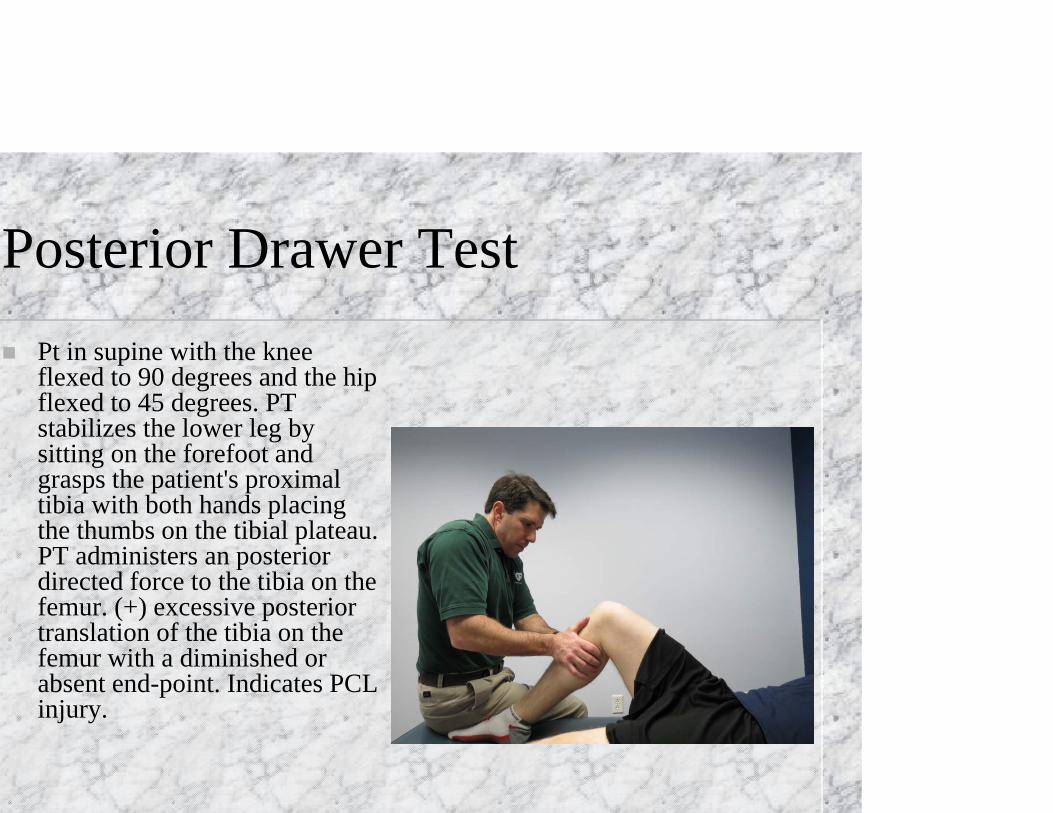
Posterior Drawer Test Pt in supine with the knee
flexed to 90 degrees and the hip flexed to 45 degrees. PT stabilizes the lower leg by sitting on the forefoot and grasps the patient's proximal tibia with both hands placing the thumbs on the tibial plateau. PT administers an posterior directed force to the tibia on the femur. (+) excessive posterior translation of the tibia on the femur with a diminished or absent end-point. Indicates PCL injury.
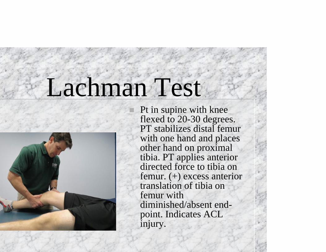
Lachman Test Pt in supine with knee
flexed to 20-30 degrees. PT stabilizes distal femur with one hand and places other hand on proximal tibia. PT applies anterior directed force to tibia on femur. (+) excess anterior translation of tibia on femur with diminished/absent end-point. Indicates ACL injury.
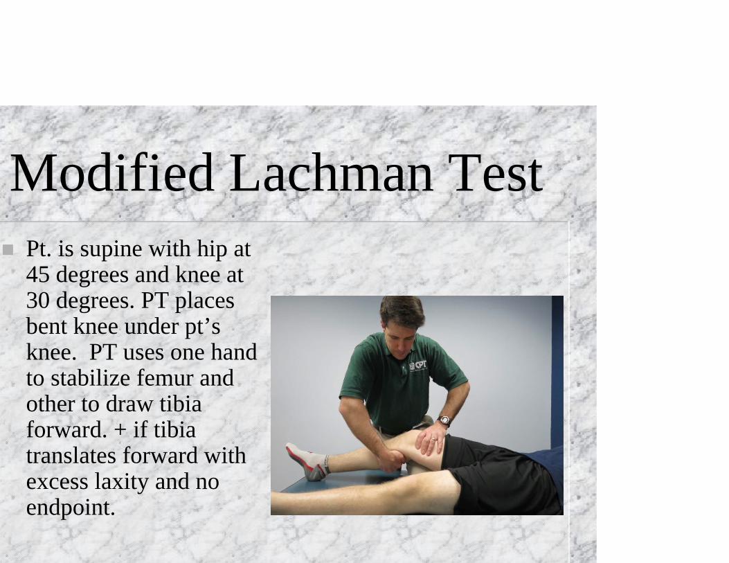
Modified Lachman Test Pt. is supine with hip at
45 degrees and knee at 30 degrees. PT places bent knee under pt’s knee. PT uses one hand to stabilize femur and other to draw tibia forward. + if tibia translates forward with excess laxity and no endpoint.
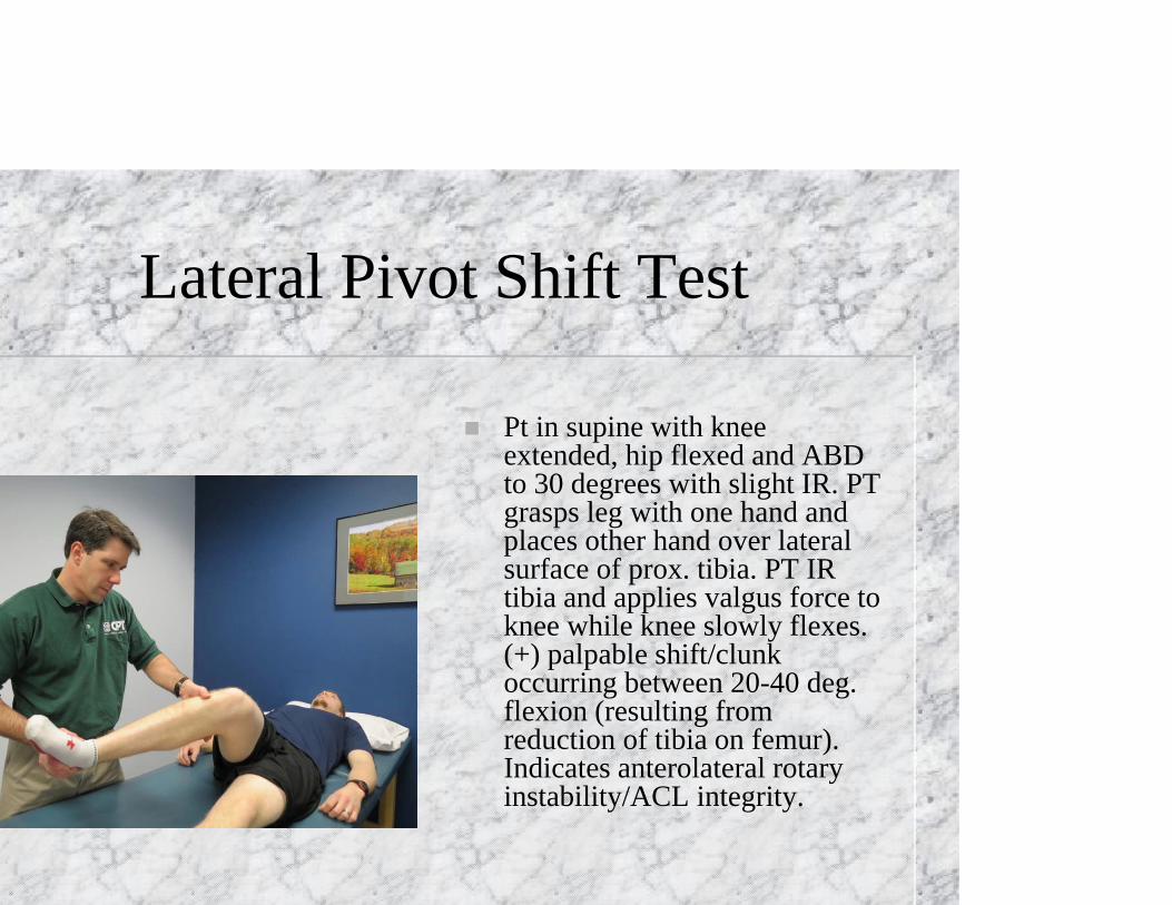
Lateral Pivot Shift Test
Pt in supine with knee extended, hip flexed and ABD to 30 degrees with slight IR. PT grasps leg with one hand and places other hand over lateral surface of prox. tibia. PT IR tibia and applies valgus force to knee while knee slowly flexes. (+) palpable shift/clunk occurring between 20-40 deg. flexion (resulting from reduction of tibia on femur). Indicates anterolateral rotary instability/ACL integrity.
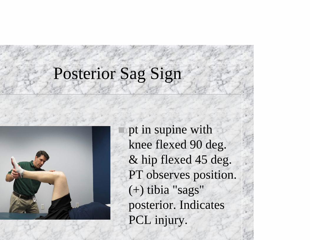
Posterior Sag Sign
pt in supine with knee flexed 90 deg. & hip flexed 45 deg. PT observes position. (+) tibia "sags" posterior. Indicates PCL injury.

Valgus Stress TestPt in supine with entire LE supported and knee flexed to 20-30 deg. PT places one hand on medial surface of ankle and other hand on lateral surface of knee. PT applies valgus force to the knee with distal hand. (+) excess valgus movement and/or pain. Indicates MCL sprain. Note: a (+) test with knee in full extension may be indicative of damage to MCL, PCL, posterior oblique ligament, posteromedial capsule
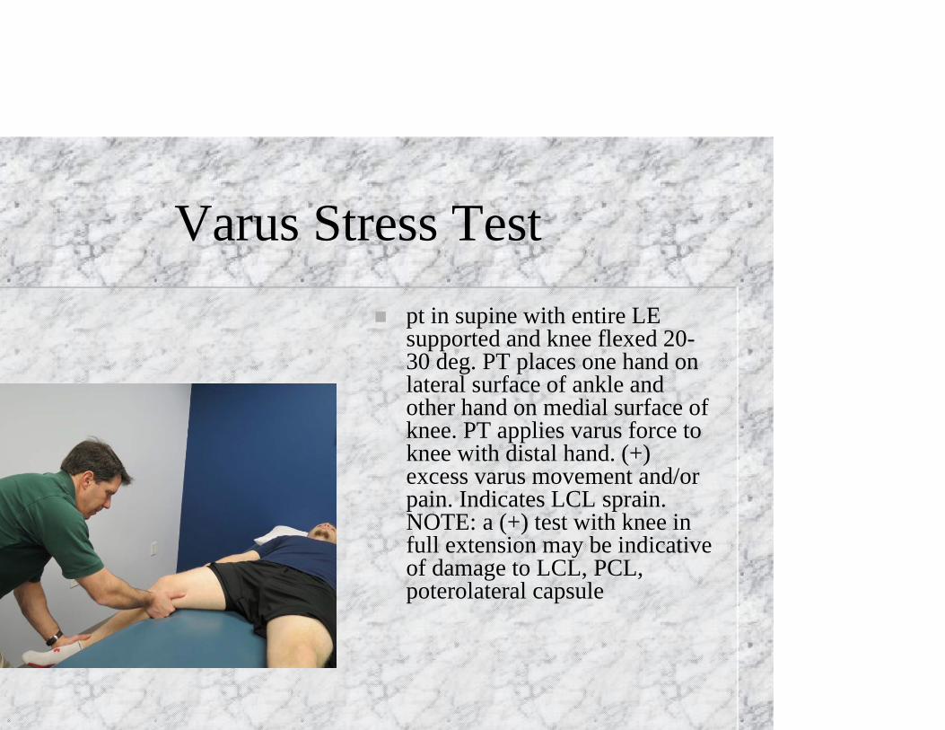
Varus Stress Test pt in supine with entire LE
supported and knee flexed 20-30 deg. PT places one hand on lateral surface of ankle and other hand on medial surface of knee. PT applies varus force to knee with distal hand. (+) excess varus movement and/or pain. Indicates LCL sprain. NOTE: a (+) test with knee in full extension may be indicative of damage to LCL, PCL, poterolateral capsule
Meniscal Tests McMurray’s test-IR of the tibia
with knee extension = torn lateral meniscus and vice-versa.Apley’s Compression
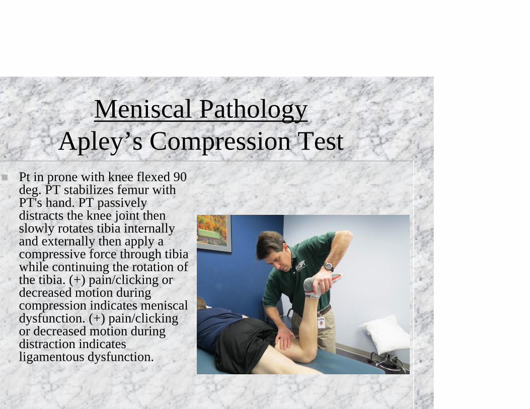
Meniscal PathologyApley’s Compression Test
Pt in prone with knee flexed 90 deg. PT stabilizes femur with PT's hand. PT passively distracts the knee joint then slowly rotates tibia internally and externally then apply a compressive force through tibia while continuing the rotation of the tibia. (+) pain/clicking or decreased motion during compression indicates meniscal dysfunction. (+) pain/clicking or decreased motion during distraction indicates ligamentous dysfunction.
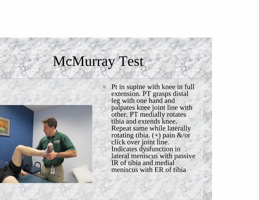
McMurray Test Pt in supine with knee in full
extension. PT grasps distal leg with one hand and palpates knee joint line with other. PT medially rotates tibia and extends knee. Repeat same while laterally rotating tibia. (+) pain &/or click over joint line. Indicates dysfunction in lateral meniscus with passive IR of tibia and medial meniscus with ER of tibia
Modified meniscal grind test Flexion & extension of the knee with a
valgus stress = compression of the lateral meniscus
Flexion & extension of the knee with a varus stress = compression of the medial meniscus
Pain with end-range flexion may = posterior horn.
Pain with end-range extension may = anterior horn
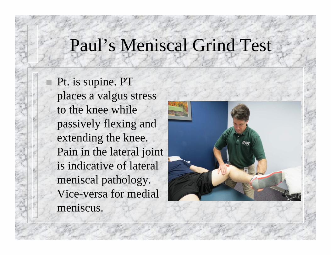
Paul’s Meniscal Grind Test
Pt. is supine. PT places a valgus stress to the knee while passively flexing and extending the knee. Pain in the lateral joint is indicative of lateral meniscal pathology. Vice-versa for medial meniscus.
Other Tests
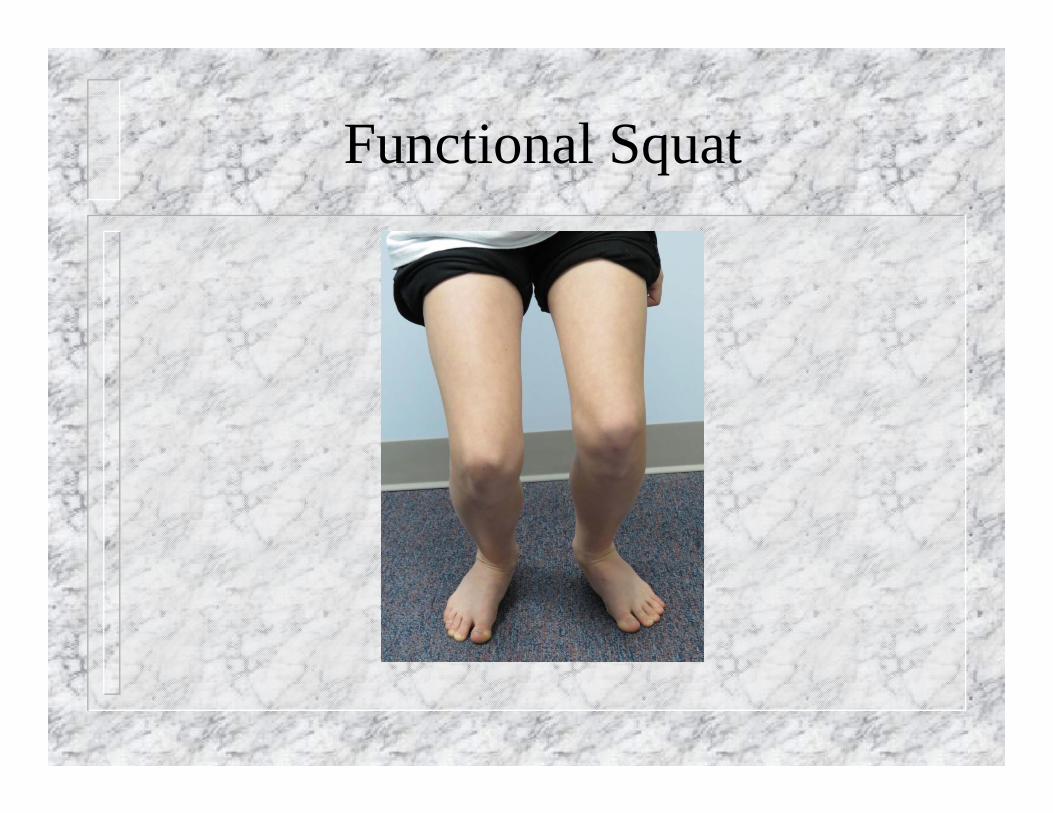
Noble Compression Test Ober’s Test Q-angle Functional Squat- Look for: -Ankle DF -Knee Valgus -Hip Varus -Inability to keep back straight
•
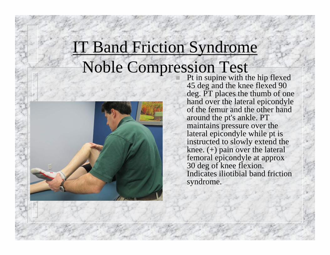
IT Band Friction SyndromeNoble Compression Test
Pt in supine with the hip flexed 45 deg and the knee flexed 90 deg. PT places the thumb of one hand over the lateral epicondyle of the femur and the other hand around the pt's ankle. PT maintains pressure over the lateral epicondyle while pt is instructed to slowly extend the knee. (+) pain over the lateral femoral epicondyle at approx 30 deg of knee flexion. Indicates iliotibial band friction syndrome.
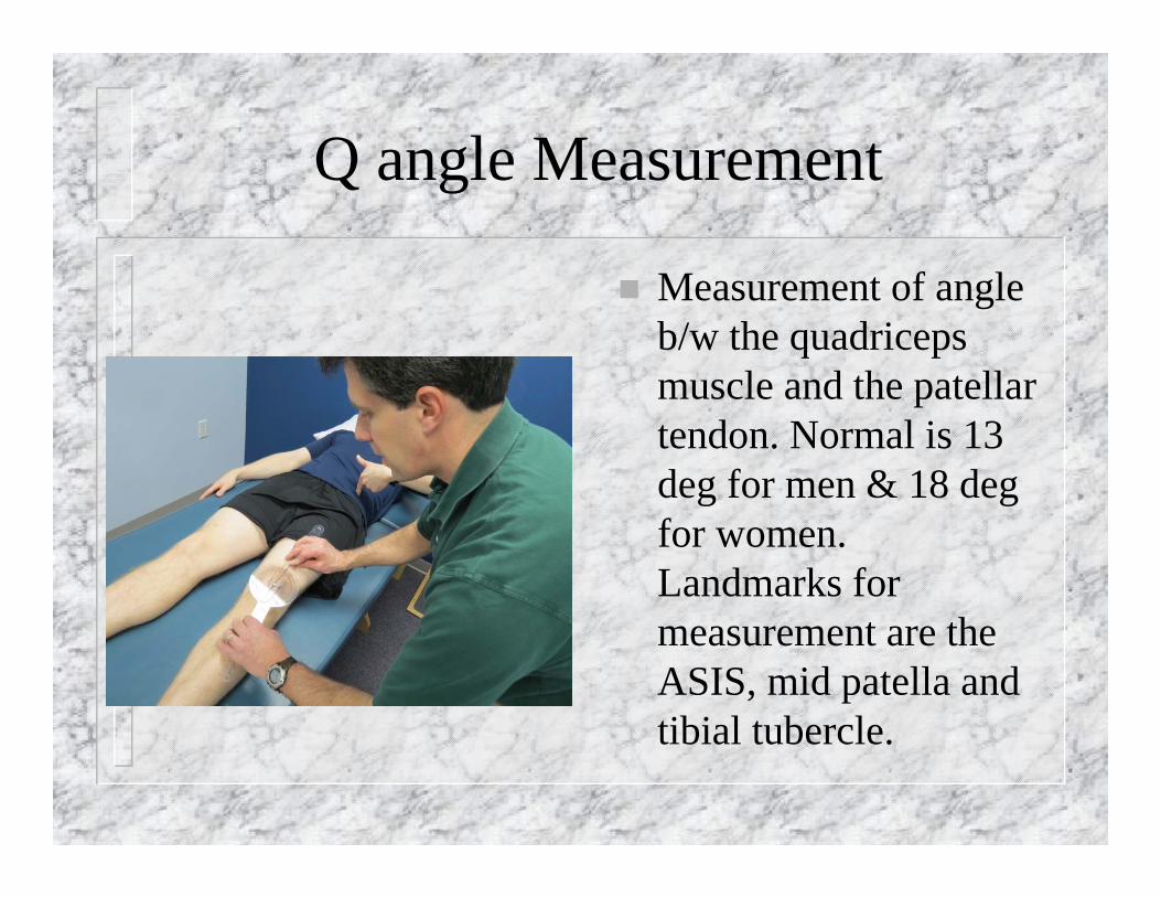
Q angle Measurement
Measurement of angle b/w the quadriceps muscle and the patellar tendon. Normal is 13 deg for men & 18 deg for women. Landmarks for measurement are the ASIS, mid patella and tibial tubercle.
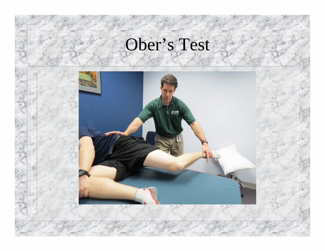
Ober’s Test
Functional Squat
Selective tissue tension tests
Must differentiate between contractile and non-contractile tissue (I.e. popliteus mm. Vs. posterior horn of lateral meniscus)