m edical i maging packet #5 chapter #22. i ntroduction medical imaging provides visual...
TRANSCRIPT

MEDICAL IMAGINGPacket #5
Chapter #22

INTRODUCTION
Medical imaging provides visual representations of body parts, tissues, or organs, for use in clinical diagnosis.
X-rays, gamma rays, high-frequency sound waves, and magnetic fields to produce the images of organs and other internal structures of the body.

RADIOLOGY
Radiology is the branch of medicine that deals with the use of radioactive substances in diagnosis (diagnostic imaging) and treatment of disease (radiation therapy).

DIAGNOSTIC IMAGING

INTRODUCTION
Electronic radiation is used to produce images of internal structures.

X-RAYS X-rays, discovered in 1895,
are used in medicine to diagnose bone fractures, dental cavities, and cancer; to locate foreign objects in the body; and to stop the spread of malignant tumors.
X-rays are of extremely short wavelength (100 nanometers to 0.001 nanometer) produced by the deceleration of charged particles or the transitions of electrons in atoms.

FLUOROSCOPY
Fluoroscopy is a dynamic x-ray imaging technique that produces a moving image over time.
It is essential for evaluating organ movement such as the beating of the heart or movement of the diaphragm.

FLUOROSCOPY II Procedures begin with the
administration of a barium mixture either by ingestion or by an enema that fills the stomach or large intestine. The barium mixture, known
as a contrast medium, like dense tissues, blocks the x-ray beam.
Fluoroscopy then reveals the location of the barium-coated lining of the stomach and intestine and enables the radiologist to observe as they contract and distend.

ANGIOGRAPHY
Angiography is the radiologic study of blood vessels. Because arteries and
veins are not normally visible in conventional x-ray studies, an iodinated compound, which is opaque to the x-ray, must be injected into the bloodstream.

ANGIOGRAPHY II There are different
types of angiograms. An arteri-ogram is an x-
ray study of the arteries A venogram is an x-ray
study of the veins. Arteriography is most
often used to show the presence and extent that arteries have become clogged and narrowed by arteriosclerosis, which can lead to strokes and heart attacks.

CT
Computed tomography (CT), also called computed axial tomography (CAT), is a scanning technique that combines computer and x-ray technologies. The computer
constructs a two-dimensional anatomic image that represents a cross-sectional slice through the body.

ULTRASOUND
Ultrasound imaging, or sonography, is a diagnostic imaging procedure that uses high frequency sound waves instead of ionizing radiation.

ULTRASOUND II During an ultrasound
examination, a lightweight transducer is placed on the patient's, skin over the part to be imaged.
The transducer produces sound waves that penetrate the skin to reach tissues and organs.
When the sound waves strike specific tissue surface, echoes are produced.
The echoes are detected by the transducer and are then electronically converted into an anatomic image that is displayed on a video screen.

ULTRASOUND III Ultrasound imaging is
commonly used in obstetrics to monitor the position and development of the fetus and also to detect any fatal abnormalities or problems during pregnancy.
However, an ultrasound is also used to show problems in other internal structures, including the gallbladder, kidney, and heart.

MRI
Magnetic resonance imaging (MRI) is a diagnostic procedure that uses a large, high-strength magnet, radio-frequency signals, and a computer to produce images.

MRI
Conducting an MRI is extremely useful in evaluating diseases of the brain and spine.
It is also used to evaluate joints, bone and soft tissue abnormalities, as well as abnormalities of the chest, abdomen, and pelvis.
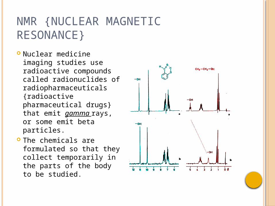
NMR {NUCLEAR MAGNETIC RESONANCE} Nuclear medicine
imaging studies use radioactive compounds called radionuclides of radiopharmaceuticals {radioactive pharmaceutical drugs} that emit gamma rays, or some emit beta particles.
The chemicals are formulated so that they collect temporarily in the parts of the body to be studied.

NMR {NUCLEAR MAGNETIC RESONANCE} For most nuclear imaging
studies, the radionuclide is injected into the patient and the images are taken with a gamma camera suspended above the patient who lies on a table.
The camera detects the gamma rays emitted from the radionuclide in the patient's body and uses this information to produce an image that shows the distribution of the radionuclide within the body.

TOMOGRAPHY The single-photon
emission computed tomography (SPECT) examination uses a computer to obtain two-dimensional images that are thin slices of internal organs such as the heart, brain, and liver.
The SPECT images can display organs with much greater detail than conventional scintigrams.

PET {POSITRON EMISSION TOMOGRAPHY} Positron emission
tomography (PET) is a more refined radio-logic technique that is used to study the metabolic activity inside an organ.
The technique has been shown to be useful in the study of brain-related disorders, such as epilepsy and Alzheimer's disease, and of the vitality of heart tissue.

EEG {ELECTROENCEPHALOGRAM}
A written recording of the electrical activity of the brain.
Electroencephalograms {EEG} are useful in studying and detecting brain disorders.

ECG {ELECTROCARDIOGRAM} The electrocardiogram
(as a paper trace or a TV monitor display) shows the changes in the voltage, detectable during the time course of the heart beat, between pairs of electrodes placed at certain points on the skin.
The basis of the ECG is that the heart, like other muscles, is triggered to contract by electrical activity.

REVIEW

REVIEW I