copyrightpsasir.upm.edu.my/id/eprint/75351/1/fpsk(m) 2016 47 ir.pdf · all material contained...
TRANSCRIPT
© COPYRIG
HT UPM
UNIVERSITI PUTRA MALAYSIA
STRUCTURAL AND BIOPHYSICAL CHARACTERISATIONS OF MeCP2 MBD MUTANTS THAT CORRELATE WITH RETT SYNDROME
CHIA JYH YEA
FPSK(M) 2016 47
© COPYRIG
HT UPMSTRUCTURAL AND BIOPHYSICAL CHARACTERISATIONS OF MeCP2
MBD MUTANTS THAT CORRELATE WITH RETT SYNDROME
By
CHIA JYH YEA
Thesis Submitted to the School of Graduate Studies, Universiti Putra Malaysia, in
Fulfilment of the Requirements for the Degree of Master of Science
January 2016

© COPYRIG
HT UPM
All material contained within the thesis, including without limitation text, logos, icons,
photographs and all other artwork, is copyright material of Universiti Putra Malaysia
unless otherwise stated. Use may be made of any material contained within the thesis
for non-commercial purposes from the copyright holder. Commercial use of material
may only be made with the express, prior, written permission of Universiti Putra
Malaysia.
Copyright © Universiti Putra Malaysia
© COPYRIG
HT UPM
i
Abstract of thesis presented to the Senate of Universiti Putra Malaysia in
fulfilment of the requirement for the Degree of Master of Science
STRUCTURAL AND BIOPHYSICAL CHARACTERISATIONS OF MeCP2
MBD MUTANTS THAT CORRELATE WITH RETT SYNDROME
By
CHIA JYH YEA
January 2016
Chairman : Ho Kok Lian, PhD
Faculty : Medicine and Health Sciences
Methyl-CpG binding protein (MBD) family consists of Methyl-CpG Binding Protein 2
(MeCP2), Methyl-CpG Binding Domain Protein 1 (MBD1), MBD2, MBD3 and MBD4
where MeCP2 is the prototype of the family. MeCP2 contains several domains: (a) a
methyl-CpG binding domain (MBD), (b) a transcriptional repression domain (TRD), (c)
two AT hooks and (d) a nuclear localisation signal (NLS). MeCP2 binds to methylated
DNA and represses the transcription of the associated genes. Mutations in MECP2 lead
to Rett syndrome (RTT), which is characterised by progressive neuro-developmental
disorder in early childhood of females. Previous studies revealed that most RTT
missense mutations alter the protein conformation and subsequently interferes the
methyl-CpG recognition. To understand how the structural changes contribute to RTT,
the 3-dimensional structure of these mutants need to be elucidated. Therefore, it is of interest to study the structure of RTT related MBD in complex with methylated DNA
using X-ray crystallography and characterised the DNA-protein binding with some
biophysical assays. Since more than 50% of the missense mutations occur within the
MBD domain. Out of the 8 hot RTT spots within this domain, RTT mutants D97E,
A140V, Y141C, P152R and D156E were included in this study. In band shift assay,
wild-type MBD complexed with DNA was significantly shifted compared to A140V
and D97E while other mutants were not significantly shifted. In SPR, wild-type MBD
showed the highest affinity towards the DNA followed by A140V (KD: 0.28 µM).
Circular dichrosim (CD) analysis revealed that the secondary structures of A140V,
Y141C, P152R and D156E are highly similar to wild-type MBD (14.7 % -helix, 25.2 %
-strand, and 60.1 % turns and unordered) except for D97E which showed 31 % -
helix, 6.5 % -strand, 62.5 % turns and unordered. The complex of MBD140V with methylated DNA was crystallised and diffracted X-ray to 2.2 Å resolution. The co-
crystal belongs to monoclinic space group C2, with unit cell parameters of a=78.66 Å,
b=53.49 Å, c=62.78 Å, ==90 ° and β=132.47 °. X-ray analysis revealed that the MBD domain was not altered by mutation of Ala-140 to Val (A140V). However,
additional water molecules were identified at the DNA-protein contact interface and around the DNA molecule. A narrow minor groove of A/T run was observed as a result
of additional bifurcated hydrogen bonds and vertical stacking of bases results from high
degree of propeller twist and heavy purine-purine stacking. Two hydration spines were
observed running down the wall of the minor groove. Each hydration spine is well

© COPYRIG
HT UPM
ii
arranged into two shells adopting a zig-zag arrangement. Hence, this finding provides
insights for the DNA geometry where the A/T run is geometrically stabilized by
extensive water network and is independent of the flanking nucleotide sequence, DNA
methylation and the bound MBD domain. The finding explores characteristics of the
methylated DNA containing A/T run, which provide the nucleotide sequence
preferences to MeCP2. In general, these additional molecular details could provide fundamental knowledge in RTT therapeutic approaches.

© COPYRIG
HT UPM
iii
Abstrak tesis yang dikemukakan kepada Senat Universiti Putra Malaysia sebagai
memenuhi keperluan untuk Ijazah Master Sains
KAJIAN STRUKTUR DAN BIOFIZIKAL MUTAN MeCP2 MBD YANG
BERKAITAN DENGAN SINDROM RETT
Oleh
CHIA JYH YEA
Januari 2016
Pengerusi : Ho Kok Lian, PhD
Fakulti : Perubatan dan Sains Kesihatan
Keluarga protein pengikat metil-CpG (MBD) terdiri daripada protein Pengikat Metil-
CpG 2 (MeCP2), pengikat metil-CpG 1 (MBD1), MBD2, MBD3 dan MBD4 di mana
MeCP2 adalah prototaip keluarga. MeCP2 mengandungi beberapa domain: (a) domain pengikat metil-CpG (MBD), (b) domain penindasan transkripsi (TRD), (c) dua
pencangkuk AT dan (d) isyarat penempatan nuklear (NLS). Protein Pengikat Metil-
CpG 2 (MeCP2) mengikat DNA bermetil dan menindas transkripsi gen yang berkaitan.
Mutasi dalam MECP2 membawa kepada sindrom Rett (RTT), yang mempunyai ciri-
ciri progresif gangguan neuron dalam perkembangan awal kanak-kanak perempuan.
Kajian sebelum ini menunjukkan bahawa kebanyakan mutasi missense RTT mengubah
komformasi protein dan seterusnya mengganggu pengenalan metil-CpG. Untuk
memahami bagaimana perubahan struktur membawa kepada RTT, struktur 3-dimensi
mutan perlu dijelaskan. Oleh itu, adalah penting untuk mengkaji struktur RTT
berkaitan dengan MBD kompleks dengan DNA bermetil menggunakan sinaran-X
penghabluran dan mencirikan dengan beberapa analisis DNA biofizikal metilasi. Lebih
daripada 50% daripada mutasi missense berkelompok dalam domain MBD. Daripada 8 lokasi penting RTT dalam domain ini, mutan RTT termasuk D97E, A140V, Y141C,
P152R dan D156E telah dimasukkan dalam kajian ini. Dalam essei anjakan jalur,
Kompleks DNA-protein MBD jenis liar meranjak dengan ketara berbanding dengan
mutan A140V dan D97E manakala anjakan jalur mutan lain tidak ketara. Dalam SPR,
MBD jenis liar mempunyai daya afiniti ke arah DNA yang paling tinggi dan diikuti
oleh mutan A140V (KD: 0.28 µM). Analisis dikreisme membulat (CD) menunjukkan
bahawa struktur sekundur mutan RTT (A140V, Y141C, P152R dan D156E) adalah
menyerupai MBD jenis liar (14.7 % α-heliks, 25.2 % β-helai, dan 60.1 % tidak tersusun)
kecuali D97E yang menunjukkan 31 % α -heliks, 6.5 % β-helai, 62.5 % tidak tersusun.
Kompleks MBD140V dengan DNA bermetil telah dihablurkan dengan menggunakan 30 %
(w/v) PEG 2000 dan hablur telah dibelau oleh sinaran-X kepada resolusi 2.2 Å. Hablur ini tergolong dalam kumpulan ruang monoklinik C2, dengan parameter unit sel = 78.66
Å, b = 53.49 Å, c = 62.78 Å, α = γ = 90 ° dan β = 132.47 °. Analisis sinaran-X
mendedahkan bahawa domain MBD tidak berubah akibat mutasi Ala-140 kepada Val
(A140V). Walau bagaimanapun, lebih banyak molekul air telah dikenalpasti di antara
muka DNA-protein dan di sekitar molekul DNA. Lurah minor dengan urutan A/T yang
berulang telah dikenalpasti dengan tambahan ikatan hidrogen bercabang dan
penindihan nukleotida secara menegak yang berpunca daripada pusingan baling-baling
© COPYRIG
HT UPM
iv
yang berdarjah tinggi dan penindihan purina. Dua spina hidrasi didapati menuruni
dinding lurah minor. Setiap spina hidrasi tersusun dalam dua lapisan dengan
penyusunan zig-zag. Oleh itu, kajian ini dapat meningkatkan pemahaman dalam
geometri DNA di mana urutan A/T yang berulang adalah stabil akibat daripada
rangkaian molekul air dan bebas daripada mengapitnya urutan nukleotida, metilasi
DNA dan domain MBD yang terikat. Penemuan ini membongkarkan ciri-ciri DNA bermetil yang mengandungi urutan A/T berulang bagi membekalkan kegemaran
jujukan nukleotida kepada MeCP2. Secara umum, Informasi molekular yang didapati
dapat menawarkan perkembangan dalam permahaman asas dalam terapeutik RTT.

© COPYRIG
HT UPM
v
ACKNOWLEDGEMENTS
With God‟s warmest blessing, there are some people who made my journey easier and
I would like to extend my greatest appreciation to those who helped me in completing
my journey. First and foremost, I would like to express my heartfelt gratitude to the
chairperson of the supervisory committee, Dr. Ho Kok Lian, for his endless support
and invaluable guidance throughout this study. He is always in an amiable manner with
his friendly gesture. All the gracious personalities of him have motivated me to become
a more down to earth and heart-warming person in the near future. In addition, his
impressive cognitive ability and intellectual maturity have also thought me the importance of critical and analytical reasoning, not only as a student point of view but
also as a researcher.
I would also like to extend my sincere appreciation to the members of the supervisory
committee, Prof. Dr. Tan Wen Siang and Assoc. Prof. Dr. Foo Hooi Ling, for their
wise opinions and constructive suggestions. Their endurance and boundless enthusiasm
in research are some of the characteristics that inspired me in the attempt to become a
diligent, persistent and successful researcher. I am also grateful to all my labmates,
especially Yoon Kam Yee; for their stimulating discussions, advice, their kind
hospitality, friendship and technical assistance. A million thanks goes to all of you who have brightened and amused me in one way or another for making my long exhausting
hours in lab a happy and pleasant one. Finally, my special thanks and deepest gratitude
are extended to my beloved family members for their understanding, perseverance,
support and endless love throughout my study.
© COPYRIG
HT UPM
© COPYRIG
HT UPM
vii
This thesis was submitted to the Senate of Universiti Putra Malaysia and has been
accepted as fulfilment of the requirement for the degree of Master of Science. The
members of the Supervisory Committee were as follows:
Ho Kok Lian, PhD
Senior Lecturer
Faculty of Medicine and Health Sciences
Universiti Putra Malaysia
(Chairperson)
Tan Wen Siang, PhD
Professor
Faculty of Biotechnology and Biomolecular Sciences
Universiti Putra Malaysia
(Member)
Foo Hooi Ling, PhD
Associate Professor
Faculty of Biotechnology and Biomolecular Sciences
Universiti Putra Malaysia (Member)
_____________________________
BUJANG KIM HUAT, PhD
Professor and Dean
School of Graduate Studies
Universiti Putra Malaysia
Date:
© COPYRIG
HT UPM
viii
Declaration by graduate student
I hereby confirm that:
this thesis is my original work
quotations, illustrations and citations have been duly referenced
the thesis has not been submitted previously or comcurrently for any other degree
at any institutions
intellectual property from the thesis and copyright of thesis are fully-owned by
Universiti Putra Malaysia, as according to the Universiti Putra Malaysia
(Research) Rules 2012;
written permission must be owned from supervisor and deputy vice –chancellor
(Research and innovation) before thesis is published (in the form of written,
printed or in electronic form) including books, journals, modules, proceedings,
popular writings, seminar papers, manuscripts, posters, reports, lecture notes,
learning modules or any other materials as stated in the Universiti Putra Malaysia
(Research) Rules 2012;
there is no plagiarism or data falsification/fabrication in the thesis, and scholarly
integrity is upheld as according to the Universiti Putra Malaysia (Graduate
Studies) Rules 2003 (Revision 2012-2013) and the Universiti Putra Malaysia
(Research) Rules 2012. The thesis has undergone plagiarism detection software
Signature: _______________________ Date: __________________
Name and Matric No.: Chia Jyh Yea , GS30076
© COPYRIG
HT UPM
ix
Declaration by Members of Supervisory Committee
This is to confirm that:
the research conducted and the writing of this thesis was under our supervision;
supervision responsibilities as stated in the Universiti Putra Malaysia (Graduate
Studies) Rules 2003 (Revision 2012-2013) are adhered to.
Signature:
Name of Chairman of Supervisory
Committee: Dr.Ho Kok Lian,
Signature:
Name of Member
of Supervisory
Committee: Professor
Dr.Tan Wen Siang
Signature:
Name of Member
of Supervisory
Committee:
Associate Professor
Dr.Foo Hooi Ling
© COPYRIG
HT UPM
x
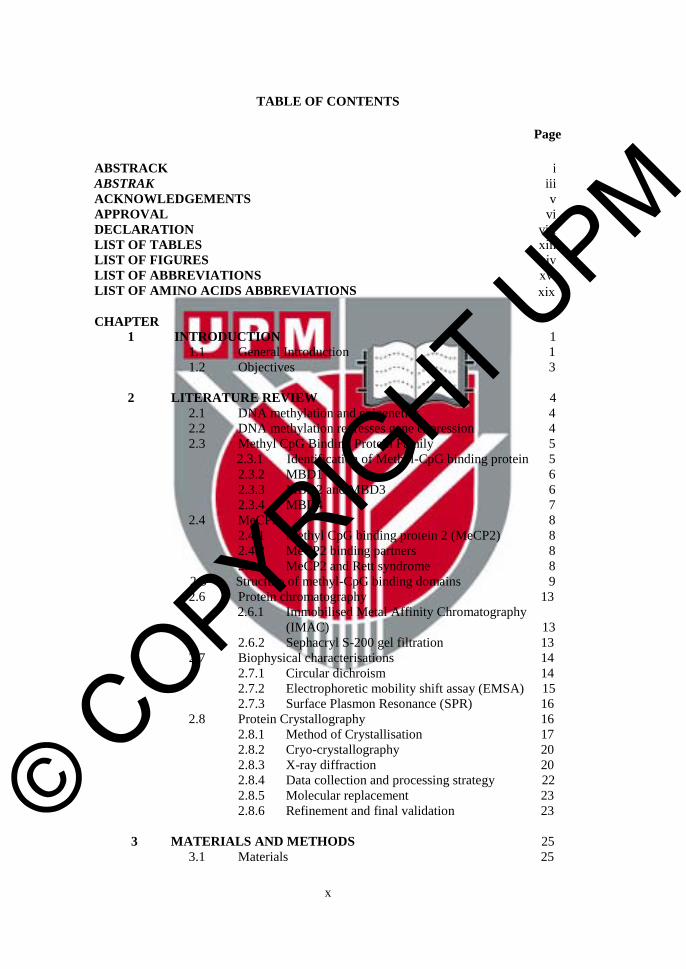
TABLE OF CONTENTS
Page
ABSTRACK i
ABSTRAK iii
ACKNOWLEDGEMENTS v
APPROVAL vi
DECLARATION viii
LIST OF TABLES xiiiLIST OF FIGURES xivLIST OF ABBREVIATIONS xviLIST OF AMINO ACIDS ABBREVIATIONS xix
CHAPTER
1 INTRODUCTION 1
1.1 General Introduction 1
1.2 Objectives 3
2 LITERATURE REVIEW 4
2.1 DNA methylation and epigenetics 4
2.2 DNA methylation represses gene expression 4
2.3 Methyl CpG Binding Protein Family 5
2.3.1 Identification of Methyl-CpG binding protein 5
2.3.2 MBD1 6
2.3.3 MBD2 and MBD3 6
2.3.4 MBD4 7
2.4 MeCP2 8
2.4.1 Methyl CpG binding protein 2 (MeCP2) 8
2.4.2 MeCP2 binding partners 8
2.4.3 MeCP2 and Rett syndrome 8
2.5 Structure of methyl-CpG binding domains 9
2.6 Protein chromatography 13
2.6.1 Immobilised Metal Affinity Chromatography
(IMAC) 13
2.6.2 Sephacryl S-200 gel filtration 13
2.7 Biophysical characterisations 14
2.7.1 Circular dichroism 14
2.7.2 Electrophoretic mobility shift assay (EMSA) 15
2.7.3 Surface Plasmon Resonance (SPR) 16
2.8 Protein Crystallography 16
2.8.1 Method of Crystallisation 17
2.8.2 Cryo-crystallography 20
2.8.3 X-ray diffraction 20
2.8.4 Data collection and processing strategy 22
2.8.5 Molecular replacement 23
2.8.6 Refinement and final validation 23
3 MATERIALS AND METHODS 25
3.1 Materials 25
© COPYRIG
HT UPM
xi
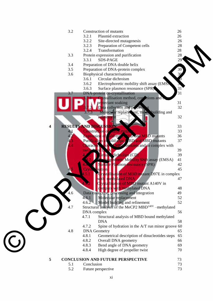
3.2 Construction of mutants 26
3.2.1 Plasmid extraction 26
3.2.2 Site-directed mutagenesis 26
3.2.3 Preparation of Competent cells 28
3.2.4 Transformation 28
3.3 Protein expression and purification 28
3.3.1 SDS-PAGE 29
3.4 Preparation of DNA double helix 30
3.5 Preparation of DNA-protein complex 30
3.6 Biophysical characterisations 30
3.6.1 Circular dichroism 30
3.6.2 Electrophoretic mobility shift assay (EMSA) 30
3.6.3 Surface plasmon resonance (SPR) 31
3.7 DNA-protein co-crystallisation 31
3.7.1 Crystallisation method, conditions and cryo-
protectant soaking 31
3.7.2 Data collection and processing 32
3.7.3 Molecular replacement, model building and
refinement 32
4 RESULTS AND DISCUSSION 33
4.1 Construction of MBD mutants 33
4.2 Expression of wild-type MBD and MBD mutants 36
4.3 Purification of wild-type MBD and MBD mutants 37
4.4 Characterisation of MBD mutants and in complex with
methylated DNA 39
4.4.1 Circular dichroism (CD) 39
4.4.2 Electrophoretic Mobility Shift assay (EMSA) 41
4.4.3 Surface Plasmon resonance (SPR) 42
4.5 Crystallisation 45
4.5.1 Crystallisation of MBD mutant D97E in complex
with methylated DNA 47
4.5.2 Crystallisation of MBD mutant A140V in
complex with methylated DNA 48
4.6 Data collection, processing and integration 49
4.6.1 Molecular replacement 52
4.6.2 Model building and refinement 52
4.7 Structural analysis of the MeCP2 MBD140V –methylated
DNA complex 56
4.7.1 Structural analysis of MBD bound methylated
DNA 58
4.7.2 Spine of hydration in the A/T run minor groove 60
4.8 DNA Geometry 65
4.8.1 Geometrical description of dinucleotides steps 65
4.8.2 Overall DNA geometry 66
4.8.3 Bend angle of DNA geometry 69
4.8.4 High degree of propeller twist 70
5 CONCLUSION AND FUTURE PERSPECTIVE 73
5.1 Conclusion 73
5.2 Future perspective 73
© COPYRIG
HT UPM
xii
REFERENCES 75
APPENDICES 85
BIODATA OF STUDENT 88
LIST OF PUBLICATIONS 89
© COPYRIG
HT UPM
xiii
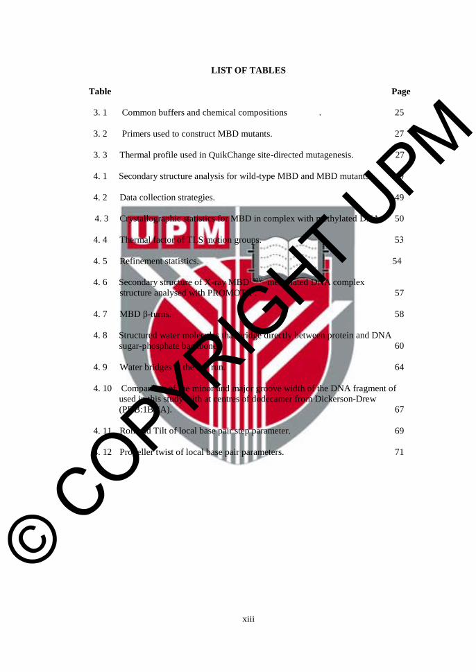
LIST OF TABLES
Table Page
3. 1 Common buffers and chemical compositions . 25
3. 2 Primers used to construct MBD mutants. 27
3. 3 Thermal profile used in QuikChange site-directed mutagenesis. 27
4. 1 Secondary structure analysis for wild-type MBD and MBD mutants. 40
4. 2 Data collection strategies. 49
4. 3 Crystallographic statistics for MBD in complex with methylated DNA. 50
4. 4 Thermal factor of TLS motion groups. 53
4. 5 Refinement statistics. 54
4. 6 Secondary structure of X-ray MBD140V–methylated DNA complex
structure analysed with PROMOTIF. 57
4. 7 MBD β-turns. 58
4. 8 Structured water molecules that bridge directly between protein and DNA
sugar-phosphate backbone. 60
4. 9 Water bridges at the AT run. 64
4. 10 Comparison of the minor and major groove width of the DNA fragment of
used in this study with at centres of dodecamer from Dickerson-Drew
(PDB:1BNA). 67
4. 11 Roll and Tilt of local base pair step parameter. 69
4. 12 Propeller twist of local base pair parameters. 71
© COPYRIG
HT UPM
xiv
LIST OF FIGURES
Figure Page
2. 1 MeCP2 function in gene regulation and chromatin remodeling. 5
2. 2 The methyl-CpG binding protein family. 5
2. 3 Hydrolytic deamination. 7
2. 4 Eight hot mutation spots within the MBD domain. 9
2. 5 Overlay of MBD domains among MBD family members. 12
2. 6 Immobilised metal affinity chromatography. 13
2. 7 Size exclusion chromatography. 14
2. 8 Far UV CD spectra associated with various type of secondary structures. 15
2. 9 Schematic illustration of protein crystallisation phase diagram. 17
2. 10 Hanging drop vapour diffusion. 18
2. 11 Sitting drop vapour diffusion. 19
2. 12 Bragg's Law. 21
2. 13 Sphere of reflection or Ewald sphere construction. 22
4. 1 Side-directed mutagenesis of MBD. 35
4. 2 SDS-PAGE [15% (w/v)] analysis of expressed wild-type MBD. 36
4. 3 SDS-PAGE [15% (w/v)] of expressed wild-type MBD and MBD mutants.36
4. 4 SDS-PAGE [15% (w/v)] analysis of protein fractions separated from MBD
mutant A140V by IMAC affinity column. 37
4. 5 SDS-PAGE [15 % (w/v)] analysis of protein fraction separated from pooled
IMAC active fraction of MBD mutant A140V. 38
4. 6 Circular dichroism spectrum of wild-type MBD and MBD mutants. 40
4. 7 Band shift assay. 42
4. 8 Sensorgram of Surface plasmon resonance. 43
4. 9 Sensorgram of the binding of wild-type MBD and MBD mutants to Biotin-
methylated DNA. 44
© COPYRIG
HT UPM
xv
4. 10 Comparison of binding levels of the wild-type MBD and the MBD mutants to
Biotin-methylated DNA. 45
4. 11 Multiple sequence alignment of MBD domain of MBD proteins. 46
4. 12 Identity and similarity of MBD domain of MBD proteins. 47
4. 13 Crystallisation of D97E mutant in complex with methylated DNA. 48
4. 14 Crystallisation of A140V mutant in complex with methylated DNA. 49
4. 15 Data collection of crystal DNA-MBD140V. 51
4. 16 Model building of DNA-MBD140V. 52
4. 17 TLS motion groups of DNA-MBD140V model. 54
4. 18 Analysis of stereochemical properties of DNA-MBD140V X-ray structure. 55
4. 19 Overall X-ray structure of MBD140V in complex with methylated DNA
at 2.18 Å. 56
4. 20 E137 is stabilised by water molecules and amide groups. 59
4. 21 Hydrogen bonds between O2' of m5C8 with W63 and W64. 59
4. 22 Overall solvent structure of MBD140V in complex with methylated DNA. 62
4. 23 Bridging system in AT run. 63
4. 24 Geometrical description of DNA parameters. 65
4. 25 Graphical output of a Curves+ analysis for DNA. 66
4. 26 Overlay of idealised B-DNA (Blue) and X-ray DNA (Cyan) with A/T run
highlighted in red. 68
4. 27 Superimposed of A/T run of MBD140V with HMGA-1. 68
4. 28 Stereo view of the superposition of the A/T run. 70
4. 29 High degree of propeller twist at AT run base pair. 72

© COPYRIG
HT UPM
xvi
LIST OF ABBREVIATIONS
α Alpha
Å Ångström
β Beta
µg microgram (10-6 g)
µL microliter (10-6 L)
µM micromolar (10-6 M)
BDNF Brain-derived neurotrophic factor
bp Basepair
BSA bovine serum albumin
ccc covalently closed circular
CCP4 Collaborative Computational Project Number 4
C-terminal carboxyl terminal
DNA Deoxy-ribonucleic acid
dNTP deoxynucleoside triphosphate
dsDNA double stranded DNA
DTT 1,4-dithiothreitol
EDTA ethylene diamine tetraacetic acid
EMSA Electrophoretic mobility shift assay
FPLC fast protein liquid chromatography
HDAC Histone deacetylase
HPLC high performance liquid chromatography
IMAC immobilised metal ion affinity chromatography
IPTG isopropyl-β-d-thiogalactopyranoside
© COPYRIG
HT UPM
xvii
K Kelvin
Kb kilobase
Kd dissociation constant
kDA kilo Dalton
LB Luria broth
m5C 5’methyl cytosine
MBD Methyl-CpG Binding Domain
MBD1 Methyl-CpG Binding Domain Protein 1
MBD2 Methyl-CpG Binding Domain Protein 2
MBD3 Methyl-CpG Binding Domain Protein 3
MBD4 Methyl-CpG Binding Domain Protein 4
MeCP2 Methyl-CpG Binding Protein 2
MBP Methyl-CpG Binding Protein
Mg milligram (10-3 g)
Min minute
NDB Nucleic Acid Database
NLS Nuclear localisation Signal
nM nanomolar (10-9 M)
NTA Nitrilotriacetic acid
OD optical density
PAGE polyacrylamide gel electrophoresis
PCM1 Protein containing MBD 1
PCR Polymerase chain reaction
PDB Protein Database Bank
PEG Polyethylene glycol
© COPYRIG
HT UPM
xviii
pI Isoelectric point
RMSD root mean square deviation
rpm revolutions per minute
RTT Rett Syndrome
s Second
SDS
SEC
sodium dodecyl sulphate
size exclusion chromatography
TBE Tris-buffered EDTA solution
TEMED Tetramethyl ethylenediamine
TRD Transcriptional repression domain
U Unit
UV Ultraviolet
v Volt
Vm Matthew’s coefficient
v/v volume/volume
w/v weight/volume
x g centrifugal force
© COPYRIG
HT UPM
xix
LIST OF AMINO ACIDS ABBREVIATIONS
One letter code Three letter code
Alanine A Ala
Arginine R Arg
Asparagine N Asn
Aspartic acid D Asp
Cysteine C Cys
Glutamic acid E Glu
Glutamine Q Gln
Glycine G Gly
Histidine H His
Isoleucine I Ile
Leucine L Leu
Lysine K Lys
Methionine M Met
Phenylalanine F Phe
Proline P Pro
Serine S Ser
Threonine T Thr
Tryptophan W Trp
Tyrosine Y Tyr
Valine V Val
© COPYRIG
HT UPM
1
CHAPTER 1
INTRODUCTION
1.1 General Introduction
DNA methylation is an epigenetic signal that affects gene regulation, genomic stability
and chromatin structure in mammalian cells (Bird, 2002; Du et al., 2015). In most cases,
this signal can be read by a family of proteins that contains a common methyl-CpG
binding domain (MBD) (Hendrich and Bird, 1998). To date, five family members,
namely MeCP2, MBD1, MBD2, MBD3 and MBD4 have been identified, in which,
MeCP2 is the prototype of this family (Hendrich and Bird, 1998). MeCP2, MBD1 and
MBD2 are able to recruit co-repressor complexes that can inhibit transcription in concert
with chromatin modifiers (Scarsdale et al., 2011). Mammalian MBD3 does not bind to
methylated DNA in vitro and in vivo due to replacement of amino acid (K43H and Y47F)
which is critical for DNA binding (Fraga et al., 2003). MBD4 contains a thymine DNA-
glycosylase at the C-terminal region that can repair G-T mismatches via hydrolytic
deamination [(Refer Section 2.3.4 and Figure 2.3) (Neddermann et al., 1996)].
MeCP2 is a transcriptional repressor that contains several domains: (a) a methyl-CpG
binding domain (MBD), (b) a transcriptional repression domain (TRD), (c) AT hooks
and (d) a nuclear localisation signal (NLS) (Lewis et al., 1992; Nan and Bird, 2001; Xu
and Pozzo-Miller, 2013). MBD domain of MeCP2 is able to recognise methyl-CpG
containing DNA. TRD domain involves in recruitment of transcriptional co-repressors
such as mSin3A and histone deacetylases (HDACs) (Bienvenu et al., 2000). Two
putative NLSs facilitate nuclear localisation, which targets the protein into the cell
nucleus (Weaving et al., 2004) and the AT hooks are believed to interact with AT rich
region of the DNA (Klose et al., 2005). In addition, the AT hooks of MeCP2 bearing
amino acid sequences 185GRGRGRP191 and 265PKKRGRKP272 (superscript indicates
amino acid number) which are highly similar to the high mobility group with the AT
hook I chromosomal protein (HMCG-I) that is capable to bind to the minor groove of
the AT stretches (A/T run) of DNA and functionally (Aravind and Landsman, 1998;
Lewis et al., 1992; Nan et al., 1993; Reeves and Nissen, 1990). Baker et al. (2013)
demonstrated that the disruption of second conserved AT hook at the C-terminal region
of MeCP2 by truncation at R270X of MeCP2 led to failure in chromatin compaction and
localization of pericentric heterochromatin domain of -thalassemia mental retardation
syndrome X-linked (ATRX); a chromatin remodelling protein, with MeCP2, and caused
the R270X mice to exhibit Rett syndrome (RTT) phenotypes which is similar to MeCP2
knock-out mice (Baker et al., 2013; Xu and Pozzo-Miller, 2013).
The MBD domain of MeCP2 is able to recognise single methyl-CpG dinucleotide (Lewis
et al., 1992). The MBD domain alone is ample for the methylated DNA binding and
mutations in the MBD domain intercept its binding to methylated sequence (Baubec et
al., 2013; Yusufzai and Wolffe, 2000). MeCP2 mutation causes Rett syndrome (RTT); a
progressive neurodevelopmental disorder in early childhood, which leads to mental
© COPYRIG
HT UPM
2
retardation in females, with a prevalence of 1 in 10,000-15,000 female births (Hagberg,
1985). RTT is caused by an X-linked mutation dominant inheritance with normally
lethality in males due to severe encephalopathy (Bianciardi et al., 2015; Bienvenu et al.,
2000; Zhao et al., 2015). Studies revealed that most RTT related missense mutations
alter the structure of the MBD domain and subsequently interrupt DNA recognition
properties (Kriaucionis and Bird, 2003; Kucukkal et al., 2015). Klose et al. (2005)
showed that an A/T run adjacent to the methyl-CpG is required to enhance the MeCP2
binding (Klose et al., 2005). Identified endogenous MeCP2 targeting genes such as brain
derived neurotropic factor (BDNF) promoter region contains high occurrences of A/T
runs closed to the methyl-CpGs (Chen et al., 2003; Martinowich et al., 2003). The A/T
run in the methylated DNA facilitated the co-crystallisation of MeCP2 MBD domain in
complex with methylated DNA used in this study. Due to the presence of AT hooks in
MeCP2 and the requirement of A/T run for maximal binding, it has been speculated that
the A/T run could interact with the AT hooks of the MeCP2. However, the characteristics
of A/T run which provide specificity for the MeCP2 to recognise the methyl groups
remained unclear. Therefore, it is of interest to elucidate the 3-dimensional structure of
RTT mutants in order to understand how the structural changes contribute to RTT and
the A/T run characteristics with MBD domain bound to its adjacent methyl-CpG
dinucleotide.
X-ray analysis of previous report on a MeCP2 MBD-methylated DNA complex revealed
that only a few residues are involved in direct contact with the DNA bases (Ho et al.,
2008). The methyl groups are recognised by the Arginine fingers of R111 and R133
while D121 is critical in maintaining the unique hydration pattern at the DNA-protein
interface. The unique water molecules distribution pattern is crucial to mediate methyl
group recognition (Ho et al., 2008). RTT mutations within the MBD domain of MeCP2,
however, are believed to alter the 3-dimensional structure of the protein and subsequently
affects DNA binding. Several critical mutations such as R111G, R133C, T158M and
D121G, which close to the DNA-protein contact region have been investigated (Free et
al., 2001; Meehan et al., 1992; Nan et al., 1993; Yusufzai and Wolffe, 2000). In this
study, other RTT mutations which are located distance from the DNA-protein contact
region have been studied. According to Wakefield and colleagues (1999), missense
mutations found in RTT usually do not specifically interrupt DNA recognition but may
result in structural changes in the domain (Bianciardi et al., 2015; Wakefield et al., 1999).
In order to further investigate the details on the structural changes and molecular
functional role, several mutants (D97E, A140V, Y141C, P152R and D156E) were
constructed and the DNA-MBD interactions were characterised with various biophysical
assays. A co-crystal structure was also elucidated, in which, more molecular details about
the DNA-protein complex have been revealed compared with previous reported structure.
In addition, the atomic details of the DNA geometry of the MBD bound A/T run are
highlighted in comparison with the A/T run of the free DNA double helices. In general,
these additional molecular details could provide fundamental knowledge in RTT
therapeutic approaches.

© COPYRIG
HT UPM
3
1.2 Objectives
The general objective of this study was to explore and understand the atomic details
of Rett mutants in complex with methylated DNA. The specific objectives were:
1. To construct MBD mutants
2. To characterise the interactions of MBD mutants and methylated DNA
3. To crystallise the MBD mutants in complex with methylated DNA
4. To solve the X-ray structure of MBD mutants in complex with methylated DNA
© COPYRIG
HT UPM
75
REFERENCES
Abrescia N.G., Thompson A., Huynh-Dinh T., Subirana J.A. (2002). Crystal structure of
an antiparallel DNA fragment with Hoogsteen base pairing. Proceedings of the
National Academy of Sciences of the United States of America 99: 2806-2811
Amir R., Dahle E.J., Toriolo D., Zoghbi H.Y. (2000). Candidate gene analysis in Rett
syndrome and the identification of 21 SNPs in Xq. American Journal of
Medical Genetics 90: 69-71
Amir R.E., Van den Veyver I.B., Wan M., Tran C.Q., Francke U., Zoghbi H.Y. (1999).
Rett syndrome is caused by mutations in X-linked MECP2, encoding methyl-
CpG-binding protein 2. Nature Genetics 23: 185-188
Aravind L., Landsman D. (1998). AT-hook motifs identified in a wide variety of DNA-
binding proteins. Nucleic Acids Research 26: 4413-4421
Baker S.A., Chen L., Wilkins A.D., Yu P., Lichtarge O., Zoghbi H.Y. (2013). An AT-
hook domain in MeCP2 determines the clinical course of Rett syndrome and
related disorders. Cell 152: 984-996
Bhattacharya S.K., Ramchandani S., Cervoni N., Szyf M. (1999). A mammalian protein
with specific demethylase activity for mCpG DNA. Nature 397: 579-583
Bienvenu T., Carrie A., de Roux N., Vinet M.C., Jonveaux P., Couvert P., Villard L.,
Arzimanoglou A., Beldjord C., Fontes M., Tardieu M., Chelly J. (2000).
MECP2 mutations account for most cases of typical forms of Rett syndrome.
Human Molecular Genetics 9: 1377-1384
Bird A. (2002). DNA methylation patterns and epigenetic memory. Genes &
Development 16: 6-21
Bird A. (2008). The methyl-CpG-binding protein MeCP2 and neurological disease.
Biochemical Society Transactions 36: 575-583
Blanchet C., Pasi M., Zakrzewska K., Lavery R. (2011). CURVES+ web server for
analyzing and visualizing the helical, backbone and groove parameters of
nucleic acid structures. Nucleic Acids Research 39: W68-73
Block H., Maertens B., Spriestersbach A., Brinker N., Kubicek J., Fabis R., Labahn J.,
Schafer F. (2009). Immobilized-metal affinity chromatography (IMAC): a
review. Methods in Enzymology 463: 439-473
Brigham-Burke M., Edwards J.R., O'Shannessy D.J. (1992). Detection of receptor-ligand
interactions using surface plasmon resonance: model studies employing the
HIV-1 gp120/CD4 interaction. Analytical Biochemistry 205: 125-131

© COPYRIG
HT UPM
76
Chayen N.E. (1998). Comparative studies of protein crystallization by vapour-diffusion
and microbatch techniques. Acta Crystallographica. Section D, Biological
Crystallography 54: 8-15
Chayen N.E., Saridakis E. (2002). Protein crystallization for genomics: towards high-
throughput optimization techniques. Acta Crystallographica. Section D,
Biological Crystallography 58: 921-927
Chen W.G., Chang Q., Lin Y., Meissner A., West A.E., Griffith E.C., Jaenisch R.,
Greenberg M.E. (2003). Derepression of BDNF transcription involves calcium-
dependent phosphorylation of MeCP2. Science (New York, N.Y.) 302: 885-889
Choi W.I., Jeon B.N., Yoon J.H., Koh D.I., Kim M.H., Yu M.Y., Lee K.M., Kim Y.,
Kim K., Hur S.S., Lee C.E., Kim K.S., Hur M.W. (2013). The proto-
oncoprotein FBI-1 interacts with MBD3 to recruit the Mi-2/NuRD-HDAC
complex and BCoR and to silence p21WAF/CDKN1A by DNA methylation.
Nucleic Acids Research 41: 6403-6420
Cramer J.M., Scarsdale J.N., Walavalkar N.M., Buchwald W.A., Ginder G.D., Williams
D.C., Jr. (2014). Probing the dynamic distribution of bound states for
methylcytosine-binding domains on DNA. The Journal of Biological Chemistry
289: 1294-1302
Crook J.M., Dunn N.R., Colman A. (2006). Repressed by a NuRD. Nature Cell Biology
8: 212-214
Cross S.H., Meehan R.R., Nan X., Bird A. (1997). A component of the transcriptional
repressor MeCP1 shares a motif with DNA methyltransferase and HRX
proteins. Nature Genetics 16: 256-259
Dickerson R.E. (1998). DNA bending: the prevalence of kinkiness and the virtues of
normality. Nucleic Acids Research 26: 1906-1926
Du Q., Luu P.L., Stirzaker, C. Clark S.J. (2015). Methyl-CpG-binding domain proteins:
Readers of the Epigenome. Epigenomics 7: 1051-1073
El Hassan M.A., Calladine C.R. (1996). Propeller-twisting of base-pairs and the
conformational mobility of dinucleotide steps in DNA. Journal of Molecular
Biology 259: 95-103
Emsley P., Cowtan K. (2004). Coot: model-building tools for molecular graphics. Acta
Crystallographica. Section D, Biological Crystallography 60: 2126-2132
Evans P. (2006). Scaling and assessment of data quality. Acta Crystallographica. Section
D, Biological Crystallography 62: 72-82
Fonfria-Subiros E., Acosta-Reyes F., Saperas N., Pous J., Subirana J.A., Campos J.L.
(2012). Crystal structure of a complex of DNA with one AT-hook of HMGA1.
PloS One 7: e37120
© COPYRIG
HT UPM
77
Fraga M.F., Ballestar E., Montoya G., Taysavang P., Wade P.A., Esteller M. (2003). The
affinity of different MBD proteins for a specific methylated locus depends on
their intrinsic binding properties. Nucleic Acids Research 31: 1765-1774
Fratini A.V., Kopka M.L., Drew H.R., Dickerson R.E. (1982). Reversible bending and
helix geometry in a B-DNA dodecamer: CGCGAATTBrCGCG. The Journal
of Biological Chemistry 257: 14686-14707
Free A., Wakefield R.I., Smith B.O., Dryden D.T., Barlow P.N., Bird A.P. (2001). DNA
recognition by the methyl-CpG binding domain of MeCP2. The Journal of
Biological Chemistry 276: 3353-3360
Fried M., Crothers D.M. (1981). Equilibria and kinetics of lac repressor-operator
interactions by polyacrylamide gel electrophoresis. Nucleic Acids Research 9:
6505-6525
Fujita N., Takebayashi S., Okumura K., Kudo S., Chiba T., Saya H., Nakao M. (1999).
Methylation-mediated transcriptional silencing in euchromatin by methyl-CpG
binding protein MBD1 isoforms. Molecular and Cellular Biology 19: 6415-
6426
Fujita N., Watanabe S., Ichimura T., Ohkuma Y., Chiba T., Saya H., Nakao M. (2003a).
MCAF mediates MBD1-dependent transcriptional repression. Molecular and
Cellular Biology 23: 2834-2843
Fujita N., Watanabe S., Ichimura T., Tsuruzoe S., Shinkai Y., Tachibana M., Chiba T.,
Nakao M. (2003b). Methyl-CpG binding domain 1 (MBD1) interacts with the
Suv39h1-HP1 heterochromatic complex for DNA methylation-based
transcriptional repression. The Journal of Biological Chemistry 278: 24132-
24138
Garman E.F., Owen R.L. (2006). Cryocooling and radiation damage in macromolecular
crystallography. Acta Crystallographica. Section D, Biological
Crystallography 62: 32-47
Ghosh R.P., Horowitz-Scherer R.A., Nikitina T., Gierasch L.M., Woodcock C.L. (2008).
Rett syndrome-causing mutations in human MeCP2 result in diverse structural
changes that impact folding and DNA interactions. The Journal of Biological
Chemistry 283: 20523-20534
Garcia-Ruiz J.M. (2003). Counterdiffusion methods for macromolecular crystallization.
Methods in Enzymology 368: 130-154
Greenfield N.J. (1996). Methods to estimate the conformation of proteins and
polypeptides from circular dichroism data. Analytical Biochemistry 235: 1-10
Haff L.A. (1978). Fractionation of water-insoluble protein using Sephacryl S-200 in
formamide. Preparative Biochemistry 8: 99-112
Hagberg B. (1985). Rett's syndrome: prevalence and impact on progressive severe
mental retardation in girls. Acta Paediatrica Scandinavica 74: 405-408
© COPYRIG
HT UPM
78
Harker D. (1956). X-ray diffraction applied to crystalline proteins. Advances in
Biological and Medical Physics 4: 1-22
Hashimoto H., Zhang X., Cheng X. (2012). Excision of thymine and 5-
hydroxymethyluracil by the MBD4 DNA glycosylase domain: structural basis
and implications for active DNA demethylation. Nucleic Acids Research 40:
8276-8284
Hendrich B., Bird A. (1998). Identification and characterization of a family of
mammalian methyl-CpG binding proteins. Molecular and Cellular Biology 18:
6538-6547
Hendrich B., Bird A. (2000). Mammalian methyltransferases and methyl-CpG-binding
domains: proteins involved in DNA methylation. Current Topics in
Microbiology and Immunology 249: 55-74
Hendrich B., Hardeland U., Ng H.H., Jiricny J., Bird A. (1999). The thymine glycosylase
MBD4 can bind to the product of deamination at methylated CpG sites. Nature
401: 301-304
Hermann A., Gowher H., Jeltsch A. (2004). Biochemistry and biology of mammalian
DNA methyltransferases. Cellular and Molecular Life Sciences : CMLS 61:
2571-2587
Ho K.L., McNae I.W., Schmiedeberg L., Klose R.J., Bird A.P., Walkinshaw M.D.
(2008). MeCP2 binding to DNA depends upon hydration at methyl-CpG.
Molecular Cell 29: 525-531
Horowitz S., Trievel R.C. (2012). Carbon-oxygen hydrogen bonding in biological
structure and function. The Journal of Biological Chemistry 287: 41576-41582
Hutchinson E.G., Thornton J.M. (1996). PROMOTIF--a program to identify and analyze
structural motifs in proteins. Protein science : A Publication of the Protein
Society 5: 212-220
Jeffery L., Nakielny S. (2004). Components of the DNA methylation system of
chromatin control are RNA-binding proteins. The Journal of Biological
Chemistry 279: 49479-49487
Jentarra G.M., Olfers S.L., Rice S.G., Srivastava N., Homanics G.E., Blue M., Naidu S.,
Narayanan V. (2010). Abnormalities of cell packing density and dendritic
complexity in the MeCP2 A140V mouse model of Rett syndrome/X-linked
mental retardation. BMC Neuroscience 11: 19
Jing M., Bowser M.T. (2011). Methods for measuring aptamer-protein equilibria: A
review. Analytica Chimica Acta 686: 9-18
Jones P.L., Veenstra G.J., Wade P.A., Vermaak D., Kass S.U., Landsberger N.,
Strouboulis J., Wolffe A.P. (1998). Methylated DNA and MeCP2 recruit
histone deacetylase to repress transcription. Nature Genetics 19: 187-191
© COPYRIG
HT UPM
79
Jorgensen H.F., Ben-Porath I., Bird A.P. (2004). Mbd1 is recruited to both methylated
and nonmethylated CpGs via distinct DNA binding domains. Molecular and
Cellular Biology 24: 3387-3395
Kabsch W. (2010). XDS. Acta Crystallographica. Section D, Biological Crystallography
66: 125-132
Kelly S.M., Jess T.J., Price N.C. (2005). How to study proteins by circular dichroism.
Biochimica et Biophysica Acta 1751: 119-139
Khrustalev V.V., Barkovsky E.V., Khrustaleva T.A. (2014). The influence of flanking
secondary structures on amino Acid content and typical lengths of 3/10 helices.
International Journal of Proteomics 2014: 360230
Klose R.J., Sarraf S.A., Schmiedeberg L., McDermott S.M., Stancheva I., Bird A.P.
(2005). DNA binding selectivity of MeCP2 due to a requirement for A/T
sequences adjacent to methyl-CpG. Molecular Cell 19: 667-678
Kriaucionis S., Bird A. (2003). DNA methylation and Rett syndrome. Human Molecular
Genetics 12 Spec No 2: R221-227
Kucukkal T.G., Yang Y., Uvarov O., Cao W., Alexov E. (2015). Impact of Rett
syndrome mutations on MeCP2 MBD stability. Biochemistry 54: 6357-6368
Laemmli U.K. (1970). Cleavage of structural proteins during the assembly of the head
of bacteriophage T4. Nature 227: 680-685
Laskowski R.A., Rullmannn J.A., MacArthur M.W., Kaptein R., Thornton J.M. (1996).
AQUA and PROCHECK-NMR: programs for checking the quality of protein
structures solved by NMR. Journal of Biomolecular NMR 8: 477-486
Leslie A.G. (2006). The integration of macromolecular diffraction data. Acta
Crystallographica. Section D, Biological Crystallography 62: 48-57
Lewis J.D., Meehan R.R., Henzel W.J., Maurer-Fogy I., Jeppesen P., Klein F., Bird A.
(1992). Purification, sequence, and cellular localization of a novel chromosomal
protein that binds to methylated DNA. Cell 69: 905-914
Li T., Jin Y., Vershon A.K., Wolberger C. (1998). Crystal structure of the
MATa1/MATalpha2 homeodomain heterodimer in complex with DNA
containing an A-tract. Nucleic Acids Research 26: 5707-5718
Lu X.J., Olson W.K. (2003). 3DNA: a software package for the analysis, rebuilding and
visualization of three-dimensional nucleic acid structures. Nucleic Acids
Research 31: 5108-5121
Ma L.Y., Wu C., Jin Y., Gao M., Li G.H., Turner D., Shen J.X., Zhang S.J., Narayanan
V., Jentarra G., Wu J. (2014). Electrophysiological phenotypes of MeCP2
A140V mutant mouse model. CNS Neuroscience & Therapeutics 20: 420-428

© COPYRIG
HT UPM
80
Magdinier F., Wolffe A.P. (2001). Selective association of the methyl-CpG binding
protein MBD2 with the silent p14/p16 locus in human neoplasia. Proceedings
of the National Academy of Sciences of the United States of America 98: 4990-
4995
Marston F.A. (1986). The purification of eukaryotic polypeptides synthesized in
Escherichia coli. The Biochemical Journal 240: 1-12
Martinowich K., Hattori D., Wu H., Fouse S., He F., Hu Y., Fan G., Sun Y.E. (2003).
DNA methylation-related chromatin remodeling in activity-dependent BDNF
gene regulation. Science (New York, N.Y.) 302: 890-893
Matthews B.W. (1968). Solvent content of protein crystals. Journal of Molecular
Biology 33: 491-497
Mcpherson A. (2004). Introduction to protein crystallization. Methods (san diego, calif.)
34: 254-265
Meehan R., Lewis J., Cross S., Nan X., Jeppesen P., Bird A. (1992). Transcriptional
repression by methylation of CpG. Journal of Cell Science. Supplement 16: 9-
14
Millar C.B., Guy J., Sansom O.J., Selfridge J., MacDougall E., Hendrich B., Keightley
P.D., Bishop S.M., Clarke A.R., Bird A. (2002). Enhanced CpG mutability and
tumorigenesis in MBD4-deficient mice. Science (New York, N.Y.) 297: 403-405
Minor W., Cymborowski M., Otwinowski Z., Chruszcz M. (2006). HKL-3000: the
integration of data reduction and structure solution--from diffraction images to
an initial model in minutes. Acta Crystallographica. Section D, Biological
Crystallography 62: 859-866
Murshudov G.N., Vagin A.A., Dodson E.J. (1997). Refinement of macromolecular
structures by the maximum-likelihood method. Acta Crystallographica. Section
D, Biological Crystallography 53: 240-255
Nan X., Bird A. (2001). The biological functions of the methyl-CpG-binding protein
MeCP2 and its implication in Rett syndrome. Brain & Development 23 Suppl
1: S32-37
Nan X., Campoy F.J., Bird A. (1997). MeCP2 is a transcriptional repressor with abundant
binding sites in genomic chromatin. Cell 88: 471-481
Nan X., Cross S., Bird A. (1998a). Gene silencing by methyl-CpG-binding proteins.
Novartis Foundation Symposium 214: 6-16; discussion 16-21, 46-50
Nan X., Hou J., Maclean A., Nasir J., Lafuente M.J., Shu X., Kriaucionis S., Bird A.
(2007). Interaction between chromatin proteins MECP2 and ATRX is disrupted
by mutations that cause inherited mental retardation. Proceedings of the
National Academy of Sciences of the United States of America 104: 2709-2714
© COPYRIG
HT UPM
81
Nan X., Meehan R.R., Bird A. (1993). Dissection of the methyl-CpG binding domain
from the chromosomal protein MeCP2. Nucleic Acids Research 21: 4886-4892
Nan X., Ng H.H., Johnson C.A., Laherty C.D., Turner B.M., Eisenman R.N., Bird A.
(1998b). Transcriptional repression by the methyl-CpG-binding protein MeCP2
involves a histone deacetylase complex. Nature 393: 386-389
Nan X., Tate P., Li E., Bird A. (1996). DNA methylation specifies chromosomal
localization of MeCP2. Molecular and Cellular Biology 16: 414-421
Neddermann P., Gallinari P., Lettieri T., Schmid D., Truong O., Hsuan J.J., Wiebauer
K., Jiricny J. (1996). Cloning and expression of human G/T mismatch-specific
thymine-DNA glycosylase. The Journal of Biological Chemistry 271: 12767-
12774
Nelson H.C., Finch J.T., Luisi B.F., Klug A. (1987). The structure of an
oligo(dA).oligo(dT) tract and its biological implications. Nature 330: 221-226
Ng J.D., Gavira J.A., Garcia-Ruiz J.M. (2003). Protein crystallization by capillary
counterdiffusion for applied crystallographic structure determination. Journal
of structural biology 142: 218-231
Ohki I., Shimotake N., Fujita N., Jee J., Ikegami T., Nakao M., Shirakawa M. (2001).
Solution structure of the methyl-CpG binding domain of human MBD1 in
complex with methylated DNA. Cell 105: 487-497
Otani J., Arita K., Kato T., Kinoshita M., Kimura H., Suetake I., Tajima S., Ariyoshi M.,
Shirakawa M. (2013). Structural basis of the versatile DNA recognition ability
of the methyl-CpG binding domain of methyl-CpG binding domain protein 4.
The Journal of Biological Chemistry 288: 6351-6362
Painter J., Merritt E.A. (2006). Optimal description of a protein structure in terms of
multiple groups undergoing TLS motion. Acta Crystallographica Section D 62:
439-450
Porath J., Carlsson J., Olsson I., Belfrage G. (1975). Metal chelate affinity
chromatography, a new approach to protein fractionation. Nature 258: 598-599
Potterton E., McNicholas S., Krissinel E., Cowtan K., Noble M. (2002). The CCP4
molecular-graphics project. Acta Crystallographica. Section D, Biological
Crystallography 58: 1955-1957
Prive G.G., Heinemann U., Chandrasegaran S., Kan L.S., Kopka M.L., Dickerson R.E.
(1987). Helix geometry, hydration, and G.A mismatch in a B-DNA decamer.
Science (New York, N.Y.) 238: 498-504
Provencher S.W., Glockner J. (1981). Estimation of globular protein secondary structure
from circular dichroism. Biochemistry 20: 33-37

© COPYRIG
HT UPM
82
Ramachandran G.N., Ramakrishnan C., Sasisekharan V. (1963). Stereochemistry of
polypeptide chain configurations. Journal of Molecular Biology 7: 95-99
Raussens V., Ruysschaert J.M., Goormaghtigh E. (2003). Protein concentration is not an
absolute prerequisite for the determination of secondary structure from circular
dichroism spectra: a new scaling method. Analytical Biochemistry 319: 114-121
Reese B.E., Bachman K.E., Baylin S.B., Rountree M.R. (2003). The methyl-CpG
binding protein MBD1 interacts with the p150 subunit of chromatin assembly
factor 1. Molecular and Cellular Biology 23: 3226-3236
Reeves R., Nissen M.S. (1990). The A.T-DNA-binding domain of mammalian high
mobility group I chromosomal proteins. A novel peptide motif for recognizing
DNA structure. The Journal of Biological Chemistry 265: 8573-8582
Robertson K.D. (2002). DNA methylation and chromatin - unraveling the tangled web.
Oncogene 21: 5361-5379
Sansom O.J., Berger J., Bishop S.M., Hendrich B., Bird A., Clarke A.R. (2003).
Deficiency of Mbd2 suppresses intestinal tumorigenesis. Nature Genetics 34:
145-147
Sansom O.J., Maddison K., Clarke A.R. (2007). Mechanisms of disease: methyl-binding
domain proteins as potential therapeutic targets in cancer. Nature Clinical
Practice. Oncology 4: 305-315
Scarsdale J.N., Webb H.D., Ginder G.D., Williams D.C., Jr. (2011). Solution structure
and dynamic analysis of chicken MBD2 methyl binding domain bound to a
target-methylated DNA sequence. Nucleic Acids Research 39: 6741-6752
Sjolund A.B., Senejani A.G., Sweasy J.B. (2013). MBD4 and TDG: multifaceted DNA
glycosylases with ever expanding biological roles. Mutation Research 743-744:
12-25
Studier F.W., Moffatt B.A. (1986). Use of bacteriophage T7 RNA polymerase to direct
selective high-level expression of cloned genes. Journal of Molecular Biology
189: 113-130
Toffaletti J., Savory J., Gitelman H.J. (1977). Use of gel filtration to examine the
distribution of calcium among serum proteins. Clinical chemistry 23: 2306-
2310
Turkenburg J.P., Dodson E.J. (1996). Modern developments in molecular replacement.
Current Opinion in Structural Biology 6: 604-610
Vagin A., Teplyakov A. (2010). Molecular replacement with MOLREP. Acta
crystallographica. Section D, Biological Crystallography 66: 22-25

© COPYRIG
HT UPM
83
Vaguine A.A., Richelle J., Wodak S.J. (1999). SFCHECK: a unified set of procedures
for evaluating the quality of macromolecular structure-factor data and their
agreement with the atomic model. Acta Crystallographica. Section D,
Biological Crystallography 55: 191-205
Venkateswaran S., McMillan H.J., Doja A., Humphreys P. (2014). Adolescent onset
cognitive regression and neuropsychiatric symptoms associated with the A140V
MECP2 mutation. Developmental Medicine and Child Neurology 56: 91-94
Wakefield R.I., Smith B.O., Nan X., Free A., Soteriou A., Uhrin D., Bird A.P., Barlow
P.N. (1999). The solution structure of the domain from MeCP2 that binds to
methylated DNA. Journal of Molecular Biology 291: 1055-1065
Walavalkar N.M., Cramer J.M., Buchwald W.A., Scarsdale J.N., Williams D.C., Jr.
(2014). Solution structure and intramolecular exchange of methyl-cytosine
binding domain protein 4 (MBD4) on DNA suggests a mechanism to scan for
mCpG/TpG mismatches. Nucleic Acids Research
Wang S., Poon G.M., Wilson W.D. (2015). Quantitative investigation of protein-nucleic
acid interactions by biosensor surface plasmon resonance. Methods Mol Biol
1334: 313-332
Watanabe S., Ichimura T., Fujita N., Tsuruzoe S., Ohki I., Shirakawa M., Kawasuji M.,
Nakao M. (2003). Methylated DNA-binding domain 1 and methylpurine-DNA
glycosylase link transcriptional repression and DNA repair in chromatin.
Proceedings of the National Academy of Sciences of the United States of
America 100: 12859-12864
Weaving L.S., Christodoulou J., Williamson S.L., Friend K.L., McKenzie O.L., Archer
H., Evans J., Clarke A., Pelka G.J., Tam P.P., Watson C., Lahooti H., Ellaway
C.J., Bennetts B., Leonard H., Gecz J. (2004). Mutations of CDKL5 cause a
severe neurodevelopmental disorder with infantile spasms and mental
retardation. American Journal of Human Genetics 75: 1079-1093
Weiss M.S., Einspahr H., Baker E.N., Dauter Z., Kaysser-Pyzalla A.R., Kostorz G.,
Larsen S. (2010). Citations in supplementary material. Acta Crystallographica.
Section D, Biological Crystallography 66: 1269-1270
Wiebauer K., Jiricny J. (1989). In vitro correction of G.T mispairs to G.C pairs in nuclear
extracts from human cells. Nature 339: 234-236
Wilchek M., Bayer E.A. (1990). Introduction to avidin-biotin technology. Methods in
Enzymology 184: 5-13
Willard H.F., Hendrich B.D. (1999). Breaking the silence in Rett syndrome. Nature
Genetics 23: 127-128
Wilson G.G., Murray N.E. (1991). Restriction and modification systems. Annual Review
of Genetics 25: 585-627
© COPYRIG
HT UPM
84
Wing R., Drew H., Takano T., Broka C., Tanaka S., Itakura K., Dickerson R.E. (1980).
Crystal structure analysis of a complete turn of B-DNA. Nature 287: 755-758
Winn M.D., Ballard C.C., Cowtan K.D., Dodson E.J., Emsley P., Evans P.R., Keegan
R.M., Krissinel E.B., Leslie A.G., McCoy A., McNicholas S.J., Murshudov
G.N., Pannu N.S., Potterton E.A., Powell H.R., Read R.J., Vagin A., Wilson
K.S. (2011). Overview of the CCP4 suite and current developments. Acta
Crystallographica. Section D, Biological Crystallography 67: 235-242
Wong E., Yang K., Kuraguchi M., Werling U., Avdievich E., Fan K., Fazzari M., Jin B.,
Brown A.M., Lipkin M., Edelmann W. (2002). Mbd4 inactivation increases
Cright-arrowT transition mutations and promotes gastrointestinal tumor
formation. Proceedings of the National Academy of Sciences of the United
States of America 99: 14937-14942
Wrinch D. (1946). Patterson distributions and native protein crystallography. Nature
157: 226
Wu P., Qiu C., Sohail A., Zhang X., Bhagwat A.S., Cheng X. (2003). Mismatch repair
in methylated DNA. Structure and activity of the mismatch-specific thymine
glycosylase domain of methyl-CpG-binding protein MBD4. The Journal of
Biological Chemistry 278: 5285-5291
Xu X., Pozzo-Miller L. (2013). A novel DNA-binding feature of MeCP2 contributes to
Rett syndrome. Frontiers in cellular neuroscience 7: 64
Yesselman J.D., Horowitz S., Brooks C.L., 3rd, Trievel R.C. (2015). Frequent side chain
methyl carbon-oxygen hydrogen bonding in proteins revealed by computational
and stereochemical analysis of neutron structures. Proteins 83: 403-410
Yusufzai T.M., Wolffe A.P. (2000). Functional consequences of Rett syndrome
mutations on human MeCP2. Nucleic Acids Research 28: 4172-4179
Zhao N., Ma D., Leong W.Y., Han J., Vandongen A., Chen T., Goh E.L. (2015). The
methyl-CpG-binding domain (MBD) is crucial for MeCP2's dysfunction-
induced defects in adult newborn neurons. Frontiers in Cellular Neuroscience
9: 158