lysine-selective molecular tweezers are cell penetrant and
TRANSCRIPT

ARTICLE
Lysine-selective molecular tweezers are cellpenetrant and concentrate in lysosomesZizheng Li1, Ibrar Siddique1, Inesa Hadrović2, Abbna Kirupakaran2, Jiwen Li3, Ye Zhang 3,4,5,
Frank-Gerrit Klärner2, Thomas Schrader2 & Gal Bitan 1,4,5✉
Lysine-selective molecular tweezers are promising drug candidates against proteinopathies,
viral infection, and bacterial biofilm. Despite demonstration of their efficacy in multiple cel-
lular and animal models, important questions regarding their mechanism of action, including
cell penetrance and intracellular distribution, have not been answered to date. The main
impediment to answering these questions has been the low intrinsic fluorescence of the main
compound tested to date, called CLR01. Here, we address these questions using new
fluorescently labeled molecular tweezers derivatives. We show that these compounds are
internalized in neurons and astrocytes, at least partially through dynamin-dependent endo-
cytosis. In addition, we demonstrate that the molecular tweezers concentrate rapidly in acidic
compartments, primarily lysosomes. Accumulation of molecular tweezers in lysosomes may
occur both through the endosomal-lysosomal pathway and via the autophagy-lysosome
pathway. Moreover, by visualizing colocalization of molecular tweezers, lysosomes, and tau
aggregates we show that lysosomes likely are the main site for the intracellular anti-amyloid
activity of molecular tweezers. These findings have important implications for the mechanism
of action of molecular tweezers in vivo, explaining how administration of low doses of the
compounds achieves high effective concentrations where they are needed, and supporting
the development of these compounds as drugs for currently cureless proteinopathies.
https://doi.org/10.1038/s42003-021-02603-2 OPEN
1 Department of Neurology, David Geffen School of Medicine, University of California, Los Angeles, Los Angeles, CA, USA. 2 Institute of Chemistry, Universityof Duisburg-Essen, Essen, Germany. 3 Department of Psychiatry and Biobehavioral Sciences, David Geffen School of Medicine, University of California, LosAngeles, Los Angeles, CA, USA. 4 Brain Research Institute, University of California, Los Angeles, Los Angeles, CA, USA. 5Molecular Biology Institute,University of California, Los Angeles, Los Angeles, CA, USA. ✉email: [email protected]
COMMUNICATIONS BIOLOGY | (2021) 4:1076 | https://doi.org/10.1038/s42003-021-02603-2 | www.nature.com/commsbio 1
1234
5678
90():,;
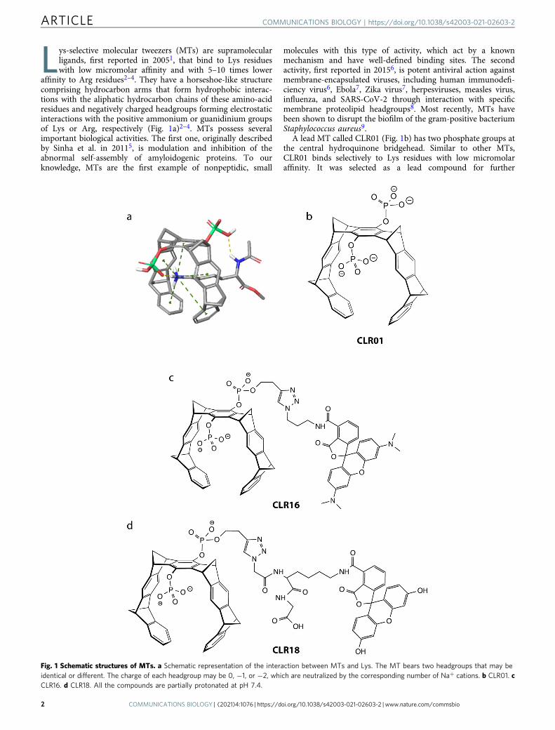
Lys-selective molecular tweezers (MTs) are supramolecularligands, first reported in 20051, that bind to Lys residueswith low micromolar affinity and with 5–10 times lower
affinity to Arg residues2–4. They have a horseshoe-like structurecomprising hydrocarbon arms that form hydrophobic interac-tions with the aliphatic hydrocarbon chains of these amino-acidresidues and negatively charged headgroups forming electrostaticinteractions with the positive ammonium or guanidinium groupsof Lys or Arg, respectively (Fig. 1a)2–4. MTs possess severalimportant biological activities. The first one, originally describedby Sinha et al. in 20115, is modulation and inhibition of theabnormal self-assembly of amyloidogenic proteins. To ourknowledge, MTs are the first example of nonpeptidic, small
molecules with this type of activity, which act by a knownmechanism and have well-defined binding sites. The secondactivity, first reported in 20156, is potent antiviral action againstmembrane-encapsulated viruses, including human immunodefi-ciency virus6, Ebola7, Zika virus7, herpesviruses, measles virus,influenza, and SARS-CoV-2 through interaction with specificmembrane proteolipid headgroups8. Most recently, MTs havebeen shown to disrupt the biofilm of the gram-positive bacteriumStaphylococcus aureus9.
A lead MT called CLR01 (Fig. 1b) has two phosphate groups atthe central hydroquinone bridgehead. Similar to other MTs,CLR01 binds selectively to Lys residues with low micromolaraffinity. It was selected as a lead compound for further
Fig. 1 Schematic structures of MTs. a Schematic representation of the interaction between MTs and Lys. The MT bears two headgroups that may beidentical or different. The charge of each headgroup may be 0, −1, or −2, which are neutralized by the corresponding number of Na+ cations. b CLR01. cCLR16. d CLR18. All the compounds are partially protonated at pH 7.4.
ARTICLE COMMUNICATIONS BIOLOGY | https://doi.org/10.1038/s42003-021-02603-2
2 COMMUNICATIONS BIOLOGY | (2021) 4:1076 | https://doi.org/10.1038/s42003-021-02603-2 | www.nature.com/commsbio

development based on its low toxicity5,10 and high activity againstboth amyloidogenic proteins and membrane-enveloped viruses. Akey feature contributing to the low toxicity of CLR01 is its highlylabile binding to Lys (and Arg) residues11 and the fact that theseresidues tend to be more exposed in unstructured and misfoldedpolypeptides than in natively folded proteins3. These character-istics allow CLR01 to disrupt effectively the relatively weakhydrophobic and electrostatic interactions mediating abnormalprotein self-assembly, without affecting normal physiologicalprocesses.
CLR01 has been found to inhibit the toxicity of various amy-loidogenic proteins, including amyloid β-protein (Aβ), α-synu-clein, islet amyloid polypeptide, and transthyretin in different celllines and primary cultures5,12–15. Using rat primary hippocampalneurons, we showed that CLR01 inhibited an Aβ42-induceddecrease in dendritic spine density, basal synaptic activity, andlong-term potentiation16. Recently, we also found that CLR01inhibited dose-dependently the prion-like propagation of tauaggregates (tau seeding) in biosensor cells17. Moreover, thepotential therapeutic effect of CLR01 has been demonstrated inmultiple animal models of various proteinopathies including thetriple-transgenic mouse model of Alzheimer’s disease (AD)16, arat model of AD18, a mouse model of tauopathy19, zebrafish12,20
and mouse15,21 models of Parkinson’s disease (PD), a lampreymodel of spinal cord injury22, and mouse models of transthyretinamyloidosis14, desmin-related cardiomyopathy23, amyotrophiclateral sclerosis24, multiple system atrophy25, and the lysosomal-storage disease Sanfilippo syndrome type A26.
Despite the promising demonstration of antiamyloid andantiviral activities in multiple cellular and animal models, untilvery recently, cell penetration/internalization of MTs has notbeen demonstrated directly. Moreover, the mechanism by whichMTs may get internalized and their cellular distribution uponinternalization, which may have important mechanistic implica-tions, currently are unknown. Early findings indicated thatCLR01 treatment reduced substantially the concentration of α-synuclein in a zebrafish model and suggested that this reductionwas mediated through alleviation of inhibition of the ubiquitin-proteasome system (UPS) by α-synuclein oligomers12. A similarfacilitation of UPS action was observed in a mouse model ofdesmin-related cardiomyopathy23. More recently, CLR01 wasfound to ameliorate significantly the pathologic phenotype in amouse model of Sanfilippo syndrome type A by a similar relievingof “clogged” lysosomes, allowing them to resume merging withautophagosomes to form autolysosomes and degrade multipleaggregated proteins, including α-synuclein, Aβ, tau, and prionprotein26. Based on these observations, it would be reasonable tohypothesize that MTs promote clearance of amyloidogenic pro-teins accumulating within cells, but how they facilitate suchclearance and what cellular mechanisms might be involved isunknown.
The main impediment to answering these questions is the lowintrinsic fluorescence of unlabeled MTs, which is too low to beobserved on the background of a cell making studying theirinteraction with cells difficult. To address this challenge andprovide insight into the intracellular distribution and mechanismof action of MTs, here we used two new fluorescently labeledderivatives—CLR16, labeled by 5-carboxytetramethylrhodamine(TAMRA, Fig. 1c) and CLR18, labeled by 6-fluorescein amidite(FAM, Fig. 1d). The pendant carboxylic group in the structure ofCLR18 is neither required for the fluorescence label nor for theMT function. It is just a consequence of our strategy to obtain aclickable FAM label. Contrary to the TAMRA fluorophore, azido-labeled FAM was not commercially available at the time thecompound was prepared for this project. Therefore, we usedFAMlysine, incorporated it into a small peptide, and attached a
terminal azide. Specifically, we started with a glycine-loadedWang resin, coupled FAM-labeled lysine, and then azidoaceticacid. Cleavage from the resin released the free carboxylic acid atthe C-terminus of this construct, which actually increases thewater solubility of the tweezer conjugate. An inherent advantageof this strategy is the introduction of a peptidic spacer betweentweezer and fluorophore. For CLR16 we purchased a TAMRAderivative with a terminal azide, allowing straightforward clickreaction with an alkyne tweezer.
Recently, we used CLR16 to demonstrate that it could beinternalized in a human oligodendroglioma cell line25, yet whe-ther the same was applicable to other cell types and the sub-cellular distribution of MTs remained unknown. Because thebrain is particularly sensitive to proteinopathies, we used here thefluorescent MTs to explore their internalization, intracellularlocalization, and mechanism of endocytosis in brain cellsincluding neurons and astrocytes.
ResultsCharacterization of CLR16 and CLR18. The concentrationdependence of the fluorescence of the two compounds wasassessed by a simple titration, which showed a linear increase ofthe fluorescence with concentration, as expected, both for thefluorescent appendage, TAMRA at 535 nm or FAM at 518 nm(Supplementary Fig. 1a, b, respectively), and for the horseshow-like structure of the MT (335 nm). The concentration dependenceof the TAMRA moiety in CLR16 (Supplementary Fig. 1a) wasshallower than that of the FAM group in CLR18, allowing formeasurement in the range 0.39–12.5 μM. Due the steeper con-centration dependence of CLR18 (Supplementary Fig. 1b), at0.39 μM it was similar to the blank, whereas at 12.5 μM themaximum fluorescence exceeded the maximum intensity of thefluorometer. These results suggested that both compounds couldbe used at 5 μM for subsequent cell-culture experiments.
To determine the pH dependence of the fluorescence, thespectrum of each compound was recorded at pH values between 3and 8 (Supplementary Fig. 1c, d). These measurements showeddistinct behaviors of the two derivatives. The fluorescence of theTAMRA group in CLR16 was maximal at pH 7 and declinedwhen the pH was decreased or increased without an apparentchange in λmax at 535 nm (Supplementary Fig. 1c). The weakfluorescence of the tweezer moiety at 335 nm decreased slightlywith the decrease in pH from 8 to 3. The fluorescence of the FAMgroup in CLR18 at 518 nm was maximal at pH 8 and declinedgradually when the pH decreased down to 5. At pH 4 or 3, thefluorescence intensity remained similar to pH 5, but the peakshifted from 518 to 570 nm (Supplementary Fig. 1d). Thisbehavior suggested that in cell-culture experiments using afluorescence microscope, at pH below 5, the fluorescence mightnot be detected because the microscope’s filter only allows awindow between 500 and 550 nm to be observed.
A possible concern is that the fluorescence labels might alterthe tweezers’ binding to Lys side chains. To test whether CLR16and CLR18 bind Lys residue in a similar manner to CLR01, i.e.,by inclusion of the Lys side chain within the tweezer’s cavity, weperformed NMR (Supplementary Fig. 2) and fluorescence(Supplementary Figs. 3 and 4) titrations with Ac-Lys-OMe. TheNMR experiments, which were done at high concentration,showed upfield shifts of the Lys side chain methylene groupsindicating inclusion inside the tweezer’s cavity, yet line broad-ening suggested that at these concentrations the labeled tweezersform clusters. In contrast, the fluorescence titrations used thetweezers at low μM concentrations, similar to the cell-cultureexperiments (see below), and showed significant quenching of theinherent tweezer fluorescence indicating that CLR16 and CLR18
COMMUNICATIONS BIOLOGY | https://doi.org/10.1038/s42003-021-02603-2 ARTICLE
COMMUNICATIONS BIOLOGY | (2021) 4:1076 | https://doi.org/10.1038/s42003-021-02603-2 | www.nature.com/commsbio 3

bound to the Lys side chain by a similar binding mode to CLR01(Fig. 1a). Interestingly, however, the binding of the fluorescenttweezers had higher affinity, in the nanomolar range, than that ofCLR01, and a second, weak binding of Ac-Lys-OMe wasobserved, likely to the fluorescent tag itself.
Fluorescently labeled MTs are internalized by neurons andastrocytes. To test whether MTs are internalized by brain cellsother than oligodendrocytes, first, we incubated undifferentiatedhuman neuroblastoma SH-SY5Y cells with CLR16 or CLR18 for24 h and examined the cells by fluorescence microscopy.Although the fluorescence spectra of CLR16 and CLR18 showedthat their λmax were relatively close (Supplementary Fig. 1), forthe cell-culture experiments, we used the microscope filtersaccording to the reported λmax of absorption and emission of theTAMRA (λab= 555, λem= 580) and FAM (λab= 494, λem= 518)groups. To visualize the cell body and nucleus, in experimentsusing the red-fluorescent CLR16 we expressed actin conjugated togreen fluorescent protein (actin-GFP) and stained the cells withHoechst dye, respectively. In the case of CLR18, the greenfluorescence of the fluorescein dye precluded the use of actin-GFPand only nuclei were stained. In both cases, we observed inter-nalization of the dye in the cells (Fig. 2 and SupplementaryMovie 1). Interestingly, outside the cells, each dye yielded diffusefluorescence in the media, whereas inside the cells, in addition tosimilar diffuse fluorescence in the cytoplasm, CLR16 (Fig. 2a) andCLR18 (Fig. 2b) appeared as bright red or green puncta,respectively. Neither compound was found in the nucleus. Thepunctate appearance of CLR16 and CLR18 in the treated cellssuggested either that the dyes formed large aggregates or that theyconcentrated in particular cellular compartments/organelles.Previously, CLR01 was found to form weak dimers at high con-centrations but not large aggregates or colloids6,27. Therefore,assuming a similar behavior by CLR16 and CLR18 we hypothe-sized that they concentrated in certain organelles and asked whatthese organelles might be.
To explore potential answers, we treated the cells with CLR16,as described above, and costained them with markers ofmitochondria, early endosomes, late endosomes, autophago-somes, or lysosomes. The colocalization of MT puncta andorganelles was quantified using the method of Manders et al.28
(Supplementary Fig. 5). Most of the experiments were done onlywith CLR16, for convenience of selecting the appropriate
wavelength filters, whereas CLR18 was used only in a few casesfor validation.
First, we incubated the cells with CLR16 in the presence ofactin-GFP and Hoechst stain, and added MitoTracker™ Deep RedFM to visualize mitochondria. These experiments showed little tono colocalization of CLR16 with mitochondria (Fig. 3a–c,Supplementary Fig. 5a). Similar experiments using the early-and late-endosome markers Rab5a and Rab7a, respectively,showed minimal colocalization of CLR16 with early endosomes(Fig. 3d–f, Supplementary Fig. 5a) and increased colocalizationwith late endosomes (Fig. 3g–i, Supplementary Fig. 5a). Thehighest colocalization was found with lysosomes, for which theoverlap between CLR16 and the dye LysoTracker™ was nearlycomplete (Fig. 4a–c, Supplementary Movie 2, SupplementaryFig. 5a). A similar analysis using CLR18 showed that nearly everypunctum of the compound in SH-SY5Y cells overlapped with alysosome. However, unlike CLR16, CLR18 fluorescence over-lapped only with a fraction of the lysosomes (Fig. 4d–f,Supplementary Fig. 5b), likely reflecting the shift in λmax from518 to 570 nm (Supplementary Fig. 1d), reducing substantiallythe detection of the compound using the green filter of themicroscope.
SH-SY5Y is a convenient cell line for initial analysis, but thecells are only an approximation of neurons, especially in theirnondifferentiated form. Therefore, we examined next theinternalization and colocalization of CLR16 with lysosomes inmouse primary hippocampal neurons. Because primary neuronsare more fragile than neuroblastoma cells, we did not label themwith actin-GFP and rather used brightfield images to observe thecell body (Fig. 4g–i, Supplementary Fig. 5c). The images showedthat CLR16 colocalized with lysosomes in the soma of theneurons, similar to its behavior in SH-SY5Y cells (Fig. 4a–c).
To test whether MTs behave similarly with other brain celltypes, we incubated mouse primary astrocytes with LysoTracker™for 16 h, then added CLR16, incubated for an additional 12 h, andvisualized the cells. These experiments showed colocalization ofCLR16 with lysosomes (Supplementary Figs. 5c and 6), similar tothe data observed in neurons.
Cellular uptake of CLR16. We asked next how MTs are taken upby cells and what path they undergo before ending up in thelysosomes. Because the negatively charged groups of the MTsmay interfere with passive permeation through the cell
Fig. 2 MTs are internalized in SH-SY5Y cells and concentrate in puncta. SH-SY5Y cells were incubated with fluorescent MTs for 24 h and visualized byfluorescence microscopy. a Cells were transiently transfected with GFP-actin (green), incubated with 5 µM CLR16 (red), and nuclei were stained withHoechst (blue). b Cells were incubated with 10 µM CLR18 (green) and nuclei were stained with Hoechst (blue).
ARTICLE COMMUNICATIONS BIOLOGY | https://doi.org/10.1038/s42003-021-02603-2
4 COMMUNICATIONS BIOLOGY | (2021) 4:1076 | https://doi.org/10.1038/s42003-021-02603-2 | www.nature.com/commsbio

membrane, we hypothesized that MTs are internalized via activeendocytosis. To investigate this possibility, we incubated SH-SY5Y cells with CLR16 in the absence or presence of increasingconcentrations of the dynamin inhibitor dynasore, which speci-fically blocks dynamin-dependent endocytosis. Dynasore is also
known to inhibit micropinocytosis29,30 by disrupting lipid rafts31
and membrane ruffling30, as well as destabilizing F-actin30. IfCLR16 internalization requires active dynamin-dependentendocytosis, it would be expected to decrease dose-dependentlywith increasing dynasore concentration.
Fig. 3 CLR16 colocalizes partially with endosomes but not mitochondria. SH-SY5Y cells were incubated with CLR16 for 24 h in the presence of specificmarkers for the different organelles. a Cells were transiently transfected with GFP-actin, incubated with 5 µM CLR16, and nuclei were stained with Hoechst.bMitochondria were stained with MitoTracker™ Deep Red FM. c Overlap of a and b shows that CLR16 does not colocalize with mitochondria. d Cells wereincubated with 5 µM CLR16 and nuclei were stained with Hoechst. e Early endosomes were visualized by transient transfection with Rab5a-GFP. f Overlapof d and e shows that CLR16 colocalizes minimally with early endosomes (highlighted in a white box). g Cells were incubated with 5 µM CLR16 and nucleiwere stained with Hoechst. h Late endosomes were visualized by transient transfection with Rab7a-GFP. i Overlap of g and h shows that CLR16 colocalizespartially with late endosomes.
COMMUNICATIONS BIOLOGY | https://doi.org/10.1038/s42003-021-02603-2 ARTICLE
COMMUNICATIONS BIOLOGY | (2021) 4:1076 | https://doi.org/10.1038/s42003-021-02603-2 | www.nature.com/commsbio 5

To assess such an effect, we expressed the late-endosomemarker Rab7a, as described above, and imaged the cells atdifferent time points after adding CLR16. We verified that underthe experimental conditions, reduction of fluorescence did notreflect cytotoxicity by dynasore (Supplementary Fig. 7). In
addition to labeling late endosomes, the diffuse fluorescence ofRab7a in the cytoplasm (Fig. 3h) helped delineate the cellboundaries.
In the absence of dynasore, at t= 5 h, both diffuse andpunctate CLR16 fluorescence were observed (Fig. 5a), which
Fig. 4 MTs colocalize strongly with lysosomes in neurons. a SH-SY5Y cells were transiently transfected with GFP-actin, incubated with 5 µM CLR16 for24 h, and nuclei were stained with Hoechst. b Lysosomes were stained with LysoTracker™ (pseudo-colored cyan). c Overlap of a and b shows strongcolocalization of CLR16 with lysosomes. d SH-SY5Y cells were incubated with 10 µM CLR18 for 24 h and nuclei were stained with Hoechst. e Lysosomeswere stained with LysoTracker™ (pseudo-colored red). f Overlap of d and e shows colocalization of CLR18 with lysosomes. g Primary mouse hippocampalneurons were incubated with 10 µM CLR16 for 24 h and nuclei were stained with Hoechst. h Lysosomes were stained with LysoTracker™ (pseudo-coloredcyan). i Overlap of g and h shows the colocalization of CLR16 with lysosomes. The neuronsʼ morphology is shown by overlap with the brightfield image.
ARTICLE COMMUNICATIONS BIOLOGY | https://doi.org/10.1038/s42003-021-02603-2
6 COMMUNICATIONS BIOLOGY | (2021) 4:1076 | https://doi.org/10.1038/s42003-021-02603-2 | www.nature.com/commsbio

colocalized to a large extent with Rab7a-GFP fluorescence(Fig. 5b), though the diffuse cytoplasmic fluorescence of CLR16and Rab7a-GFP partially obscured the puncta. In contrast, in thepresence of 120 μM dynasore, little or no CLR16 fluorescence wasobserved in the cells (Fig. 5c, d). Quantitation of the averageCLR16 fluorescence intensity in the cells confirmed a time-dependent increase, which was reduced dose-dependently bydynasore at each time point (Fig. 5e). To further quantify thecolocalization of CLR16 and late endosomes, the diffusefluorescence in both the green and the red channels was removedby using haze-reduction settings in the microscope software.Under these conditions, substantial overlap of CLR16 and Rab7a-GFP was observed in the puncta in the absence of dynasore(Fig. 5f, g, yellow), whereas in the presence of 120 μM dynasorethe vast majority of the puncta were green (Fig. 5h, i),demonstrating the inhibition of CLR16 endocytosis. Quantitationwas performed by counting the puncta manually and calculatingthe fraction of late endosomes colocalized with CLR16 (yellowpuncta) in all the late endosomes (yellow+ green puncta) inrepresentative fields of view as a function of time and dynasoreconcentration. In all cases, we found a time-dependent increase inthe percentage of late endosomes colocalized with CLR16between 3 and 6 h, which was reduced dose-dependently by
dynasore (Fig. 5j). These results suggested that dynamin-dependent endocytosis is a major pathway by which CLR16entered the cells, although partial entry by clathrin-mediatedendocytosis, passive diffusion, or other mechanisms could not beruled out.
Few puncta showed red fluorescence only, suggesting thatduring the time frame of this experiment, 6 h, Rab7a marks bothlate endosomes and endolysosomes formed by fusion of the lateendosomes with the lysosomes. This is in contrast to the partialcolocalizaion of CLR16 with late endosomes observed at 18 h(Fig. 3g–i). Presumably, at the later time point, Rab7a had beenrecycled to late endosomes and no longer colocalizes withlysosomes.
To further explore the CLR16 endocytosis pathway, wemeasured the time-dependence of CLR16 colocalization withearly endosomes, late endosomes, and (endo)lysosomes for 18 husing live-cell imaging. To secure enough time for microscopesetup, the first time point was taken 30 min after adding CLR16and to minimize cellular damage caused by the fluorescent-microscope laser, the image-capture interval was set to 20 min.The degree of CLR16 colocalization with the cytoplasm, earlyendosomal, late endosomal, and lysosomal compartments wasdefined first using the appropriate marker and then CLR16
Fig. 5 Dynasore inhibits CLR16 internalization. SH-SY5Y cells were transfected with Rab7a-GFP, and incubated with 5 µM CLR16 and differentconcentrations of dynasore. a–d Representative images at t= 5 h after adding CLR16. e Quantification of the average fluorescence intensity of CLR16(N= 3 arbitrarily chosen fields of view in the same experiment). Fluorescent images obtained using haze-reduction settings either overlapping withbrightfield images (f, h) or alone (g, i) in the absence (f, g) or presence (h, i) of 120 μM dynasore. j Quantification of the percentage of late endosomesoverlapping with CLR16 in all the late endosomes (N= 3 arbitrarily chosen fields of view in the same experiment). The data are presented as mean ± SD.
COMMUNICATIONS BIOLOGY | https://doi.org/10.1038/s42003-021-02603-2 ARTICLE
COMMUNICATIONS BIOLOGY | (2021) 4:1076 | https://doi.org/10.1038/s42003-021-02603-2 | www.nature.com/commsbio 7

fluorescence intensity was measured within the regions of overlap(the cytoplasm was defined as the intracellular diffuse fluores-cence excluding bright puncta). The first time point in thecytoplasm was set as baseline, which was subtracted from allother data points.
At the earliest time point, 30 min, the highest fluorescence wasin the Rab7a-positive puncta, corresponding to late endosomes/endolysosomes, which was about twice the signal in the Rab5a-positive puncta (early endosomes) and ~25% higher than in theLysoTracker™-positive puncta (Fig. 6a), suggesting that as CLR16gets internalized, it traffics quickly from the early endosomes tothe late endosomes, and at a slower rate to the lysosomes, likelyvia fusion of late endosomes with lysosomes to form endolyso-somes. Thus, the colocalization of CLR16 with Rab7a plateauedafter ~4 h under these experimental conditions, whereas thecolocalization with LysoTracker™ continued to increase andsurpassed that of Rab7a at ~8 h. At this time point, thefluorescence in the early endosomes also plateaued, whereas inthe LysoTracker™-positive puncta, the CLR16 fluorescencereached a plateau value only at 16 h (Fig. 6a). The maximalfluorescence was calculated as the average of the last 2 h ofmeasurement and was 29.9 ± 0.5 in the cytoplasm, 33.4 ± 0.7 inthe early endosomes, 45.0 ± 0.8 in the Rab7a-positive lateendosomes/endolysosomes, and 54.4 ± 0.6 in the LysoTracker™-positive lysosomes (Fig. 6b).
CLR16 colocalizes with autophagosomes. As drug candidates forproteinopathies, MTs are thought to exert their therapeutic effectby binding primarily to Lys residues in misfolded proteins andinhibiting their self-assembly. One of the ways by which cellsremove misfolded proteins is by sequestering them in autopha-gosomes, which then fuse with lysosomes to form autolysosomeswhere these proteins are to be degraded32. Therefore, we asked ifin addition to accumulating in endolysosomes via the endosome-lysosome pathway, as discussed above, MTs also might be presentin autophagosomes. To explore this possibility, we expressedGFP-tagged p62 as a marker of autophagosomes in SH-SY5Y cellsand treated the cells with CLR16 in the absence or presence ofchloroquine, which inhibits the fusion of autophagosomes withlysosomes, leading to accumulation of autophagosomes. Live-cell,
time-lapse imaging showed that in the absence of chloroquine,although CLR16 puncta were apparent (Fig. 7a, c) the timeresolution of the experiment was not sufficient for observing theautophagosomes (Fig. 7b, c), whereas visualization in the pre-sence of chloroquine showed clearly that CLR16 colocalized withautophagosomes, although to a lower extent than with lysosomes(Fig. 7d–f, Supplementary Fig. 5a).
CLR16 colocalizes with tau aggregates in lysosomes. In a recentstudy17, we showed that CLR01 inhibited tau seeding in biosensorHEK293 cells expressing the four-repeat domain of tau carryingthe disease-associated P301S substitution and conjugated to cyanfluorescent protein or yellow fluorescent protein33. However,because CLR01’s fluorescence is too weak, as discussed above, itsinteraction with tau in the biosensor cells could not be demon-strated. Here, to test whether MTs colocalize with tau aggregates,we treated the same biosensor cells with CLR16 after inducingintracellular tau aggregation using a brain extract of an 8-month-old PS19 mouse. The PS19 line is a tauopathy mouse modelexpressing human tau carrying the same P301S substitution.Fluorescence microscopy showed that the cells contained both taupuncta (Fig. 8a) and large tau aggregates (Fig. 8e). CLR16 colo-calized with the puncta (Fig. 8b, d, arrowheads), but not with thelarge aggregates (Fig. 8f, h, arrows). The puncta also colocalizedwith lysosomes (Fig. 8c, d), whereas the large aggregates did not(Fig. 8g, h), suggesting that MTs facilitate tau clearance by dis-sociating tau oligomers and small aggregates primarily in lyso-somes, rather than by a direct effect on large, insoluble tauaggregates, such as neurofibrillary tangles.
Indirect evidence for colocalization of CLR01 with lysosomes.The TAMRA and FAM fluorophores render CLR16 and CLR18,respectively, larger and more hydrophobic than CLR01 (Fig. 1),which may affect their ability to enter cells and their intracellularlocalization compared to CLR01. Therefore, to gain insight intothe putative internalization and subcellular localization of CLR01,we asked whether competition of the fluorescently labeled twee-zers by the unlabeled CLR01 could provide indirect evidence forendocytosis of CLR01 and a possible preference for lysosomallocalization. As the binding titrations (Supplementary Fig. 3)
Fig. 6 Time-lapse quantification of CLR16 fluorescence at lysosomes, early endosomes, and late endosomes. SH-SY5Y cells were either transfected withRab5a-GFP or Rab7a-GFP, or treated with LysoTrackerTM, to define the area of early endosomes, late endosomes, or lysosomes, respectively. a Fivemicromolar CLR16 was added and time-lapse images were captured every 20min. The mean CLR16 fluorescence in each cell compartment was quantified(N= 3 arbitrarily chosen fields of view in the same experiment). b The maximal fluorescence was calculated as the average of the last 2 h of measurement(N= 6 time points). The data are shown as mean ± SD. P values were calculated by a one-way ANOVA.
ARTICLE COMMUNICATIONS BIOLOGY | https://doi.org/10.1038/s42003-021-02603-2
8 COMMUNICATIONS BIOLOGY | (2021) 4:1076 | https://doi.org/10.1038/s42003-021-02603-2 | www.nature.com/commsbio

suggested that CLR16 bound Lys more avidly than CLR01, weexpected the competition to be weak, which means that a rela-tively large excess of CLR01 would be needed to observe such acompetition. Thus, we conducted live-cell, time-lapse imaging ofSH-SY5Y cells cotreated with LysoTracker™, CLR16, and up to 40times higher concentrations of CLR01 with monitoring of the
LysoTracker™ and CLR16 fluorescence. If CLR01 competes withCLR16 for entering cells and/or lysosomes, the degree of overlapbetween LysoTracker™ and CLR16 would decrease with increasedCLR01 concentration.
As a precaution, we tested first whether CLR01 might quenchthe fluorescence of CLR16 in vitro. We titrated CLR01 into a
Fig. 7 CLR16 colocalizes with autophagosomes. SH-SY5Y cells were transfected with GFP-p62, incubated with 5 µM CLR16 in the absence (a–c) orpresence (d–f) of chloroquine for 24 h, and stained with Hoechst.
Fig. 8 CLR16 colocalizes with tau aggregates in lysosomes. Biosensor HEK293 cells were treated with a brain extract from a PS19 transgenic mouse toinduce tau aggregation, Then, 5 µM CLR16 and LysoTracker were added. a–d CLR16 colocalized with small tau puncta at lysosomes (white arrowheads).e–h CLR16 did not colocalize with large tau aggregates (white arrows).
COMMUNICATIONS BIOLOGY | https://doi.org/10.1038/s42003-021-02603-2 ARTICLE
COMMUNICATIONS BIOLOGY | (2021) 4:1076 | https://doi.org/10.1038/s42003-021-02603-2 | www.nature.com/commsbio 9

5-µM solution of CLR16 in cell-culture medium and measuredTAMRA emission using a fluorometer (Supplementary Fig. 8a).CLR01 quenched the fluorescence of CLR16 weakly, reaching~10% intensity reduction at 40-fold excess (SupplementaryFig. 8b). With that in mind, to determine the effect of CLR01on CLR16 fluorescence in the SH-SY5Y cells, we quantified themean TAMRA fluorescence intensity in the area defined by theactin-GFP signal (cytoplasm) and separately in the area definedby the LysoTracker™ fluorescence (lysosomes) at each time point(Fig. 9a, b). The plateau value was calculated as the averagefluorescence in the last 2 h of the measurement and wasnormalized to the control condition (no CLR01 added, Fig. 9c,d). The plateau CLR16 fluorescence was reduced by nearly 30% incytoplasm, and 40% in the lysosomes in the presence of 40-foldexcess CLR01. These results indicated that only part of thereduction in fluorescence could be attributed to the quenchingobserved in cell-culture medium, suggesting that CLR01 com-peted with CLR16 for entry into the cells and colocalization withlysosomes.
DiscussionMTs are promising drug candidates for proteinopathies showingbeneficial therapeutic effects in multiple cell-culture and animalmodels2–4. Entry of these compounds into cells has been
postulated but not demonstrated until very recently, and theirintracellular distribution has been unknown. Recently, we showedthat CLR16 crossed the plasma membrane and appeared aspuncta in the cytoplasm of oligodendrocytes25. Here, wedemonstrate that CLR16 and a related derivative, CLR18, enterneuronal SH-SY5Y cells, HEK293 cells, primary mouse hippo-campal neurons, and primary mouse astrocytes (Figs. 2, 4, 5, and9, Supplementary Movie 1, Supplementary Fig. 5). Moreover, weshow that shortly after addition to the medium, upon entry intothe cells, the compounds accumulate in acidic compartments,primarily lysosomes (Figs. 4, 5, and 9, Supplementary Movie 2,and Supplementary Fig. 5). We also found that after 18 h ofincubation with SH-SY5Y cells, CLR16 colocalized weakly withearly endosomes (Fig. 3d–f) and moderately with late endosomes(Fig. 3g–i). Kinetic analysis suggested that CLR16 might accu-mulate first in the late endosomes and gradually move to thelysosomes until its concentration in the lysosomes surpasses theconcentration in the late endosomes (Fig. 6). Alternatively, thekinetic data, in combination with the dynasore-inhibitionexperiments (Fig. 5), suggest a transition from the late endo-somes to endolysosomes, which still contain Rab7a at 6 h (Fig. 5),but not after 18 h of incubation (Fig. 3g–i). CLR16 was found tocolocalize partially also with autophagosomes (Fig. 7), suggestingthat it may accumulate in lysosomes via two different pathways—
Fig. 9 Excess CLR01 competes with cell internalization and lysosomal localization of CLR16. SH-SY5Y cells were transfected with actin-GFP and treatedwith LysoTrackerTM to visualize the cytoplasm and lysosomes, respectively. Cells were pretreated with different concentrations of CLR01 4 h prior to theaddition of 5 µM CLR16. Time-lapse images of three fields of view for each time points were measured every 20min and averaged. Cytoplasmic (a) andlysosomal (b) CLR16 fluorescence were quantified (N= 3 arbitrarily chosen fields of view in the same experiment). The plateau fluorescence ofcytoplasmic (c) and lysosomal (d) CLR16 was calculated as the average of the last 2 h of measurement (N= 6 time points). The percentage offluorescence intensity compared to control cells not treated with CLR01 is shown. The data are presented as mean ± SD.
ARTICLE COMMUNICATIONS BIOLOGY | https://doi.org/10.1038/s42003-021-02603-2
10 COMMUNICATIONS BIOLOGY | (2021) 4:1076 | https://doi.org/10.1038/s42003-021-02603-2 | www.nature.com/commsbio

endocytosis leading from the extracellular space to early endo-somes, late endosomes, and endolysosomes, or after entering thecell, autophagosomes, and then autolysosomes (Fig. 10).
These pathways are characterized generally by a gradualdecrease in pH. The extracellular space and cytoplasm have aneutral or slightly basic pH (7.0–7.4). The pH of early endosomesis ~6.5, that of late endosomes is ~5.5, and the lysosomes have apH of ~4.534,35. Autophagosome have a wider range of pH, from4.5 to 6.5. Our results suggest that the accumulation of CLR16correlates with a decrease in pH. Interestingly, the fluorescenceintensity of the compound decreases with the decrease in pH,suggesting that the CLR16 fluorescence detected in these orga-nelles is an underrepresentation of its actual concentration.Accumulation in acidic compartments is typical for weakly basic,lipophilic compounds, e.g., hydrophobic amines, which areknown to diffuse through the membrane into acidic cellularcompartment in their neutral state and get trapped inside due toprotonation that renders them ionic and no longer membranepermeable36. MTs have the opposite chemical character, they arenegatively charged at moderately acidic and basic pH and becomeneutral at pH ~ 2. Thus, their trapping at the moderately acidicpH of the lysosomes may seem paradoxical. A plausible expla-nation for this trapping is the binding of MTs to Lys residues inproteins or peptides that become trapped in the acidic com-partments, though the exact mechanism will require furtherinvestigation.
The presence of CLR16 and CLR18 in the cytoplasm already atthe earliest time points we measured, together with the datademonstrating internalization via dynamin-dependent endocy-tosis, suggests either that a fraction of the compounds coulddiffuse into the cell directly through the plasma membrane or thatupon entry through endocytosis, a fraction of the endocytosedMT escapes from the organelles into the cytoplasm. Anotherpossibility is receptor-mediated, dynamin-independent pathways
of cell entry. In support of the existence of alternative pathways,CLR16’s cell entry and lysosomal accumulation was not blockedcompletely even at the highest concentration of dynasore.
The goal of our study was to elucidate the cellular localizationof MTs, most of which, such as CLR01, cannot be observeddirectly because their intrinsic fluorescence is too weak. However,the large and hydrophobic TAMRA and FAM fluorophores inCLR16 and CLR18, respectively, may affect the cellular dis-tribution differently from smaller and more hydrophilic MTs. Toaddress this question, we tested whether CLR01 might competewith CLR16 for the same cellular compartments, as a way toobserve indirectly the localization of CLR01. As CLR16 bindsmore avidly to Lys than CLR01, competition experiments aredifficult and require a large excess of the competitor, yet we arenot aware of better alternatives for addressing this question. Wefound that the cytoplasmic and the lysosomal CLR16 fluorescencewere reduced by ~30% and ~40%, respectively, in the presence of40-fold excess CLR01 (Fig. 9b, d). We also found that CLR01quenches CLR16’s fluorescence, but by only up to 10% (Supple-mentary Fig. 8). Our results show that CLR01 partially displacedCLR16 under these conditions, and therefore a plausible inter-pretation is that CLR01 behaves similarly to CLR16 in terms ofcell entry and lysosomal accumulation.
Multiple studies have shown that misfolded protein seeds areinternalized via endocytosis in proteinopathies37–39. Degradationof misfolded proteins by endolysosomes is important for inhi-biting cell-to-cell, prion-like spreading of the seeds38,40. Anotherimportant mechanism for degradation of abnormal proteinaggregates is autophagy32. However, in proteinopathies, the cel-lular protein degradation and clearance mechanisms graduallybecome overwhelmed by the accumulating misfolded andaggregated proteins and eventually fail, leading to cell degenera-tion and death41. MTs have been shown to inhibit seeding andtoxicity in cell culture3,17 and reduce the accumulation of the
Fig. 10 Schematic summary of MTs internalization and cellular distribution. MTs enter the cells via dynamin-dependent endocytosis and potentiallyother mechanisms. They then move from early endosomes to late endosomes to lysosomes, where they reach the highest concentration. Alternatively,they also may end up in autolysosomes after initially colocalizing with autophagosomes.
COMMUNICATIONS BIOLOGY | https://doi.org/10.1038/s42003-021-02603-2 ARTICLE
COMMUNICATIONS BIOLOGY | (2021) 4:1076 | https://doi.org/10.1038/s42003-021-02603-2 | www.nature.com/commsbio 11

offending proteins in multiple animal models4,15,18,21,23–25.However, it has been unclear how very low concentrations of thecompounds, particularly in the brain10, still yield substantialtherapeutic effects.
In the first mouse study examining CLR01, 28-day sub-cutaneous administration of the compound in the triple-transgenic mouse model of AD42 at 0.04 mg kg−1 per day,which was estimated to yield low nM to high pM concentrationsin the brain10, led to a robust reduction in both amyloid plaquesand neurofibrillary tangles16. Similarly, treatment of a mousemodel of PD with the same dose of CLR01 administered sub-cutaneously twice weekly led to rescue of dopaminergic neuronsand significant amelioration of motor deficits15. Part of theexplanation is accumulation of the compound in the brain overtime due to a brain-clearance rate that is much slower than theclearance of the compound from the blood10, yet a remainingquestion has been how MTs achieve sufficient effective con-centrations to prevent aberrant protein self-assembly and facil-itate clearance of the rogue protein oligomers and aggregates inthe context of a cell, where so many competing Lys/Arg bindingsites are present.
Part of the answer has been provided recently by revisiting thequestion of the blood–brain barrier penetration of CLR01. Pre-viously, following intravenous administration, the brain-to-bloodratio was found to be 2–3% in wild-type mice, the triple-transgenic mouse model of AD10, and the Thy1-aSyn model ofPD21. However, in the recent study examining the effect of0.04 mg kg−1 per day CLR01 administered twice weekly in adifferent mouse model of PD, the brain-to-blood ratio of CLR01was measured following subcutaneous administration and wasfound to be an order of magnitude higher than when the com-pound was administered intravenously15, likely due to a slowrelease of the compound from the subcutaneous adipose tissueinto the bloodstream.
The results presented here provide another missing piece of thepuzzle. Assuming a similar behavior of CLR16 and CLR01, whichis supported by the competition data presented in Fig. 9, ratherthan binding to every potential binding site in the cell, MTs arequickly confined into the very organelles into which the cellsdirect abnormal protein aggregates for clearance—endolysosomesand autolysosomes. As these organelles occupy roughly 1–2% ofthe cell volume, our data suggest that the effective concentrationof MTs is could be two orders of magnitude higher in thesecompartments compared to what would be predicted if theydiffused freely throughout the cell (and the extracellular space).Presumably, this allows MTs to disrupt the abnormal proteinassemblies efficiently and facilitate their clearance even when theMTs are administered at a dose as low as 0.04 mg kg−1, as wasdone in the AD16 and PD15 mouse models.
The colocalization experiments in tau-biosensor cells (Fig. 8)support this putative mechanism. The images show colocalizationof CLR16 (underrepresented due to the low pH) with tau inlysosomes, but not with large tau aggregates outside the lyso-somes, suggesting that within the lysosomal compartment, MTscan inhibit tau aggregation and dissociate small tau aggregates. Asaccumulation of aggregated tau and other amyloidogenic proteinshas been shown to impair lysosome function43,44, binding of MTsto these proteins in the lysosome apparently allows dissociation oftheir oligomers and aggregates and restoration of the lysosomefunction, thus accelerating the clearance of the aberrant assem-blies. Indeed, a recent study in a mouse model of Sanfilipposyndrome type A showed massive lysosomal storage of amyloi-dogenic proteins including α-synuclein, Aβ, tau, and PrP in thebrain, leading to mislocalization of the lysosomes in the neuronalperikarya and preventing their fusion with autophagosomes,which concentrated in the cell periphery and axons26. A daily
injection of CLR01 for 4.5 months restored lysosomal functionand autophagy flux and ameliorated the neuroinflammation andmemory deficits in this mouse model. Our findings that MTscolocalize with tau in lysosomes are consistent with these resultsand provide insight into the mechanism by which CLR01 restoreslysosomal function.
Important questions remain to be answered, including thespecific mechanisms that lead to accumulation of MTs in acidiccompartments and the actual concentration of the molecules inthese compartments. Nonetheless, our study delineates the waysby which MTs are internalized in living cells and become rapidlycompartmentalized in organelles responsible for protein degra-dation, allowing the MTs to achieve high concentrations exactlywhere they are needed. This information supports the develop-ment of MTs as therapeutic drugs and may be used in futurestudies aimed at designing compounds with improved anti-amyloid activity.
Materials and methodsSynthesis of MTs. CLR01 and CLR16 were prepared as described previously25,45.To synthesize CLR18, monophosphate monobutyl phosphate tweezer 1 (Supple-mentary Fig. 9a, 10 mg, 12.8 µmol) was dissolved in 2 mL 1:1tetrahydrofuran–water (THF/H2O) mixture in a 5 mL round-bottom flask togetherwith the Gly-Lys-Azac-FAM peptide 2 (10.3 mg, 16 µmol). Freshly distilled diiso-propylethylamine (11.3 µL) was added to a previously degassed solution. Subse-quently, a copper sulfate solution (8.3 mg CuSO4·5H2O, 33 µmol in 1 mL water)was mixed with a sodium ascorbate solution (13 mg, 66 µmol in 1 mL water) andthe catalytic brew was immediately added to the reaction solution. The reactionmixture was stirred for 16 h at room temperature (RT). Quenching proceeded byaddition of HCl (2.5 %, 5 mL, Supplementary Fig. 9a). Formation of a yellowprecipitate was observed immediately after addition of the acid. The suspensionwas filtered directly through a D4-fritted funnel and the collected solid was sub-sequently washed with HCl (2.5%, 2 × 2 mL) and water (2 × 1 mL). The crudeproduct was rinsed with distilled THF from the fritted funnel and dried on a rotaryevaporator. Further purification proceeded via reverse-phase preparative HPLC(MeCN/H2O= 1/1 gradient). CLR18 was as obtained as a yellow solid (16 mg,11.24 µmol, 88 % yield).
1H NMR (600MHz, DMSO-d6, Supplementary Fig. 9b) δ 8.88 (s, 2H), 8.57 (s,2H), 8.48 (s, 1H), 8.38 (s, 2H), 8.26 (d, J= 8.1 Hz, 1H), 8.06 (d, J= 8.2 Hz, 1H),7.93 (s, 1H, triazol-H), 7.32 (s, 2H), 7.13–6.99 (m, 8H), 6.79–6.72 (m, 4H), 6.67 (s,2H), 6.57 (s, 4H), 5.11 (s, 2H), 4.29 (s, 6H), 4.05 (t, J= 13.0 Hz, 5H), 3.95 (s, 2H),3.73 (s, 2H), 3.57 (s, 1H), 2.83 (s, 3H), 2.37 – 2.10 (m, 10H), 2.00 (d, J= 12.0 Hz,2H), 1.75 (s, 2H), 1.60 (s, 4H), 1.40 (s, 4H), 1.23 (s, 2H).
13C NMR (151MHz, DMSO-d6) δ 168.70, 165.05, 160.23, 152.32, 150.75,150.72, 150.67, 147.27, 136.86, 129.58, 126.96, 124.86, 124.62, 123.76, 121.88,117.22, 116.99, 113.20, 102.73, 69.19, 68.14, 50.78, 50.75, 50.70, 48.48, 48.38, 41.15,31.18, 25.60, 23.05.
HRMS; m/z [M+H] +: 1423.3752 calc., 1423.3790 obs.
Fluorescence measurements of CLR16 and CLR18 at different concentrationsand pH values. Fluorescence spectra were recorded using a Hitachi F-4500Fluorescence Spectrophotometer. The fluorescence emission was measured in therange 300–700 nm with excitation at 285 nm. Background spectra of buffer alonewere subtracted from all the spectra, which therefore are presented as ΔI as afunction of wavelength. Each data point is presented as mean ± SD of three tech-nical replicates. For determining the fluorescence intensity change with con-centration, CLR16 or CLR18 were dissolved in PBS (0.137M NaCl, 0.0027M KCl,0.011 M sodium phosphate, pH 7.4) at a concentration of 12.5 µM and dilutedserially in the same buffer.
For determining the dependence of the fluorescence intensity on the solution’spH, phosphate-citrate buffers at pH 3–8 were prepared by mixing 0.2 M Na2HPO4
and 0.1 M of citric acid and adjusting the pH using NaOH or HCl, as needed.CLR16 or CLR18 were dissolved in each buffer at a final concentration of 5 μM.
NMR titration. 1H NMR titration was performed using CLR16 (host) and Ac-Lys-OMe (guest) to monitor changes in the chemical shift of the Lys alkyl side chaincaused by host/guest interactions. Six hundred microliters of 0.38 mM Ac-Lys-OMe in a 10 mM sodium phosphate, pH 7.4, was placed in an NMR tube and the1H-spectrum was recorded. Keeping the guest concentration constant, CLR16 wastitrated into the solution at concentrations ranging from 0.02 to 3.87 mM. The 1HNMR spectrum was recorded after each addition and the chemical shift changes ofthe Lys side chain methylene protons were determined.
ARTICLE COMMUNICATIONS BIOLOGY | https://doi.org/10.1038/s42003-021-02603-2
12 COMMUNICATIONS BIOLOGY | (2021) 4:1076 | https://doi.org/10.1038/s42003-021-02603-2 | www.nature.com/commsbio

Host–guest fluorescence titration. Fluorescence titrations were performed usingthe hosts CLR16 (3.32 µM) or CLR18 (7.76 µM) dissolved in 10 mM sodiumphosphate, pH= 7.4, and the guest Ac-Lys-OMe to monitor fluorescencequenching of the tweezer moiety (λem= 339–342 nm) upon host/guest interaction.The host concentration was kept constant. Seven hundred microliters of the hostsolution was placed in a quartz cuvette and the fluorescence spectrum was mea-sured using λex= 284 nm. The guest solution was added stepwise up to 50–90equivalents. After each addition, the fluorescence spectrum was recorded. ΔI atλmax (339–342 nm) was plotted against Ac-Lys-OMe concentrations and thebinding constant was calculated using a nonlinear regression in SigmaPlot 10.
Cell culture. Undifferentiated human neuroblastoma SH-SY5Y cells were pur-chased from ATCC and were cultured in Dulbecco’s modified Eagle’s medium(DMEM)/F12 medium (Gibco, 11320033) supplemented with 10% fetal bovineserum (FBS) and a penicillin–streptomycin solution (100 units mL−1 of penicillinand 100 µg mL−1 of streptomycin, Caisson Labs, PSL01). Tau-biosensor cells33
were obtained from ATCC and cultured in DMEM supplemented with GlutaMAX(4mM of L-alanyl-L-glutamine, Gibco 10569044), 10% FBS, andpenicillin–streptomycin. Cells were kept under a 5% CO2 atmosphere at 37 °C. Celllines were used as is and were not authenticated. The cell lines were not tested formycoplasma contamination.
Experiments using primary cultures were carried out in accordance withNational Research Council Guide for the Care and Use of Laboratory Animals,approved by the University of California-Los Angeles Institutional Animal CareUse Committee, and performed with strict adherence to the guidelines set out inthe National Institutes of Health Guide for the Care and Use of LaboratoryAnimals.
Primary mouse hippocampal neurons were derived from postnatal day 1wild-type (C57Bl/6 × C3H) F1 mice. Isolated hippocampi were incubated in a0.5 mg mL−1 papain/0.6 µg mL−1 DNAase solution for 20 min at 37 °C. Then, thetissue was washed and triturated by pipetting 20 times through filtered 1000-µLtips. Dissociated cells were collected by centrifugation (200 × g, 3 min at 25 °C) andresuspended in complete medium comprising neurobasal medium (Gibco, 21103)supplemented with 2% B-27 Plus (Gibco A3582801), Antibiotic-Antimycotic(100 units mL−1 of penicillin, 100 µg mL−1 of streptomycin, and 250 ng mL−1 ofamphotericin B, Gibco 15240062), and GlutaMAX. Cells from both hippocampi ofeach mouse were pooled together and plated in two wells of an eight-wellchambered coverglass (Thermo, 155409PK). The coverslips were coated using a0.5 mg mL−1 poly-ornithine solution (Sigma, P8638), followed by coating with5 µg mL−1 laminin (Corning, 354232) at RT for 16 h. Nonneuronal cells wereremoved by addition of 2 μΜ cytosine arabinoside (Acros Organics, AC449560010)after 72 h in culture. Antibiotics-depleted complete medium was replenished twiceweekly, replacing 1/3 of volume each time. Cells were used for imaging on day 14 ofthe culture.
Mouse astrocytes were purified and cultured as described previously46. Three150-mm diameter petri dishes were coated first with species-specific secondaryantibodies47 and then with antibodies against CD45 (BD 550539), a hybridomasupernate against the O4 antigen47, and an antibody against HepaCAM (R&DSystems, MAB4108), respectively. Cerebral cortices were dissected from postnatalday 2 C57Bl/6 pups, and digested with papain to obtain a single-cell suspension.Microglia, macrophages, and oligodendrocyte precursor cells were depleted andastrocytes were purified by incubating the suspension sequentially on the CD45antibody, anti-O4 hybridoma supernate, and HepaCAM antibody-coated petridishes. Purified astrocytes were plated on poly-D-lysine coated plastic coverslips ina serum-free medium containing DMEM (Life Technologies, 11960069),Neurobasal (Life Technologies, 21103049), sodium pyruvate (Life Technologies,11360070), SATO48, glutamine (Life Technologies, 25030081), N-acetyl cysteine(Sigma, A8199), and heparin binding epidermal growth factor (Sigma, E4643).
Fluorescence microscopy cell imaging. Live-cell, time-lapse images wereobtained using a BZ-X710 fluorescence microscope (Keyence). CLR16 or CLR18were added immediately before initiating imaging and images were captured every20 min. In experiments imaging CLR16 in the presence of CLR01, CLR01 wasadded 4 h before adding CLR16. Cell nuclei were stained with Hoechst dye. Forlysosome and mitochondria labeling, LysoTracker™ Deep Red (Invitrogen, L12492)or MitoTracker™ Deep Red FM (Invitrogen, M22426) were added to the culturemedium at final concentrations of 200 or 100 nM, respectively, 30 min beforeinitiating imaging. To visualize the actin cytoskeleton, early endosomes, or lateendosomes, actin-GFP, Rab5a-GFP, or Rab7a-GFP, respectively, were expressed inSH-SY5Y cells by treating the cells with the corresponding CellLight reagents(Invitrogen, C10582, C10586, C10588, respectively). These reagents are ready-to-use transient transfection mixtures of DNA constructs packaged in baculovirus. Sixmicroliters of undiluted solution were added to 200 µL cell-culture medium perwell. Cells were used for imaging after 16 h of incubation. To visualize autopha-gosomes, p62-GFP was expressed in SH-SY5Y cells using a Premo™ AutophagySensor p62-GFP Kit (Invitrogen, P36240). This reagent uses the same system as theCellLight reagents so the same concentration was used for GFP-p62 expression.Cells were pretreated with 60 µM of chloroquine for 5 h to let autophagosomesaccumulate before adding CLR16. 3-D images and movies were created by
combining Z-stacked images using the 3-D image viewer of BZ-X analyzer soft-ware. In all cases, measurements from three fields of view in each experiment wereused for statistical analysis.
Endocytosis inhibition. SH-SY5Y cells were transfected with the late-endosomemarker GFP-Rab7a as described above. Sixteen hours after transfection, cells weretreated with 0, 40, 80, or 120 µM of the endocytosis inhibitor dynasore (Abcam,ab120192) and 5 μM CLR16. Before imaging the cells, the culture medium wasremoved and the cells were washed with PBS to remove any CLR16 in the mediumor on the surface of the cell. Cells were imaged every hour between 3 and 6 h. Forquantitation, three areas were chosen randomly from different fields of view foreach time point and each well. For collection of haze-reduced images, specificsettings (Image/Image filter/Haze-reduction/setting-5) of the microscope were usedto remove any background fluorescence including fluorescence in the cytoplasm, sothat only bright, punctate fluorescence was visible. The percentage of CLR16colocalized with Rab7a was calculated by dividing the number of yellow puncta(CLR16 in late endosomes/endolysosomes) by the total number of yellow andgreen puncta (Rab7a) × 100.
Cytotoxicity. Cell viability was measured using modifications of previously pub-lished protocols12,49. Briefly, SH-SY5Y cells were plated at 4 × 10⁴ cells in 96-wellplates in 100 μL of serum-supplemented medium. Dynasore at 20, 30, 40, 60, 80,100, 110, or 120 μΜ was added to the cells in six technical replicates and incubatedfor 6 h. Control cells were incubated in the absence or dynasore. At the completionof the incubation period, 10 μL of PrestoBlue (Invitrogen) was added to each welland incubated for 10 min at 37 °C. Fluorescence was read with λex= 560 nm andλem= 590 nm using a BioTek, Synergy HT plate reader.
Tau seeding. Tau seeds were prepared from PS19-mouse hippocampal extractsand added to biosensor cells for measurement of intracellular tau aggregation asdescribed previously19. CLR16 was added 24 h after seeding.
Fluorescence titration of CLR16 by CLR01. CLR16 fluorescence were measuredusing an F-4500 Fluorescence Spectrophotometer (Hitachi). CLR16 was added tothe cell-culture medium at a final concentration of 5 µM and increasing amounts ofCLR01 were added and mixed by pipetting. The fluorescence emission was mea-sured between 560 and 700 nm. Background emission from the cell-culture med-ium was subtracted. The peak emission was calculated as the average of themeasurements between 573 and 575 nm.
Image analysis. Images were analyzed using BZ-X Analyzer (Keyence) andImageJ50. For the quantification of fluorescence intensity, images were capturedusing appropriate filters and thresholds were set to select high fluorescenceintensity areas. First, the organelle area was defined using the appropriate marker.When defining the cytoplasm area, the corresponding organelle areas were overlaidonto the actin-GFP images and excluded. The area selections then were overlaidonto the CLR16 image and the average CLR16 fluorescence intensity in those twosets of areas were measured. The measurement was done for each time point andthe results were plotted against time. Nonlinear curves were fitted in Prism 7.0e,using the “One site – specific binding” algorithm. The plateau values were definedas the average of the measurements during the last 2 h. For quantifying the colo-calization of MT puncta and organelles, first, the background of the images wassubtracted using ImageJ’s rolling ball algorithm. Next, JACoP plugin51 for ImageJwas used to obtain the Manders’ coefficients M1 and M228. M1 shows the fractionof MT puncta overlapping organelles, whereas M2 shows the fraction of organellesoverlapping MT puncta. Manders’ coefficients range from 0 to 1, where 0 means nooverlap and 1 means perfect overlap.
Statistics and reproducibility. Experiments were conducted at a minimum ofthree independent biological replicates unless otherwise indicated. The number ofdata points is indicated in the figure legends where appropriate. Statistical analysiswas conducted using GraphPad Prism 9.2 unless otherwise indicated.
Reporting summary. Further information on research design is available in the NatureResearch Reporting Summary linked to this article.
Data availabilitySource data for all the graphs included in this paper are available as Supplementary Datain Excel format. All other data are available from the corresponding author uponreasonable request.
Received: 2 February 2021; Accepted: 24 August 2021;
COMMUNICATIONS BIOLOGY | https://doi.org/10.1038/s42003-021-02603-2 ARTICLE
COMMUNICATIONS BIOLOGY | (2021) 4:1076 | https://doi.org/10.1038/s42003-021-02603-2 | www.nature.com/commsbio 13

References1. Fokkens, M., Schrader, T. & Klärner, F. G. A molecular tweezer for lysine and
arginine. J. Am. Chem. Soc. 127, 14415–14421 (2005).2. Attar, A. & Bitan, G. Disrupting self-assembly and toxicity of amyloidogenic
protein oligomers by “molecular tweezers”—from the test tube to animalmodels. Curr. Pharm. Des. 20, 2469–2483 (2014).
3. Schrader, T., Bitan, G. & Klärner, F. G. Molecular tweezers for lysine andarginine—powerful inhibitors of pathologic protein aggregation. Chem.Commun. 52, 11318–11334 (2016).
4. Hadrovic, I., Rebmann, P., Klärner, F. G., Bitan, G. & Schrader, T. Molecularlysine tweezers counteract aberrant protein aggregation. Front. Chem. 7, 657(2019).
5. Sinha, S. et al. Lysine-specific molecular tweezers are broad-spectruminhibitors of assembly and toxicity of amyloid proteins. J. Am. Chem. Soc. 133,16958–16969 (2011).
6. Lump, E. et al. A molecular tweezer antagonizes seminal amyloids and HIVinfection. eLife 4, https://doi.org/10.7554/eLife.05397 (2015).
7. Röcker, A. E. et al. The molecular tweezer CLR01 inhibits Ebola and Zika virusinfection. Antivir. Res. 152, 26–35 (2018).
8. Weil, T. et al. Supramolecular mechanism of viral envelope disruption bymolecular tweezers. J. Am. Chem. Soc. https://doi.org/10.1021/jacs.0c06400(2020).
9. Malishev, R. et al. Inhibition of Staphylococcus aureus biofilm-formingfunctional amyloid by molecular tweezers. Cell Chem. Biol. https://doi.org/10.1016/j.chembiol.2021.03.013 (2021).
10. Attar, A., Chan, W. T., Klärner, F. G., Schrader, T. & Bitan, G. Safety andpharmacological characterization of the molecular tweezer CLR01—a broad-spectrum inhibitor of amyloid proteins’ toxicity. BMC Pharmacol. Toxicol. 15,23 (2014).
11. Bier, D. et al. Molecular tweezers modulate 14-3-3 protein-proteininteractions. Nat. Chem. 5, 234–239 (2013).
12. Prabhudesai, S. et al. A novel “molecular tweezer” inhibitor of α-synucleinneurotoxicity in vitro and in vivo. Neurotherapeutics 9, 464–476 (2012).
13. Lopes, D. H. et al. Molecular tweezers inhibit islet amyloid polypeptideassembly and toxicity by a new mechanism. ACS Chem. Biol. 10, 1555–1569(2015).
14. Ferreira, N. et al. Molecular tweezers targeting transthyretin amyloidosis.Neurotherapeutics 11, 450–461 (2014).
15. Bengoa-Vergniory, N. et al. CLR01 protects dopaminergic neurons in vitroand in mouse models of Parkinson’s disease. Nat. Commun. 11, 4885 (2020).
16. Attar, A. et al. Protection of primary neurons and mouse brain fromAlzheimer’s pathology by molecular tweezers. Brain 135, 3735–3748 (2012).
17. Despres, C. et al. Major differences between the self-assembly and seedingbehavior of heparin-induced and in vitro phosphorylated tau and theirmodulation by potential inhibitors. ACS Chem. Biol. 14, 1363–1379 (2019).
18. Malik, R. et al. Using molecular tweezers to remodel abnormal protein self-assembly and inhibit the toxicity of amyloidogenic proteins. Methods Mol.Biol. 1777, 369–386 (2018).
19. Di, J. et al. The molecular tweezer CLR01 improves behavioral deficits andreduces tau pathology in P301S-tau transgenic mice. Alzheimers Res. Ther. 13,6 (2021).
20. Lulla, A. et al. Neurotoxicity of the Parkinson disease-associated pesticideziram is synuclein-dependent in zebrafish embryos. Environ. Health Perspect.124, 1766–1775 (2016).
21. Richter, F. et al. A molecular tweezer ameliorates motor deficits in miceoverexpressing α-synuclein. Neurotherapeutics 14, 1107–1119 (2017).
22. Fogerson, S. M. et al. Reducing synuclein accumulation improves neuronalsurvival after spinal cord injury. Exp. Neurol. 278, 105–115 (2016).
23. Xu, N. et al. Inhibition of mutant αB crystallin-induced protein aggregation bya molecular tweezer. J. Am. Heart Assoc. 6, e006182 (2017).
24. Malik, R. et al. The molecular tweezer CLR01 inhibits aberrant superoxidedismutase 1 (SOD1) self-assembly in vitro and in the G93A-SOD1 mousemodel of ALS. J. Biol. Chem. 294, 3501–3513 (2019).
25. Herrera-Vaquero, M. et al. The molecular tweezer CLR01 reduces aggregated,pathologic, and seeding-competent α-synuclein in experimental multiplesystem atrophy. Biochim. Biophys. Acta Mol. Basis Dis. 1865, 165513 (2019).
26. Monaco, A. et al. The amyloid inhibitor CLR01 relieves autophagy andameliorates neuropathology in a severe lysosomal storage disease. Mol. Ther.28, 1167–1176 (2020).
27. Dutt, S. et al. Molecular tweezers with varying anions: a comparative study. J.Org. Chem. 78, 6721–6734 (2013).
28. Manders, E. M. M., Verbeek, F. J. & Aten, J. A. Measurement ofcolocalization of objects in dual-color confocal images. J. Microsc. 169,375–382 (1993).
29. Persaud, A., Cormerais, Y., Pouyssegur, J. & Rotin, D. Dynamin inhibitorsblock activation of mTORC1 by amino acids independently of dynamin. J. CellSci. 131, https://doi.org/10.1242/jcs.211755 (2018).
30. Park, R. J. et al. Dynamin triple knockout cells reveal off target effects ofcommonly used dynamin inhibitors. J. Cell Sci. 126, 5305–5312 (2013).
31. Preta, G., Lotti, V., Cronin, J. G. & Sheldon, I. M. Protective role of thedynamin inhibitor dynasore against the cholesterol-dependent cytolysin ofTrueperella pyogenes. FASEB J. 29, 1516–1528 (2015).
32. Menzies, F. M. et al. Autophagy and neurodegeneration: pathogenicmechanisms and therapeutic opportunities. Neuron 93, 1015–1034 (2017).
33. Holmes, B. B. et al. Proteopathic tau seeding predicts tauopathy in vivo. Proc.Natl Acad. Sci. USA 111, E4376–E4385 (2014).
34. Hu, Y. B., Dammer, E. B., Ren, R. J. & Wang, G. The endosomal-lysosomalsystem: from acidification and cargo sorting to neurodegeneration. Transl.Neurodegener. 4, 18 (2015).
35. Maulucci, G. et al. Quantitative analysis of autophagic flux by confocal pH-imaging of autophagic intermediates. Autophagy 11, 1905–1916 (2015).
36. de Duve, C. et al. Commentary. Lysosomotropic agents. Biochem. Pharmacol.23, 2495–2531 (1974).
37. Münch, C., O’Brien, J. & Bertolotti, A. Prion-like propagation of mutantsuperoxide dismutase-1 misfolding in neuronal cells. Proc. Natl Acad. Sci. USA108, 3548–3553 (2011).
38. Jiang, P., Gan, M., Yen, S. H., McLean, P. J. & Dickson, D. W. Impaired endo-lysosomal membrane integrity accelerates the seeding progression of alpha-synuclein aggregates. Sci. Rep. 7, 7690 (2017).
39. Guo, J. L. & Lee, V. M. Seeding of normal Tau by pathological Tau conformersdrives pathogenesis of Alzheimer-like tangles. J. Biol. Chem. 286, 15317–15331(2011).
40. Chen, J. J. et al. Compromised function of the ESCRT pathway promotesendolysosomal escape of tau seeds and propagation of tau aggregation. J. Biol.Chem. https://doi.org/10.1074/jbc.RA119.009432 (2019).
41. Ciechanover, A. & Kwon, Y. T. Degradation of misfolded proteins inneurodegenerative diseases: therapeutic targets and strategies. Exp. Mol. Med.47, e147 (2015).
42. Oddo, S. et al. Triple-transgenic model of Alzheimer’s disease with plaquesand tangles: intracellular Aβ and synaptic dysfunction. Neuron 39, 409–421(2003).
43. Collin, L. et al. Neuronal uptake of tau/pS422 antibody and reducedprogression of tau pathology in a mouse model of Alzheimer’s disease. Brain137, 2834–2846 (2014).
44. Monaco, A. & Fraldi, A. Protein aggregation and dysfunction of autophagy-lysosomal pathway: a vicious cycle in lysosomal storage diseases. Front. Mol.Neurosci. 13, 37 (2020).
45. Talbiersky, P., Bastkowski, F., Klärner, F. G. & Schrader, T. Molecular clip andtweezer introduce new mechanisms of enzyme inhibition. J. Am. Chem. Soc.130, 9824–9828 (2008).
46. Li, J. et al. Astrocyte-to-astrocyte contact and a positive feedback loop ofgrowth factor signaling regulate astrocyte maturation. Glia 67, 1571–1597(2019).
47. Zhang, Y. et al. Purification and characterization of progenitor and maturehuman astrocytes reveals transcriptional and functional differences withmouse. Neuron 89, 37–53 (2016).
48. Foo, L. C. et al. Development of a method for the purification and culture ofrodent astrocytes. Neuron 71, 799–811 (2011).
49. Mason, A. J. et al. Different inhibitors of Aβ42-induced toxicity have distinctmetal-ion dependency. ACS Chem. Neurosci. https://doi.org/10.1021/acschemneuro.0c00192 (2020).
50. Abramoff, M. D., Magelhaes, P. J. & Ram, S. J. Image processing with ImageJ.Biophotonics Int. 11, 36–42 (2004).
51. Bolte, S. & Cordelieres, F. P. A guided tour into subcellular colocalizationanalysis in light microscopy. J. Microsc. 224, 213–232 (2006).
AcknowledgementsThe work was supported by NIH/NIA grants R01 AG050721 and RF1 AG054000 and theDeutsche Forschungsgemeinschaft DFG provided generous funding of the CollaborativeResearch Centre CRC 1093 Supramolecular Chemistry on Proteins.
Author contributionsG.B. and Z.L. designed the study. I.H. and A.K. synthesized the fluorescently labeledMTs. J.L. and Y.Z. provided mouse primary astrocytes. Z.L., I.S., and A.K. performed theexperiments. Z.L., I.S., A.K., T.S., and G.B. analyzed the data and wrote the manuscript.F.-G.K. and T.S. provided materials and critical reading of the manuscript.
Competing interestsG.B. and T.S. are inventors of International Patent PCT/EP2010/000437, US Patent No.US 8481484 B2, European Patent No. EP 2493859 A1, and International Patent Appli-cation No. PCT/US2019/039943, US Patent Application No. 17/255,963 protecting thecomposition of matter and methods of use of MTs. The remaining authors declare nocompeting interests.
ARTICLE COMMUNICATIONS BIOLOGY | https://doi.org/10.1038/s42003-021-02603-2
14 COMMUNICATIONS BIOLOGY | (2021) 4:1076 | https://doi.org/10.1038/s42003-021-02603-2 | www.nature.com/commsbio

Additional informationSupplementary information The online version contains supplementary materialavailable at https://doi.org/10.1038/s42003-021-02603-2.
Correspondence and requests for materials should be addressed to Gal Bitan.
Peer review information Communications Biology thanks Jeanne Leblond Chain, andthe other, anonymous, reviewer(s) for their contribution to the peer review of this work.Primary Handling Editors: Eve Rogers. Peer reviewer reports are available.
Reprints and permission information is available at http://www.nature.com/reprints
Publisher’s note Springer Nature remains neutral with regard to jurisdictional claims inpublished maps and institutional affiliations.
Open Access This article is licensed under a Creative CommonsAttribution 4.0 International License, which permits use, sharing,
adaptation, distribution and reproduction in any medium or format, as long as you giveappropriate credit to the original author(s) and the source, provide a link to the CreativeCommons license, and indicate if changes were made. The images or other third partymaterial in this article are included in the article’s Creative Commons license, unlessindicated otherwise in a credit line to the material. If material is not included in thearticle’s Creative Commons license and your intended use is not permitted by statutoryregulation or exceeds the permitted use, you will need to obtain permission directly fromthe copyright holder. To view a copy of this license, visit http://creativecommons.org/licenses/by/4.0/.
© The Author(s) 2021
COMMUNICATIONS BIOLOGY | https://doi.org/10.1038/s42003-021-02603-2 ARTICLE
COMMUNICATIONS BIOLOGY | (2021) 4:1076 | https://doi.org/10.1038/s42003-021-02603-2 | www.nature.com/commsbio 15