lymphoproliferative activity of methimazole: free sh group dependency
TRANSCRIPT
Pergamon 03063623(95)0001 l-9
Gen. Phnrmac. Vol. 26, No. 6, pp. 136H367, 1995 Copyright Q 1995 Else&r Science Ltd
Printed in Great Britain. All rights reserved 0306-3623/95 $9.50 + 0.00
Lymphoproliferative Activity of Methimazole: Free SH Group Dependency
M. L. CHABERNAUD, J. FATIMI, J. F. LAGORCE, F. SEGURE, J. BUXERAUD and C. RABY*
Department of Organic and Therapeutic Chemistry, Faculty of Pharmacy of the University of Limoges, 2 rue du Dr Marcland, 87025 Limoges Cedex, France
/Fax: 55 43 58 011
(Received 27 October 1994)
Abstract-l. The comparative effects of methimazole (MTI), an antithyroid drug, and its S-methyl derivate (MMTI), were studied in vitro on the lymphoproliferative response to lectin in order to point out the free SH group importance. The cell cycle analysis was performed by flow cytometry after cellular DNA staining by propidium iodide.
2. We showed that MT1 enhanced the PHA-induced DNA synthesis phase (P < 0.05 from 1 to 100 pm) whereas MMTI had no significant activity. The free SH group seems to be necessary to the MT1 immunomodulatory activity.
Key Words Methimazole, lymphoproliferative activity, flowcytometry, phase transfer catalysis, imkunomodulatory activity
INTRODUCTION
Antithyroid drugs, and methimazole (MTI) in par- ticular, are commonly used in the treatment of Graves’ disease and their mechanism of action is shown (Raby et al., 1990). This autoimmune hyper- thyroidism results from the action of specific im- munoglobulins directed against the TSH receptor (Bach et al., 1982; Burman et al., 1985). This drug not only possesses strong antithyroid activity, but it also helps remission (Allannic et al., 1990; Romaldini et al., 1983). Its immunomodulatory activity was stud- ied in uitro(McGregor et al., 1980; Hallengren et al., 1980; Okabe et al., 1983; Sharma et al., 1987) on the lymphoproliferative response to lectins. MT1 in- creased the tritiated thymidine incorporation as well as reducing agents, /3 mercaptoethanol, L-cysteine, glutathione and sulfites (Chen et al., 1972; Fanger et al., 1970), which also contain a sulfur atom.
In this study we would like to investigate the reducing character and also the effect of the two pairs of the upper level’s free electrons of SH group on the immunomodulatory activity of MTI. To facilitate this we synthesized the S-methyl-derivative of methi- mazole (MMTI). The effects of these two molecules
*To whom all correspondence should be addressed.
were assessed by cellular DNA content analysis using flow cytometry.
MATERIALS AND METHODS
Chemicals
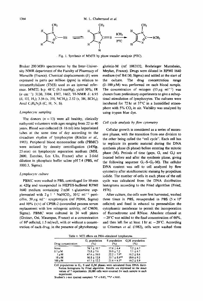
MT1 (l-methyl-2-thioimidazole) ref M8506) was obtained from Sigma, (Saint-Quentin Fallavier, France). MMTI was synthesized in our laboratory by phase transfer catalysis (Fig. 1) according to Kister et al. (1979): 0.05mol of methyl iodide ref 6769-2 (Fluka, Saint-Quentin Fallavier, France) is added to 0.05 mol of I-methyl-2-thioimidazole dissolved in a mixture of 150 ml of benzene plus 15 ml of sodium hydroxide at 40%, with tetrabutylammonium- bromide ref 8686-O (Fluka, France) as catalyst (0.003 mol). The reaction is carried out for 6 hr at 60°C with vigorous stirring. After decantation the organic phase is distilled under reduced pressure (0.5 mmHg).
The non-corrected boiling point was determined under reduced pressure at 0.5 mmHg. Elemental analyses were performed by the Central Microanaly- sis Department of the CNRS at Vernaison (RhBne, France). IR spectra were determined using a Beck- man 4750 IR spectrophotometer. ‘H-NMR spectra were recorded in solution in deuterochloroform on a
1363
1364 M. L. Chabemaud et al.
H
x
H
1 ‘c-SH 9 4 H N H
Fig. 1. Synthesis of MMTI by phase transfer catalysis (PTC).
Bruker 200 MHz spectrometer by the Inter-Univer- sity NMR department of the Faculty of Pharmacy of Marseille (France). Chemical displacements (6) were expressed in parts per million (ppm) in relation to tetramethylsilane (TMS) used as an internal refer- ence. MMTI: b.p. 48°C (0.5 mmHg); yield 30%; IR (rr cm-‘): 3130, 3104, 1507, 1462; ‘H-NMR 6: 6.95 (d, lH, H,); 3.56 (s, 3H, NCH,); 2.52 (s, 3H, SCH& Anal CSH,N,S (C, H, N, S).
Lymphocyte sampling
The donors (n = 13) were all healthy, clinically euthyroid volunteers with ages ranging from 22 to 40 years. Blood was collected (8-16 ml) into heparinized tubes at the same time of day according to the circadium rhythm of lymphocytes (Ritchie er al., 1983). Peripheral blood mononuclear cells (PBMC) were isolated by density centrifugation (145Og, 25min) on lymphocyte separation medium (MSL 2000, Eurobio, Les Ulis, France) after a 2-fold dilution in phosphate buffer saline pH 7.4 (PBS, ref 1000.3, Sigma).
Lymphocyte culture
PBMC were washed in PBS, centrifuged for 10 min at 420g and resuspended in HEPES-buffered RPM1 1640 medium containing 2 mM L-glutamine sup- plemented with 2 g 1-l NaHCO,, 50 U ml-’ peni- cillin, 50 pg ml- ’ streptomycin (ref PO906, Sigma) and 10% (v/v) of CPSR-2 (controlled process serum replacement with low mitogenic activity, ref C9030, Sigma). PBMC were cultured in 24 well plates (Greiner, Osi, Maurepas, France) at a concentration of lo6 cells/ml, 1.5 ml/well, with or without a concen- tration of each drug, in the presence of phytohemag-
glutinin-M (ref 1082132, Boehringer Mannheim, Meylan, France). Drugs were diluted in RPM1 1640 medium (ref R4130, Sigma) and added at the start of the culture. The drug concentration range (0-1OOpM) was performed on each blood sample. The concentration of mitogen (15 pg ml-‘) was chosen from preliminary experiments to give a subop- timal stimulation of lymphocytes. The cultures were incubated for 72 hr at 37°C in a humidified atmos- phere with 5% COZ in air. Viability was analysed by using trypan blue dye.
Cell cycle analysis by flow cytometry
Cellular growth is considered as a series of succes- sive phases, with the transition from one division to the other being called the “cell cycle”. Each cell has to replicate its genetic material during the DNA synthesis phase (S phase) before entering the mitotic phase (M). Periods of time (gaps, Gi and GJ are located before and after the synthesis phase, giving the following sequence G,-S-G,-M). The cellular DNA content was cell to cell analyzed by flow cytometry after stoichiometric staining by propidium iodide. The number of cells in each phase of the cell cycle was calculated from the DNA distribution histograms according to the Fried algorithm (Fried, 1976).
After culture, the cells were first harvested, washed three times in PBS, resuspended in PBS (5 x lo6 cells/ml) and fixed in ethanol to permeabilize the cytoplasmic membrane to permit the incorporation of fluorochrome and RNase. Absolute ethanol at - 20°C was added to the final concentration of 66%, and then left for at least 1 hr at -20°C. According to Crissman et al. (1982), cells were washed three
Table I. MT1 effects on PHA-stimulated lvmohocvtes
Drug concentration
None 0.1 PM
1pM IOpM
1OOuM
G, population S population G,M population (%) W) (“/)
74.7 f 10.7 17.8 + 6.4 7.2 + 6.7 73.8 k 1.6 20.0 f 7.2 7.1 + 6.7 67.6 k 10.0 22.2 f 1.9’ 10.2 f 8.6 66.8 k 12.8 23.7 f 8.6*’ 10.6 k 9.2 61.1 + 12.2 21.6 + 8.5* 10.2 + 8.9
Cell populations in G,, S and G,M phases were calculated from DNA distri- bution histograms by Fried algorithm. Results are expressed as the mean values of 7 experiments. 20,000 cells were counted for each sample in each experiment.
Student’s t-test (paired samples): *P < 0.05, **P < 0.01.
Methimazole SCH, and lymphocytes
Table 2. 1-methyl-2methylthioimidazole effects on PHA-stimulated lymphocytes
G, population S population G,M population Drue. concentration (%) (%) (%)
1365
None 68.4 + 10.2 24.4 + 8.6 1.2 k 2.2 0.1 pM 68.4 + 9.6 24.1 f 7.5 7.5 f 2.8
lpM 69.5 f 9.5 23.5 + 7.5 7.0 f 2.5 10pM 69.6 f 9.2 22.4 f 5.4 8.0 + 4.0
10O~tM 68.5 f 6.6 27.3 f 7.4 4.2 f 1.5”
Cell populations in G,, S and C&M phases were calculated from DNA distri- bution histograms by Fried algorithm. Results are expressed as the mean values of 6 experiments. 20,000 cells were counted for all samples in each experiment.
Student’s I-test (paired samples): P < 0.01.
times in PBS and adjusted to the final concentration of 8 x 10’ cells/ml in PBS, propidium iodide (PI, ref P1304, Molecular Probes Inc., Eugene, OR, U.S.A., 18pg ml-‘), and RNase A (ref 109169, Boehringer Mannheim, 30 U ml-‘) in order to obtain shoichio- metric staining of DNA. The preparation was kept in the dark for 20 min at room temperature, and at least 1 hr at 4°C before flow cytometric analysis.
Flow cytometry
Samples were analyzed in an Ortho SOH flow cytometer (Ortho Diagnostic Systems, U.S.A.) equipped with an argon laser (Innova 90-4, Coherent, U.S.A.). Power outputs and wavelength were 400 mW at 488 nm. Data were processed by a computer (MCA 300, Briiker, France) connected to the cytometer. Samples were gated on forward angle (FAS) and right angle (RAS) light scatters to exclude debris and clumps, and select the cell population of interest. Twenty thousand gated cells were analyzed in each experiment. The red fluorescence emitted by PI (DNA) was collected through a 615 nm long pass filter. The fluorescence signal was subjected to linear amplification.
Statistics
The significance of the results was then assessed by paired comparison Student’s t test on differences of population size.
**
0 0.1 I 10 IOOkM
Drug concentration
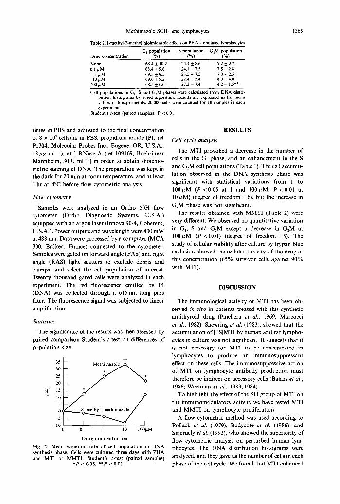
Fig. 2. Mean variation rate of cell population in DNA synthesis phase. Cells were cultured three days with PHA and MT1 or MMTI. Student’s r-test (paired samples)
*P < 0.05, **p < 0.01.
RESULTS
Cell cycle analysis
The MT1 provoked a decrease in the number of cells in the G, phase, and an enhancement in the S and G,M cell populations (Table 1). The cell accumu- lation observed in the DNA synthesis phase was significant with statistical variations from 1 to 100pM (P < 0.05 at 1 and lOOpM, P < 0.01 at 10 PM) (degree of freedom = 6), but the increase in G,M phase was not significant.
The results obtained with MMTI (Table 2) were very different. We observed no quantitative variation in G,, S and GrM except a decrease in G2M at 100pM (P < 0.01) (degree of freedom = 5). The study of cellular viability after culture by trypan blue exclusion showed the cellular toxicity of the drug at this concentration (65% survivor cells against 90% with MTI).
DISCUSSION
The immunological activity of MT1 has been ob- served in vivo in patients treated with this synthetic antithyroid drug (Pinchera et al., 1969; Marcocci et al., 1982). Shewring et al. (1983) showed that the accumulation of [35S]MTI by human and rat lympho- cytes in culture was not significant. It suggests that it is not necessary for MT1 to be concentrated in lymphocytes to produce an immunosuppressant effect on these cells. The immunosuppressive action of MT1 on lymphocyte antibody production must therefore be indirect on accessory cells (Balazs et al., 1986; Weetman et al., 1983, 1984).
To highlight the effect of the SH group of MT1 on the immunomodulatory activity we have tested MT1 and MMTI on lymphocyte proliferation.
A flow cytometric method was used according to Pollack et al. (1979), Bodycote et al. (1986), and Smerdely et al. (1993), who showed the superiority of flow cytometric analysis on perturbed human lym- phocytes. The DNA distribution histograms were analyzed, and they gave us the number of cells in each phase of the cell cycle. We found that MT1 enhanced
1366 M. L. Chabernaud et al.
the PHA-induced synthesis phase which is in agree- ment with results obtained previously (Hallengren et al., 1980; Okabe et al., 1983; Sharma et al., 1987; Smerdely et al., 1993). The quantification of the number of cells in each phase of the cycle, G,-S-GrM showed that although the cells entered the cell cycle, they tended to accumulate in the S phase, which was also reflected by statistical analysis. It should be noted, that the effect did not increase in a dose-response manner, and even at 10m6M, MT1 was found to have a significant activity. The concen- trations used were within the range of concentrations measured in humans receiving long-term treatment with carbimazole (Jansson et al., 1983; Skellern et al., 1974). A systemic effect of this drug cannot therefore be excluded.
The results obtained with MMTI showed no sig- nificant activity of this drug on the various phases of the cell cycle within concentrations of O.l-100pM (Fig. 2). At 100 PM the trypan blue dye test showed a decrease of cell viability. The cytotoxicity of the drug is widely marked on the most transformed cells, and the GrM population is underestimated for the benefit of the G, and S populations. The cells in G1 being the most sturdy, their estimation is right.
As with /I-mercaptoethanol, sulfites, gluthatione, and L-cysteine, the MT1 increased the lymphoblastic transformation. This study showed the free SH group relationship activity, perhaps the MT1 thiol group interconverted in biochemical oxidation-reduction reactions as L-cysteine which is modified in its dithioether form. The disulfide linkages of cysteine units, as well as MT1 units, are responsible for the reducing character. The S-methylation of the MT1 removes the possibility of disulfide linkages and so strongly decreases its reducing character. This is one explanation of the inability for lymphoblastic trans- formation of the lymphocytes treated by MMTI.
In conclusion, the results of this study confirm the activity of MT1 on lymphoblastic transformation, and demonstrate that MMTI cannot stimulate lym- phocytes. Although the immunomodulatory activity of this drug seems to be dependent on the reducing character and the free SH group. We showed that S-methylation increases cell toxicity.
Acknowledgements-We would like to thank Mrs Marie- Helene Ratinaud and Chantal Jayat for skilful technical assistance with the flow cytometry.
REFERENCES
Allannic H., Fauchet R.. Oraiazzi J., Madec A. M., Genetet B., Lorcy Y., Le Guerier A. M., Delambre C. and Derennes V. (1990) Antithvroid drugs and Graves’ dis- ease: a prospective randomized evalu&on of the efficacity of treatment duration. J. clin. Endocr. Metab. 70,675-679.
Bach J. F. and Morel E. (1982) L’extension du concept de maladie par auto-anticorps anti-recepteurs. Nouv. Press. Med. 11, 1845-1847.
Balazs C., Kiss E., LeGvey A. and Farid N. R. (1986) The immunosuppressive effect of methimazole on cell-medi- ated immunity is mediated by its capacity to inhibit peroxidase and to scavenge free oxygen radicals. Clin. Endocr. 25, 7-16.
Bodycote J. and Wolff S. (1986) Metabolic breakdown of [3H]thymidine and the inability to measure human lymphocyte proliferation by incorporation of radioactivitv. Proc. natn. Acad. Sci. U.S.A. 83. 4749-4753. -
Burman K. D. and Baker J. R. (1985) Immune mechanisms in Graves’ disease. Endocr. Rev. 6. 183-220.
Chen C. and Hirsch J. G. (1972) The effects of mercap- toethanol and of peritoneal macrophages on the antibody- forming capacity of nonadherent mouse spleen cells in vitro. J. exp. Med. 136, 604-617.
Crissman H. A. and Steinkamp J. A. (1982) Rapid, one step staining procedures for analysis of cellular DNA and protein by single and dual laser flow cytometry. Cytometry 3, 84-90.
Fanger M. W., Hart D. A., Wells J. V. and Nisonoff A. (1970) Enhancement by reducing agents of the transform- ation of human and rabbit peripheral lymphocytes. J. Immun. 105, 1043-1045.
Fried J. (1976) Method for the quantitative evaluation of data from flow microfluorometry. Comput. Biomed. Res. 9, 263-276.
Hallengren B., Forsgren A. and Melander A. (1980) Effects of antithyroid drugs on lymphocyte function in vitro. J. clin. Endocr. Metab. 51. 298-301.
Jansson R., Dahlberg P. A., Johansson H. and Lindstrom B. (1983) Intrathyroidal concentrations of methimazole in patients with Graves’ disease. J. clin. Endocr. Metab. 57, 129-132.
Kister J., Assef G., Mille G. and Metzger J. (1979) Synthbe et etude du rearrangement SRoNR des diazoles-1,3: alkyl-1 alkylthio-2 (allylthio, arylthio, cycloalkylthio) im- idazole. Partie I. Synthtse et etudes physicochimiques. Can. J. Chem. 57, 813-821.
McGregor A. M., Petersen M. M., McLachlan S. M., Rooke P., Rees Smith B. and Hall R. (1980) Carbimazole and the autoimmune response in Graves’ disease. New Engl. J. Med. 303, 302-307.
Marcocci C., Chiovatto L., Mariotti S. and Pinchera A. (1982) Changes in circulating thyroid autoantibody levels during and after therapy with methimazole in patients with Graves’ disease. i Endocr. Invest. 5, 13-19:
Okabe N.. Inoue K. and Mori R. (1983) Effects of antithv- roid drugs on lymphocyte proliferative responses to lectins: relationship between insulin autoimmune syndrome and methimazole. J. clin. Lab. Immun. 11, 167-171.
Pinchera A., Liberti P., Martin0 E., Fenzi G. F., Grass0 L., Rovis L., Baschieri L. and Doria G. (1969) Effects of antithyroid therapy on the long-acting thyroid stimulator and the antithyroglobulin antibodies. J. c/in. Endocr. Metab. 29, 231-238.
Pollack A., Bagwell C. B., Hudson J. L. and Irvin G. L. (1979) Differences in flow cytometry and )H-thymidine analysis of perturbed human lymphocytes. J. Histochem. Cytochem. 27, 486-490.
Raby C., Lagorce J. F., Jambut-Absil A. C., Buxeraud J. and Catanzano G. (1990) The mechanism of action of syn- thetic antithyroid drugs: iodine complexation during oxi- dation of iodine. Endocrinology 126, 1683-1691.
Ritchie A. W. S., Oswald I., Micklem H. S., Boyd J. E., Elton R. A., Jazwinska E. and James K. (1983) Circadian variation of lymphocyte subpopulations: a study with monoclonal antibodies. Br. Med. J. 286, 1773-1775.
Methimazole SCH, and lymphocytes 1367
Romaldini J. H., Bromberg N., Werner R. S., Tanaka L. metabolite of carbimazole, in hyperthyroid patients. Br. M., Rodrigues H. F., Werner M. C., Farah C. S. and Reis J. clin. Pharmac. 1, 265-269. L. C. F. (1983) Comparison of effects of high and low Smerdely P., Pitsiavas V. and Boyages S. C. (1993) Methi- dosage regimens of antithyroid drugs in the management mazole inhibits FRTL-5 thyroid cell proliferation by of Graves’ hyperthyroidism. J. clin. Endow. Metab. 57, inducing S-phase arrest of the cell cycle. Endocrinology 563-570. 133, 2403-2406.
Sharma B. S. and Elias A. N. (1987) Effects of methimazole on human lymphocyte proliferation and natural killer cell activity. Gen. Pharmac. 18, 449-453.
Shewring G. S. and Lazarus J. H. (1983) The accumulation of [35S]methimazole by human and rat lymphocytes. Acta Endocr. 102, 68-70.
Weetman A. P., McGregor A. M. and Hall R. (1983) Methimazole inhibits thyroid autoantibody production by an action on accessory cells. Clin. Immun. Immunopath. 28, 39-45.
Skellern G. G., Stenlake J. B., Williams W. D. and McLarty D. G. (1974) Plasma concentrations of methimazole, a
Weetman A. P., Holt M. E., Campbell A. K., Hall R. and McGregor A. M. (1984) Methimazole and generation of oxygen radicals by monocytes: potential role in immuno- suppression. Br. Med. J. 288, 518-520.