lymphoma: a heterogeneous disease - jsvpjsvp.jp/activity/pdf/tumor9_2.pdfextranodal - other...
TRANSCRIPT

Lymphoma: a heterogeneousdisease
Peter F. Moore
Application of the WHO Lymphoma Classification Scheme

WHO - classificationExtension of REAL classification - 1994 ILSG
Boadened to include myeloid, mast cell andhistiocytic neoplasia
Disease entities defined:
Lineage and postulated cell of origin
Morphology and Immunophenotype
Genetic features and Clinical features

WHO - classification
Lymphoid neoplasia: B cell, T cell, NK celland Hodgkins lymphoma
Lymphomas and leukemias consideredtogether - may be manifestations of thesame tumor
B-CLL and B cell small lymphocytic lymphoma
Lymphoblastic lymphoma and lymphoblasticleukemia

WHO - classificationB and T/NK lymphomas:
Precursor cell lymphomas
Mature cell lymphomas
Non-Hodgkin lymphomas -
Distinct diseases
Distinctive clinical features/epidemiology
Distinctive responses the therapy.

WHO classification of tumors ofhematopoietic and lymphoid tissues
Proponents of other schemes have endorsedWHO classification
First true world-wide consensus classificationscheme for hematologic malignancies
ACVP initiative - Lymphoma Study Group -investigated suitability of WHO scheme foranimal lymphomas (led by Dr. Ted Valli)
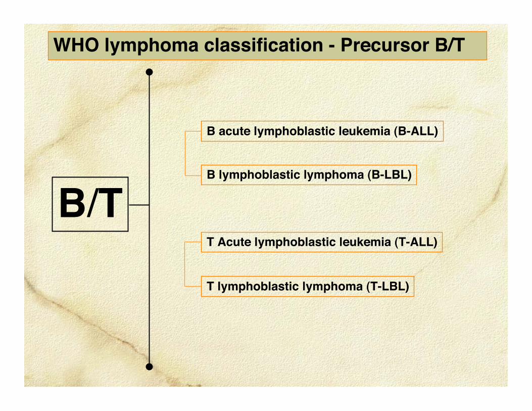
B lymphoblastic lymphoma (B-LBL)
B acute lymphoblastic leukemia (B-ALL)
WHO lymphoma classification - Precursor B/T
B/TT Acute lymphoblastic leukemia (T-ALL)
T lymphoblastic lymphoma (T-LBL)

WHO Lymphoma classification - Mature B cell
Diffuse large BCLCentroblasticImmunoblasticT cell/histiocyte richAnaplastic
Marginal zone BCLNodalSplenicMALT
Mantle cell BCL
Extramedullary Plasmacytoma
Follicular BCL
Multiple myeloma
B-CLL/Small lymphocytic BCL
Burkitt-like BCL
B
Indolent - initially

Extranodal - otherEnteropathy associated TCLHepatosplenic TCLPeripheral TCL - unspecified
LGL leukemia T-LGL CLLT-LGL ALL
Mycosis fungoidesPagetoid reticulosisSézary syndromePeripheral TCL - unspecified
Cutaneous TCL
Nodal TCL
Peripheral TCL - unspecifiedT-zone TCLAnaplastic large TCLAngioimmunoblastic TCL
WHO Lymphoma classification - Mature T cell
T
LGL lymphoma Indolent - initially
Indolent - some forms

Integrative diagnostics - Leukocytic diseases
Clinical and clinico-pathological data
Morphological data - histology/cytology
Immunophenotyping - reagent panels
Molecular assessments - antigen receptorclonality for lymphoma

Lymphocyte Development
and
Antigen receptor gene rearrangement

T cell receptor gene rearrangement
TCRG - molecular clonality target (rearranged in γδ and αβ T cells)
Variable Joining Constant
TdT n base addition
TCRG/TCRD TCRA/TCRB
TCRαβ
CD3
TCRγδ
CD3
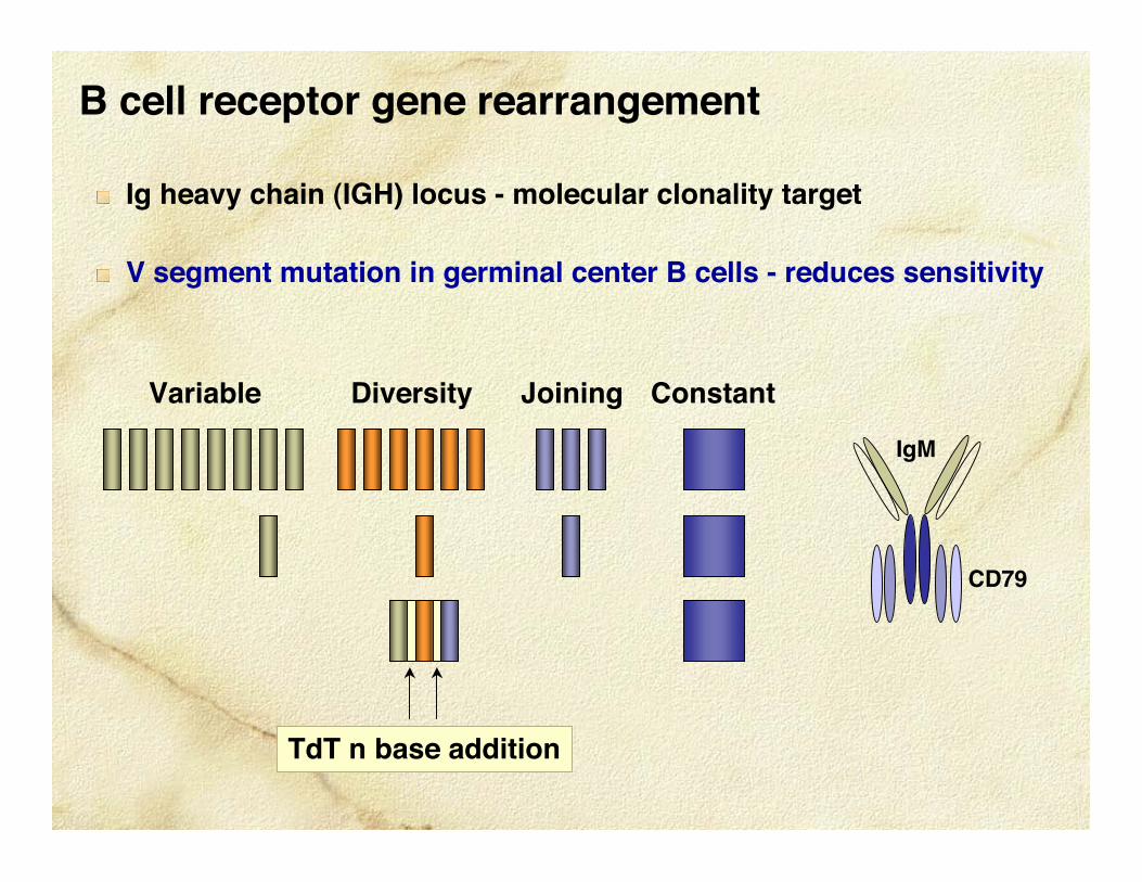
B cell receptor gene rearrangement
Variable Joining ConstantDiversity
TdT n base addition
IgM
CD79
Ig heavy chain (IGH) locus - molecular clonality target
V segment mutation in germinal center B cells - reduces sensitivity

Feline TCRG V-N-J alignment CDR3 region
3ʼ V segment J segmentCDR3
5ʼ primer 3ʼ primerPCR
variability

Ag receptor gene rearrangement - indications
Morphological, cytological, immuno-phenotypic properties inconclusive
Lack of architectural effacement in organizedlymphoid tissue - MZL or TZL
Lamina proprial or intra-epithelial lymphocytosisin the small intestine
Lympho-histiocytic proliferations in skin

387403 - Bernese Mtn dog, MC, 6 yrs - masses ondigit, carpus, mandible.
Dec 07 - DX#1: Histiocytic dermatitis
Mar 08 - DX#2: Histiocytic sarcoma
May 08 - DX#3: Reactive histiocytosis
Canine “inflamed” T cell lymphoma (PTCL)


387403 - Bernese mountain dog, MC, 6 yearsDX: Non-epitheliotropic T cell lymphoma (& lympho-histiocytic dermatitis)
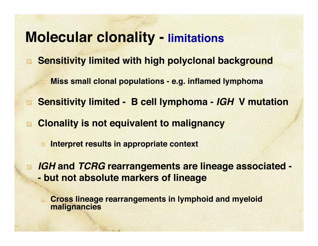
Molecular clonality - limitations
Sensitivity limited with high polyclonal background
Miss small clonal populations - e.g. inflamed lymphoma
Sensitivity limited - B cell lymphoma - IGH V mutation
Clonality is not equivalent to malignancy
Interpret results in appropriate context
IGH and TCRG rearrangements are lineage associated -- but not absolute markers of lineage
Cross lineage rearrangements in lymphoid and myeloidmalignancies

B lymphoblastic lymphoma (B-LBL)
B acute lymphoblastic leukemia (B-ALL)
WHO lymphoma classification - Precursor B/T
B/TT Acute lymphoblastic leukemia (T-ALL)
T lymphoblastic lymphoma (T-LBL)

T-lymphoblastic lymphoma (T-LBL)
Mass lesion : T-LBL
mediastinum, LNs, spleen, other sites
Predominance of blood/BM involvement: T-ALL

T-lymphoblastic lymphomaOrigin: Precursor T lymphoblast
Hypercalcemia a common feature
High grade rapidly progressive
Loss Cfa 11 in high- grade TCL
P16 (Rb) deletion/inactivation in all cases


WHO Lymphoma classification - Mature B cell
Diffuse large BCLCentroblasticImmunoblasticT cell/histiocyte richAnaplastic
Marginal zone BCLNodalSplenicMALT
Mantle cell BCL
Extramedullary Plasmacytoma
Follicular BCL
Multiple myeloma
B-CLL/Small lymphocytic BCL
Burkitt-like BCL
B
Indolent - initially

Ig affinity maturation
Ig class switch
IGH-V mutation
Mantle zone
Light zone
Dark zone
Germinal Center Responses
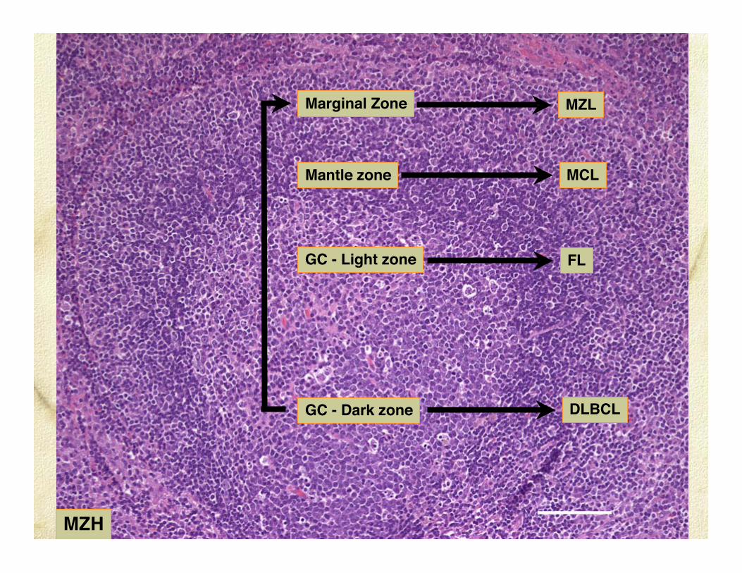
Marginal Zone
GC - Dark zone
Mantle zone
MZL
MCL
DLBCL
GC - Light zone FL
MZH

Diffuse Large B cell Lymphoma (DLBCL)
Centroblastic
Immunoblastic
T cell/histiocyte rich
Anaplastic

Diffuse Large B cell Lymphoma
Origin: centroblasts in GC dark zone
Lymph nodes; spleen; extranodal
Most prevalent lymphoma in dogs
High grade lymphoma - high proliferativefraction

Spleen - diffuse large B cell lymphoma
Canine spleen - splenomegaly due to white pulp infiltration/obliteration

DLBCL
Dark zone

DLBCL - Centroblastic

DLBCL - Immunoblastic

00B1609 - QH mare, 11yrs - Skin masses 2yrs
amyloid
DLBCL - T cell rich

CD3
CD79a

T cell rich B cell lymphoma - equine skin
2565 2565
native denatured
PC PC+ +1609 1609

CD20
DLBCL - Anaplastic

Marginal zone lymphoma (MZL)
Nodal - most common
Spleen - solitary mass and/or diffuse
Extranodal - MALT lymphoma - rare

Marginal zone lymphomaOrigin: LN - perifollicular MZ B cells (chronic
follicular hyperplasia) (dogs)
Splenic MZ B cells (dogs)
BALT and NALT - cats - rare
DX: architecture + cytologic characteristics
DDX - nodular hyperplasia when spleen involved
Indolent lymphoma - low proliferative fraction
May evolve into DLBCL

Spleen - marginal zone lymphoma
MZL- solitary mass and diffuseinvolvement. Perifollicularmarginal zones slowly coalesce.

Marginal zone hyperplasia - lymph node

MZL

MZL

CD20
CD3
MZL

MZL

Follicular lymphoma (FL)
Nodal - most common
Splenic
Extranodal

Follicular lymphomaOrigin: Centrocytes in GC light zone
DX: architecture + cytologic characteristics
Indolent B cell lymphoma - low proliferativefraction
May evolve into DLBCL
Human: t(14:18) - BCL2 gene rearranged

FL
Light zone
Dark zone

FL

FL-3b

CD20
FL - liver - dog

FL-2

Mantle cell lymphoma (MCL)Nodal
Spleen - 3 dogs (clonal IGH)
Bone marrow
Extranodal - GI tract
Mantle cell lymphomaOrigin: B cell from inner mantle zone
DX: architecture + cytologic characteristics
Solitary nodular mass in the spleen of dogs
DDX: splenic nodular hyperplasia in dogs
Indolent B cell lymphoma - low proliferativefraction - dogs; more aggressive in humans - esp.blastoid variant
Human: CD5+, BCL2+, Cyclin D1+

MCL

MCL

MCL
FDC

Extranodal - otherEnteropathy associated TCLHepatosplenic TCLPeripheral TCL - unspecified
LGL leukemia T-LGL CLLT-LGL ALL
Mycosis fungoidesPagetoid reticulosisSézary syndromePeripheral TCL - unspecified
Cutaneous TCL
Nodal TCL
Peripheral TCL - unspecifiedT-zone TCLAnaplastic large TCLAngioimmunoblastic TCL
WHO Lymphoma classification - Mature T cell
T
LGL lymphoma Indolent - initially
Indolent - some forms

Peripheral T cell lymphoma - PTCL
Heterogeneous group
Nodal
Skin (non-epitheliotropic TCL)
Generalized (2o leukemia common)

Peripheral T cell lymphoma - PTCL
Origin: Peripheral T cells
High-grade lymphoma - high proliferative fraction
Cytology extremely variable
Inflamed lymphoma - esp. cutaneous PTCL
DDX: Reactive (cutaneous) histiocytosis
P16 (Rb) deletion/inactivation in all cases

NE-CTCL

081002

CD3081002

T-zone lymphoma (TZL)
Nodal
Human - variant of PTCL - i.e. high-gradelymphoma

T-zone lymphomaOrigin: Peripheral T cells
Variable LN involvement (1, 2 or generalized)
Indolent lymphoma (dogs) - years
Low proliferative fraction - mitotic rate low - (ifnot - PTCL)
2o leukemia observed - prognosis unaffected
DDX: paracortical hyperplasia (TCRG clonality)Marginal zone BCL (MZL) - requires IHC

TZL
F
F
F
F
paracortex

TZL

Hepatosplenic lymphoma (HS-TCL)
Spleen
Liver
Bone marrow
Generalized lymphadenopathy lacking

Hepatosplenic T cell lymphoma
Origin: splenic red pulp γδ T cell
Cytology - LGL. Usually TCRγδ+ CD11d+
2o hemophagocytic syndrome common(CD11d+ macrophages activated);malignant T cells erythrophagocytic
Clinical - aggressive course, anemia,thrombocytopenia (immune mediated??)
DDX: hemophagocytic histiocytic sarcoma

CD11d
WPMZ
RP
T-CLL - LGL type
Hepatosplenic lymphoma
Hemophagocytic HS
Splenic red pulp - CD11d+ diseases

CD11d
Liver HS-TCL

CD11dBone Marrow HS-TCL

Lymphocyte Trafficking
and
Tissue Localization of Disease
Lymphomas of skin and gut

T cell lymphomas of skin and gut
Marked species differences
incidence
behavior
immunophenotype

αβ T cells
Naïve T cells - exported from the thymus
Recirculate between blood and lymph nodes
Effector memory T cells - wide migratory range
Recirculate between blood and cutaneous or mucosalsites
Central memory T cells - retain migratory path ofnaïve T cells

Lymphocyte recruitment - to skin
How are the migratory pathways of naïvelymphocytes redirected to skin?
Dendritic Cell Imprinting

Naïve T cell
LN Paracortex
Dendritic Cell
Home to SKIN
Memory T cell
CCR4
αLβ2
E-selectin CCL17
Dermal endothelium
CLA
Fucosyl Tr VII

Extranodal - otherEnteropathy associated TCLHepatosplenic TCLPeripheral TCL - unspecified
LGL leukemia T-LGL CLLT-LGL ALL
Mycosis fungoidesPagetoid reticulosisSézary syndromePeripheral TCL - unspecified
Cutaneous TCL
Nodal TCL
Peripheral TCL - unspecifiedT-zone TCLAnaplastic large TCLAngioimmunoblastic TCL
WHO Lymphoma classification - Mature T cell
T
LGL lymphoma Indolent - initially
Indolent - some forms

Cutaneous LymphomaEpitheliotropic TCL
Mycosis fungoides
Pagetoid reticulosis
Sézary syndrome
Non-epitheliotropic PTCL
Non-T non-B lymphoma
B cell lymphoma (Diffuse large BCL)
Plasmacytoma

Skin homing T cell lymphoma

Epitheliotropic T cell lymphoma - skin
Mycosis fungoides (MF) - lesions confined to skin forextended period - clinical course up to 4 yrs.
MF is a disease of skin homing memory T cells
Dissemination initially occurs within the skin andskin draining lymph nodes
Evidence of dissemination - identical T cell clonefound in multiple skin sites

Classical MF

Pagetoid MF

CD3CD3
Epidermis Hair follicle

Canine Mycosis Fungoides
Immunophenotype
Consistent expression of CD3 (n = 56)
CD8+ (80% cases) or CD4-CD8- (20% cases)
Memory cell phenotype (CD45+CD45RA-CD49d+)
Marked contrast to human MF - TCRαβ+CD4+

Canine Mycosis Fungoides
T CELL RECEPTOR USAGE?
Development program for TCR specific probes
Mab specific for TCRαβ and TCRγδ developed

Canine MF - TCR Expression
TCR immunophenotype in MF all forms
TCRαβ+ 21 cases (40%)
TCRγδ+ 32 cases (60%)
Canine MF involves γδ T cells at much higherincidence than human MF

Canine MF - TCR Expression
Classical MF: TCRαβ+ ≈ TCRγδ+ (n=38)
Pagetoid MF: TCRγδ+ (n=15)

TCR αβ TCR γδ
CD3
Pagetoid MF: a lymphoma of γδ T cells

Canine MF - pagetoid reticulosis
Clonal origin from resident epidermal γδ T cells
Exclusive expression of TCRgd
Prolonged expansion entirely within the epidermis

Extranodal - otherEnteropathy associated TCLHepatosplenic TCLPeripheral TCL - unspecified
LGL leukemia T-LGL CLLT-LGL ALL
Mycosis fungoidesPagetoid reticulosisSézary syndromePeripheral TCL - unspecified
Cutaneous TCL
Nodal TCL
Peripheral TCL - unspecifiedT-zone TCLAnaplastic large TCLAngioimmunoblastic TCL
WHO Lymphoma classification - Mature T cell
T
LGL lymphoma Indolent - initially
Indolent - some forms

Gastrointestinal lymphomaEnteropathy associated TCL (EATCL)
small cell
large cell
LGL
Diffuse large BCL

Enteropathy associated TCL
Origin: intestinal homing T cell (IEL or LPL)
Small intestine - high prevalence in cats
IBD: precursor lesion in most cats -distinction (TCRG clonality)
Small cell - indolent
Large cell (LGL) - aggressive high grade
Architecture - mucosal or transmural

Mucosal homing T cell lymphoma

IELs - distinctive phenotypic subsets versus PBL
Expression of β7 integrins (α4β7) linked to mucosalhoming
Feline IEL (30%) granulated - perforin, granzymes
CD8αα T cells predominate - role in immunesurveillance
Feline IEL (70%+) express the mucosal integrin -CD103 (αEβ7)
Feline small intestine: diffuse MALT

04B0314 -Feline, DLH, FS, 13 yrs
Mucosal epitheliotropic T cell lymphoma - duodenum - endoscopic

native denatured
DuoD DuoDStom Stompc + +pc
TCRG clonality - 04B0314 -Feline, DLH, FS, 13 yrs Endoscopic biopsy - duodenum and stomach

eTCL small cell

Mucosal lymphoma - cytology
L-GL3
Large lymph2
Small lymph68
S-GL12
85

Mucosal lymphoma - survival
Dead 13.9 ± 13.1m (0.5 - 46m, n=29)
Alive 24.2 ± 17.1m (4 - 51m, n=16)

Jejunal mass: transmural LGL T cell lymphoma

eTCL - LGL

eTCL - LGL LGL leukemia

Transmural T cell lymphoma - survival
Dead 2.9 ± 4.8m (0.1 - 15m)
Alive 28m
L-GL9
L-lymph2 2m & 60m
S-lymph1
L-GL 2m1

LymphomaWHO classification scheme is applicable to canine(feline) lymphoma
Basic immunophenotyping often needed (B/T)
Molecular clonality necessary in some instances(TZL, MZL, MCL, FL and T cell/histiocyte rich BCL)
Recognition of homogeneous lymphoma groupswill lead to tailored therapies and discovery ofunderlying molecular defects

ThanksJapanese Society of Veterinary Pathology
Colleagues - Ted Valli, Bill Vernau, Paola Roccabianca, VerenaAffolter, Barbara Hirt, Jenny Woo, Dimitri Danilenko, SeanMcDonough, Thierry Olivry, Paul Rossitto, Petra Graham,Sandra Kosten, Eric Cavanaugh
Funding - LABL, Center for Companion Animal Health, Centerfor Equine Health, Morris Animal Foundation, MacDonald-Rivasgrant, ICOS Corporation.