lymphoedema
TRANSCRIPT

LYMPHOEDEMA

• Lymphoedema may be defined as abnormal limb swelling caused by the accumulation of increased amounts of high protein ISF secondary to defective lymphatic drainage in the presence of (near) normal net capillary filtration.

Risk factors: Upper limb/trunk lymphoedema
• Surgery for lymph node dissection. Breast surgey.
• Radiotherapy to breast or to axilary, im or subclaviclar lymph nodes.
• Wound complications• Cancer• Obesity
• Hypertension
• Chronic skin disorders• Air travel• Congenital predisposition
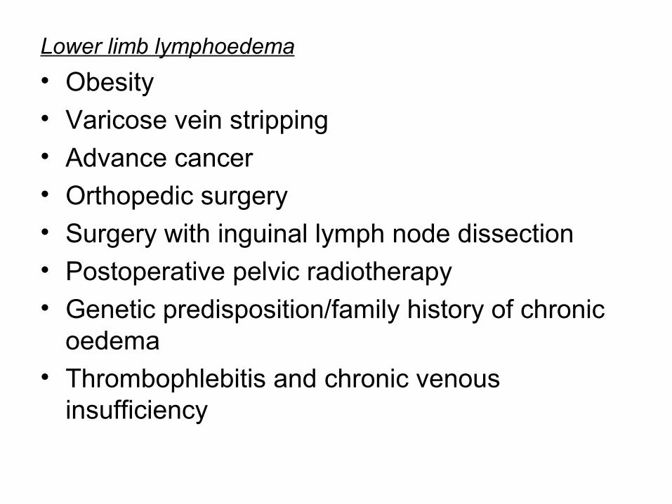
Lower limb lymphoedema
• Obesity• Varicose vein stripping• Advance cancer• Orthopedic surgery• Surgery with inguinal lymph node dissection• Postoperative pelvic radiotherapy
• Genetic predisposition/family history of chronic oedema
• Thrombophlebitis and chronic venous insufficiency
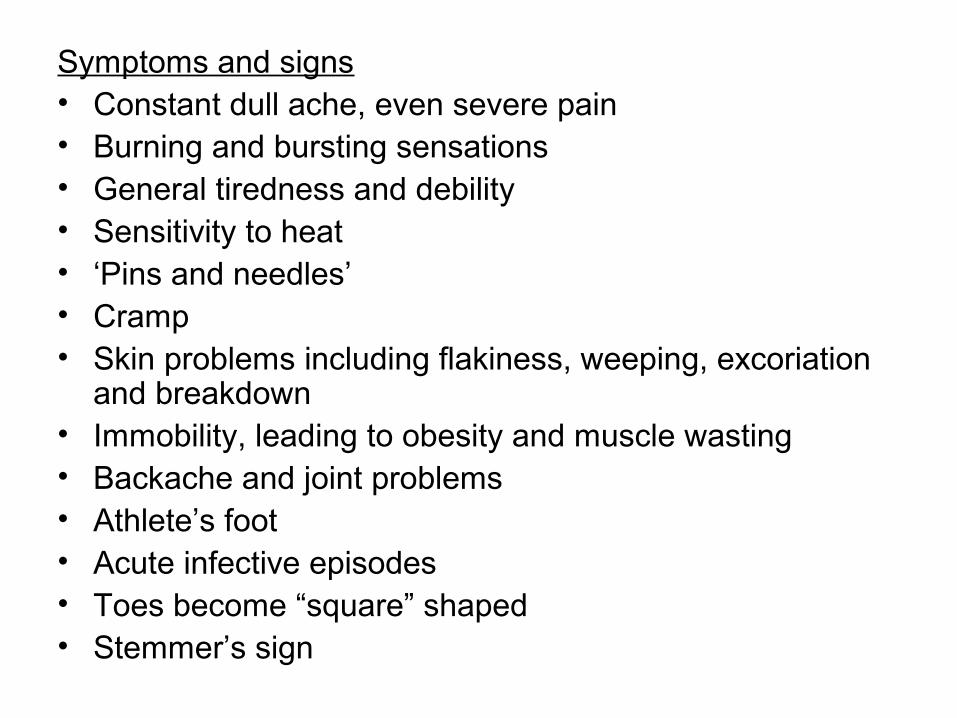
Symptoms and signs• Constant dull ache, even severe pain• Burning and bursting sensations• General tiredness and debility• Sensitivity to heat• ‘Pins and needles’• Cramp• Skin problems including flakiness, weeping, excoriation
and breakdown• Immobility, leading to obesity and muscle wasting• Backache and joint problems• Athlete’s foot• Acute infective episodes• Toes become “square” shaped• Stemmer’s sign

Classification
• Primary lymphoedema, in which the cause is unknown (or at least uncertain and unproven); it is thought to be caused by ‘congenital lymphatic dysplasia’
• Secondary or acquired lymphoedema, in which there is a clear underlying cause.

Clinical classification


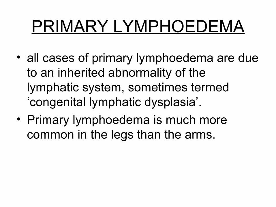
PRIMARY LYMPHOEDEMA
• all cases of primary lymphoedema are due to an inherited abnormality of the lymphatic system, sometimes termed ‘congenital lymphatic dysplasia’.
• Primary lymphoedema is much more common in the legs than the arms.
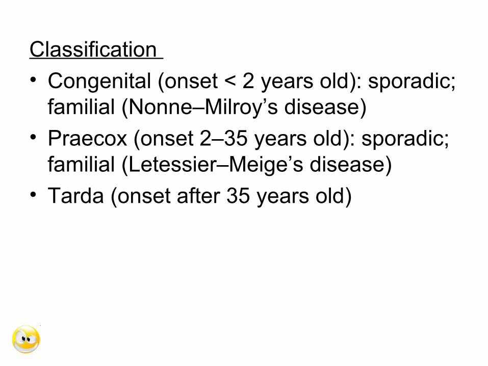
Classification
• Congenital (onset < 2 years old): sporadic; familial (Nonne–Milroy’s disease)
• Praecox (onset 2–35 years old): sporadic; familial (Letessier–Meige’s disease)
• Tarda (onset after 35 years old)


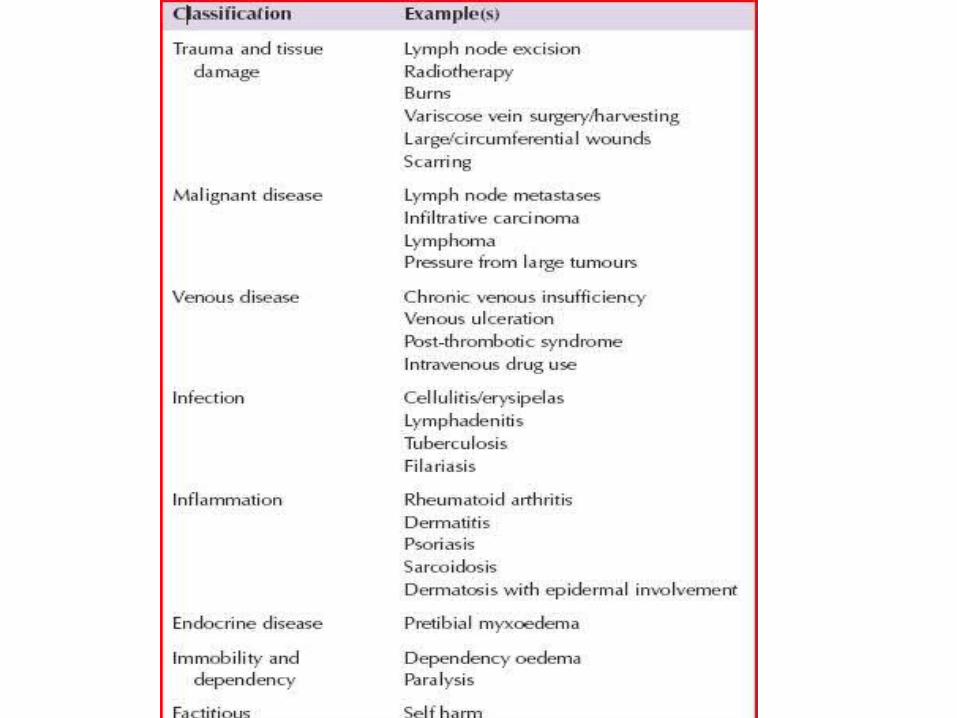
SECONDARY LYMPHOEDEMA
• This is the most common form of lymphoedema. There are several well-recognised causes including infection, inflammation, neoplasia and trauma


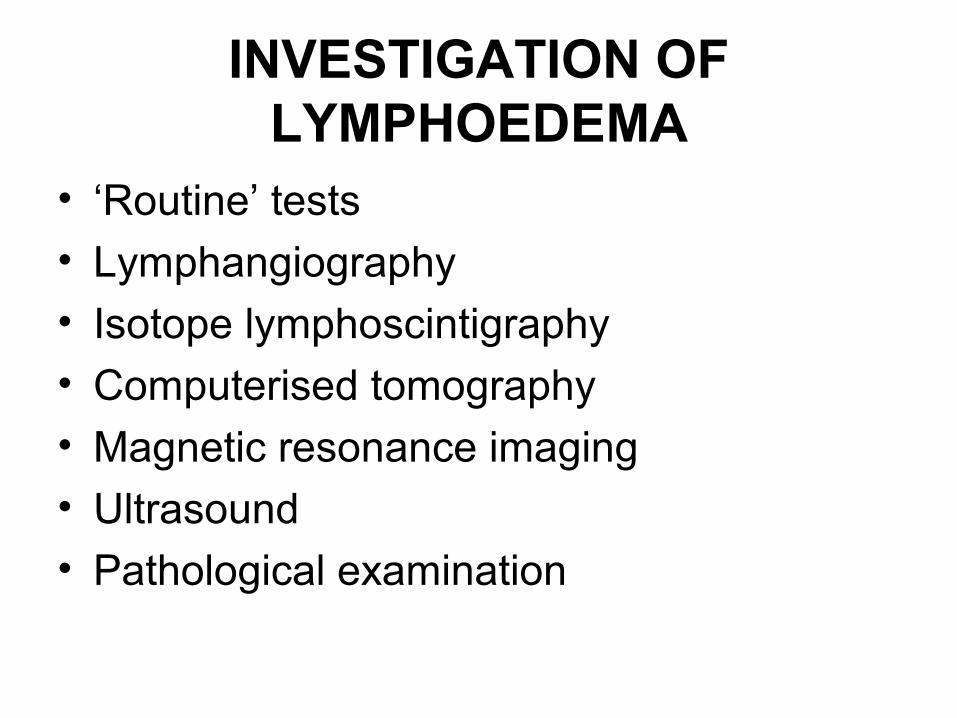
INVESTIGATION OF LYMPHOEDEMA
• ‘Routine’ tests
• Lymphangiography
• Isotope lymphoscintigraphy
• Computerised tomography
• Magnetic resonance imaging
• Ultrasound
• Pathological examination


MANAGEMENT OF LYMPHOEDEMAInitial evaluation of the patient with lymphoedema• History (age of onset, location, progression, exacerbating
and relieving features)• Past medical history including cancer history• Family history• Obesity • Complications (venous, arterial, skin, joint, neurological,
malignant)• Assessment of physical, emotional and psychosocial
symptoms• Social circumstances (mobility, housing, education, work)• Special needs (footwear, clothing, compression garments) • Previous and current treatment• Pain control• Compliance with therapy and ability to self-care
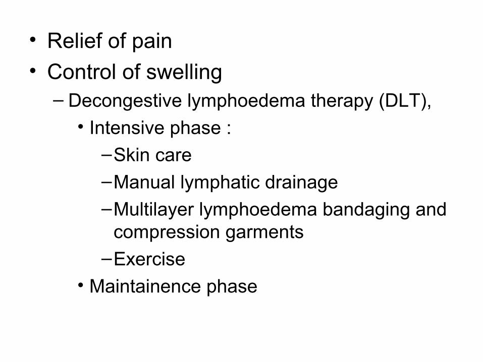
• Relief of pain
• Control of swelling– Decongestive lymphoedema therapy (DLT),
• Intensive phase :–Skin care–Manual lymphatic drainage–Multilayer lymphoedema bandaging and
compression garments–Exercise
• Maintainence phase

Surgery• Only a small minority of patients with lymphoedema
benefit from surgery. Operations fall into two categories:• Bypass procedures
– The rare patient with proximal ilioinguinal lymphatic obstruction and normal distal lymphatic channels might benefit, at least in theory, from lymphatic bypass.
• Limb Reduction procedures.
– Sistrunk– Homans– Thompson– Charles


