lymphatic system examination
TRANSCRIPT
Evaluation of Lymphadenopathy &Splenomegaly
Daniel Eshetu
Lymphatic system Introduction Mechanism and causes of lymphadenopathy Approach to lymphadenopathy: Hx, P/E, Lab
Splenomegaly Introduction Causes of splenomegaly Evaluation of splenomegaly: Hx, P/E, Lab studies,
Imaging... Evaluation of swellings (Lumps)
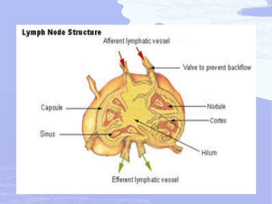
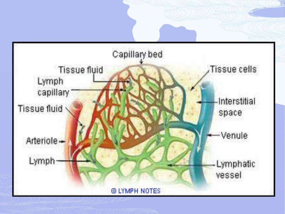
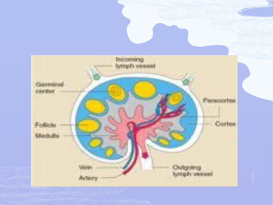
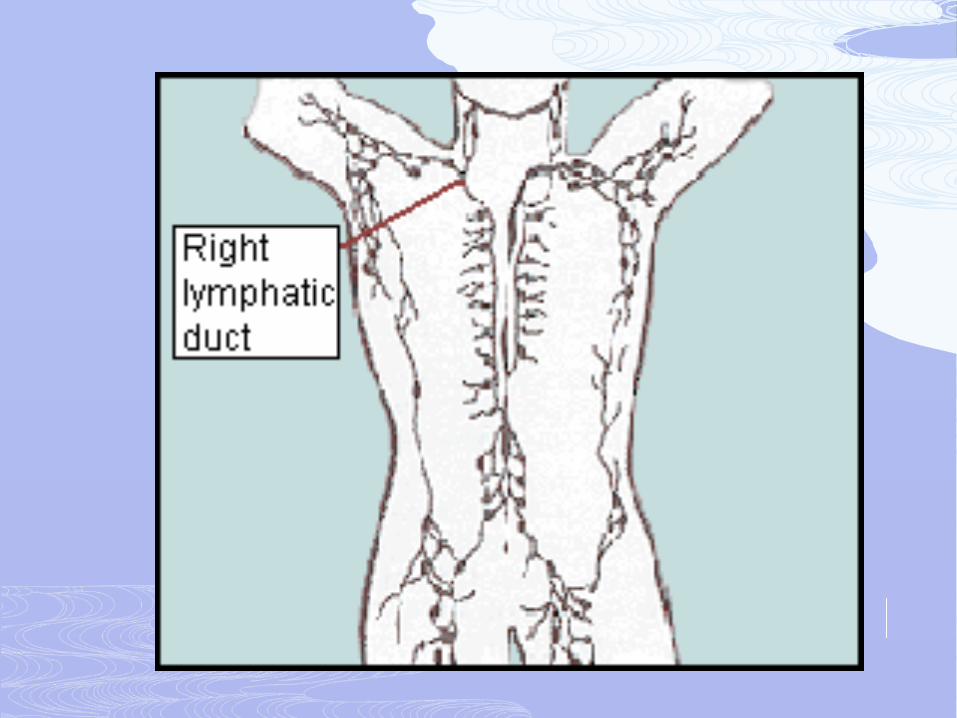
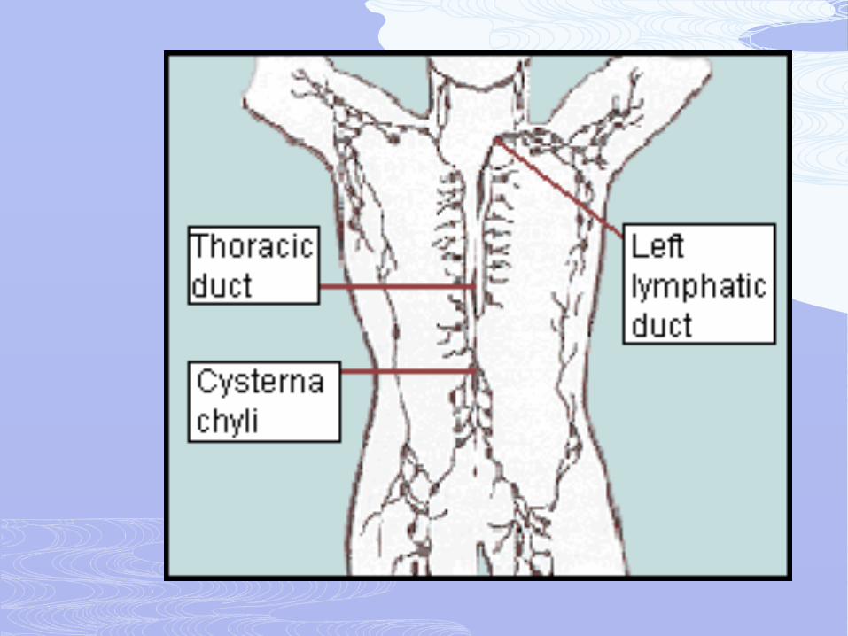
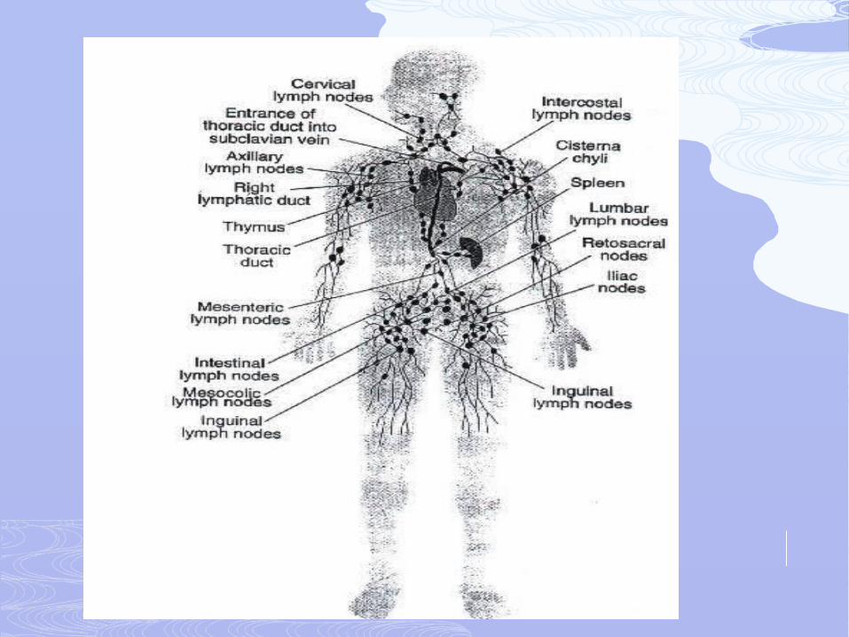
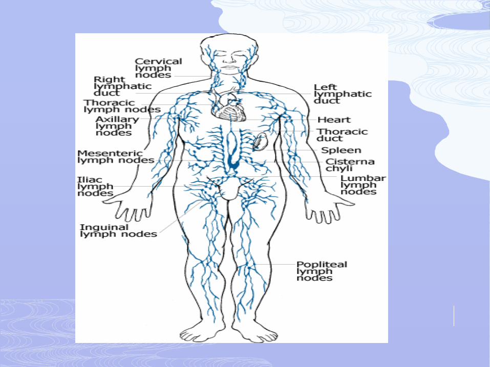
Lymphatic/ lymphoid system comprises the lymph, the lymphatic vessels, lymph nodes, the thymus and the spleen.
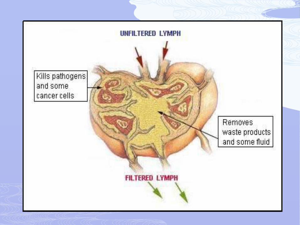
Function of the lymphatic system
it is responsible for the removal of interstitial fluid from tissues
it absorbs and transports fatty acids and fats as chyle from the digestive system
it transports white blood cells to and from the lymph nodes into the bones
The lymph transports antigen-presenting cells (APCs), such as dendritic cells, to the lymph nodes where an immune response is stimulated
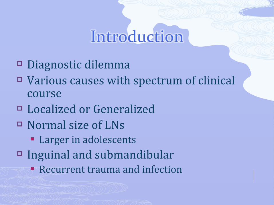
Diagnostic dilemma Various causes with spectrum of clinical
course Localized or Generalized Normal size of LNs
Larger in adolescents Inguinal and submandibular
Recurrent trauma and infection
Benign proliferation of residential cells HIV Infection
Infiltration by inflammatory cells Infection – lymphadenitis Auto-immune conditions- SLE
In situ proliferation of Malignant lymphocytes Lymphomas
Infiltration of lymph nodes by metastatic malignant cells Breast cancer Colorectal cancer Lung cancer
Infiltration of lymph nodes by metabolite-laden macrophages: Lipid storage diseases
Infectious Viral Bacterial Fungal Chlamydial Parasitic Rickettsial
Immunologic diseases Malignant diseases
Hematologic Metastatic
Lipid storage diseases Endocrine diseases Other disorders
Focused history Sx of anemia Infection Bleeding
Duration of lymphadenopathy Acute vs Chronic
Progression of the lymphadenopathy Waxing & weaning Slow vs fast Involvement of adjacent or distant LN
Associated symptoms Pain Fever, hotness Sx of obstruction
Localizing symptoms of infections and malignancy Draining sinus Hotness and local pain
Exposures Radiation Chemotherapy Other agents: pets
Constitutional symptoms Travel history
Endemic areas Medications associated with LAP
Anticonvulsants Drugs which cause LAP with serum sickness
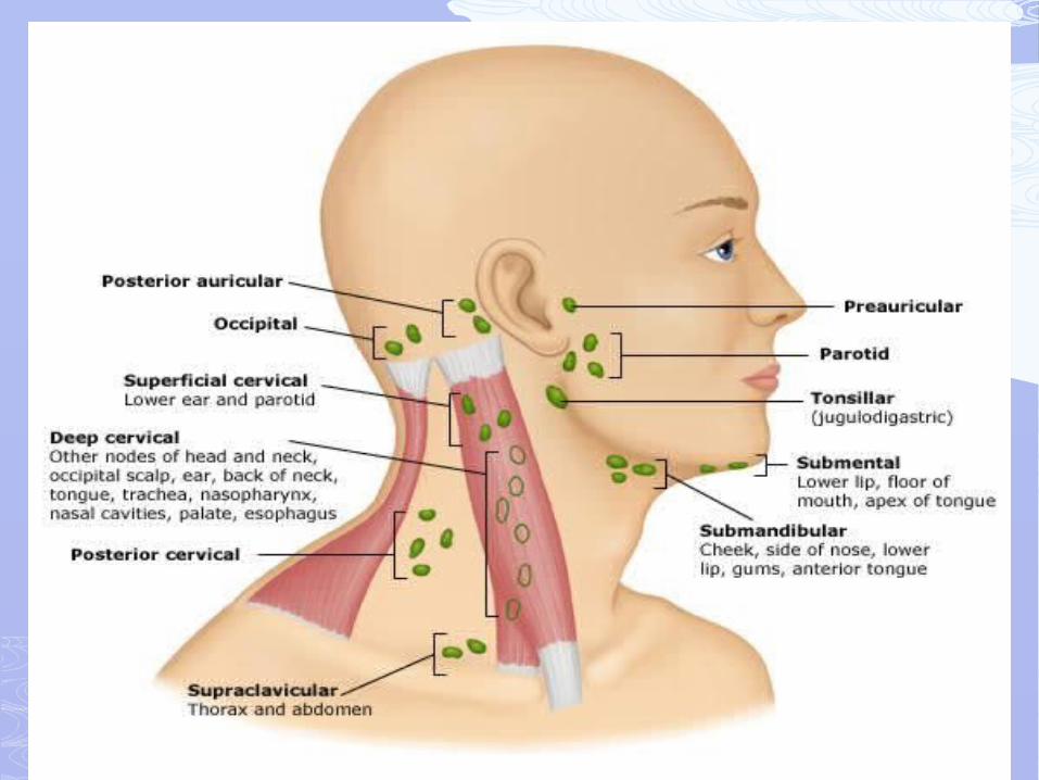
Complete physical examination is vital Distribution
Localized Regional generalized
Symmetry
Features characteristic of the lymph node Location Size Consistency Fixation Tenderness
Splenomegaly hepatomegaly
• Components– Various laboratory and serologic tests– Imaging– Lymph node biopsy– Bone Marrow study – Other biopsies. – ? Empirical treatment
• Depend on various factors– Age– Duration– Localized/regional/generalized– Epidemiology and the clinical setting
Laboratory tests CBC & Peripheral Smear ESR HIV RPR/VDRL ANA Heterophile Antibody tests LDH & other tests according to the setting as
well as importance
Imaging study for the purpose of Defining size & distribution more precisely Distinguishing from other similar structure Staging Guiding for FNA
Imaging study includes Chest X-Ray Ultrasonography & Doppler Nuclear/Isotope scans CT-Scan MRI PET/SPECT
• Types of biopsy– Open biopsy– Fine Needle Aspiration– Core Needle Biopsy
• Choice of LN & type of biopsy– The most diseased– Supraclavicular/cervical/axillary/inguinal– If single go for open biopsy as much as possible– accessability
• Possible studies from the specimen– Pathological– Immunochemistry/immunophenotype– Genetic/molecular studies
Spleen is one of the lymphoid organs which is also called reticuloendothelial system.
Splenomegaly is common clinical condition & it is never normal
Various causes with diagnostic challenge Other condition
Massive splenomegaly Splenic infarction Ruptured spleen Splenic abscess Functional hyposplenism/ asplenia Hypersplenism
Lies in the Peritoneal cavity in the left upper quadrant.
Adjacent to 9th- 11th rib, stomach, colon and pancreas.
Weight Male= 80-200g Female= 70-180g Average = 150g (0.2% of Body Weight)
Palpability and size Not palpable normal ( children, adolescents, thin
adults) Soft organ unless infiltrated
Participates in cellular and humoral immunity Removes senescent and/or poorly deformable
red cells, bacteria, and other particulates from the circulation
Under abnormal circumstances the spleen may become the site of extramedullary hematopoiesis
Approximately one-third of circulating platelets are sequestered in the spleen, where they are in equilibrium with circulating platelets
Splenic abnormalities can include Increased function (hypersplenism) Decreased to absent function (hyposplenism,
asplenia) Abscess, infarction, calcification, cysts Traumatic or atraumatic rupture Enlargement (Splenomegaly)
Splenic engorgement with sequestration Chronic inflammation or infection Lipid deposition Congenital condition Splenic infiltration
1.Congestive Cirrhosis Heart Failure Thrombosis of portal, hepatic and splenic veins
2.Malignancy Lymphomas, usually indolent Leukemias Myeloproliferative Disorders Primary splenic tumours Metastatic solid tumours
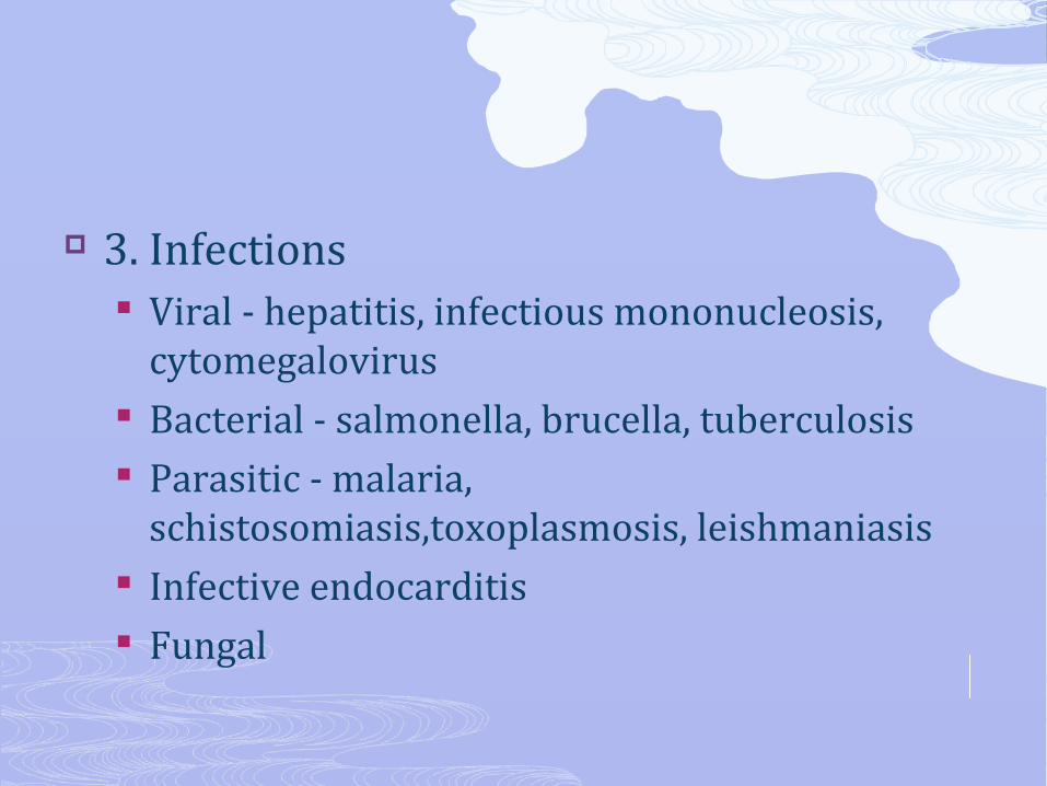
3. Infections Viral - hepatitis, infectious mononucleosis,
cytomegalovirus Bacterial - salmonella, brucella, tuberculosis Parasitic - malaria,
schistosomiasis,toxoplasmosis, leishmaniasis Infective endocarditis Fungal
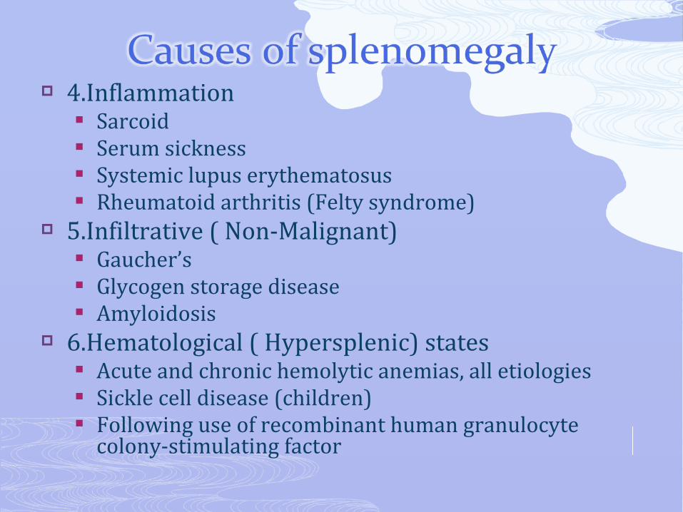
4.Inflammation Sarcoid Serum sickness Systemic lupus erythematosus Rheumatoid arthritis (Felty syndrome)
5.Infiltrative ( Non-Malignant) Gaucher’s Glycogen storage disease Amyloidosis
6.Hematological ( Hypersplenic) states Acute and chronic hemolytic anemias, all etiologies Sickle cell disease (children) Following use of recombinant human granulocyte
colony-stimulating factor
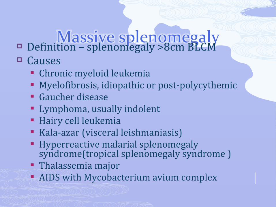
Definition – splenomegaly >8cm BLCM Causes
Chronic myeloid leukemia Myelofibrosis, idiopathic or post-polycythemic Gaucher disease Lymphoma, usually indolent Hairy cell leukemia Kala-azar (visceral leishmaniasis) Hyperreactive malarial splenomegaly
syndrome(tropical splenomegaly syndrome ) Thalassemia major AIDS with Mycobacterium avium complex
History Physical Examination Laboratory studies Imaging Additional studies
Biopsy
Symptoms of splenomegaly Pain, a sense of fullness, or discomfort in the left
upper quadrant Pain referred to the left shoulder Early satiety, due to encroachment on the
adjacent stomach Focused history Underlying conditions Constitutional symptoms Travel history
Complete physical examination Cardinal steps in spleen/ abdominal exam
Inspection Palpation
Bimanual Ballottement Middleton’s method ( palpation from above)
Percussion Nixon’s Method Castell’s Method Percussion of the Traube’s semilunar space
Auscultation
CBC & Peripheral Smear ESR HIV RPR/VDRL ANA, RF Heterophile Antibody tests LDH & other tests according to the setting as well as
importance
Spleen CT scanning magnetic resonance imaging ultrasound Tc-99m sulfur colloid scintigraphy 18F-FDG PET
Other sites CXR CHEST CT
Splenic Biopsy Aspiration Following splenectomy Laparascopy
Liver biopsy Bone Marrow Aspiration/Bone Marrow
Biopsy Biopsy from other sites
• If there is an unusual lump anywhere in the body note the following– Site/appearance– Size in diameter– Shape & nature of surface skin– Fixation– Consistency– Tenderness– Pulsation & bruit(auscultation)– Transillumination in a darkened room

Like us on facebook.com/habeshaentertainment101
follow me @danieleshetu99
Habesha Entertainment http://habeshaentertainment.blogspot.com