lupus erythematosus and nutrition: a …amybrown/lupus-scientificarticle.pdflupus erythematosus and...
TRANSCRIPT

LUPUS ERYTHEMATOSUS AND NUTRITION: A REVIEW OF THE LITERATURE
Author: 2004 Amy C. Brown, PhD, RD Department of Human Nutrition, Food, & Animal Sciences University of Hawaii @ Manoa 1955 East-West Road, Rm 216 Honolulu, HI 96822 808/956-3846 ♦ 808/956-4883 (fax) [email protected]

Lupus Erythematosus and Nutrition: A Review of the Literature Amy C. Brown, PhD, RD*
The purpose of this paper was to search the scientific literature for dietary
compounds that alleviate or exacerbate symptoms of lupus erythematosus (LE) in both
animal and human models. A detailed literature review was undertaken to find articles
showing a relationship between LE and nutrition by using MEDLINE/INDEX MEDICUS
(1950 - March 2000) for English-language articles, followed by cross-referencing.
Aggravating substances appear to include excess calories, excess protein, high fat
(especially saturated and omega-6 polyunsaturated fatty acids), zinc, iron, and L-
canavanine found in alfalfa tablets. Possible beneficial dietary compounds include vitamin
E, vitamin A (beta-carotene), selenium, fish oils (omega-3 polyunsaturated fatty acids),
evening primrose oil, flaxseed, a plant herb (Tripterygium wilfordii), DHEA
(dehydroepiandrosterone), and calcium plus vitamin D (if taking corticosteroids). Some
people with systemic LE placed on food allergy elimination diets reported improvement in
their LE symptoms, however, this may be related to a decrease of other substances in the
diet. Also, although no direct evidence was reported on the beneficial effects of either
bromelain or a vegetarian diet (possibly allowing fish), it is suggested that they might be
beneficial. Limitations to this research are that the findings are based on relatively few
studies, many of which were without control groups or extrapolated from animal models.
No large-scale studies have been done with LE patients to substantiate the benefit, if any,
of these individual dietary interventions, and if they were conducted, the remission and
exacerbation pattern of LE may interfere with elucidating their effectiveness. Also, dietary
changes should not be attempted without a physician's approval/monitoring.
* Department of Human Nutrition, Food, & Animal Sciences, University of Hawaii at Manoa, Honolulu, HI.
Address reprint requests to Amy C. Brown, PhD, RD, Department of Human Nutrition, Food, & Animal Sciences, University of
Hawaii @ Manoa, Honolulu, HI 96822. Email: [email protected], 808/956-3846.

Lupus Erythematosus and Nutrition: A Review of the Literature
The relationship between nutrition and lupus erythematosus (LE) remains elusive
especially since most autoimmune diseases are multifactorial in origin with genetic,
environmental, hormonal, viral, and psychoneurological influences all playing a role.1,2 It is
known that no specific diet for the treatment of LE exists; however, a review of the literature
investigating the influence of nutrition in both animals and humans suggests that certain
substances in the diet may aggravate or alleviate LE symptoms (Tables 1 & 2).
This article elaborates on many of the studies behind the various dietary
compounds listed in Tables 1 & 2 that may aggravate or alleviate LE symptoms. Although,
much of the research presented in this review article is based on animal studies, those
involving human experiments are also discussed. Human subjects are designated as
having discoid or systemic lupus erythematosus when specified in the literature, or "LE" if
no such designation was provided.
Possible Harmful Substances
Excess Energy. Most animal studies suggest that energy restriction ameliorates
autoimmune disease3,4 and increases longevity in New Zealand black or white mice
(NZB/NZW) which spontaneously develop an autoimmune disease resembling systemic
LE.5-8 The disease in these mice is manifested by synthesized antibodies to double
stranded DNA, high levels of circulating immune complexes (a marker of SLE clinical
activity), and deposition of the later in the renal glomerulus. As a result, glomerulonephritis
is the major cause of death with mortality rates averaging 50 percent by about 8.5 months.9
Caloric restriction delays the onset of glomerulonephritis in these mice, however,
this dietary manipulation is severe and initiated early - parameters that cannot be
duplicated in the human model since caloric restrictions would be equivalent to 25-35
percent or more of total intake prior to adolescence. The degree of energy restriction in
one mice study was even higher at 60 percent (at 2 months of age) versus the control
group allowed to feed ad libitum. At 14 months, the percentage of these mice still living
that had not died due to renal disease was 100 and 0 percent respectively.10 Although

3
there are no studies showing the effect of caloric restriction in humans, Kipen reported that
SLE disease activity was associated with an increase in body mass index (BMI) over a
three year period in pre-menopausal women (n=55).11
In terms of animal models, the exact mechanisms by which energy restriction
benefits autoimmune conditions are still being explored. Safai-Kutti reported a reduction in
circulating immune complexes, occurring in mice eating an energy-restricted diet,12 while
Chandraseker observed a decrease in pro-inflammatory cytokines.13 Mizutani reported
that reducing calories to 32 percent or less than controls in autoimmune-prone mice
resulted in decreased immunoprecipitates, less coronary vascular lesions, and fewer
glomerular lesions.14 Restricting calories in mice was reported by Meydani to also reduce
prostaglandin E2 (PGE2) synthesis, another compound known for its proinflammatory
effects.15 One study restricting calories in mice and comparing them to a control group
reported a delay or inhibition of Sjogren's syndrome abnormalities, increased
immunosuppressive transforming growth hormone beta-1, and decreased cytokines.12
Excess Protein. Low-protein diets are also known to improve survival rates in
autoimmune mice.6 Mice fed a moderately restricted protein diet experienced longer
lasting immunologic functions and delayed development of autoimmunity when compared
to mice fed a normal protein diet.16
These results are not surprising since high protein intakes have been commonly
associated with acceleration of kidney damage in both autoimmune-prone humans and
experimental animals.17 Protein restriction has long been the standard treatment for renal
failure.18
After establishing that the amount of dietary protein influenced the outcome of
autoimmune-prone mice, researchers focused on specific types of protein. One study
indicated that limiting proteins containing high levels of phenylalanine and tyrosine, such as
those found in beef and dairy products, is beneficial to mice with a systemic LE-type
condition.4 Carr and others reported that 12 of 15 mice fed a casein-free diet were still
alive at 10 months, compared to only 1 in 10 mice on the control diet, and that casein-free
mice had less anti-DNA antibody and immunoreactants in the glomeruli.19 Another amino
4
acid in question is tryptophan because elevated urinary excretion levels of tryptophan
metabolites were reported in 11 discoid lupus patients.20 Researchers have also
suggested that tryptophan breakdown products may lead to autoantibody production,21
and a research study investigating this possibility determined that a tryptophan-deficient
diet fed to lupus animals resulted in longer survival times.22
The average American ingests about 100 grams of protein a day, an amount that
can be reduced by almost half in healthy people without jeopardizing their protein
requirements. The Recommended Daily Allowances (RDA - 1989) for protein are 50
grams for women and 63 grams for men in the 25-50 years age group. Vegetarian diets
often automatically reduce dietary protein, and there was a case study reported of a patient
with SLE that went on a vegetarian diet (zero percent animal protein). Her antibody titers
returned to normal, urinary protein excretion decreased, and serum albumin rose. Although
protein dropped from 97 to 32 grams, so did calories (2295 to 1216) and grams of fat (70
to 50).23 The caloric reduction along with the fact that this is a single-case study and that
the possibility of remission exists does not warrant any dietary recommendation, however,
further study in this area is suggested.
High Fat (especially saturated fat and omega-6 polyunsaturated fatty acids). Diets
high in overall fat were associated with more severe autoimmune disease and decreased
life span in mice compared to a control group, whereas low fat diets were reported to
retard the development of disease.24,25
The type of dietary fat also dramatically affects the onset of autoimmune disease in
mice particularly if it consists of saturated or omega-6 polyunsaturated fatty acids.
Alexander reported that by 10 months of age, the percentage of mice still alive was; 94
percent of the fish oil (omega-3 polyunsaturated fatty acids) group, 35 percent of the mice
fed corn oil (omega-6 polyunsaturated fatty acids), and zero percent of the group fed
saturated fat in the form of lard (n-9 fatty acids).26 Fernandes supported these results with
a study showing that omega-3 fatty acids lowered the severity of autoimmune disease in
mice, while both saturated (n-9) and polyunsaturated (n-6) dietary lipids exacerbated the
disease.27

5
Autoimmune-prone mice fed saturated fats experience more severe nephritis and
glomerular pathology.28,29 Several researchers have reported that mice fed saturated fat
diets produced higher levels of autoantibodies than those on low fat or high unsaturated fat
diets.28,30,31 High fat diets in mice have also been reported to increase proteinuria,24,32
prostaglandin (PGE2) production, cytokine levels (interleukin 6),33 and macrophage
function.24 These types of results have led some researchers to suggest that dietary fat,
especially saturated fat, restriction may be an effective therapeutic approach to murine
lupus nephritis.29
Several studies suggest that limiting essential omega-6 fatty acids and/or zinc may
suppress immune response, and therefore flare-ups. Hurd and Gilliam postulated that
some of the prostaglandins or related products of arachidonic acid (prostacyclin,
thromboxane, or lipoxgenase pathway products) may be necessary for the full expression
of autoimmunity.35 Essential fatty acid deficiency in mice resulted in an increased survival
time, delay in antibody production, and less severe renal disease.34-36 However,
researchers of another study reported that the survival of mice (MRL/l) was not affected by
polyunsaturated fatty acid deficiency.37 In studies that do show positive results, limiting
essential fatty acids may be beneficial because less are then available for the synthesis of
specific prostaglandins responsible for inflammation.
Thorner conducted one of the few human studies in which SLE patients reduced
their omega-6 polyunsaturated fatty acid intake. After one year, the number of patients with
active SLE dropped from 11 to 3. Spontaneous improvement, placebo effects, and lack of
a control group must be considered as possible influences in this and other studies
investigating the role of diet on LE symptoms. However, the researchers suggested the
possibility of reducing the intake of omega-6 polyunsaturated fatty acids as a non-
pharmacological approach to the treatment of patients with SLE.38 Foods high in omega-6
fatty acids are listed in Table 4.
Zinc. Zinc is important for enhancing the immune response, and MRL/1 mice on zinc-
deficient diets were reported to have increased survival times. Researchers observed a
decrease in lymphoproliferation,39 and a delayed expression of autoantibodies.40 It has
6
been suggested that zinc deprivation results in increased serum corticosteroids which may
contribute to the decreased number of autoimmune disease symptoms.39
Iron. Only one animal study suggests that high iron intakes, seven times the requirement,
in mice resulted in high proteinuria, renal histopathology, and mortality. The researchers
theorized that excess iron may enhance the Haber-Weiss reaction causing free radical
damage of the tissues. During an inflammatory response, neutrophils and macrophages
release superoxide (O2 ) and hydrogen peroxide (H2O2), and the reaction of these
compounds with iron produces a highly toxic hydroxyl radical (OH ).41 A source of excess
iron for humans is ingestion of pre-natal vitamin/mineral supplements that often contain 30-
60 mg (Recommended Dietary Allowance (RDA) for women = 15 mg/day).
Alfalfa (L-Canavanine). Researchers studying the cholesterol-lowering effect of alfalfa
seeds observed signs of SLE-like symptoms in both laboratory animals and a few human
case studies.42-44 Two human patients were reported to experience symptoms of malaise,
lethargy, depression, and arthralgias after ingesting 8-15 alfalfa tablets daily.45 In vitro
experiments suggest that L-canavanine, an amino acid in alfalfa products, acts on
suppressor-inducer T cells to regulate antibody synthesis and lymphocyte proliferation.46
Feeding L-canavanine to autoimmune mice resulted in increased antibody production and
higher renal histology scores.47 However, the alfalfa tablets of one manufacturer tested
negative for canavanine (with a detection limit of 5 ppm), and alanine, an amino acid, has
previously been mistaken for canavanine.48
Possible Beneficial Substances
Vitamin E. Although animal studies on MRL/lpr mice show that vitamin E treatment delays
the onset of autoimmunity and extends mean survival time,49 treating LE patients with
vitamin E continues to be controversial. Vitamin E studies related to LE first started to
appear in the late 1940s, and a historical overview of the literature reveals that large
vitamin E doses may be beneficial in some cases, while dosages below 300 IU may not be
7
sufficient.50 For example, four discoid LE patients in one study, receiving 900-1600 IU of
vitamin E daily, showed partial or complete clearing of rashes, while two patients receiving
only 300 IU daily had no benefit.51 Other human studies reporting either positive 5,52-57
or negative 58-61 results of vitamin E on discoid LE lesions are listed in Table 3.
Investigators warn that "while the effect of mixed tocopherols in LE is apparently
profound, often rapid and at times almost specific, the fact remains that recurrences are
not uncommon."62 Also, Vitamin E is a fat soluble vitamin that acts as an anticoagulant at
the high dosages used in these studies, much higher than the Reference Daily Intake (RDI)
of 30 IU (9 mg) alpha-TE. Dietary sources of vitamin E are listed in Table 4.
Vitamin A. Vitamin A-deficient LE animals were reported to experience more severe
lupus-like symptoms. Researchers attributed this observation to increased hyper-
gammaglobulinemia and an earlier onset of autoantibodies, both naturally occurring
thymocytoxic autoantibodies and IgM anti-erythrocyte antibodies.63 Three patients whose
skin lesions flared with sun exposure were given 50 mg of beta-carotene three times daily,
and experienced a clearing of all lesions starting within one week of treatment.64 Other
researchers reported that very high levels of vitamin A (100,000 U daily for 2 weeks) in
SLE patients resulted in an enhancement of antibody-dependent cell-mediated cytotoxicity,
natural killer cell activity and blastogenic response to mitogens.65
However patients should be cautious with extremely high levels of vitamin A unless they are
water-soluble, because ingesting excess vitamin A from animal sources may result in one
or more of the following symptoms: anemia, headache, dry skin, hair loss, nausea, lack of
appetite, bone pain, stunted growth in infants/children, pseudohydrocephalus, and death.
Table 4 lists vegetable sources high in vitamin A (beta-carotene). Although excess beta-
carotene from plant sources does not result in the symptoms elicited from animal sources,
it can produce hypercarotenemia turning the skin slightly orange.
Selenium (Se). Anti-inflammatory properties have been attributed to selenium, a natural
antioxidant.66 Supplementing the diets of auto-immune mice with selenium increases their
survival time, and although the mechanism by which selenium exerted this effect is unclear,

8
there is a significantly higher level of natural killer cell activity in the selenium-supplemented
mice.67 It was also observed that low levels of blood glutathione-peroxidase (GSH-Px)
exist in some patients with systemic LE, and that GSH-Px activity increased slowly after
administering tablets containing 0.2 mg selenium (Na2SeO3) and 10 mg tocopherol
succinate for 6-8 weeks. Some researchers have suggested that physicians could check
GSH-Px activity and consider selenium and vitamin E supplementation in people with LE
or other conditions such as severe psoriasis, eczema, dermatitis herpetiforms, and liver
disease.60 Again, warnings against high intakes of selenium should be given to patients
since toxicity results in symptoms of diarrhea, vomiting, hair and nail loss, and lesions of
the nervous system and skin. Dietary sources of selenium are listed in Table 4.
Fish Oils. Fish oils retard, but do not entirely prevent, lupus-like disorders found in
autoimmune-prone mice. These mice eventually develop the illness, but at a slower rate
than controls. Fish oil supplementation appears to have an anti-inflammatory effect,68 and
prolongs the life of autoimmune-prone mice.7,69-76 The increased life span might be due to
delayed onset of renal disease since mice introduced to a fish oil diet as weanlings had an
almost total protection against renal disease. One possible mechanism related to the
beneficial effect of fish oil in autoimmune-prone mice may be related to its high omega-3
fatty acid content - eicosapentaenoic acid (EPA) and docosahexaenoic acid (DHA).
Omega-3 fatty acids inhibit the production of eicosanoids (pro-inflammatory
compounds such as prostaglandin E2 (PGE2) and leukotriene B4 (LTB4)), while fatty acids
from the omega-6 series have the opposite effect.77-80 Arachidonic acid, an omega-6 fatty
acid, is metabolized into pro-inflammatory eicosanoids.81-84 Omega-3 fatty acids can
displace arachidonic acid in the cell membranes,70,74 resulting in less PGE2 formation,
and compete with arachidonic acid for cyclooxygenase and lipoxygenase enzymes.69 This
competition shifts production to the non-inflammatory series-3 prostaglandins and series-5
leukotrienes that have been suggested to directly suppress immunologic and or
inflammatory mediators of murine lupus.69,70,80 Specifically, there is a reduced synthesis of
endogenous dienoic cyclooxygenase metabolites and leukotriene 4 (LT4), while increased
synthesis of trienoic PG and leukotriene 5 (LT5).69 Another factor is that omega-3 fatty

9
acids are poor substrates for cyclooxygenase which is the rate-limiting step in the
synthesis of prostaglandins, particularly PGE2.78
Omega-3 fatty acids may also inhibit the inflammatory response by decreasing T-
cell activity and cytokine (stimulate prostaglandin production) concentration.85-87 Normally,
inflammation activates T cells and cytokines at the site of tissue injury and in the
circulation.82 Certain types of cytokines are also involved in peroxidation which is a
common final pathway in much of the tissue damage seen in intense inflammation.
Cytokines (IL-1 and TNF-alpha) generate H2O2 and O2- in mesangial cells and
macrophages, and these can result in free radical damage to the tissues. Reactive oxygen
intermediates (ROI), implicated in immune-complex mediated glomerulonephritis, affect
glomerular filtration rate, impair sieving, and inhibit renal function. ROIs may react with
polyunsaturated fatty acids in cell membranes resulting in derivatives that attract
inflammatory cells that can further secrete inflammatory cytokines and growth factors.
Peroxidation of lipid membranes also leads to altered fluidity, and may change ion
transport and enzyme activities in target tissues such as renal cells.82 To help protect
vessels and organs from the damage of excess inflammation, some researchers have
suggested the administration of antioxidants such as vitamin E and selenium.81
The majority of animals studies show omega-3 fatty acids alleviating the severity of
autoimmune disease, but Table x shows only modest anti-inflammatory effects have been
reported in humans with LE.88-90 However, omega-3 fatty acids have been reported to
improve blood lipid values which is of benefit to patients with SLE who have a higher rate
of premature atherosclerosis than the general population.89,91,92 Despite the controversy
over whether or not omega-3 fatty acids benefit humans with autoimmune conditions,
Kinsella stated that hospital nutritional support, such as enteral and parenteral formulas in
addition to intravenous emulsions, may need to be modified for use in patients with
inflammatory reactions such as lupus erythematosus, rheumatoid arthritis, and multiple
sclerosis.93 Dietary sources of omega-3 fatty acids (EPA/DHA) are listed in Table 4.
Bromelain. Although no animal or human studies have been conducted on bromelain
related to LE, this complex of proteases from the pineapple plant has been known to act as

10
an anti-inflammatory agent.94
Evening Primrose Oil (EPO). EPO was reported to increase survival time in autoimmune
mice,95 and this may be due to its gamma-linolenic acid (19%) content from which PGE1 is
formed. Several studies support the role of PGE1 treatment alone in delaying the onset
and severity of lupus in autoimmune animals.96-98 This beneficial effect of PGE1 might be
due its anti-inflammatory effects via membrane stabilization and lowering lymphocyte
activity.99 Feeding rodents at least 5-10 g gamma-linolenic acid/100 g total fatty acids has
been shown to decrease lymphocyte proliferation and natural killer cell activity.100 In
addition, a derivative of gamma-linolenic acid has been postulated to block the
transformation of arachidonic acid to leukotrienes that have proinflammatory effects.101
Flaxseed. Two studies, one with mice and the other with human subjects, suggest that
flaxseed may be beneficial. A 15 percent flaxseed diet provided to mice, and compared to
a control diet, resulted in decreased proteinuria, spleen lymphocyte proliferation, and
mortality. Flaxseed also appeared to preserve glomerular filtration rate (GFR).102 Eight
humans with SLE were given 30 grams of flaxseed mixed in with their cereal or juice
(tomato or orange), and were reported to have improved renal function defined by
decreased proteinuria, decreased serum creatinine, and increased creatinine clearance.
Also noted in these human subjects, was the ability of flaxseed to inhibit platelet activating
factor (PAF) induced platelet aggregation.103 PAF, a participant in the inflammatory
response, is often elevated in LE patients. Studies in lupus animal models suggest that
inhibiting PAF resulted in decreased proteinuria and increased survival times.
Flaxseed is one of the richest food sources for lignans which are natural antagonists
to PAF receptors. This plant food is also high in an omega-3 fatty acid, alpha-linoleic
acid.103 It has been reported that the beneficial affect of these and possible other
compounds in flaxseed is best achieved by ingesting it in its whole form, rather than in its
oil (linseed oil) or defatted form.104 Regardless of the form ingested, patients should be
cautioned of possible allergic reactions.105

11
Plant Herb. Tripterygium wilfordii hook F (TWH), also known as Thunder God Vine, is a
plant that has been used in China for more than 2000 years to treat SLE and rheumatoid
arthritis.106-109 In vitro tests support this traditional practice through evidence that the plant
has immunosuppressive qualities.110 Ramagolam reported that TWH inhibited
lymphoproliferation (mitogen-stimulated), production of cytokines by moncytes and
lymphocytes, and prostaglandin E2 production via the cyclooxygenase (COX-2) pathway.111
Despite the promising benefit of TWH, it is difficult to evaluate the use of herbal
therapies since their apparent successes and sometimes serious side-effects are often
not documented, particularly for renal patients. Reported side-effects of TWH include
gastrointestinal upset, infertility, and suppression of lymphocyte proliferation. At least one
case was reported of a previously healthy young man who died of shock possibly related to
cardiac toxicity.112 As with most herbs, its use during pregnancy or lactation is not
recommended; a report exists of a woman taking TWH during pregnancy and giving birth
to a child with a protrusion in the lower back of its head (occipital
meningoencephaloecele).113
DHEA (dehydroepiandrosterone). Although not a nutrient or a dietary supplement, this
steroid hormone can be purchased over-the-counter. Animal studies with autoimmune-
prone mice have shown that DHEA produces similar results to those obtained with caloric
restriction - decreased antibody synthesis and prolonged survival rates.114-116 In humans, a
double-blind, placebo-controlled study of 28 SLE patients taking DHEA (200 mg/day) for 3
months resulted in decreased lupus flares, SLE Disease Activity Index scores, disease
activity (assessed by physicians and patients), and prednisone dosages. The researchers
observed only mild acne as a side-effect and concluded that DHEA may be a useful
therapeutic agent for the treatment of mild to moderate SLE.117 Several other human
studies, however these without control groups, also indicated that DHEA might be
beneficial to patients with LE.118-121
Immune responses are often influenced by sex hormones.122 Androgens naturally
suppress the immune system and concomitant inflammation, while estrogens can do
either, although they usually accelerate autoimmunity.123 DHEA is an androgen and an

12
intermediate compound in testosterone synthesis. Women with autoimmune diseases like
LE and rheumatoid arthritis have lower plasma androgens than controls, and researchers
theorize that the ingestion of weak androgens, like DHEA, may improve the clinical
manifestations of the disease.124,125 The androgenic nature of DHEA in women
taking over 50 mg a day dictates that a doctor's supervision and caution should be elicited.
Minor side-effects at this dosage in women include acne, facial hair growth, menstrual
changes, and improved mood, however, these symptoms disappeared after DHEA intake
was stopped. Short-term studies usually report no serious side-effects in humans
ingesting DHEA,126 but long-term trials have not been performed and androgen
replacement remains in the realm of clinical investigation.127 There is the possibility that
DHEA can convert into certain sex hormones,128 and there is speculation about its safety
since it has been linked to hepatic cancer in rats.129
Food Elimination Diets. Some researchers have reported that LE patients may be more
prone to food allergies.130,131 Several case studies indicate that systemic LE patients
experience remissions following food elimination diets.132,133
Calcium Plus Vitamin D (if taking corticosteroids). Calcium and vitamin D are not
reported to alleviate symptoms of LE, however, they are recommended as part of the
treatment against osteoporosis, the most serious side-effect of long-term corticosteroid
therapy.134 The long-term use of corticosteroids, the most commonly prescribed
immunosuppressants,135 are responsible for an estimated 20 percent of the 20 million
osteoporosis cases in the United States. One in four of these patients experiences a
fracture,136 but unlike other forms of osteoporosis, the majority of corticosteroid-induced
osteoporosis fractures are at the spine.134 This is a particular concern for SLE patients
that have been reported to have reduced bone mineral densities compared to matched
healthy controls.137,138
Cushing first associated skeletal mass loss with hypercortisolism in 1932,139

13
however, patients prescribed long-term corticosteroid therapy are not always 1) informed
of the osteoporosis risk, or 2) provided any form of osteoporosis prophylaxis.
Researchers in one study reported that only 5.6 percent of 214 patients in a British hospital
receiving corticosteroid therapy (37 percent for four or more months) were given treatment
to help delay the onset of osteoporosis.140
To combat the long-term negative side-effects of corticosteroids, the American
College of Rheumatology (ARC) has formulated optimal medical management guidelines
to reduce the risk of bone loss in patients. Preventative treatment should begin as soon as
long-term corticosteroid therapy is started and include baseline bone mineral test, lowest
effective dosage, hormone replacement therapy, medication, reducing risks for falls,
lifestyle (weight-bearing exercise, and avoiding smoking, immobilization, and
amenorrhea), and nutrient supplementation (supplements for calcium (up to 1500 mg) and
vitamin D (20 ug (800 IU), less in children)).136 Another approach would be to initially seek
these nutrients through food sources and not to exceed supplementation in excess of 10 ug
(400 IU) for vitamin D. Other nutrient factors to reduce include eliminating excess protein,
salt, alcohol, or caffeine. Side-effects of excess calcium supplements include constipation,
headaches, calcification of the soft tissues, and certain kidney stones. Vitamin D
supplements should also not be taken in excess, because they have been reported to
cause headache, nausea, calcification of the soft tissues and bone, a tendency toward
kidney stones, and in children - possible stunted growth, mental retardation, and death by
renal failure.
Conclusion
No dietary recommendations currently exist for LE patients, however, physician
researchers postulated over fifteen years ago that diet might be one of the possible future
therapies for people with LE.141 Tables 1 & 2 provide tentative dietary suggestions based
on a literature review. Patients with LE may benefit from a balanced diet limited in calories
and fat (especially saturated and omega-6 polyunsaturated fatty acids), containing rich
sources of vitamin E, vitamin A (beta-carotene), selenium, and calcium. Supplements of

14
fish oil, evening primrose oil, flaxseed, a plant herb (Tripterygium wilfordii), DHEA (under a
physician's care), and calcium plus vitamin D (if taking corticosteroids) may also be
beneficial. Foods high in omega-3 polyunsaturated fatty acids are recommended and
include fish oils, fatty fish, certain vegetable oils such as walnut and canola, and soybeans.
Conversely, foods to be avoided that contain omega-6 polyunsaturated fatty acids are
vegetable oils made from corn, cottonseed, poppyseed, safflower, sesame, soybean,
sunflower, and walnut. People with LE may also benefit by avoiding supplements
containing protein, omega-6 polyunsaturated fatty acids, zinc, and iron. Avoiding an
excess of foods rich in these compounds might possibly be beneficial which would consist
of limiting meats (protein), dairy (protein), oysters (zinc), Brazil nuts (zinc), and enriched
grains and cereals, including breakfast cereals (zinc and iron). It may also be judicious to
avoid alfalfa tablets or alfalfa in any form including sprouts. Remissions have been
reported in people with LE going on food elimination diets, and perhaps these could be
tried by LE patients in an attempt to alleviate flare-ups or eliminate the possibility of any
existing food allergies. Further investigation should be conducted on the possible
beneficial use of bromelain and vegetarian diets in people with LE.
Again, these tentative dietary suggestions are based on a literature review and the
nature of remissions occurring in people with LE along with any medications make it
difficult to evaluate their effectiveness. The purpose of this paper was to elucidate from the
scientific literature the dietary compounds that alleviate or exacerbate symptoms of LE in
both animal and human models. Extrapolations are tenuous at best, and the lack of control
groups and/or use of animal studies sheds questionable query on the results. However, an
ample array of research has been conducted and the results are summarized in the form of
Tables 1 & 2. This compilation is based on a limited number of studies and no large scale
studies have been done with LE patients to substantiate the benefit, if any, of these dietary
interventions. Nevertheless, the possibility exists that patients with LE may benefit by
incorporating one or more of these dietary modifications with the approval/monitoring of a
physician.142
15
References
1. Talal, N. Sex hormones and modulation of immune response in SLE. Arthritis Rheum 8:23, 1982.
2. Mongey A, Hess EV. Drug and environmental effects of the induction of autoimmunity. J Lab Clin Med 122(6):652-657, 1993.
3. Fernandes G, Friend P, Yunis EJ, Good RA. Influence of dietary restriction on immunologic function and renal disease in (NZB x
NZW) F1 mice. Proc Natl Acad Sci 75:1500-1504, 1978.
4. Corman LC. The role of diet in animal models of systemic lupus erythematosus: possible implications for human lupus. Semin
Arthritis Rheum 15(1):61-69, 1985.
5. Jolly CA, Fernandes G. Diet modulates Th-1 and Th-2 cytokine production in the peripheral blood of lupus-prone mice. J Clin
Immunol 19(3):172-178, 1999.
6. Weindruch R, Walford RL. Dietary restriction in mice beginning at one year of age: effect on life span and spontaneous cancer
incidence. Science 152:1415-1418, 1982.
7. Troyer DA, Chandrasekar B, Thinnes T, et al. Effects of energy intake on type 1 plasminogen activator inhibitor levels in glomeruli
of lupus-prone B/W mice. Am J Pathol 146(1):111-120, 1995.
8. Troyer DA, Chandrasekar B, Barnes JL, Fernandes G. Calorie restriction decreases platelet-derived growth factor (PDGF)-A and
thrombin receptor mRNA expression in autoimmune murine lupus nephritis. Clin Exp Immunol 108(1):58-62, 1997.
9. Andrews BS, Eisenberg RA, Theofilopoulos AN, et al. Spontaneous murine lupus-like syndromes. Clinical and
immunopathological manifestations in several strains. J Exp Med 148:1198-1215, 1978.
10. Urao M, Ueda G, Abe M, et al. Food restriction inhibits an autoimmune disease resembling systemic lupus erythematosus in (NZB
x NZW) F1 mice. J Nutr 125(9):2316-2324, 1995.
11. Kipen Y, Briganti Em, Strauss BJ, et al. Three year follow -up of body composition changes in pre-menopausal women with
systemic lupus erythematosus. Rheumatology 38(1):59-65, 1999.
12. Safai-Kutti S, Fernandes G, Wang Y, et al. Reduction of circulating immune complexes by calorie restriction in (NZB x NZW) F1
mice. Clin Immunol Immunopathol 15:293-300, 1980.
13. Chandrasekar B, McGuff HS, Aufdermorte TB, et al. Effects of calorie restriction on transforming growth factor beta-1 and
proinflammatory cytokines in murine Sjogren's syndrome. Clin Immun Immunopath 70(3):291-296, 1995.
14. Mizutani H, Engelman RW, Kinjoh K, et al. Calorie restriction prevents the occlusive coronary vascular disease of autoimmune
(NZW x BXSB) F1 mice. Proc Natl Acad Sci USA 91(10):4402-4406, 1994.
15. Meydani SN, Lipman R, Blumber JB, et al. Dietary energy restriction decreases ex vivo spleen prostaglandin E2 synthesis in
Emory mice. J Nutr 120(1):112-115, 1990.
16. Good RA, Fernandes G, Yunis EJ, et al. Nutritional deficiency, immunologic function, and disease. Am J Pathol 84(3):599-614,

16
1976.
17. Johnson BC, Gajjar A, Kubo C, et al. Calories versus protein in onset of renal disease in NZB x NZW mice. Proc Natl Acad Sci
USA 83(15):5659-5662, 1986.
18. Ihle BU, Becker GJ, Whitworth JA, et al. The effect of protein restriction on the progression of renal insufficiency. N Engl J Med
321:1773-1777, 1989.
19. Carr R, Forsyth S, Sadi D. Abnormal responses to ingested substances in murine systemic lupus erythematosus: apparent effect
of a casein-free diet on the development of systemic lupus erythematosus in NZB/W mice. J Rheumatol 14(Suppl.13):158-165,
1987.
20. Mandell EH, Appleton HD. Tryptophan metabolism. Results of studies in discoid lupus erythematosus. Arch Dermatol 94:358-
360, 1966.
21. McCormick JP, Fischer JR, Pachlatko JP. Characterization of a cell-lethal product from the photooxidation of tryptophan:
Hydrogen peroxide. Science 191:468-469, 1976.
22. Dubois EL. Lupus Erythematosus. Magraw Hill, 2nd edition, p.121, 1966.
23. Shigemasa C, Tanaka T, Mashiba H. Effect of vegetarian diet on systemic lupus erythematosus. Lancet 339(8802):1177, 1992.
24. Lin BF, Huang CH, Chiang BL, Jeng SJ. Dietary fat influences Ia antigen expression, cytokines and prostaglandin E2 production of
immune cells in autoimmune-prone NZB x NZW F1 mice. Br J Nutr 75(5):711-722, 1996.
25. Swanson CA, Levy JA, Morrow WJ. Effect of low dietary lipid on the development of Sjogren's syndrome and haematological
abnormalities in (NZB x NZW)F1 mice. Ann Rheum Dis 48(9):765-770, 1989.
26. Alexander NJ, Smythe NL, Jokinen MP. The type of dietary fat affects the severity of autoimmune disease in NZB/NZW mice. Am
J Pathol 127(1):106-121, 1987.
27. Fernandes G, Venkatramna J, Khare A, et al. Modulation of gene expression in autoimmune disease and aging by food restriction
and dietary lipids. Proc Soc Exp Biol Med 193(1):16-22, 1990.
28. Levy JA, Ibrahim AB, Shirai T, et al. Dietary fat affects immune response, production of antiviral factors, and immune complex
disease in NZB/NZW mice. Proc Natl Acad Sci USA 79(6):1974-1978, 1982.
29. Yumura W, Hattori S, Morrow WJ, et al. Dietary fat and immune function. II. Effects on immune complex nephritis in (NZB x
NZW)F1 mice. J Immunol 135(6):3864-3868, 1985.
30. Erickson KL, Adams DA, Scibienski RJ. Dietary fat acid modulation of murine B-cell responsiveness. J Nutr 116(9):1830-1840,
1986.
31. Jyonouchi H, Sun S, Goodman D, et al. Dietary fatty acid modulates actions of nucleotides on humoral immune responses.
Nutrition 11(5):437-443, 1995.
32. Morrow WJ, Homsy J, Swanson CA, et al. Dietary fat influences the expression of autoimmune disease in MRL/lpr/lpr mice.
17
Immunology 59(3):439-443, 1986.
33. Yaqoob P, Calder PC. The effects of dietary lipid manipulation on the production of murine T cell-derived cytokines. Cytokine
7(6):548-553, 1995.
34. Lucus JA, Ahmed SA, Casey ML, et al. Prevention of autoantibody formation and prolonged survival in New Zealand black/New
Zealand white F1 mice fed dehydroisoandrosterone. J Clin Invest 75:2091-2093, 1985.
35. Hurd ER, Gilliam JN. Beneficial effect of an essential fatty acid deficient diet in NZB/NZW F1 mice. J Invest Dermatol 77:381-384,
1981.
36. Watson J, Godfrey D, Stimson WH, et al. The therapeutic effects of dietary fatty acid supplementation in the autoimmune disease
of the MRL-mp-lpr/lpr mouse. Int J Immunopharmacol 10(4):467-471, 1988.
37. Westberg G, Tarkowski A, Svalander C. Effect of eicosapentaenoic acid rich menhaden oil and MaxEPA on the autoimmune
disease of Mrl/l mice. Int Arch Allergy Appl Immunol 88(4):454-461, 1989.
38. Thorner A, Walldius G, Nilsson E, et al. Beneficial effects of reduced intake of polyunsaturated fatty acids in the diet for one year
in patients with systemic lupus erythematosus [letter]. Ann Rheum Dis 49(2):134, 1990.
39. Beach RS, Gershwin ME, Hurley LS. Nutritional factors and immunity. III. Zinc deprivation versus restricted food intake in MRL/1
mice - the distinction between interacting dietary influences. J Immunol 129(6):2686-2692, 1982.
40. Gershwin ME, Lentz DR, Beach RS, Hurley LS. Nutritional factors and autoimmunity. IV. Dietary vitamin A deprivation induces a
selective increase in IgM autoantibodies and hypergammaglobulinemia in New Zealand black mice. J Immunol 133(1):222-226,
1984.
41. Leiter LM, Reugh KR, Racis SP, Sherman AR. Iron status alters murine systemic lupus erythematosus. J Nutr 125:474-484,
1995.
42. Podell RN. Systemic lupus erythematosus. Does diet play a causative role? Postgrad Med 75(1):251-254, 1984.
43. Malonow MR, Bardara EJ, Pirofsky B, et al. Systemic lupus erythematosus-like syndrome in monkeys fed alfalfa sprouts: Role of
a nonprotein amino acid. Science 216:415-417, 1982.
44. Montanaro A, Bardana EJ. Dietary amino acid-induced systemic lupus erythematosus. Rheum Dis Clin North Am 17(2):323-
332, 1991.
45. Roberts JL, Hayashi JA. Exacerbation of SLE associated with alfalfa ingestion. N Eng J Med 308(22):1361, 1983.
46. Morimoto I, Shiozawa S, Tanaka Y, Fujita T. L-canavanine acts on suppressor-inducer T cells to regulate antibody synthesis:
lymphocytes of systemic lupus erythematosus patients are specifically unresponsive to L-canavanine. Clin Immunol
Immunopathol 55(1):97-108, 1990.
47. Prete PE. Effects of L-canavanine on immune functioning in normal and autoimmune mice: disordered B-cell function by a dietary
amino acid in the immunoregulation of autoimmune disease. Can J Physiol Pharmacol 63(7):843-854, 1985.

18
48. Whittam J, Jenson C, Hudson T. Alfalfa, vitamin E, and autoimmune disorders. Am J Clin Nutr 62(5):1025-1026, 1995.
49. Weimann BJ, Hermann D. Inhibition of autoimmune deterioration in MRL/lpr mice by vitamin E. Int J Vitam Nutr Res 69(4):255-
261, 1999.
50. Sweet RD. Vitamin E in collagenoses [letter]. Lancet 2:310, 1948.
51. Ayers S, Mihan R. Is vitamin E involved in the autoimmune mechanism? Cutis 21:321-325, 1978.
52. Burgess JF, Pritchard JE. Tocopherols (vitamin E). Treatment of lupus erythematosus; preliminary report. Arch Derm Syph
57:953-964, 1948.
53. Welch AL. Treatment by combined use of massive amounts of pantothenic acid and vitamin E in lupus erythematosus. Arch
Dermatol 70:181, 1954.
54. Ayres S, Mihan R. Lupus erythematosus and vitamin E: an effective and nontoxic therapy. Cutis 23:49-54, 1979.
55. Grubb E, Hagerman G. Our experiences with vitamin E treatment. Acta Derm Venereol 32:256-258, 1952.
56. Shinskii GE, Telegina KA, Shephovotsava VV. Ex perience with vitamin E in the treatment of lupus erythematosus. Dermato
Venerol 36:64, 1962.
57. Silver SH, Feigenbaum HL. Chronic discoid lupus erythematosus successfully treated with vitamin E. Arch Dermatol 61:163,
1950.
58. Morgan J. A note on the treatment of lupus erythematosus with vitamin E. Br J Dermatol 63:224-225, 1951.
59. Sawicky HH. Therapy of lupus erythematosus. Arch Dermatol 61:163, 1950.
60. Pascher F, Sawicky HH, Silverberg MG, et al. Tocopherols (vitamin E) for discoid lupus erythematosus and other dermatoses. J
Invest Dermatol 17:261-263, 1951.
61. Yell JA, Burge S, Wojnarowska F. Vitamin E and discoid lupus erythematosus. Lupus 1(5):303-305, 1992.
62. Juhlin L, Edqvist LE, Ekman LG, et al. Blood glutathione-peroxidase levels in skin diseases: Effect of selenium and vitamin E
treatment. Acta Derm Venereol 62:211-214, 1982.
63. Gershwin ME, Lentz DR, Beach RS, Hurley LS. Nutritional factors and autoimmunity. IV. Dietary vitamin A deprivation induces a
selective increase in IgM autoantibodies and hypergammaglobulinemia in New Zealand Black mice. J Immunol 133(1):222-226,
1984.
64. Newbold PCH. Beta-carotene in the treatment of discoid lupus erythematosus. Br J Dermatol 95:100-101, 1976.
65. Gergely P, Csaky L, Gonzalez-Cabello P. Immunological effects of retinoids. Tokai J Exp Clin Med 15(2-3):235-239, 1990.
66. Spallholz JE. Anti-inflammatory, immunologic and carcinostatic attributes of selenium in experimental animals. In: Phillips & Baetz,
eds. Diet and Resistance to Disease. New York, NY: Plenum, pg.43, 1980.
67. O'Dell JR, McGivern JP, Kay HD, Klassen LW. Improved survival in murine lupus as the result of selenium supplementation. Clin
Exp Immunol 73:322-327, 1988.

19
68. James MJ, Cleland LG, Gibson RA, Hawkes JS. Interaction between fish and vegetable oils in relation to rat leucocyte leudotriene
production. J Nutr 121:631-637, 1991.
69. Wofsy D. New approaches to treating systemic lupus erythematosus [medical staff conference]. West J Med 147:181-186,
1987.
70. Kelley VE, Ferretti A, Izui S, Strom TB. A fish oil diet rich in eicosapentaenoic acid reduces cyclooxygenase metabolites, and
suppresses lupus in MRL-1pr mice. J Immunol 134(3):1914-1919, 1985.
71. Watson J, Godfrey D, Stimson WH, et al. The therapeutic effects of dietary fatty acid supplementation in the autoimmune disease
of the MRL-mp-1pr/1pr mouse. Int J Immunopharmacol 10(4):467-471, 1988.
72. Robinson DR, Prickett JD, Polisson R, et al. The protective effect of dietary fish oil on murine lupus. Prostaglandins 30(1)51-75,
1985.
73. Chandrasekar B, Troyer DA, Venkatraman JT, Fernandes G. Dietary omega-3 lipids delay the onset and progression of
autoimmune lupus nephritis by inhibiting transforming growth factor beta mRNA and protein expression. J Autoimmun 8:381-
393, 1995.
74. Fernandes G, Bysani C, Venkatraman JT, et al. Increased TGF-beta and decreased oncogene expression by omega-3 fatty
acids in the spleen delays onset of autoimmune disease in B/W mice. J Immunol 152(12):5979-5987, 1994.
75. Fernandes G, Chandrasekar B, Luan X, Troyer DA. Modulation of antioxidant enzymes and programmed cell death by n-3 fatty
acids. Lipids 31(Suppl):S91-S96, 1996.
76. Reifen R, Blank M, Afek A, et al. Dietary polyunsaturated fatty acids decrease anti-DNA and anti-cardiolipin antibodies production
in idiotype induced mouse model of systemic lupus erythematosus. Lupus 7(3):192-197, 1998.
77. Hardardottir I, Kinsella JE. Tumor necrosis factor production by murine resident peritoneal macrophages is enhanced by dietary
n-3 polyunsaturated fatty acids. Biochem Biophys Acta 1095(3):187-195, 1991.
78. Henderson CD, Black HS, Wolf JE. Influence of omega-3 and omega-6 fatty acid sources on prostaglandin levels in mice. Lipids
24(6):502-505, 1989.
79. Scharschmidt L, Miller M, Holthofer H, et al. A fish oil diet preserves renal function in nephrotoxic serum nephritis. J Lab Clin
Med 115(4):405-414, 1990.
80. Kinsella JE, Lokesh B. Dietary lipids, eicosanoids, and the immune system. Crit Care Med 18:S94-S113, 1990.
81. Haw MP, Bell SJ, Blackburn GL. Potential of parenteral and enteral nutrition in inflammation and immune dysfunction: A new
challenge for dietitians. J Am Diet Assoc 91:701-706, 709, 1991.
82. Chandrasekar B, Fernandes G. Decreased pro-inflammatory ctyokines and increased antioxidant enzyme gene expression by
w -3 lipids in murine lupus nephritis. Biochem Biophys Res Commun 200(2):893-898, 1994.
83. Robinson DR, Xu LL, Tateno S, et al. Suppression of autoimmune disease by dietary n-3 fatty acids. J Lipid Res 34(8):1435-

20
1444, 1993.
84. Lokesh BR, Hsieh HL, Kinsella JE. Peritoneal macrophages from mice fed dietary (n-3) polyunsaturated fatty acids secrete low
levels of prostaglandins. J Nutr 116(12):2547-2552, 1986.
85. Robinson DR, Urakaze M, Huang R, et al. Dietary marine lipids suppress continuous expression of interleukin-1 beta gene
transcription. Lipids 31(Suppl):S23-S31, 1996.
86. Blok WL, Katan MB, van der Meer JW. Modulation of inflammation and cytokine production by dietary (n-3) fatty acids. J Nutr
126(6):1515-1533, 1996.
87. Erikson KL, Hubbard NE. Dietary fish oil modulation of macrophage tumoricidal activity. Nutrition 12(1 Suppl):S34-S38, 1996.
88. Westberg G, Tarkdowski A. Effect of maxEPA in patients with SLE. Scand J Rheumatol 19(2):137-143, 1990.
89. Clark WF, Parbtani A, Huff M, et al. Omega-3 fatty acid supplementation in systemic lupus nephritis. Kidney International 36:653-
660, 1989.
90. Clark WF, Parbtani A, Naylor CD, et al. Fish oil in lupus nephritis: Clinical findings and methodological implications. Kidney Int
44(1):75-86, 1993.
91. Layne KS, Goh YK, Jumpsen JA, et al. Normal subjects consuming physiological levels of 18:3(n=3) and 20:5(n=3) from flaxseed
or fish oils have characteristic differences in plasma lipid and lipoprotein fatty acid levels. J Nutr 126(9):2130-2140, 1996.
92. Ilowite NT, Copperman N, Leicht T, et al. Effects of dietary modification and fish oil supplementation on dyslipoproteinemia in
pediatric systemic lupus erythematosus. J Rheumatol 22(3):1347-1351, 1995.
93. Kinesella JE. Dietary polyunsaturated fatty acids affect inflammatory, immune functions. The Nutrition Report 8(10):1, 1990.
94. Lotz-Winter H. On the pharmacology of bromelain: an update with special regard to animal studies on dose-dependent effects.
Planta Med 56(3):249-253, 1990.
95. Godfrey DG, Stimson WH, Watson J, et al. Effects of dietary supplementation on autoimmunity in the MRL/lpr mouse: a preliminary
investigation. Ann Rheum Dis 45(12):1019-1024, 1986.
96. Hurd ER, Johnston JM, Okita JR, et al. Prevention of glomerulonephritis and prolonged survival in New Zealand black/New
Zealand white F1 hybrid mice fed an essential fatty acid-deficient diet. J Clin Invest 67:476-485, 1981.
97. Zurier RB, Sayadoff DM, Torrey SB, Rothfield NF. Prostaglandin E1 treatment of NZB/NZW mice. I. Prolonged survival of female
mice. Arthritis Rheum 20:723-728, 1977.
98. Zurier RB, Damjanov I, Sayadoff DM, Rothfield NF. Prostaglandin E1 treatment of NZB/NZW F hybrid mice. II. Prevention of
glomerulonephritis. Arthritis Rheum 20:1449-1456, 1977.
99. Leslie C, Meydani S, Cathcart ES, et al. Enhancement of B-cell function in fish oil fed, arthritis susceptible mice. Proc Am Soc
Exp Biol 43:1991, 1984.
100. Peterson LD, Thies F, Calder PC. Dose-dependent effects of dietary gamma-linolenic acid on rat spleen lymphocyte functions.

21
Prostaglandins Leukot Essent Fatty Acids 61(1):19-24, 1999.
101. Belch JJ, Hill A. Evening primrose oil and borage oil in rheumatologic conditions. Am J Clin Nutr 71(1 Suppl):352S-356S, 2000.
102. Hall AV, Parbtani A, Clark WF, Spanner E, et al. Abrogation of MRL/lpr lupus nephritis by dietary flaxseed. Am J Kid Dis
22(2):326-332, 1993.
103. Clark WF, Parbtani A, Huff MW, et al. Flaxseed: A potential treatment for lupus nephritis. Kidney Int 48(2):475-480, 1995.
104. Parbtani A, Clark WF. Flaxseed and its components in renal disease. In: Cunnane S, Thompson LU. Flaxseed in Human
Nutrition. American Oil Chemists Society Press. pp.262-278, 1995.
105. Alonso L, Marcos ML, Blanco JG, et al. Anaphylaxis caused by linseed (flaxseed) intake. J Allergy Clin Immunol 98(2):469-
470, 1996.
106. Tao X, Lipsky PE. The Chinese anti-inflammatory and immunosuppressive herbal remedy Tripterygium wilfordii Hook F. Rheum
Dis Clin North Am 26(1):29-50, 2000.
107. Tao X, Sun Y, Zhang N. Treatment of rheumatoid arthritis with low doses of Tripterygium wilfordii. Chin J Integrated Tradit
West Med 10:289-291, 1990.
108. Kao NL, Richmond GW, Moy JN. Resolution of severe lupus nephritis associated with Tripterygium wilfordii hook F ingestion.
Arthritis Rheum 36:1751-1752, 1993.
109. Ye RG, Ren GH, Li HQ. Therapy of integrated traditional Chinese medicine and Western medicine on 74 lupus nephritis. Chung
Kuo Chung His I Chieh Ho Tsa Chih 14(6):343-345, 324, 1994.
110. Ho LJ, Chang DM, Chang ML, et al. Mechanism of immunosuppression of the antiirheumatic herb TWHf in human T cells. J
Rheumatol 26(1):14-24, 1999.
111. Ramgolam V, Ang SG, Lai YH, et al. Traditional Chinese medicines as immunosuppressive agents. Ann Acad Med Singapore
29(1):11-6, 2000.
112. Chou WC, Wu CC, Yang PC, Lee YT. Hypovolemic shock and mortality after ingestion of Tripterygium wilfordii hook F.: a case
report. Int J Cardiol 49(2):173-177, 1995.
113. Takei A, Nagashima G, Suzuki R, et al. Meningoencephalocele associated with Tripterygium wilfordii treatment. Pediatr
Neurosurg 27(1):45-48, 1997.
114. Yang BC, Liu CW, Chen YC, and CK Yu. Exogenous dehydroepiandrosterone modified the expression of T helper related
cytokines in NZB/NZW F1 mice. Immunol Invest 27(4-5):291-302, 1998.
115. Matsunaga A, Miller BC, Cottam GL. Dehydroisoandrosterone prevention of autoimmune disease in NZB/W F1 mice: lack of an
effect on associated immunological abnormalities. Biochim Biophys Acta 992(3):265-271, 1989.
116. Miller BC, Lau HW, Tyler NE, Cottam GL. Liver composition and lipid metabolism in NZB/W F1 female mice fed
dehydroisoandrosterone. Biochim Biophys Acta 962(1):25-36, 1989.

22
117. Van Vollenhoven RF, Engleman EG, McGuire JL. Dehydroisoandrosterone in systemic lupus erythematosus. Results of a double-
blind, placebo-controlled, randomized clinical trial. Arthritis Rheum 38(12):1826-1831, 1995.
118. Van Vollenhoven RF, Park JL, Genovese MC, et al. A double-blind, placebo-controlled, clinical trial of dehydroepiandrosterone in
severe systemic lupus erythematosus. Lupus 8(3):181-187, 1999.
119. Van Vollenhoven RF, Morabito LM, Engleman EG, McGuire JL. Treatment of systemic lupus erythematosus with
dehydroepiandrosterone: 50 patients treated up to 12 months. J Rheumatol 25(2):285-289, 1998.
120. Barry NN, McGuire JL, Van Vollenhoven RF. Dehydroepiandrosterone in systemic lupus erythmatosus: relationship between
dosage, serum levels, and clinical response. J Rheumatol 25(12):2352-2356, 1998.
121. Suzuki T, Suzuki N, Engleman EG, et al. Low serum levels of dehydroepiandrosterone may cause deficient IL-2 production by
lymphocytes in patients with systemic lupus erythematosus. Clin Exp Immunol 99(2):251-255, 1995.
122. Steinberg AD, Melez KA, Raveche ES, et al. Approach to the study of the role of sex hormones in autoimmunity. Arthritis Rheum
22(11):1170-1176, 1979.
123. Van Vollenhoven RF, McGuire JL. Estrogen, progesterone, and testosterone: can they be used to treat autoimmune disease?
Cleve Clin J Med 61(4):276-284, 1994.
124. Lahita RG. The connective tissue diseases and the overall influence of gender. Int J Fertil Menopausal Stud 41(2):156-165,
1996.
125. Suzuki T, Suzuki N, Sakane T. Hormones and lupus: defective dehydroepiandrosterone activity induces impaired interleukin-2
activity of T lymphocytes in patients with systemic lupus erythematosus. Ann Med Interne (Paris) 147(4):248-252, 1996.
126. Morales AJ, Nolan JJ, Nelson JC, Yen SS. Effects of replacement dose of dehydroepiandrosterone in men and women of
advancing age. J Clin Endocrinol Metab 78(6):1360-1367, 1994.
127. Casson PR, Carson SA. Androgen replacement therapy in women: myths and realities. Int J Fertil Menopausal Stud 41(4):412-
422, 1996.
128. Schwartz AG, Lewbart ML, Pashko LL. Novel dehydroepiandrosterone analogues with enhanced biological activity and reduced
side effects in mice and rats. Cancer Res 48(17):4817-4822, 1988.
129. Rao MS, Subbarao V, Yeldandi AV, Reddy JK. Hepatocarcinogenity of dehydroepiandrosterone in the rat. Cancer Res
52(10):2977-2979, 1992.
130. Carr RI, Wold RT, Farr RS. Antibodies to bovine gamma globulin (BCG): and the occurrence of a BCG-like substance in systemic
lupus erythematosus sera. J Allergy Clin Immunol 50(1):18-30, 1972.
131. Diumenjo MS, et al. Allergic manifestations of systemic lupus erythematosus. Allergol Immunopathol (Madr) 13(4):323-326,
1985.
132. Cooke HM, Reading CM. Dietary intervention in systemic lupus erythematosus: 4 cases of clinical remission and reversal of
23
abnormal pathology. Int Clin Nutr Rev 5(4):166-176, 1985.
133. Rea WJ, Brown OD. Mechanisms of environmental vascular triggering. Clin Ecology 3(3):122-128, 1985.
134. Sambrook PN. Corticosteroid induced osteoporosis. J Rheumatol Suppl 45:19-22, 1996.
135. Berchtold P, Seitz M. Immunosuppression - a tightrope walk between iatrogenic harm and therapy. Schweiz Med Wochenschr
126(38):1603-1609, 1996.
136. Skolnick AA. Rheumatologist issue guidelines for preventing and treating corticosteroid-induced osteoporosis. JAMA 277(2):98-
99, 1997.
137. Gilboe IM, Kvien TK, Haugeberg G, Husby G. Bone mineral density in systemic lupus erythematosus: comparison with rheumatoid
arthritis and healthy controls. Ann Rheum Dis 59(2):110-115, 2000.
138. Kipen Y, Buchbinder R, Forbes A, et al. Prevalence of reduced bone mineral density in systemic lupus erythematosus and the
role of steroids. J Rheumatol 24(10):1922-1929, 1997.
139. Cushing H. The basophil adenomas of the pituitary body and their clinical manifestations. Bull Johns Hopkins Hosp 50:137-195,
1932.
140. Peat ID, Healy S, Reid DM, Ralston SH. Steroid induced osteoporosis: an opportunity for prevention. Ann Rheum Dis 54(1):66-
68, 1995.
141. Miller ML, Magilavy DB, Warren RW. The immunologic basis of lupus. Pediatr Clin North Am 33(5):1191-1202, 1986.
142. Danieli MG, Candela M. Diet and autoimmunity. Recenti Prog Med 81(7-8):532-538, 1990.

Table 1
POSSIBLE HARMFUL DIETARY SUBSTANCES RELATED TO
LUPUS ERYTHEMATOSUS
Possible
Harmful Substances Suggested Maximum Daily Intakesa
Excess Energy 2400-2600 calories (men)/ 1600 calories (women)b
Excess Protein 63 g (men)/50 g (women)
High Fat (especially saturated 30% of calories/65 g (total fat)
& polyunsaturated omega-6 fatty acids) 10% of kcalories/20 g (saturated fat)
Zinc 15 mg (men)/12 mg (women)
Iron 10 mg (men)/15 mg (women)
L-canavanine (alfalfa tablets) NA
a Based on the
1989 Recommended Dietary Allowances (RDA) for adults 25-50 yrs; 1997 Dietary Reference Intakes (DRI); Reference Daily
Intakes (RDI) and Daily Reference Values (DRV).
b Note: These values represent the average daily caloric intake of Americans (the majority of which are overweight) and are
below the RDA values for men (2900 kcalories) and women (2200 kcalories).

Table 2
POSSIBLE BENEFICIAL DIETARY SUBSTANCES RELATED TO
LUPUS ERYTHEMATOSUS
Possible
Beneficial Substances Daily Intakesa
Vitamin Eb 30 IU/9 mg alpha-TE/(400-1500 IU/130-500mg)
Vitamin A (beta-carotene) 5000 IU/1000 ug RE
Selenium 70 ug
Fish Oilsc (omega-3 fatty acids) (1.5 - 3 g of EPA/DHA)
Evening Primrose Oil (5 g)
Flaxseed (30 g)
Plant Herbd (Tripterygium wilfordii) (10 mg – side-effects?)
DHEAe (dehydroepiandrosterone) (200 mg - side-effects?)
Food Allergy Elimination Diets NA
Calcium (if taking corticosteroids) 1000 mg
Plus Vitamin D 400 IU/10 ug
a Based on the Reference Daily Intakes (RDI). Amounts in ( ) represent tentative research data (refer to review).
b High dosages of vitamin E act as an anticoagulant.
c Most fish oil capsules contain about 300 mg of omega-3 fatty acids, so about 2-3 tablets/meal will yield 1.8 - 2.7 g.
d Previously reported side-effects include, but are not limited to, gastrointestinal upset, infertility, suppression of lymphocyte
proliferation, and possible cardiac toxicity and birth defects.
e Caution: People should not take DHEA unless under the care of their physician who approves such a regimen. The benefits of
DHEA reported in people with lupus occurred at high, and questionable, intakes of 200 mg/day. DHEA is an androgenic with male
hormonal influences, and dosages as low as 50 mg/day have been reported to cause minor side-effects such as acne, facial
hair growth, menstrual changes, and improved mood. There are also animal studies in which DHEA appears to cause liver
cancer in rats.
Table 3. Selected human studies on the positive and negative results of vitamin E on LE lesions.

POSITIVE RESULTS
REFERENCE DOSAGE (per day) RESULTS
52 600 mg tocopherol (natural mixed)
24/25 improved
53 1000-2000 mg tocopherol (synthetic & natural) + Ca pantothenate (10-15 g) or Na pantothenate (5-10 g)
Complete clearing of a majority of 67 subjects
54 300-1200 IU tocopherol + topical vitamin E
4/7 showed excellent improvement
55 300-400 mg L-tocopherol 47 subjects - beneficial only in those with recent lesions
56 150-250 mg synthetic vitamin E 17/25 recovered, but 2 relapes within 6 months
57 150 mg alpha-tocopherol followed by 300 mg + intramuscular injections
1 patient with severe facial lesions showed improvement after 1 month
NEGATIVE RESULTS
REFERENCE DOSAGE RESULTS
58 600 IU mixed natural tocopherols 2/9 improved
59 600 mg tocopherol + 400mg intramuscularly (twice weekly)
6/45 improved
60 204 mg dl, alpha-tocopherol acetate + 50 mg dl, alpha-tocopherol acetate or 400 mg mixed tocopherols twice weekly
5/45 improved
61 1200 mg tocopherol 0/7 improved
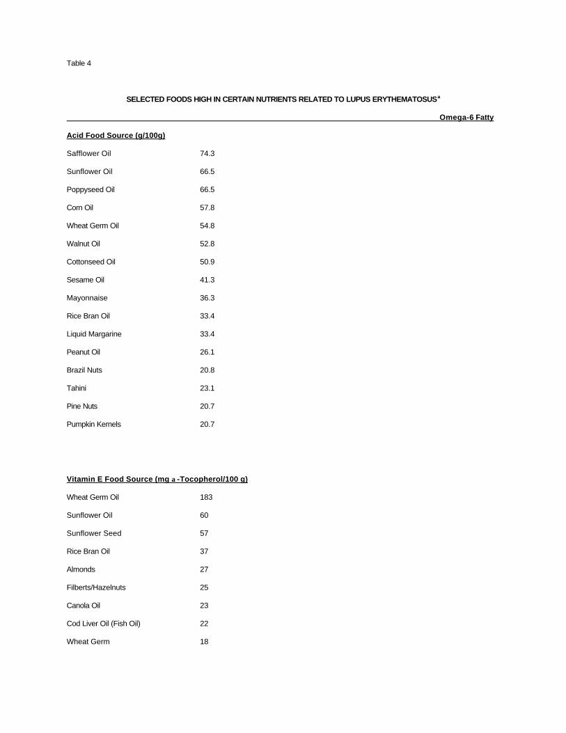
Table 4
SELECTED FOODS HIGH IN CERTAIN NUTRIENTS RELATED TO LUPUS ERYTHEMATOSUSa
Omega-6 Fatty
Acid Food Source (g/100g)
Safflower Oil 74.3
Sunflower Oil 66.5
Poppyseed Oil 66.5
Corn Oil 57.8
Wheat Germ Oil 54.8
Walnut Oil 52.8
Cottonseed Oil 50.9
Sesame Oil 41.3
Mayonnaise 36.3
Rice Bran Oil 33.4
Liquid Margarine 33.4
Peanut Oil 26.1
Brazil Nuts 20.8
Tahini 23.1
Pine Nuts 20.7
Pumpkin Kernels 20.7
Vitamin E Food Source (mg α-Tocopherol/100 g)
Wheat Germ Oil 183
Sunflower Oil 60
Sunflower Seed 57
Rice Bran Oil 37
Almonds 27
Filberts/Hazelnuts 25
Canola Oil 23
Cod Liver Oil (Fish Oil) 22
Wheat Germ 18

Beta-Carotene
Food Source (RE/100 g cooked, unless noted)
Carrot Juice (canned) 2575
Carrots (raw) 2454
Sweet Potato 2182
Shallots (raw) 1250
Mixed Vegetables (canned) 1164
Pumpkin 1082
Spinach 819
Kale 740
Apricot Halves (dried) 723
Collard Greens 598
Red Bell Pepper (raw) 570
Selenium
Food Source (ug/100 g)
Pike 190
Carp 159
Herring 141
Rainbow Trout 124
Wheat Germ 101
Crayfish/Crawdads 100
Anchovies 90
Scallops 82
Tuna (in water) 80
Sunflower Seeds 78
Lobster 77
Octopus 75
Oysters 72
Chicken Livers 71
Whole Wheat Flour 71
Rainbow Trout 71
Salmon 60
Liverwurst - Pork 58

Sardines 57
Pork Sirloin 52
Omega-3 Fatty
Acid Food Source (g/100 g)
OILS/NUTS*
Sardine Oil 22.2
Cod Liver Oil 18.8
Walnut Oil 10.4
Canola Oil 8.0
Wheat Germ Oil 6.9
Walnuts 6.8
Soybean Oil 6.8
Mayonnaise 4.7
FISH/SHELLFISH/SOYBEANS
Mackerel 1.9
Sablefish 1.9
Salmon (Chinook) 1.9
Whitefish 1.9
Herring 1.7
Bluefin Tuna 1.5
Soy Nuts/Soybeans 1.5
Atlantic Sardines in Oil 1.5
Oysters 1.4
Rainbow Trout 1.2
Swordfish 1.1
Sea Bass 1.0
Scallops 1.0*
Most fish oil capsules contain about 300 mg of omega-3 fatty acids; 180 mg EPA & 120 mg DHA, so 3 tablets/meal
yields 2.7 g a Nutrient analysis based on Food
Processor Plus (Version 6.0), ESHA Research, Salem, OR.
Table 5. Selected human studies on the benefit of fish oils for LE symptoms (clinically & serologically).
REFERENCE DOSAGE (per day) RESULTS

88 (2.3 g EPA/1.4 g DHA)a
8/17 treatment and 2/17 controls improved in the 1st 3 months. No significant difference after 6 months.
89 6 or 18 g fish oil (1.8 or 5.4 g EPA/DHA)
12 subjects - no significant improvement in immune complex, anti-DNA titer, or prostacyclin (PGI2)
90 15 g fish oil (4.4 g EPA/DHA)
21 subjects - no significant improvement in renal function or disease activity
a "Fish oil" dosages are often reported instead of the active components - EPA/DHA.