lung tumours - university of pretoria · lung tumours ¾small cell ... malignant lung tumours 1....
TRANSCRIPT

07/03/2007 1
Lung Tumours
Dr Emil Beltchev
07/03/2007 2
Lung CancerPrimary carcinoma of the lung was an uncommoncancer until the 1930s. At that time a dramaticincrease in the incidence of lung cancerbegan that has not yet abated. Although the overall incidence of lung cancer has dramatically
increased over the past 30 years, the relative incidence of squamous cell carcinoma has decreased, and adenocarcinoma has become the dominant cell type—a phenomenon that has been temporally associated with the changes in tobacco blends and the use of filters in cigarettes
Lung cancer is now the most common cause of cancer mortality in both males and females.

07/03/2007 3
Classification of the malignant lung tumours
Small cell carcinomaNon small cell carcinomasMesotheliomaMetastases from distant sites (Breast,GIT,Ovarian Ca,Melanoma)

07/03/2007 4
Non malignant causes of a mass in the lung
Radiographic artefact- eg nipple shadow Infection – aspergilloma (X-ray reveals dense round ball, capped by slim meniscus of air, in a cavity)
Connective tissue disease- Wegenersgranulomatosis.(necrotizing granulomatous angiitis involving the lungs. X-ray reveals diffuse or nodular infiltrates that may resemble malignant metastases.)

07/03/2007 5
Aetiology of malignant lung tumours
Smoking-80% lung cancer occur in active or former smokers.5% occur in passive smokers.Much less frequent causes of lung cancer are exposure to:Asbestos (associated with Mesothelioma)RadonPolycyclic aromatic hydrocarbonsNikelChromateInorganic arsenicals
07/03/2007 6
WHO pathological classification
A. Squamous Cell Ca 30%B. Small Cell Ca 20%C. Adenocarcinama 40%:
-Acinar-Papillary-Bronchoalveolar-Mucinous
D. Large Cell Ca E. Mixed

07/03/2007 7
For the purpose of management lung cancers are grouped as :
o Non-small cell lung cancer (NSCLC)
o Small cell lung cancer (SCLC)
07/03/2007 8
Clinical presentations of malignant lung tumours
1. Symptoms:Depend on the size, location and metastasesa. Coughb. Haemoptisisc. Dyspnead. Chest paine. Recurrent chest infectionsf. Dysphagiag. Hoarseness

07/03/2007 9
2.Signs-Local and paraneoplasticA. Local Signs:
Chest sign- nil or signs of collapse/consolidation/formation of abscessHorner’s syndrom (miosis, ptosis, enophthalmos, and anhydrosis)
Pancoast’s syndrom(lower brachial plexopathy, Horner’s syndrom,shoulder pain)
Superior vena cava syndrom (Swelling of the neck, face, and arms especially in the morning, headache, visual disturbance. Fixed engorgement of external and internal jugular veins, collateral veins over the chest wall)
Supraclavicular mass/Lymph node

07/03/2007 10
B. Paraneoplastic signs
ClubbingHypertrophic pulmonary osteoartropathy(bone and joint pain, periostal inflamation and elevation on the X-ray,affects the distal end of the long bones, elevated ALP, respond to Aspirin and NSAID)
Proximal myopathyPeripheral neuropathyEaton-Lambert myastenic syndrom (weakness affecting the limbs and sparing the ocular and bulbar muscle.Pt. Complain of weakness and pain in the proximal limbs, paresthesias,dry mouth, impotents and ptosis, deep tendon reflexes-redused)
IADH secretion

07/03/2007 11
Investigations
1. Chest X-ray (AP and lateral)2. Sputum Cytology3. Tissue biopsy :
Biopsy of massBronchoscopyCT-guided biopsyMediastinotomyThoracotomy

07/03/2007 12
It is essential to make a definitive diagnosis prior to the treatment.This might be difficult. Cytology
can be misleading.An exception is a life
threatening SVC syndrome –Radiation prior to the
diagnosis.

07/03/2007 13
Staging1. Different staging systems used for different
tumours.2. Tests required for adequate staging:
FBC/UKE/LFT/Ca- may reveal biochemical paraneoplastic phenomena, eg hyponatremia, hypercalcaemia, or suggest sites of metastases.CT chest and abdomen : size of the tumour, site, relationship to the chest wall, fissures, mediastinal structures, diaphragm, lymphnodes>1cm, involvement of the liver and adrenals.Bone scan- if symptoms suggestive of bone metastases (pain, Elevated ALP and Ca)CT of the brain – if CNS signsMUGA or heart sonar if suspicious of underlying heart disease and cardiotoxic chemotherapy planned.
07/03/2007 14
1. Non small cell lung cancer (NSCLC)
TNM Staging T- Size of the tumour, structures it invades,
closeness to carina. T1-4N- Nodes (ipsilateral, bilateral, position of
nodes) N1-3M- Presence of distant metastases. M0-1Stage 1-4

07/03/2007 15
Stage TNM Survival Rate (%)
Revised staging system
IA T1, N0, M0 >70
IB T2, N0, M0 60
IIA T1, N1, M0 50
IIB T2, N1, M0 30
T3, N0–N1, M0 40
IIIA T1–T3, N2, M0 10–30
IIIB Any T4, any N3, M0 <10
IV Any M1 <5

07/03/2007 16
2. Small cell lung cancer
Limited or Extensive stage
1. Limited- tumour confined to ipsilateralthorax and nodes and able to fit in one radiation field.
2. Extensive- disease which can not fit in one radiation field.

07/03/2007 17
3. Mesothelioma
Number of different systems including TNM and Buchart classification.

07/03/2007 18
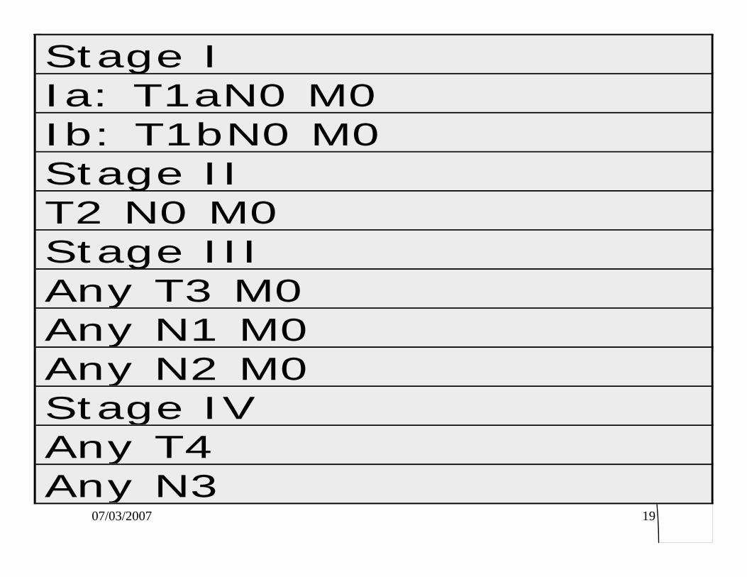
TNM staging system for MPM
T- reflects the invaded structures by the tumour. T1 limited to the pleura;T2 invades the diaphragmal muscle or pulmonary parenchima; T3 is defined as locally advanced and resectable,T4 is defined as locally advanced unresectable
N-Reflects the involved lymphnode N0-3M-Reflects the distant metastases. M0-1
07/03/2007 19
Stage IIa: T1aN0 M0Ib: T1bN0 M0Stage IIT2 N0 M0Stage IIIAny T3 M0Any N1 M0Any N2 M0Stage IVAny T4Any N3

07/03/2007 20
Performance status (PS) WHO performance status score:0-no symptoms1-Symptoms but normal activity2-Symptoms causing the pt. to lie in bed<50%
of the day3-Symptoms causing the pt. to lie in bed>50%
of the day4-The pt is bedridden

07/03/2007 21
PS is a very important concept in oncology.
The PS together with the stage of the disease helps the oncologist to decide whether the patient will
benefit from chemotherapyNSCLC and mesothelioma, PS>2 do
very poorly on chemotherapy

07/03/2007 22
Individual tumors in more details
1. SCLC2. NSCLC3. Mesothelioma

07/03/2007 23
1. Small cell lung cancer (SCLC)
It is one of the most aggressive, fast growing tumorsWithout treatment the median survival is 6-9 weeksRefer the patient as soon as you have the diagnosis

07/03/2007 24
ChemotherapyMainstay of treatment, because of the chemo-responsiveness of the SCLC and frequent dissemination at the time of diagnosisVarious regimens exist (Adriamycin-based, Cisplatin-based, Ifosfomide-based)Substantial activity has been shown by several newagents, including the taxanes (paclitaxel, docetaxel) and the topoisomerase I inhibitors (topotecan, irinotecan), gemcitabine, andvinorelbine.Response rates:75-90% for limited stage and 75% for extensive stage50% of limited stage have complete response (CR)25% CR for the extensive stage

07/03/2007 25
Cisplatin-based:
Cisplatin 80 mg/m day 1 and etoposide 80 mg/m days 1, 2, 3
Cisplatin 25 mg/m days 1, 2, 3 and etoposide 100 mg/m days 1, 2, 3
Doxorubicin-based
Cyclophosphamide 1000 mg/m day 1 and doxorubicin 45 mg/m day 1 and
vincristine 1.4 mg/m day 1
Ifosfamide-based
Ifosfamide 1200 mg/m days 1, 2, 3, 4 and etoposide 75 mg/m days 1, 2, 3, 4
and cisplatin 20 mg/m days 1, 2, 3, 4
Mesna required for ifosfamide.

07/03/2007 26
Despite the excellent response rates, cure is very unusualMedian survival for Limited stage is 14 months and for extensive stage 7 months

07/03/2007 27
Radiation60% of the relapses after chemotherapy are in the thorax.TI reduces the risk of relapse by 50% Has role in SVCS and spinal cord compressionHigh risk of brain metastases in SCLC(20% have brain involvement at diagnosis, 80% have brain involvement at death)- prophylactic cranial RT increases 3 year survival by 5% and is usually given if the patient is in CR post chemotherapy Palliative RT – short course of irradiatoin to either the primary tumor or site of metastases can provide useful symptom control.
07/03/2007 28
2. Non small cell lung cancer
AdenocarcinomaSquamous cell carcinomaLarge cell carcinoma

07/03/2007 29
More slow growing than SCLC.
Surgery and radiotherapy have more important role than in SCLC.

07/03/2007 30
Treatment of NSCLC
Surgical removal of NSCLC continues to offer best possibility of cure. Consequently, each patient should be considered where possible for surgical treatment, although advanced stage and significant co-morbidity will preclude this option in many patients.

07/03/2007 31
First group: stage1,2,3A. Surgically resectable
Surgery (lobectomy) is the treatment of choice-60-80% cureRadiotherapy if contraindication for surgery-20%cureAdjuvant chemotherapy may have a role in stage 1,2 and 3A providing survival advantage.
07/03/2007 32
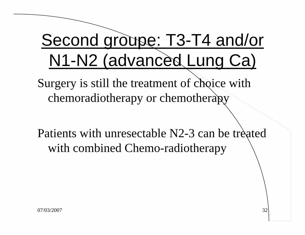
Second groupe: T3-T4 and/or N1-N2 (advanced Lung Ca)
Surgery is still the treatment of choice with chemoradiotherapy or chemotherapy
Patients with unresectable N2-3 can be treated with combined Chemo-radiotherapy

07/03/2007 33
Third group: Metastaticdisease (M1)
Radiation therapy or chemotherapy for palliation of symptoms
Patients previosly treated with platinum based chemotherapy may have survival benefit and symptoms control from Taxol and epidermal growth factor receptor inhibitor.

07/03/2007 34
Stage 4 NSCLC
Chemotherapy improves survival (10% 1year if untreated vs 30% if treated)Different regimens are used most platinum-basedUnfortunately median survival remains poor(10 months)Palliation is very important-adequate pain control, pleurodesis for recurrent pleural effusion, drugs for dispnoea.
07/03/2007 35
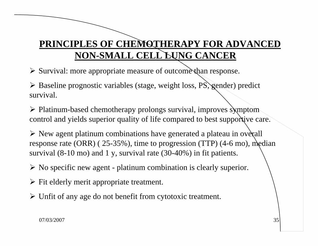
PRINCIPLES OF CHEMOTHERAPY FOR ADVANCED NON-SMALL CELL LUNG CANCER
Survival: more appropriate measure of outcome than response.
Baseline prognostic variables (stage, weight loss, PS, gender) predict survival.
Platinum-based chemotherapy prolongs survival, improves symptom control and yields superior quality of life compared to best supportive care.
New agent platinum combinations have generated a plateau in overall response rate (ORR) ( 25-35%), time to progression (TTP) (4-6 mo), median survival (8-10 mo) and 1 y, survival rate (30-40%) in fit patients.
No specific new agent - platinum combination is clearly superior.
Fit elderly merit appropriate treatment.
Unfit of any age do not benefit from cytotoxic treatment.

07/03/2007 36
3.Mesothelioma1. Malignant pleural mesothelioma (MPM) is an
aggressive tumour arising from the serosal lining of the chest and abdomen with survival rates less then 1 year reported following diagnosis
2. M/F ratio-5:13. High correlation with asbestos exposure4. Other causative agents include:
RadiationThorium dioxideSilicate fibersSimian virus 40(SV40),discovered as contaminant of early poliovorus vaccines.

07/03/2007 37
PathologyThree distinctive subtypes have been identified:1. Epithelial (50% of all cases)2. Sarcomatouse3. Mixed histologies
Distinguishing mesothelioma from other intrathoracic malignancies such as adenocarcinoma is important and requires assistance of experienced pathologist.
07/03/2007 38
Treatment1.Localized MPM
Surgery if technically possiblePalliative RT
2.Extensive MPMAim is to palliate symptomsRadiotherapy/PleurodesisSingle agent chemotherapy has 10-20% response rateNew agents show more promises Alimta, GemzarAlimta +Cisplatin improves survival and quality of life in comparison with cisplatin alone
07/03/2007 39
Prognosis for MPM remains very poor
Median survival - 6 to 12 months

07/03/2007 40
Take home message
Have a high index of suspicion in smoker with suggestive symptomsHigh correlation between MPM and asbestos exposure (>15 yrs. Prior)Do appropriate diagnostic tests quickly and try to get histological confirmation as soon as possible Refer the patient early- Chemotherapy and/or Radiotherapy improves survival and quality of life.

07/03/2007 41
Novel therapeutic approaches
Gene therapy ImmunotherapyVEGF& EGFR inhibitors PRESENTATION

07/03/2007 42