lung cancer - isdscotland.org · data definitions for the national minimum core dataset for lung...
TRANSCRIPT

Data Definitions for the National Minimum Core Dataset for Lung Cancer. Developed by ISD Scotland 2014
Lung Cancer
Data Definitions for the National Minimum Core Dataset to Support the Introduction of Lung Cancer Quality Performance Indicators
Definitions developed by ISD Scotland in collaboration with the Lung Quality Performance Indicator Development Group
Version 3.3: March 2019
To be used in conjunction with:
1. Lung Clinical Quality Performance Indicators v3.0 (February 2017) 2. Lung QPI Dataset Validations (latest published version) 3. Lung Measurability of Quality Performance Indicators (latest published version)

Data Definitions for the National Minimum Core Dataset for Lung Cancer. Developed by ISD Scotland 2014
DOCUMENT CONTROL SHEET Key Information
Title Lung Cancer – Data Definitions for the National Minimum Core Dataset to support the introduction of Lung QPIs
Date Published/Issued March 2019
Date Effective From 1
st January 2017 (TNM 8 changes effective from
1st
January 2018)
Version/Issue Number v3.3
Document Type Guidance
Document Status Final
Standard Audience NHS staff involved in implementing and recording Lung Quality Performance Indicators.
Cross References Lung Quality Performance Indicators Lung Measurability of Quality Performance Indicators
Author Information Services Division of NHS National Services Scotland
Revision History
Version Date Summary of Changes Name Changes Marked
V1.1 December
2013
Changes agreed out with review to support data
collection.
David Early, ISD
See page xii
V1.2 February
2014 Changes to version and
referencing. David Early,
ISD n/a
V2.0 March 2014
Changes agreed at 9 month review. Changes to be applied for patients diagnosed from 1
st
April 2014
David Early, ISD
See page x
V2.1 July 2014 Changes agreed outwith review to support data
collection
Charlotte Anthony
ISD See page x
V2.2 November
2014
Changes agreed outwith review to support data
collection. Jane Garrett See page x
V2.3 June 2015 Changes agreed at baseline
review Jane Garrett See page ix
V2.4 July 2015 Change agreed outwith review Karen Heatlie
See page ix
V2.5 April 2016 Change agreed outwith review Charlotte
Anthony ISD See page ix
V3.0 March 2017
Changes agreed at Formal Review
Charlotte Anthony ISD
See page vi
V3.1 November
2017 Changes agreed outwith
review Charlotte
Anthony ISD See page iv
V3.2 January
2018 Changes agreed outwith
review Hannah
Ebbins ISD See page iii
V3.3 March 2019
Changes agreed outwith review
Jane Garrett, ISD
See page iii

Data Definitions for the National Minimum Core Dataset for Lung Cancer. Developed by ISD Scotland 2014
CONTENTS
PREFACE .............................................................................................................................. i NOTES FOR IMPLEMENTATION OF CHANGES .................................................................ii CONVENTIONS .....................................................................................................................ii REVISIONS TO DATASET ................................................................................................... iii CRITERIA FOR INCLUSION OF PATIENTS IN AUDIT ...................................................... xiii DATABASE SPECIFICATION ............................................................................................. xiv Section 1: Demographic Items ........................................................................................... 1 Person Family Name (at Diagnosis) ...................................................................................... 2 Person Given Name .............................................................................................................. 3 Patient Postcode at Diagnosis .............................................................................................. 4 Date of Birth .......................................................................................................................... 5 Person Sex at Birth ............................................................................................................... 6 CHI Number .......................................................................................................................... 7 Section 2: Pre-treatment Imaging & Staging Investigations ............................................ 8 Date of CT Thorax ................................................................................................................ 9 Date of Bronchoscopy {Lung Cancer} ................................................................................. 10 Seen by Clinical Nurse Specialist {Lung Cancer/ Mesothelioma} ........................................ 11 Location of Diagnosis {Cancer} ........................................................................................... 12 Date of Diagnosis {Cancer} ................................................................................................. 13 Site of Origin of Primary Tumour {Cancer} .......................................................................... 14 Origin of Tumour ................................................................................................................. 15 Histological/Cytological Diagnosis {Lung Cancer} (Pre-Treatment) ..................................... 16 Date of Histological / Cytological Diagnosis {Cancer} .......................................................... 18 Epidermal Growth Factor Receptor (EGFR) Status ............................................................. 19 Oncogenic Anaplastic Lymphoma Kinase (ALK) Status ...................................................... 21 PD-L1 Status ...................................................................................................................... 22 Date of Integrated FDG-PET/CT (PET/CT) Scan (Pre-treatment) ....................................... 23 Mediastinal/Supraclavicular (SCF) Node Results at FDG-PET/CT (PET/CT) Scan ............. 24 Mediastinal/SCF Sampling Results (pre-treatment) ............................................................. 25 Synchronous Primary Tumours ........................................................................................... 26 TNM Tumour Classification (Clinical) {Lung Cancer} ........................................................... 27 TNM Nodal Classification (Clinical) {Lung Cancer} .............................................................. 30 TNM Metastases Classification (Clinical) {Lung Cancer} ..................................................... 31 TNM Tumour Classification (Clinical) {Pleural Mesothelioma} ............................................. 32 TNM Nodal Classification (Clinical) {Pleural Mesothelioma} ................................................ 34 TNM Metastases Classification (Clinical) {Pleural Mesothelioma} ....................................... 35 Brain Imaging ...................................................................................................................... 36 Date of Brain Imaging (Pre-treatment) ................................................................................ 37 WHO/ ECOG Performance Status ...................................................................................... 38 Date Discussed by Care Team (MDT) ................................................................................ 39 Type of First Cancer Treatment .......................................................................................... 40 Date of First Cancer Treatment ........................................................................................... 41 Date of Definitive Treatment {Lung Cancer} ........................................................................ 42 Section 3: Surgery ............................................................................................................ 43 Location Code {Cancer Surgery} ......................................................................................... 44 Definitive Surgery Performed {Lung Cancer} ....................................................................... 46 Date of Surgery ................................................................................................................... 47 Surgical Approach ............................................................................................................... 48 Histological/Cytological Diagnosis Following Surgery {Lung Cancer} .................................. 49 Section 4: Pathological Details ........................................................................................ 51 TNM Tumour Classification (Pathological) {Lung Cancer} ................................................... 52

Data Definitions for the National Minimum Core Dataset for Lung Cancer. Developed by ISD Scotland 2014
TNM Nodal Classification (Pathological) {Lung Cancer} ...................................................... 54 TNM Metastases Classification (Pathological) {Lung Cancer} ............................................. 55 TNM Tumour Classification (Pathological) {Pleural Mesothelioma} ..................................... 56 TNM Nodal Classification (Pathological) {Pleural Mesothelioma} ........................................ 58 TNM Metastases Classification (Pathological) {Pleural Mesothelioma} ............................... 59 N2 Lymph Node Stations .................................................................................................... 60 Section 5: Oncology ......................................................................................................... 61 Radiotherapy Course Type (1-3) ......................................................................................... 62 Stereotactic Ablative Radiotherapy (SABR) ........................................................................ 64 Site of Radiotherapy (Courses 1- 3) .................................................................................... 65 Radiotherapy Dose: Total Administered {Cancer} 1-3 ......................................................... 66 Radiotherapy Fractions: Total Administered {Cancer} 1-3 ................................................... 67 Date Treatment Started {Cancer} (Radiotherapy) 1-3 .......................................................... 68 Date Treatment Completed {Cancer} (Radiotherapy) 1-3 .................................................... 69 Type of Systemic Anti-Cancer Therapy (SACT) 1-3 ............................................................ 70 Systemic Therapy Agent 1-3 {Lung Cancer} ....................................................................... 71 Date Treatment Started Systemic Anti-Cancer Therapy (SACT) {Cancer} 1-3 .................... 73 Date Treatment Completed Systemic Anti-Cancer Therapy (SACT) {Cancer} 1-3 ............... 74 Section 6: Clinical Trials ................................................................................................... 75 Patient Entered into Clinical Trial ........................................................................................ 76 Section 7: Death Details ................................................................................................... 77 Date of Death ...................................................................................................................... 78

Data Definitions for the National Minimum Core Dataset for Lung Cancer. Developed by ISD Scotland 2013
i
PREFACE Following the publication of Better Cancer Care: An Action Plan in October 2008, the Scottish Government established the Scottish Cancer Taskforce to oversee its implementation. The NHS Scotland Healthcare Quality Strategy in 2010 expands on this by articulating quality ambitions. A quality measurement framework has been developed setting out measures and targets which will be used to monitor, challenge, manage and report progress. Part of this strategy is the development of quality performance indicators (QPIs) to drive quality improvement in cancer care throughout NHS Scotland. As high quality data are required to enable comparisons over time and between regions, it is important that national data definitions are used to facilitate consistent data collection. National data definitions already in use have been used as much as possible to allow electronic data capture, thereby minimising duplication of data collection. Where national data definitions do not already exist, definitions used in other systems have been incorporated. To ensure that findings are comparable across Scotland, the national dataset and data definitions in conjunction with the final quality performance indicators were agreed through public engagement and are now ready for implementation for patients diagnosed from 1st April 2013. Lung Cancer remains one of the major causes of death in Scotland. Treatment challenges arise from both co morbidity and extent of disease at time of diagnosis, with the majority of patients presenting with too advanced disease for curative intent. The particular challenge for all is often the balance of survival benefits over quality of life especially when cure is not an option. Multidisciplinary team working is important to enable that the best information is given to the patient and carers to help them make the correct decision for them. This involves being as accurate as possible about stage assessment, both to ensure that more aggressive radical treatment is offered to those that have most to gain and also to prevent interventions being performed where the patient with poor performance status is unlikely to benefit. The use of combination chemoradiotherapy is increasing for specific subgroups, with challenges about increasing morbidity, often in patient groups with already significant comorbidity from heart and lung disease and reduced performance status. Pathology is playing an increasing role in deciding on appropriate treatment option. This not only includes accurate staging of the mediastinum to determine appropriate radical treatment options but also in the subtyping of lung cancer, especially non-small cell lung cancer and for identifying specific gene mutations. It is our intention that the QPIs that we have produced reflect the emerging evidence of care required to help deliver appropriate treatment to patients diagnosed with lung cancer in Scotland. They have been developed as a collaborative approach involving multiple disciplines from all of the regions of Scotland to try and improve standards of care. Carrie Featherstone Consultant Clinical Oncologist

Data Definitions for the National Minimum Core Dataset for Lung Cancer. Developed by ISD Scotland 2013
ii
NOTES FOR IMPLEMENTATION OF CHANGES The following changes should be implemented for all patients who are diagnosed with Lung cancer on or after 1st January 2017, who are eligible for inclusion in the Lung cancer audit. Changes to definitions fall into the following categories:
to address problems with ongoing audit and standardise data definitions, where feasible, between different cancer sites
to address problems with existing definitions
to allow Quality Performance Indicators to be measured and reported against
If you have difficulties in using individual definitions within this document please contact General Enquiries on the collection of the National Minimum Core Dataset If you have any comments on the attached data definitions ISD would welcome your feedback. Please contact: [email protected]
CONVENTIONS The layout for each item is standard as shown below where it is applicable: Common Name(s): Main Source of Data Item Standard: Definition: Field Name: Field Type: Field Length: Notes for Users: Codes and Values: Related Data Item(s): Notes by Users: In addition the following two conventions have been used in the document:
{curly brackets} - definition relates to one specific named data set
'described elsewhere' - indicates there is a definition for the named item within this document

Data Definitions for the National Minimum Core Dataset for Lung Cancer. Developed by ISD Scotland 2013
iii
REVISIONS TO DATASET Revisions to Dataset Outwith Review (March 2019) Systemic Therapy Agent 1-3 {Lung Cancer} - Field Name amend ‘CHEMTYPE’ to ‘CHEMAGENT’ Histological/Cytological Diagnosis {Lung Cancer} (Pre-Treatment) - Notes for Users add ‘Pathologists often state 'favouring' a certain tumour sub-type and this should be documented as recorded rather than Not Otherwise Specified 'NOS’. Histological/Cytological Diagnosis Following Surgery {Lung Cancer} -Notes for Users add ‘Pathologists often state 'favouring' a certain tumour sub-type and this should be documented as recorded rather than Not Otherwise Specified 'NOS’. TNM Tumour Classification (Clinical) {Lung Cancer} -Implement this change from 1/1/2019 Notes for Users add ‘If the size of the tumour is not specified as pT2a or pT2b then it should be recorded as pT2a’; Codes and Values table remove T1, T2 TNM Tumour Classification (Pathological) {Lung Cancer} - Implement this change from 1/1/2019 Notes for Users add ‘If the size of the tumour is not specified as pT2a or pT2b then it should be recorded as pT2a’; Codes and Values table remove T1, T2 Revisions to Dataset Outwith Review (January 2018) TNM Tumour Classification (Clinical) {Lung Cancer} - Standard and definition changed from Seventh Edition, 2009 to Eighth Edition 2017 - Add code and value ‘T1mi - Minimally invasive adenocarcinoma’ - Amend code description T1a to ‘Tumour ≤ 1cm in greatest dimension.’ - Amend code description T1b to ‘Tumour > 1cm – ≤ 2cm in greatest dimension.’ - Add code and value ‘T1c - Tumour >2cm - ≤3cm in greatest dimension.’ - Amend code description T2 to ‘Tumour > 3cm to ≤ 5cm, or tumour with any of the following features: Involves main bronchus regardless of distance to the carina, but without involvement of carina, or Invades visceral pleura or associated with atelectasis or obstructive pneumonitis that extends to the hilar region either involving part of or the entire lung. - Amend code description T2a to ‘Tumour > 3cm – ≤ 4cm in greatest dimension.’ - Amend code description T2b to ‘Tumour > 4cm – ≤ 5cm in greatest dimension.’ - Amend code description T3 to ‘Tumour more than > 5cm - ≤ 7cm in greatest dimension or directly invades any of the following: parietal pleura, chest wall (including superior sulcus tumour), phrenic nerve, or parietal pericardium; or separate tumour nodule(s) in the same lobe as the primary.’ - Amend code description T4 to ‘Tumour > 7cm or of any size that invades any of the following: diaphragm, mediastinum, heart, great vessels, trachea, recurrent laryngeal nerve, oesophagus, vertebral body, carina; or separate tumour nodule(s) in a different ipsilateral lobe to that of the primary’. TNM Nodal Classification (Clinical) {Lung Cancer} - Standard and definition changed from Seventh Edition, 2009 to Eighth Edition 2017 TNM Metastases Classification (Clinical) {Lung Cancer} - Standard and definition changed from Seventh Edition, 2009 to Eighth Edition 2017 - Codes and Values table add code and value ‘M1 – Distant Metastasis’

Data Definitions for the National Minimum Core Dataset for Lung Cancer. Developed by ISD Scotland 2013
iv
- Amend code description M1a to include ‘or pericardial’, explanatory note changed from ‘the effusion should be excluded as a staging element and the patient should be classified as M0’ to ‘the effusion should be excluded as a staging descriptor.’ - Amend code description M1b to ‘Single extrathoracic metastasis in a single organ’ - Add code and value ‘M1c - Multiple extrathoracic metastasis in a single or multiple organs’ TNM Tumour Classification (Clinical) {Pleural Mesothelioma} - Standard and definition changed from Seventh Edition, 2009 to Eighth Edition 2017, Codes and Values table amend code description T1 to include ‘mediastinal or diaphragmatic pleura’. Delete codes and values T1a and T1b Amend code description T2 to ‘the ipsilateral pleural (parietal or visceral pleura), with at least one of the following:’ Amend code description T3 to ‘ipsilateral pleural (parietal or visceral pleura), with at least one of the following:’ Amend code description T4 to ‘(parietal or visceral pleura), with at least one of the following:
chest wall, with or without associated rib destruction (diffuse or multifocal)
peritoneum (via direct transdiaphragmatic extension)
contralateral pleura
mediastinal organs (oesophagus, trachea, heart, great vessels)
vertebra, neuroforamen, spinal cord
internal surface of the pericardium (transmural invasion with or without a pericardial effusion)
TNM Nodal Classification (Clinical) {Pleural Mesothelioma} - Standard and definition changed from Seventh Edition, 2009 to Eighth Edition 2017 Codes and Values table amend code description N1 to ‘Metastasis to ipsilateral intrathoracic lymph nodes (includes ipsilateral bronchopulmonary, hilar, subcarinal, paratracheal, aortopulmonary, paraesophageal, peridiaphragmatic, pericardial fat pad, intercostal and internal mammary nodes)’. Amend code description N2 to ‘Metastasis to contralateral intrathoracic lymph nodes. Metastases to ipsilateral or contralateral supraclavicular lymph nodes’ Delete code and value N3 TNM Metastases Classification (Clinical) {Pleural Mesothelioma} - Standard and definition changed from Seventh Edition, 2009 to Eighth Edition 2017 TNM Tumour Classification (Pathological) {Lung Cancer} - Standard changed from Seventh Edition, 2009 to Eighth Edition 2017, Codes and Values table add code and value ‘pT1mi - Minimally invasive adenocarcinoma’ Amend code description pT1a to ‘Tumour ≤ 1cm in greatest dimension.’ Amend code description pT1b to ‘Tumour > 1cm – ≤ 2cm in greatest dimension.’ Add code and value ‘pT1c - Tumour >2cm - ≤3cm in greatest dimension.’ Amend code description pT2 to ‘Tumour > 3cm to ≤ 5cm, or tumour with any of the following features: Involves main bronchus regardless of distance to the carina, but without involvement of carina, or Invades visceral pleura or associated with atelectasis or obstructive pneumonitis that extends to the hilar region either involving part of or the entire lung. Amend code description pT2a to ‘Tumour > 3cm – ≤ 4cm in greatest dimension.’ Amend code description pT2b to ‘Tumour > 4cm – ≤ 5cm in greatest dimension.’ Amend code description pT3 to ‘Tumour more than > 5cm - ≤ 7cm in greatest dimension or directly invades any of the following: parietal pleura, chest wall
Data Definitions for the National Minimum Core Dataset for Lung Cancer. Developed by ISD Scotland 2013
v
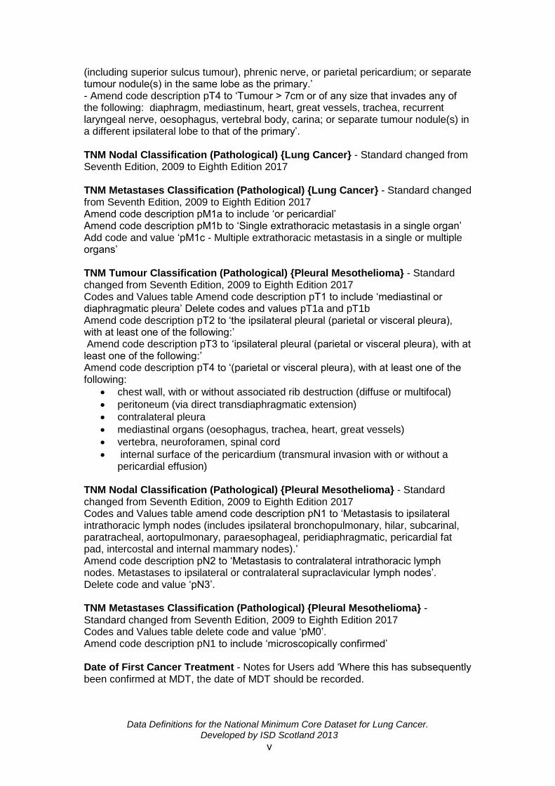
(including superior sulcus tumour), phrenic nerve, or parietal pericardium; or separate tumour nodule(s) in the same lobe as the primary.’ - Amend code description pT4 to ‘Tumour > 7cm or of any size that invades any of the following: diaphragm, mediastinum, heart, great vessels, trachea, recurrent laryngeal nerve, oesophagus, vertebral body, carina; or separate tumour nodule(s) in a different ipsilateral lobe to that of the primary’. TNM Nodal Classification (Pathological) {Lung Cancer} - Standard changed from Seventh Edition, 2009 to Eighth Edition 2017 TNM Metastases Classification (Pathological) {Lung Cancer} - Standard changed from Seventh Edition, 2009 to Eighth Edition 2017 Amend code description pM1a to include ‘or pericardial’ Amend code description pM1b to ‘Single extrathoracic metastasis in a single organ’ Add code and value ‘pM1c - Multiple extrathoracic metastasis in a single or multiple organs’ TNM Tumour Classification (Pathological) {Pleural Mesothelioma} - Standard changed from Seventh Edition, 2009 to Eighth Edition 2017 Codes and Values table Amend code description pT1 to include ‘mediastinal or diaphragmatic pleura’ Delete codes and values pT1a and pT1b Amend code description pT2 to ‘the ipsilateral pleural (parietal or visceral pleura), with at least one of the following:’ Amend code description pT3 to ‘ipsilateral pleural (parietal or visceral pleura), with at least one of the following:’ Amend code description pT4 to ‘(parietal or visceral pleura), with at least one of the following:
chest wall, with or without associated rib destruction (diffuse or multifocal)
peritoneum (via direct transdiaphragmatic extension)
contralateral pleura
mediastinal organs (oesophagus, trachea, heart, great vessels)
vertebra, neuroforamen, spinal cord
internal surface of the pericardium (transmural invasion with or without a pericardial effusion)
TNM Nodal Classification (Pathological) {Pleural Mesothelioma} - Standard changed from Seventh Edition, 2009 to Eighth Edition 2017 Codes and Values table amend code description pN1 to ‘Metastasis to ipsilateral intrathoracic lymph nodes (includes ipsilateral bronchopulmonary, hilar, subcarinal, paratracheal, aortopulmonary, paraesophageal, peridiaphragmatic, pericardial fat pad, intercostal and internal mammary nodes).’ Amend code description pN2 to ‘Metastasis to contralateral intrathoracic lymph nodes. Metastases to ipsilateral or contralateral supraclavicular lymph nodes’. Delete code and value ‘pN3’. TNM Metastases Classification (Pathological) {Pleural Mesothelioma} - Standard changed from Seventh Edition, 2009 to Eighth Edition 2017 Codes and Values table delete code and value ‘pM0’. Amend code description pN1 to include ‘microscopically confirmed’ Date of First Cancer Treatment - Notes for Users add ‘Where this has subsequently been confirmed at MDT, the date of MDT should be recorded.
Data Definitions for the National Minimum Core Dataset for Lung Cancer. Developed by ISD Scotland 2013
vi
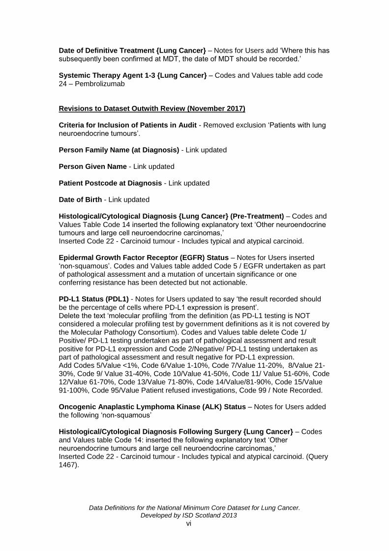
Date of Definitive Treatment {Lung Cancer} – Notes for Users add ‘Where this has subsequently been confirmed at MDT, the date of MDT should be recorded.’ Systemic Therapy Agent 1-3 {Lung Cancer} – Codes and Values table add code 24 – Pembrolizumab Revisions to Dataset Outwith Review (November 2017) Criteria for Inclusion of Patients in Audit - Removed exclusion ‘Patients with lung neuroendocrine tumours’. Person Family Name (at Diagnosis) - Link updated Person Given Name - Link updated Patient Postcode at Diagnosis - Link updated Date of Birth - Link updated Histological/Cytological Diagnosis {Lung Cancer} (Pre-Treatment) – Codes and Values Table Code 14 inserted the following explanatory text ‘Other neuroendocrine tumours and large cell neuroendocrine carcinomas,’ Inserted Code 22 - Carcinoid tumour - Includes typical and atypical carcinoid. Epidermal Growth Factor Receptor (EGFR) Status – Notes for Users inserted ‘non-squamous’. Codes and Values table added Code 5 / EGFR undertaken as part of pathological assessment and a mutation of uncertain significance or one conferring resistance has been detected but not actionable. PD-L1 Status (PDL1) - Notes for Users updated to say ‘the result recorded should be the percentage of cells where PD-L1 expression is present’. Delete the text ‘molecular profiling ‘from the definition (as PD-L1 testing is NOT considered a molecular profiling test by government definitions as it is not covered by the Molecular Pathology Consortium). Codes and Values table delete Code 1/ Positive/ PD-L1 testing undertaken as part of pathological assessment and result positive for PD-L1 expression and Code 2/Negative/ PD-L1 testing undertaken as part of pathological assessment and result negative for PD-L1 expression. Add Codes 5/Value <1%, Code 6/Value 1-10%, Code 7/Value 11-20%, 8/Value 21-30%, Code 9/ Value 31-40%, Code 10/Value 41-50%, Code 11/ Value 51-60%, Code 12/Value 61-70%, Code 13/Value 71-80%, Code 14/Value/81-90%, Code 15/Value 91-100%, Code 95/Value Patient refused investigations, Code 99 / Note Recorded. Oncogenic Anaplastic Lymphoma Kinase (ALK) Status – Notes for Users added the following ‘non-squamous’ Histological/Cytological Diagnosis Following Surgery {Lung Cancer} – Codes and Values table Code 14: inserted the following explanatory text ‘Other neuroendocrine tumours and large cell neuroendocrine carcinomas,’ Inserted Code 22 - Carcinoid tumour - Includes typical and atypical carcinoid. (Query 1467).

Data Definitions for the National Minimum Core Dataset for Lung Cancer. Developed by ISD Scotland 2013
vii
Revisions to Dataset following Formal Review (March 2017) The following addition has been made to facilitate the recording of data. This change will take effect for patients diagnosed from 1st January 2017. Criteria for inclusion of Patients in Audit – Exclude added bullet point ‘Patients with lung neuroendocrine tumours’. Origin of Tumour - Notes for Users add Required for QPI(s) 1-16’. Histological/Cytological Diagnosis {Lung Cancer} (Pre-Treatment) - Notes for Users Required for QPI(s)’14, 15’. Delete ’13’. Codes and Values table delete Code 14 ‘Other neuroendocrine tumours and large cell neuroendocrine carcinomas,’ from the explanatory text. Code 22: delete code, description and explanatory note. Date of Histological / Cytological Diagnosis {Cancer} – Add New Data Item. Epidermal Growth Factor Receptor (EGFR) Status – Definition add text ‘molecular profiling’. Delete text ‘predictive marker’. Notes for Users: replace ‘, specific tests e.g. EGFR, and’ with ‘. Specific tests e.g. EGFR, are’. Delete text ‘adenocarcinoma’. Oncogenic Anaplastic Lymphoma Kinase (ALK) Status – Add new Data Item PD-L1 Status (PDL1) – Add New Data Item Date of Integrated FDG-PET/CT (PET/CT) Scan (Pre-treatment) - Notes for Users remove QPI 5. Mediastinal/SCF Sampling Results (pre-treatment) - Notes for Users add ‘is’ to first sentence. TNM Tumour Classification (Clinical) {Lung Cancer} - Notes for Users Required for QPI(s) delete ‘4,8,12’ and add ‘9,10,11,14’. TNM Nodal Classification (Clinical) {Lung Cancer} - Notes for Users: Required for QPI(s) delete ‘4,8,12’ and add ‘6,10,11,14,16’. TNM Metastases Classification (Clinical) {Lung Cancer} - Notes for Users: Required for QPI(s) delete ‘4,12’ and add ‘5, 6,10,11,14’. Codes and Values Table delete ‘code M1’ and value ‘distant metastases’. TNM Metastases Classification (Clinical) {Pleural Mesothelioma} - Definition delete ‘sixth’ edition and replace with ‘seventh’. Brain Imaging – Add New Data Item. Date of Brain Imaging (Pre-treatment) – Add New Data Item. WHO/ ECOG Performance Status - Notes for Users Required for QPI ‘2’. Type of First Cancer Treatment - Notes for Users Required for QPI(s) delete ‘2’ and add ‘1, 10, 11, 12 & 14’. Codes and Values Table code 2, explanatory note – add text ‘SABR’. Date of First Cancer Treatment - Notes for Users add QPI(s) 4 & 15. Also used for the’.

Data Definitions for the National Minimum Core Dataset for Lung Cancer. Developed by ISD Scotland 2013
viii
Date of Definitive Treatment {Lung Cancer} - Notes for Users Delete QPI 3 and add 1. Location Code {Cancer Surgery} - Notes for Users delete ‘QPI 10’ and add ‘analysis purposes’. Definitive Surgery Performed {Lung Cancer} - Notes for Users Required for QPI(s) ‘2,14,15,16. Date of Surgery - Notes for Users delete QPI ‘6’. Histological/Cytological Diagnosis Following Surgery {Lung Cancer} - Notes for Users delete QPI(s) ‘8,9,10,11’. Codes and Values Table - Code 14, Explanatory note – delete text ‘Other neuroendocrine tumours and large cell neuroendocrine carcinomas’. Delete entry for code 22. TNM Tumour Classification (Pathological) {Lung Cancer} - Notes for Users remove QPI ‘2’. TNM Nodal Classification (Pathological) {Lung Cancer} - Notes for Users remove QPI ‘2’ TNM Metastases Classification (Pathological) {Lung Cancer} - Notes for Users remove QPI ‘2’ Radiotherapy Course Type (1-3) - Notes for Users add QPI ‘6,15,16’ and delete ‘11’. Notes for Users add ‘Actual treatment should be recorded. Where patients receive radiotherapy with radical intent (either radical radiotherapy or chemoradiotherapy) and treatment is abandoned prior to receiving full dosage, this should be recorded as palliative’. Delete ‘If patients receiving radiotherapy with radical intent (either radical radiotherapy or chemoradiotherapy) abandon treatment before full dosage is received, this should still be recorded as radical radiotherapy/ chemoradiotherapy’ (unpublished change). Codes and Values Table code 2 Explanatory note, add text ‘Includes SABR. Stereotactic Ablative Radiotherapy (SABR) - Add New Data Item Radiotherapy Dose: Total Administered {Cancer} 1-3 - Notes for Users remove QPI(s) ‘8,10’. Date Treatment Started {Cancer} (Radiotherapy) 1-3 - Notes for Users Remove QPI(s) ‘4, 11, 12’. Type of Systemic Anti-Cancer Therapy (SACT) 1-3 - Notes for Users remove QPI(s) ‘8,9’ and add ’15, 16’. Notes for Users, add ‘Actual treatment should be recorded. Where patients receive chemoradiotherapy and treatment is abandoned prior to receiving full dosage, this should be recorded as palliative’. Systemic Therapy Agent 1-3 {Lung Cancer} - Notes for Users remove QPI ‘11’. Codes and Values Table delete entry for codes ‘9, 13, 14, 98’ and add codes and values ‘20 Crizotinib, 21 Afatinib, 22 Other biological agent, 23 Other chemotherapy agent’.

Data Definitions for the National Minimum Core Dataset for Lung Cancer. Developed by ISD Scotland 2013
ix
Date Treatment Started Systemic Anti-Cancer Therapy (SACT) {Cancer} 1-3 - Notes for Users remove QPI ‘5’ Revisions to Dataset Outwith Review (April 2016) The following addition has been made to facilitate the recording of data. This change will take effect for patients diagnosed from 1st April 2015. Date of Diagnosis – Notes for Users change CT scan to chest imaging Revisions to Dataset Outwith Review (July 2015) Date of Diagnosis – Notes for Users add ‘The date recorded is the date on which the suspicion of cancer was first raised by the earliest relevant investigation (where the diagnosis was subsequently confirmed), i.e. the investigation which led to the decision to treat’ (effective from 1st April 2015)’ and remove ‘The date recorded is the date of the first investigative procedure that confirms a diagnosis of lung cancer whether done radiologically or histologically’.
Revisions to Dataset following Baseline Review (June 2015)
Dataset Location of Diagnosis {Cancer} – Codes and Values Table removed X1010=Not applicable Epidermal Growth Factor Receptor (EGFR) Status - Notes for Users amended to adenocarcinoma instead of non-squamous Mediastinal/SCF Sampling Results (pre-treatment) – Notes for users add ‘video-assisted thoracoscopic surgery (VATs) sampling’ and for Methods of sampling for mediastinal nodes Total Number of Hilar Lymph Nodes Examined Microscopically – Remove Data Item Total Number of Mediastinal Lymph Nodes Examined Microscopically – Remove Data Item N2 Lymph Node Stations – Add New Data Item Histological/Cytological Diagnosis {Lung Cancer} (Pre-Treatment) – Codes and Values table move explanatory note ‘Includes large cell carcinoma and undifferentiated, pleomorphic, sarcomatoid or anaplastic carcinoma’ from ‘code 13’ to code ‘14’ Histological/Cytological Diagnosis Following Surgery {Lung Cancer} – Codes and Values table move explanatory note ‘Includes large cell carcinoma and undifferentiated, pleomorphic, sarcomatoid or anaplastic carcinoma’ from ‘code 13’ to code ‘14’

Data Definitions for the National Minimum Core Dataset for Lung Cancer. Developed by ISD Scotland 2013
x
Database Specification N2 Lymph Node Stations – Add new Data Item Total Number of Hilar Lymph Nodes Examined Microscopically – Remove Data Item Total Number of Mediastinal Lymph Nodes Examined Microscopically – Remove Data Item. Revisions to Dataset Outwith Review (November 2014) Date of Diagnosis {Cancer} - Notes for Users add ‘this may be the date of the CT scan where suspicion of lung cancer was raised and subsequently confirmed’ Therapy Agent 1-3 {Lung Cancer} - Codes and Values table remove terminology 'palliative and neoadjuvant' Revisions to Dataset Outwith Review (June 2014) Database Specification
Origin of Tumour - Field Length corrected from 2 to 1
Date of Definitive Treatment {Lung Cancer} - Add new Data Item Field Name DEFTREATDATE, Field Type: Date, Field Length 10. Dataset
Origin of Tumour - Field Length changed to 1
Date of Definitive Treatment {Lung Cancer} - Add New Data Item Revisions to Dataset Following 9 Month Review (March 2014) Database Specification - Corrected missing fields and page references updated. Dataset
Seen by Clinical Nurse Specialist {Lung Cancer/Mesothelioma} - Notes for Users add: “In some settings, the clinical nurse specialist seen by the patient may be a palliative care nurse. This should be coded as ‘1: Yes’.”
Epidermal Growth Factor Receptor (EGFR) Status – Codes and Values Table added ‘4: Inconclusive’ to table of codes and values.
Mediastinal/SCF Sampling Results (Pre-treatment) - Notes for Users added “Where there is definitive evidence of distant metastases, or where there are no nodes to sample, record as ’96: Not applicable. Codes and Values Table added ‘3: Inconclusive’.

Data Definitions for the National Minimum Core Dataset for Lung Cancer. Developed by ISD Scotland 2013
xi
TNM Tumour Classification (Clinical) {Lung Cancer} – Notes for Users amended “This is a pre/non-operative classification as defined prior to first treatment and documented in patient notes” and “In cases where there are multiple or synchronous tumours, the tumour with the poorest prognosis should be recorded.” Codes and Values Table removed ‘Tis: carcinoma in situ’ as these are excluded from the dataset.
TNM Nodal Classification (Clinical) {Lung Cancer} - Notes for users amended “This is a pre/non-operative classification as defined prior to first treatment and documented in patient notes and “In cases where there are multiple or synchronous tumours, the tumour with the poorest prognosis should be recorded.”
TNM Metastases Classification (Clinical) {Lung Cancer} – Notes for Users amended: “This is a pre/non-operative classification as defined prior to first treatment and documented in patient notes” and “In cases where there are multiple or synchronous tumours, the tumour with the poorest prognosis should be recorded.”
TNM Tumour Classification (Clinical) {Pleural Mesothelioma} – Notes for Users amended “This is a pre/non-operative classification as defined prior to first treatment and documented in patient notes” and “In cases where there are multiple or synchronous tumours, the tumour with the poorest prognosis should be recorded.”
TNM Nodal Classification (Clinical) {Pleural Mesothelioma} – Notes for Users amended “This is a pre/non-operative classification as defined prior to first treatment and documented in patient notes” and “In cases where there are multiple or synchronous tumours, the tumour with the poorest prognosis should be recorded.”
TNM Metastases Classification (Clinical) {Pleural Mesothelioma} – Notes for Users “This is a pre/non-operative classification as defined prior to first treatment and documented in patient notes” and “In cases where there are multiple or synchronous tumours, the tumour with the poorest prognosis should be recorded.”
Location Code {Cancer Surgery} – Notes for Users amended ‘Each location has a location code, which is maintained jointly by ISD: http://www.isdscotland.org/Products-and-Services/Data-Definitions-and-References/National-Reference-Files/index.asp’ and ‘Location must be viewed as an address and not a code. If any new locations arise where NHS healthcare is delivered/administered, please ensure that the Reference Files Team at ISD is informed using form LOC-NEW (which can be downloaded from the website below) so that a new code may be issued as appropriate. http://www.isdscotland.org/Products-and-Services/Data-Definitions-and-References/National-Reference-Files/SMR-Reference-Files/’
Date of Surgery – Notes for Users amended notes for users: removed ‘All treatments given as part of the initial treatment plan.’ Added: ‘Patients treated within 6 months of a patient initially refusing investigation or whose initial treatment is ‘Watch and Wait’ can also be recorded.’
TNM Tumour Classification (Pathological) {Lung Cancer} - Codes and Values Table removed ‘Tis: carcinoma in situ’ as these are excluded from the dataset.
Radiotherapy Course Type (1-3) – Notes for Users amended “Patients treated within 6 months of initially refusing investigation or whose initial treatment is ‘Watch and Wait’ can also be recorded.”
Data Definitions for the National Minimum Core Dataset for Lung Cancer. Developed by ISD Scotland 2013
xii
Date Treatment Completed {Cancer} (Radiotherapy) 1-3 – Filed Name amended from ‘RCOMPDATE11’ to ‘RCOMPDATE1’.
Revisions to Dataset Outwith Review (December 2013)
Date Treatment Completed Systemic Anti-Cancer Therapy (SACT) {Cancer) 1-3 - Notes for Users from ‘This is the last dose of the last cycle of course of chemotherapy, or biological therapy’ to ‘This is the first day of the last cycle of a course of SACT’.

Data Definitions for the National Minimum Core Dataset for Lung Cancer. Developed by ISD Scotland 2013
xiii
CRITERIA FOR INCLUSION OF PATIENTS IN AUDIT To facilitate national comparisons the same patients must be audited throughout Scotland. The following eligibility criteria have been documented for this purpose. Include:
All patients with a confirmed new primary cancer of the bronchus, lung or trachea or mesothelioma of the pleura, peritoneum, pericardium and other sites (see page 18 Site of Tumour, for ICD-10 codes that are included.
Including all patients who have:
Had a previous primary malignancy of any site or a concurrent primary malignancy of another site.
Exclude:
Patients with metastatic lung disease from another primary cancer site
Patients where the origin of the primary is uncertain
Patients with tumour type sarcoma or lymphoma
Patients with recurrent disease (as opposed to a new primary)
Patients with carcinoma in situ
Patients, at date of diagnosis, under 16 years of age i.e. up to 15 years 364 days.
Patients where the only record of their cancer is from a death certificate (DCO).
Patients with normal residence outwith Scotland.
Patients whose definitive cancer treatment was privately funded or undertaken outwith NHS Scotland.
NB:
Only treatments as part of the initial treatment plan should be recorded.
Patients treated within 6 months of a patient initially refusing further investigation or whose initial treatment is ‘Watch and Wait’ can also be recorded.

Data Definitions for the National Minimum Core Dataset for Lung Cancer. Developed by ISD Scotland 2013
xiv
DATABASE SPECIFICATION DOWNLOAD FORMAT
To assist with downloading data to ISD for the National Quality Assurance Programme and other agreed activities, all sites should be able export data according to the following specification.
Data Item Field Name Field Type Size Page
Section 1: Demographic Items 1
Person Family Name (at Diagnosis)
PATSNAME Characters 35 2
Person Given Name PATFNAME Characters 35 3
Patient Postcode at Diagnosis PATPCODE Characters 8 4
Date of Birth DOB Date 10 5
Person Sex at Birth SEX Integer 2 6
CHI Number CHINUM Integer 10 7
Section 2:Pre-treatment Imaging & Staging Investigations 8
Date of CT Thorax CTHORAXDATE Date (DD/MM/CCYY)
10 9
Date of Bronchoscopy {Lung Cancer}
BDATE Date (DD/MM/CCYY)
10 10
Seen by Clinical Nurse Specialist {Lung Cancer/ Mesothelioma}
CNS Integer 2 11
Location of Diagnosis {Cancer} HOSP Characters 5 12
Date of Diagnosis {Cancer} DIAGDATE Date (DD/MM/CCYY)
10 13
Site of Origin of Primary Tumour {Cancer}
SITE Characters 4 14
Origin of Tumour ORIGIN Integer 1 15
Histological/Cytological Diagnosis {Lung Cancer} (Pre-Treatment)
HIST Integer 2 16
Date of Histological/Cytological Diagnosis {Cancer}
HISTDATE Date (DD/MM/CCYY)
10 18
Epidermal Growth Factor Receptor (EGFR) Status
EGFR Integer 2 19
Oncogenic Anaplastic Lymphoma Kinase (ALK) Status
ALK Integer 2 20
PD-L1 Status PDL1 Integer 2 21
Date of Integrated FDG-PET/CT (PET/CT) Scan (Pre-treatment)
PETDATE Date (DD/MM/CCYY)
10 22
Mediastinal/Supraclavicular (SCF) Node Results at FDG-PET/CT (PET/CT) Scan
MEDPET Integer 2 23
Mediastinal/SCF Sampling Results (pre-treatment)
MEDSAMP Integer 2 24

Data Definitions for the National Minimum Core Dataset for Lung Cancer. Developed by ISD Scotland 2013
xv
Synchronous Primary Tumours MULTIPLE Characters
2 25
TNM Tumour Classification (Clinical) {Lung Cancer}
TLUNG Characters 3 26
TNM Nodal Classification (Clinical) {Lung}
NLUNG Characters 2 28
TNM Metastases Classification (Clinical) {Lung Cancer}
MLUNG Characters 3 29
TNM Tumour Classification (Clinical) {Pleural Mesothelioma}
TMESO Characters 3 30
TNM Nodal Classification (Clinical) {Pleural Mesothelioma}
NMESO Characters 2 32
TNM Metastases Classification (Clinical) {Pleural Mesothelioma}
MMESO Characters 2 33
Brain Imaging BRAIN Characters 2 34
Date of Brain Imaging (Pre-treatment)
BRAINDATE Date (DD/MM/CCYY)
10 35
WHO/ECOG Performance Status
PSTATUS Integer 1 36
Date Discussed by Care Team (MDT)
MDTDATE Date (DD/MM/CCYY)
10 37
Type of First Cancer Treatment MODE1 Integer 2 38
Date of First Cancer Treatment FIRSTTREATDATE Date (DD/MM/CCYY)
10 39
Date of Definitive Treatment {Lung Cancer}
DEFTREATDATE Date (DD/MM/CCYY)
10 40
Section 3: Surgery 41
Location Code {Cancer Surgery} HOSPSURG Characters 5 42
Definitive Surgery Performed {Lung Cancer}
SURGTYPE Integer 2 44
Date of Surgery DSURG Date (DD/MM/CCYY)
10 45
Surgical Approach APPROACH Integer 2 46
Histological/Cytological Diagnosis Following Surgery {Lung Cancer}
HISTSURG Integer 2 47
Section 4: Pathological Details 49
TNM Tumour Classification (Pathological) {Lung Cancer}
PTLUNG Characters 3 50
TNM Nodal Classification (Pathological) {Lung Cancer}
PNLUNG Characters 2 52
TNM Metastases Classification (Pathological) {Lung Cancer}
PMLUNG Characters 3 53
TNM Tumour Classification (Pathological) {Pleural Mesothelioma}
PTMESO Characters 3 54

Data Definitions for the National Minimum Core Dataset for Lung Cancer. Developed by ISD Scotland 2013
xvi
TNM Nodal Classification (Pathological) {Pleural Mesothelioma}
PNMESO Characters 2 56
TNM Metastases Classification (Pathological) {Pleural Mesothelioma}
PMMESO Characters 2 57
N2 Lymph Node Stations N2NODES Integer 2 58
Section 5: Oncology 59
Radiotherapy Course Type (1-3 RADIOTYPE1 Integer 2 60
Radiotherapy Course Type (1-3 RADIOTYPE2 Integer 2 60
Radiotherapy Course Type (1-3 RADIOTYPE3 Integer 2 60
Stereotactic Ablative Radiotherapy (SABR)
SABR Integer 2 62
Site of Radiotherapy (Courses 1-3) RADIOSITE1 Integer 2 63
Site of Radiotherapy (Courses 1-3) RADIOSITE2 Integer 2 63
Site of Radiotherapy (Courses 1-3) RADIOSITE3 Integer 2 63
Radiotherapy Dose: Total Administered {Cancer} 1-3
TOTDOSE1 Float nnn.nnn 7 64
Radiotherapy Dose: Total Administered {Cancer} 1-3
TOTDOSE2 Float nnn.nnn 7 64
Radiotherapy Dose: Total Administered {Cancer} 1-3
TOTDOSE3 Float nnn.nnn 7 64
Radiotherapy Fractions: Total Administered {Cancer} 1-3
FRACTIONS1 Float 4 65
Radiotherapy Fractions: Total Administered {Cancer} 1-3
FRACTIONS2 Integer 3 65
Radiotherapy Fractions: Total Administered {Cancer} 1-3
FRACTIONS3 Integer 3 65
Date Treatment Started {Cancer} (Radiotherapy) 1-3
RSRTDATE1 Date (DD/MM/CCYY)
10 66
Date Treatment Started {Cancer} (Radiotherapy) 1-3
RSRTDATE2 Date (DD/MM/CCYY)
10 66
Date Treatment Started {Cancer} (Radiotherapy) 1-3
RSRTDATE3 Date (DD/MM/CCYY)
10 66
Date Treatment Completed {Cancer} (Radiotherapy) 1-3
RCOMPDATE1 Date (DD/MM/CCYY)
10 67
Date Treatment Completed {Cancer} (Radiotherapy) 1-3
RCOMPDATE2 Date (DD/MM/CCYY)
10 67
Date Treatment Completed {Cancer} (Radiotherapy) 1-3
RCOMPDATE3 Date (DD/MM/CCYY)
10 67
Type of Systemic Anti-Cancer Therapy (SACT) 1-3
CHEMTYPE1 Integer 2 68
Type of Systemic Anti-Cancer Therapy (SACT) 1-3
CHEMTYPE2 Integer 2 68
Type of Systemic Anti-Cancer Therapy (SACT) 1-3
CHEMTYPE3 Integer 2 68
Systemic Therapy Agent 1-3 {Lung Cancer}
CHEMAGENT1 Integer 2 69

Data Definitions for the National Minimum Core Dataset for Lung Cancer. Developed by ISD Scotland 2013
xvii
Systemic Therapy Agent 1-3 {Lung Cancer}
CHEMAGENT2 Integer 2 69
Systemic Therapy Agent 1-3 {Lung Cancer}
CHEMAGENT3 Integer 2 69
Date Treatment Started Systemic Anti-Cancer Therapy (SACT) {Cancer} 1-3
CHEMDATE1 Date (DD/MM/CCYY)
10 70
Date Treatment Started Systemic Anti-Cancer Therapy (SACT) {Cancer} 1-3
CHEMDATE2 Date (DD/MM/CCYY)
10 70
Date Treatment Started Systemic Anti-Cancer Therapy (SACT) {Cancer} 1-3
CHEMDATE3 Date (DD/MM/CCYY)
10 70
Date Treatment Completed Systemic Anti-Cancer Therapy (SACT) {Cancer} 1-3
CHEMENDATE1 Date (DD/MM/CCYY)
10 71
Date Treatment Completed Systemic Anti-Cancer Therapy (SACT) {Cancer} 1-3
CHEMENDATE2 Date (DD/MM/CCYY)
10 71
Date Treatment Completed Systemic Anti-Cancer Therapy (SACT) {Cancer} 1-3
CHEMENDATE3 Date (DD/MM/CCYY)
10 71
Section 6: Clinical Trials 72
Patient Entered into Clinical Trial
TRIAL Integer 2 73
Section 7: Death Details 74
Date of Death DOD Date (DD/MM/CCYY)
10 75

Data Definitions for the National Minimum Core Dataset for Lung Cancer. Developed by ISD Scotland 2013
1
Section 1: Demographic Items

Data Definitions for the National Minimum Core Dataset for Lung Cancer. Developed by ISD Scotland 2013
2
Person Family Name (at Diagnosis) Common Name(s): Surname, Family name Main Source of Data Item Standard: Government Data Standards Catalogue
Definition: That part of a person's name which is used to describe family, clan, tribal
group, or marital association at the time of diagnosis. Field Name: PATSNAME Field Type: Characters Field Length: 35
Notes for Users: The surname of a person represents that part of the name of a person indicating the family group of which the person is part. It should be noted that in Western culture this is normally the latter part of the name of a person. However, this is not necessarily true of all cultures. This will, of course, give rise to some problems in the representation of the name. This is resolved by including the data item Name Element Position in the structured name indicating the order of the name elements. From SMR Definitions and Codes
Notes by Users:

Data Definitions for the National Minimum Core Dataset for Lung Cancer. Developed by ISD Scotland 2013
3
Person Given Name
Common Name(s): Forename, Given Name, Personal Name Main Source of Data Item Standard: Government Data Standards Catalogue Definition: The forename or given name of a person.
Field Name: PATFNAME Field Type: Characters Field Length: 35 Notes for Users: The first forename of a person represents that part of the name of a person which after the surname is the principal identifier of a person. Where the person's preferred forename is not the first forename, the related data item 'Preferred Forename' should be used to indicate this.
Notes by Users:
Data Definitions for the National Minimum Core Dataset for Lung Cancer. Developed by ISD Scotland 2013
4
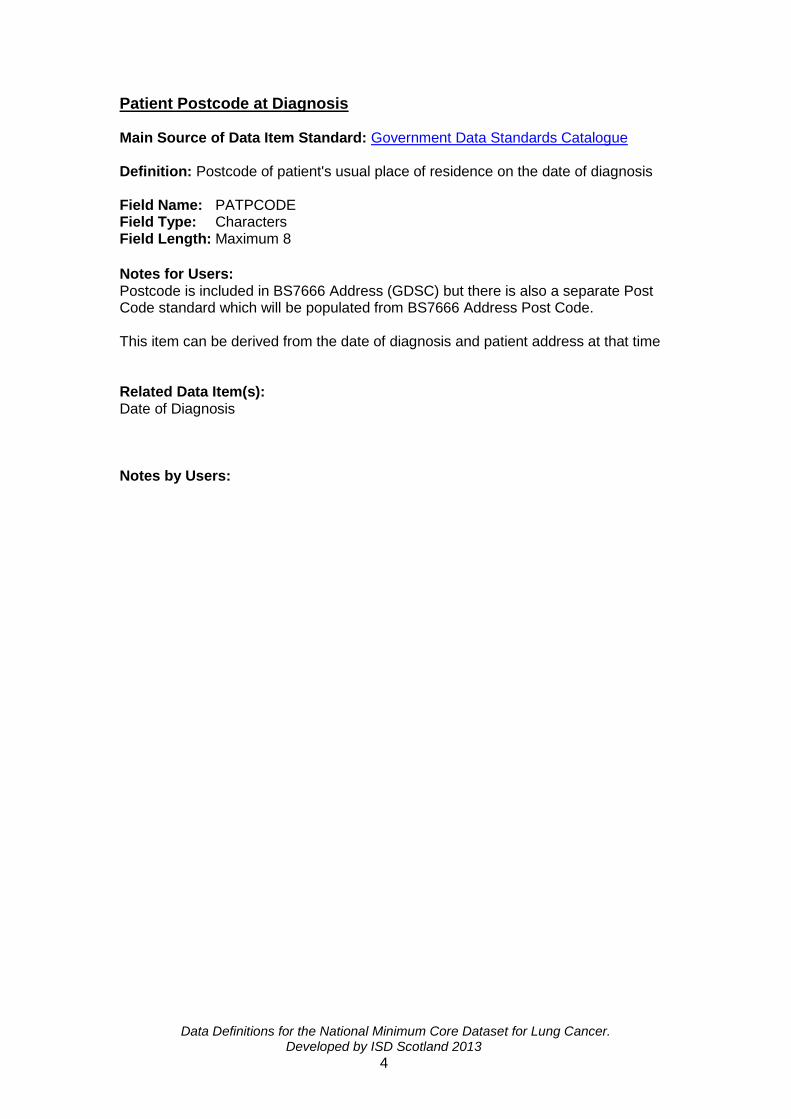
Patient Postcode at Diagnosis Main Source of Data Item Standard: Government Data Standards Catalogue Definition: Postcode of patient's usual place of residence on the date of diagnosis Field Name: PATPCODE Field Type: Characters Field Length: Maximum 8
Notes for Users: Postcode is included in BS7666 Address (GDSC) but there is also a separate Post Code standard which will be populated from BS7666 Address Post Code. This item can be derived from the date of diagnosis and patient address at that time Related Data Item(s): Date of Diagnosis Notes by Users:

Data Definitions for the National Minimum Core Dataset for Lung Cancer. Developed by ISD Scotland 2013
5
Date of Birth Main source of Data Item Standard: Government Data Standards Catalogue Definition: The date on which a person was born or is officially deemed to have been born, as recorded on the Birth Certificate. . Field Name: DOB Field Type: Date (DD/MM/CCYY) Field Length: 10 Notes for Users: If the patient's date of birth is recorded differently on different occasions, the most frequently used or latest date should be recorded. The patient's full date of birth inclusive of the century should be recorded. The format should be DD/MM/CCYY e.g. 01/02/2011. Related Data Item(s): CHI Number Notes by Users:

Data Definitions for the National Minimum Core Dataset for Lung Cancer. Developed by ISD Scotland 2013
6
Person Sex at Birth Common Name(s): Sex at Birth Main Source of Data Item Standard of Standard: Derived from the nearest equivalent Government Data Standards Catalogue standard ‘Person Gender at Registration’ Definition: This is a factual statement, as far as is known, about the phenotypic (biological) sex of the person at birth Field Name: SEX Field Type: Integer Field Length: 2 Notes for Users: A person’s sex has clinical implications, both in terms of the individual’s health and the health care provided to them. In the majority of cases, the phenotypic (biological) sex and genotypic sex are the same and the phenotypic sex is usually easily determined. In a small number of cases, accurate determination of genotype may be required. Codes and Values:
Code Value Explanatory Notes
1 Male
2 Female
9 Not specified/Indeterminate Where it has not been possible to determine if the person is male or female at birth, e.g. intersex / hermaphrodite.
99 Not recorded
Related Data Item(s): CHI Number Notes by Users:

Data Definitions for the National Minimum Core Dataset for Lung Cancer. Developed by ISD Scotland 2013
7
CHI Number Main Source of Data Item Standard of Standard: Scottish Executive Health Department. Definition: The Community Health Index (CHI) is a population register, which is used in Scotland for health care purposes. The CHI number uniquely identifies a person on the index. Field Name: CHINUM Field Type: Characters Field Length: 10 Notes for Users: The Community Health Index (CHI) is a computer based population index whose main function at present is to support primary care services. CHI contains details of all Scottish residents registered with a General Practitioner and was originally envisaged and implemented as a population-based index to help assess the success of immunisation and screening programmes. It is therefore closely integrated with systems for child health, cervical cytology and breast screening call and recall…It is intended that this number, the Scottish equivalent of the new NHS number in England and Wales, should become the Unique Patient Identifier throughout the NHS in Scotland. From Designed to Care - Scottish Office The CHI number is a unique numeric identifier, allocated to each patient on first registration with the system. The CHI number is a 10-character code consisting of the 6-digit date of birth (DDMMYY), two digits, a 9th digit which is always even for females and odd for males and an arithmetical check digit. (ISD, Information Services, NHS National Services Scotland) The CHI number should always be used to identify a patient. However, Health record identifiers, such as hospital numbers in Patient Administration Systems (PAS), may be used locally, in conjunction with the CHI number or in the absence of the CHI number, to track patients and their records.
Although there may be no number when a patient presents for treatment, there must be an allocation at some point in the episode of care as CHI is mandatory on all clinical communications. Non-Scottish patients and other temporary residents can have a CHI number allocated if required but it is envisaged that future development may allow the identifying number used in other UK countries to be used in Scotland. Related Data Item(s): Date of Birth, Person Sex at Birth. Notes by Users:
Data Definitions for the National Minimum Core Dataset for Lung Cancer. Developed by ISD Scotland 2013
8
Section 2: Pre-treatment Imaging & Staging Investigations

Data Definitions for the National Minimum Core Dataset for Lung Cancer. Developed by ISD Scotland 2013
9
Date of CT Thorax Main Source of Data Item Standard: The National Cancer Audit Datasets developed by the regional Cancer Networks supported by Information Services. Definition: The date the CT of the thorax was performed for staging and assessment. Field Name: CTHORAXDATE Field Type: Date (DD/MM/CCYY). Field Length: 10 Notes for Users: Required for QPI: 3 If the patient has more than one CT of thorax to diagnose lung cancer the date of the first procedure is recorded. Date CT Pulmonary angiogram (CTPA) can be recorded to diagnose lung cancer. If the exact date of the CT thorax is not documented, record as 09/09/0909 (Not recorded). If CT thorax was not performed, record as 10/10/1010 (Not applicable). Codes and Values: Related Data Items: Notes by Users:

Data Definitions for the National Minimum Core Dataset for Lung Cancer. Developed by ISD Scotland 2013
10
Date of Bronchoscopy {Lung Cancer} Main Source of Data Item Standard: The National Cancer Audit Datasets developed by the regional Cancer Networks supported by Information Services. Definition: The date of bronchoscopy is the date the procedure was performed for the purposes of investigating a possible diagnosis of lung cancer. Field Name: BDATE Field Type: Date (DD/MM/CCYY). Field Length: 10 Notes for Users: Required for QPI: 3 If the patient has more than one bronchoscopy to diagnose lung cancer, the date of the first procedure is recorded. If the exact date of the bronchoscopy is not documented, record as 09/09/0909 (Not recorded). If bronchoscopy was not performed, record as 10/10/1010 (Not applicable). Codes and Values: Related Data Items: Notes by Users:

Data Definitions for the National Minimum Core Dataset for Lung Cancer. Developed by ISD Scotland 2013
11
Seen by Clinical Nurse Specialist {Lung Cancer/ Mesothelioma} Main Source of Data Item Standard: The National Cancer Audit Datasets developed by the regional Cancer Networks supported by Information Services. Definition: A record to determine if the patient was seen by a clinical nurse specialist during their journey for the investigation and management of their cancer. Field Name: CNS Field Type: Integer Field Length: 2 Notes for Users: Required for NLCA In this context a clinical nurse specialist is a nurse who has specific expertise in the care and support of patients with cancer. In some settings, the clinical nurse specialist seen by the patient may be a palliative care nurse. This should be coded as ‘1: Yes’. Codes and Values:
Code Value
1 Yes
2 No
99 Not recorded
Related Data Items: Notes by Users:
Data Definitions for the National Minimum Core Dataset for Lung Cancer. Developed by ISD Scotland 2013
12
Location of Diagnosis {Cancer} Main Source of Data Item Standard: The National Audit Cancer Datasets developed by the regional Cancer Networks supported by Information Services. Definition: The patient's hospital of investigation in which the diagnosis of cancer was first made. Field Name: HOSP Field Type: Characters Field Length: 5 Notes for Users: Required for analysis purposes and clarifying responsibility for data collection. Details of location codes for hospitals can be found in the "Definitions and Codes for the NHS in Scotland" manual produced by ISD Scotland. Location codes for hospitals are five character codes maintained by ISD Scotland and the General Register Office (Scotland).The first character denotes the health board, the next three are assigned and the fifth denotes the type of location (H=hospital) e.g. A111H=Crosshouse Hospital G107H=Glasgow Royal Infirmary X9999=Not recorded If a patient was provisionally diagnosed at one hospital but transferred to another for confirmation of the diagnosis only e.g. biopsy, then returns to the original hospital, the first hospital should be recorded as the Location of diagnosis. Codes and Values: Related Data Items: Date of Diagnosis {Cancer} Histological/Cytological Diagnosis {Lung Cancer} Notes by Users:

Data Definitions for the National Minimum Core Dataset for Lung Cancer. Developed by ISD Scotland 2013
13
Date of Diagnosis {Cancer} Main Source of Data Item Standard: The National Cancer Audit Datasets developed by the regional Cancer Networks supported by Information Services. Definition: The date on which the cancer was first diagnosed whether by histology, cytology, immunology, cytogenetics or clinical (including radiological) methods. Field Name: DIAGDATE Field Type: Date (DD/MM/CCYY) Field length: 10 Notes for Users: Required for national survival analysis and national comparative analysis. The date recorded is the date on which the suspicion of cancer was first raised by the earliest relevant investigation (where the diagnosis was subsequently confirmed), i.e. the investigation which led to the decision to treat (effective from 1st April 2015).
This may be the date of the chest imaging where suspicion of lung cancer was raised and subsequently confirmed. If the exact date is not documented, record as 09/09/0909. The date recorded is the date the procedure was performed, not the date the report was issued.
Codes and Values: Related Data Items: Location of Diagnosis (Cancer) Notes by Users:

Data Definitions for the National Minimum Core Dataset for Lung Cancer. Developed by ISD Scotland 2013
14
Site of Origin of Primary Tumour {Cancer} Main Source of Data Item Standard: The World Health Organisation (WHO) and the Cancer Registration New Data definitions for Socrates (August 1999 Version 8.0).
Definition: The anatomical site of origin of the primary tumour according to the International Classification of Diseases (ICD-10). Field Name: SITE Field Type: Characters ICD-10 () Field length: 5
Notes for Users: Required for QPI(s): 1 – 13. For ICD-10, tumours should be assigned to the subcategory that includes the point of origin of the tumour. A tumour that overlaps the boundaries of two or more subcategories and whose point of origin cannot be determined should be classified as subcategory ‘C34.8’.It should be noted that this subcategory should only be used where it is impossible to identify the specific site of origin of the tumour. Codes and Values: ICD-1O Code
Value Notes on Inclusion
C33.X Malignant neoplasm of trachea
C34.0 Malignant neoplasm of bronchus and lung. Main bronchus
Includes: Carina, Hilus (of lung)
C34.1 Upper lobe, bronchus or lung
C34.2 Middle lobe, bronchus or lung
C34.3 Lower lobe, bronchus or lung
C34.8 Overlapping lesion of bronchus and lung
C34.9 Bronchus or lung, unspecified
C45.0 Mesothelioma of pleura
C45.1 Mesothelioma of peritoneum
Includes: Mesentery, Mesocolon, Omentum, Peritoneum (parietal)(pelvic) Excludes: other malignant neoplasms of peritoneum
C45.2 Mesothelioma of pericardium Excludes: other malignant neoplasms of pericardium.
C45.7 Mesothelioma of other sites
C45.9 Mesothelioma, unspecified
C99.X Not recorded

Data Definitions for the National Minimum Core Dataset for Lung Cancer. Developed by ISD Scotland 2013
15
Origin of Tumour Main Source of Data Item Standard: The National Cancer Audit Datasets developed by the regional Cancer Networks supported by Information Services. Definition: The origin of the primary tumour as detected clinically (including imaging).
Field Name: ORIGIN Field Type: Integer Field Length: 2
Notes for Users: Required for DCE and QPI(s): 1-16 . Codes and Values:
Code Value Explanatory Notes
1 Lung carcinoma
2 Mesothelioma Includes: Pleural, peritoneal, pericardial and other types
Related Data Items: Notes by Users:

Data Definitions for the National Minimum Core Dataset for Lung Cancer. Developed by ISD Scotland 2013
16
Histological/Cytological Diagnosis {Lung Cancer} (Pre-Treatment) Main Source of Data Item Standard: The National Cancer Audit Datasets developed by the Scottish Pathology Network supported by Information Services. Definition: This is the histological/cytological microscopic examination of the specimen by a pathologist to determine the presence of malignancy and the classification of the malignant tumour prior to surgery. Field Name: HIST Field Type: Integer Field Length: 2 Notes for Users: Required for QPI(s): 2, 4, 5, 6, 7, 8, 9, 10, 11, 12, 14, 15 A pathological diagnosis should be obtained from biopsy. If subtype is unknown use code 13 or 21 to record if tumour type is SCLC or NSCLC. Adequate tissue sampling should be undertaken, ensuring appropriate balance of risk to patients, to allow for pathological diagnosis including tumour sub-typing and analysis of predictive markers (NICE 2011 Lung Cancer: The diagnosis and treatment of lung cancer. April 2011. CG121 http://www.nice.org.uk/nicemedia/live/13465/54202/54202.pdf). There may be more than one biopsy/histology report. If there is a discrepancy between reports of cytology and histology, the histology report should be recorded as the definitive report. The WHO Classification is intended primarily for use with surgically resected cases (surgical resections pathology recorded elsewhere) and cannot be applied in full to small biopsy/cytology diagnosis. Consequently, a proportion of cases on biopsy/cytology specimens will be reported as “non-small cell carcinoma" (NSCLC), as this is as specific a diagnosis as may be possible on the material available Allocation to tumour subtype or variant category may not be achievable on diagnostic samples.
If a report is no more specific than “malignant cells” and does not further classify the tumour as carcinoma or other type of malignancy, the histology should be recorded as "other malignancies". Findings reported as “carcinoma, NOS” should also be recorded as "other malignancies. Pathologists often state 'favouring' a certain tumour sub-type and this should be documented as recorded rather than Not Otherwise Specified 'NOS' Pathology taken within 6 months of a patient initially refusing further investigation or whose initial treatment is ‘Watch and Wait’ can also be recorded.

Data Definitions for the National Minimum Core Dataset for Lung Cancer. Developed by ISD Scotland 2013
17
Codes and Values:
Code Description Explanatory Note
11 Squamous NSCLC Includes all variants
12 Adenocarcinoma NSCLC Includes: acinar, papillary, bronchiolo-alveolar, solid, signet ring cell and mucus cell types or patterns
13 NSCLC, not otherwise specified (NOS)
14 Other specific non-small cell carcinomas
NSCLC Includes: Other neuroendocrine tumours and large cell neuroendocrine carcinomas, salivary-type carcinomas Includes large cell carcinoma and undifferentiated, pleomorphic, sarcomatoid or anaplastic carcinoma
21 Small cell carcinoma (SCLC)
SCLC Includes:
22 Carcinoid tumour Includes typical and atypical carcinoid
31 Combination of non-small cell components
NSCLC Includes adenosquamous carcinoma and other mixed NSCLC-type cases
32 Small cell/non-small cell components
SCLC
41 Other malignancies (including malignancy NOS)
Includes cases reported as ‘carcinoma, NOS’ and metastatic tumours
42 Mesothelioma Unspecified Mesothelioma
43 Epithelioid Mesothelioma Mesothelioma
44 Sarcomatoid/Spindle Cell Mesothelioma
Mesothelioma
45 Biphasic Mesothelioma Mesothelioma
8 Negative histology
95 Patient refused investigation
96 Not applicable e.g. no pathology carried out
99 Not recorded
Related Data Items: Location of Diagnosis (Cancer) Date of Diagnosis (Cancer)

Data Definitions for the National Minimum Core Dataset for Lung Cancer. Developed by ISD Scotland 2013
18
Date of Histological / Cytological Diagnosis {Cancer} Main Source of Data Item Standard: The National Cancer Audit Datasets developed by the regional Cancer Networks supported by Information Services. Definition: The date on which the lung cancer was first diagnosed whether by histology or cytology. Field Name: HISTDATE Field Type: Date (DD/MM/CCYY) Field length: 10 Notes for Users: Required for QPI 15. There may be more than one biopsy/histology report. If there is a discrepancy between reports of cytology and histology, the histology report should be recorded as the definitive report. If no cytological or histological diagnosis was made, record as 10/10/1010 (Not applicable). If the exact date is not documented, record as 09/09/0909 (Not recorded). The date recorded is the date the procedure was performed, not the date the report was issued
Codes and Values: Related Data Items: Location of Diagnosis {Cancer} Notes by Users:

Data Definitions for the National Minimum Core Dataset for Lung Cancer. Developed by ISD Scotland 2013
19
Epidermal Growth Factor Receptor (EGFR) Status Main Source of Data Item Standard: The National Cancer Audit Datasets developed by the regional Cancer Networks supported by the Information Services. Definition: A record of the outcome of an epidermal growth factor receptor (EGFR) molecular profiling test, as part of the pathological assessment, taken prior to treatment. Field Name: EGFR Field Type: Integer Field Length: 2 Notes for Users: Required for QPI(s): 2, 11
There are structures on the surface of many types of cancer cells, known as epidermal growth factor receptors (EGFRs). The receptors allow epidermal growth factor (EGF), a particular protein present in the body, to attach to them. When EGF attaches to the receptor, it becomes activated and causes chemical processes to occur inside the cell that make it grow and divide more quickly. Erlotinib is an EGFR inhibitor, which has been accepted for use in Scotland by the SMC. This prevents the receptor from being activated and stops the cancer cells from growing so quickly.
Drugs known as EGFR antagonists attach themselves to the EGF receptor on the cell, and prevent the receptor from being activated. This can help to stop the cancer cells from growing so quickly.
Some drug treatments work best if they are targeted on the basis of histological subtype/predictive markers. Specific tests e.g. EGFR, are therefore required to predict whether targeted treatments are likely to be effective. EGFR should be undertaken on all patients with stage IIIB or IV non-squamous NSCLC.
Codes and Values:
Code Value Explanatory Notes
1 Positive EGFR undertaken as part of pathological assessment and a sensitising mutation has been detected.
2 Negative EGFR undertaken as part of pathological assessment and no EGFR mutations are detected..
3 Not done
4 Inconclusive Biopsy sample insufficient, test failed.
5 Undertaken
EGFR undertaken as part of pathological assessment and a mutation of uncertain significance or one conferring resistance has been detected but not actionable.
95 Patient refused investigations
99 Not recorded
Related Data Item(s): (pathological or clinical?) TNM Tumour Classification (Clinical) {Lung Cancer}

Data Definitions for the National Minimum Core Dataset for Lung Cancer. Developed by ISD Scotland 2013
20
TNM Nodal Classification (Clinical) {Lung Cancer} TNM Metastases Classification (Clinical) {Lung Cancer}

Data Definitions for the National Minimum Core Dataset for Lung Cancer. Developed by ISD Scotland 2013
21
Oncogenic Anaplastic Lymphoma Kinase (ALK) Status Main Source of Data Item Standard: The National Cancer Audit Datasets developed by the regional Cancer Networks supported by the Information Services. Definition: A record of the outcome of an oncogenic anaplastic lymphoma kinase (ALK) molecular profiling test, as part of the pathological assessment, taken prior to treatment. Field Name: ALK Field Type: Integer Field Length: 2 Notes for Users: Required for QPI(s): 2, 11
The ALK gene rearrangement produces an abnormal ALK protein that causes the cells to grow and spread. Drugs known as tyrosine kinase inhibitors (TKI) target ALK protein and can stop the cells growing.
Some drug treatments work best if they are targeted on the basis of histological subtype/predictive markers. Specific tests e.g. ALK, are therefore required to predict whether targeted treatments are likely to be effective. ALK should be undertaken on all patients with stage IIIB or IV non-squamous NSCLC.
Codes and Values:
Code Value Explanatory Notes
1 Positive ALK testing undertaken as part of pathological assessment and result positive for ALK fusion gene.
2 Negative ALK testing undertaken as part of pathological assessment and result negative for ALK fusion gene.
3 Not done
4 Inconclusive Biopsy sample insufficient, test failed.
95 Patient refused investigations
99 Not recorded

Data Definitions for the National Minimum Core Dataset for Lung Cancer. Developed by ISD Scotland 2013
22
PD-L1 Status Main Source of Data Item Standard: The National Cancer Audit Datasets developed by the regional Cancer Networks supported by the Information Services. Definition: A record of the outcome of an oncogenic PD-L1 test, as part of the pathological assessment, taken prior to treatment. Field Name: PDL1 Field Type: Integer Field Length: 2 Notes for Users: Required for QPI(s):
T-cells are part of the immune system and play a role in attacking cancer cells within the body. The PD-L1 expression attaches to a receptor on a T-cell which then prevents this process.
Some drug treatments work best if they are targeted on the basis of histological subtype/predictive markers, specific tests e.g. PD-L1, and therefore are required to predict whether targeted treatments are likely to be effective. The result recorded should be the percentage of cells where PD-L1 expression is present.
Codes and Values:
Code Value Explanatory Notes
3 Not done
4 Inconclusive Biopsy sample insufficient, test failed.
5 <1%
6 1-10%
7 11-20%
8 21-30%
9 31-40%
10 41-50%
11 51-60%
12 61-70%
13 71-80%
14 81-90%
15 91-100%
95 Patient refused investigations
99 Not recorded

Data Definitions for the National Minimum Core Dataset for Lung Cancer. Developed by ISD Scotland 2013
23
Date of Integrated FDG-PET/CT (PET/CT) Scan (Pre-treatment) Main Source of Data Item Standard: The National Cancer Audit Datasets developed by the regional Cancer Networks supported by Information Services. Definition: This denotes the date of the integrated FDG-PET/CT (PET/CT) scan was performed for staging and assessment. Field Name: PETDATE Field Type: Date (DD/MM/CCYY). Field Length: 10 Notes for Users: Required for QPI: 4 A PET CT scan should be completed and reported by the multi-disciplinary team (MDT) for patients with NSCLC who are being considered for treatment with curative intent. If the patient has more than one PET/CT scan the date of the first procedure is recorded. If the exact date of the PET/CT Scan is not documented, record as 09/09/0909. If PET/CT scan was not performed, e.g. if patients has SCLC, record as 10/10/1010 (not applicable). Related Data Item(s): Mediastinal/Supraclavicular (SCF) node results at FDG-PET/CT (PET/CT) Scan

Data Definitions for the National Minimum Core Dataset for Lung Cancer. Developed by ISD Scotland 2013
24
Mediastinal/Supraclavicular (SCF) Node Results at FDG-PET/CT (PET/CT) Scan Main Source of Data Item Standard: The National Cancer Audit Datasets developed by the regional Cancer Networks supported by the Information Services. Definition: A record of the results of mediastinal/SCF node evaluation (N2/N3) as determined by PET/CT imaging. Field Name: MEDPET Field Type: Integer Field Length: 2 Notes for Users: Required for QPI: 5 Results recorded are based on PET CT scan results as reported by radiologist/MDT, not on the sampling of mediastinal/SCF nodes. Codes and values:
Code Value Explanatory Notes
1 Positive mediastinal/SCF nodes identified
Positive mediastinal/SCF nodes noted on PET CT report (N2/N3 disease recorded in radiology report)
2 No positive nodes identified No positive mediastinal/SCF nodes noted on PET CT report (N0/N1 disease recorded in radiology report).
95 Patient refused investigation
96 Not applicable e.g. no PET CT scan undertaken
99 Not Recorded
Related Data Item(s): Mediastinal/SCF Sampling Results (pre-treatment)

Data Definitions for the National Minimum Core Dataset for Lung Cancer. Developed by ISD Scotland 2013
25
Mediastinal/SCF Sampling Results (pre-treatment) Main Source of Data Item Standard: The National Cancer Audit Datasets developed by the regional Cancer Networks supported by the Information Services. Definition: A record to determine if mediastinal/supraclavicular fossa (SCF) nodes were sampled. Field Name: MEDSAMP Field Type: Integer Field Length: 2 Notes for Users: Required for QPI: 5 Sampling is not required if there is definitive distant metastatic disease. Where there is definitive evidence of distant metastases, or where there are no nodes to sample, record as ’96: Not applicable’. Methods of sampling for mediastinal nodes are - Endobronchial Ultrasound (EBUS), mediastinoscopy, mediastinotomy and video-assisted thoracoscopic surgery (VATs) sampling. Methods of sampling for SCF nodes are - ultrasound guidance or direct FNA (if palpable). Codes and Values:
Code Value
1 Yes
2 No
3 Inconclusive e.g. failed attempt
95 Patient refused investigation
96 Not applicable
99 Not recorded
Related Data Item(s): TNM Metastases Classification (Clinical) {Lung Cancer}

Data Definitions for the National Minimum Core Dataset for Lung Cancer. Developed by ISD Scotland 2013
26
Synchronous Primary Tumours Main Source of Data Item Standard: The National Cancer Audit Datasets developed by the regional Cancer Networks supported by Information Services. Definition: This denotes whether or not synchronous primary tumours are present. Field Name: MULTIPLE Field Type: Characters Field length: 2 Notes for Users: Required for comparative analysis. This refers to the presence of synchronous primary tumours which may be in the same lung (ipsilateral) or involving both sides (Bilateral). Record the presence or absence of synchronous tumours. Codes and Values:
Code Description Explanatory Notes
1a Ipsilateral
1b Bilateral
2 Absent
99 Not recorded
Related Data Items:
Notes by Users:

Data Definitions for the National Minimum Core Dataset for Lung Cancer. Developed by ISD Scotland 2013
27
TNM Tumour Classification (Clinical) {Lung Cancer} Common name: Clinical TNM Tumour Classification (Lung Cancer) Main Source of Data Item Standard: TNM Classification (TNM Classification of Malignant Tumours, Eighth Edition, UICC, 2017). Definition: The size and extent of the tumour as determined by pre-treatment investigations (not pathological), coded according to the official TNM Classification (TNM Classification of Malignant Tumours, Eighth Edition, 2017). Field Name: TLUNG Field Type: Characters Field length: 3 Notes for Users: Required for QPI(s): 2, 9, 10, 11 & 14 Clinical TNM is derived from all the clinical, radiological and biochemical results prior to treatment. The TNM system is base on the assessment of three components (T tumour, N node and M metastases) and the addition of numbers after the letter components to indicate the extent of the malignant disease. This is a pre/non-operative classification as defined prior to first treatment and documented in patient notes. In cases where there are multiple or synchronous tumours, the tumour with the poorest prognosis should be recorded.
To adhere to the stage grouping in the TNM classification, recording the subdivision codes ‘a’ and ‘b’ in the codes and values table for T1 and T2 tumours is recommended. If the size of the tumour is not specified as pT2a or pT2b then it should be recorded as pT2a.

Data Definitions for the National Minimum Core Dataset for Lung Cancer. Developed by ISD Scotland 2013
28
Codes and Values:
Code Value Explanatory Notes
T0 No evidence of primary tumour
T1mi Minimally invasive adenocarcinoma
Solitary adenocarcinoma (not more than 3cm in greatest dimension)., with a pre-dominantly lepidic pattern and not more than 5mm invasion in greatest dimension in any one focus)
T1a Tumour ≤ 1cm in greatest dimension1.
T1b Tumour > 1cm – ≤ 2cm in greatest dimension1.
T1c Tumour >2cm - ≤3cm in greatest dimension1.
T2a > 3cm – ≤ 4cm in greatest dimension.
T2b > 4cm – ≤ 5cm in greatest dimension.
T3
Tumour > 5cm - ≤ 7cm in greatest dimension or one that directly invades any of the following: parietal pleura, chest wall (including superior sulcus tumours), phrenic nerve, , or parietal pericardium; or separate tumour nodules(s) in the same lobe as the primary.
T4
Tumour > 7cm or of any size that invades any of the following: diaphragm, mediastinum, heart, great vessels, trachea, recurrent laryngeal nerve, oesophagus, vertebral body, carina; separate tumour nodule(s) in a different ipsilateral lobe to that of the primary.
TX
Primary tumour cannot be assessed, or tumour proven by presence of malignant cells in sputum or bronchial washings but not visualised by imaging or bronchoscopy.
96 T Classification Not applicable Not lung cancer
99 T Classification Not recorded
Related data items:

Data Definitions for the National Minimum Core Dataset for Lung Cancer. Developed by ISD Scotland 2013
29
TNM Nodal Classification (Clinical) {Lung Cancer} TNM Metastases Classification (Clinical) {Lung Cancer}

Data Definitions for the National Minimum Core Dataset for Lung Cancer. Developed by ISD Scotland 2013
30
TNM Nodal Classification (Clinical) {Lung Cancer} Common name: Clinical TNM Nodal Classification (Lung Cancer). Main Source of Data Item Standard: TNM Classification (TNM Classification of Malignant Tumours, Eighth Edition, UICC, 2017). Definition: The extent of regional lymph node metastases as determined by pre-treatment investigations (not pathological), coded according to the official TNM Classification (TNM Classification of Malignant Tumours, Eighth Edition, 2017). Field Name: NLUNG Field Type: Characters Field length: 2 Notes for Users: Required for QPI(s): 2, 6, 9, 10, 11, 14 & 16 Clinical TNM is derived from all the clinical, radiological and biochemical results prior to treatment. The TNM system is base on the assessment of three components (T tumour, N node and M metastases) and the addition of numbers after the letter components to indicate the extent of the malignant disease. This is a pre/non-operative classification as defined prior to first treatment and documented in patient notes. In cases where there are multiple or synchronous tumours, the tumour with the poorest prognosis should be recorded.
Codes and Values:
Code Value Explanatory Notes
N0 No regional lymph node metastasis.
N1 Metastasis in ipsilateral peribronchial and/or ipsilateral hilar lymph nodes and intrapulmonary nodes, including involvement by direct extension.
N2 Metastasis in ipsilateral mediastinal and/or subcarinal lymph node(s).
N3 Metastasis in contralateral mediastinal, contralateral hilar, ipsilateral or contralateral scalene, or supraclavicular lymph node(s).
NX Regional lymph nodes cannot be assessed (e.g. previously removed).
96 Not applicable
99 Not recorded
Related Data items: TNM Tumour Classification (Clinical) {Lung Cancer} TNM Metastases Classification (Clinical) {Lung Cancer}

Data Definitions for the National Minimum Core Dataset for Lung Cancer. Developed by ISD Scotland 2013
31
TNM Metastases Classification (Clinical) {Lung Cancer} Common name: Clinical TNM Metastases Classification (Lung Cancer). Main Source of Data Item Standard: TNM Classification (TNM Classification of Malignant Tumours, Eighth Edition, UICC, 2017). Definition: The extent of metastatic spread of the tumour as determined by pre-treatment investigations (not pathological), coded according to the official TNM Classification (TNM Classification of Malignant Tumours, Eighth Edition, 2017). Field Name: MLUNG Field Type: Characters Field length: 3 Notes for Users: Required for QPI(s): 2, 5, 6, 8, 9, 10, 11 & 14 Clinical TNM is derived from all the clinical, radiological and biochemical results prior to treatment. The TNM system is base on the assessment of three components (T tumour, N node and M metastases) and the addition of numbers after the letter components to indicate the extent of the malignant disease. This is a pre/non-operative classification as defined prior to first treatment and documented in patient notes. In cases where there are multiple or synchronous tumours, the tumour with the poorest prognosis should be recorded. To adhere to the stage grouping in the TNM classification, recording the subdivision codes ‘a’ and ‘b’ in the codes and values table for M1 tumours is recommended. Codes and Values:
Code Value Explanatory Notes
M0 No distant metastasis
M1 Distant metastasis
M1a
Separate tumour nodule(s) in a contralateral lobe; tumour with pleural or pericardial nodules or malignant pleural or pericardial effusion
3.
3Most pleural (pericardial) effusions with lung cancer are due to
tumour. In a few patients, multiple microscopical examinations of pleural (pericardial) fluid are negative for tumour, and the fluid is non-bloody and is not an exudate. Where these elements and clinical judgement dictate that the effusion is not related to the tumour, the effusion should be excluded as a staging descriptor.
M1b Single extrathoracic metastasis in a single organ
This includes involvement of a single non-regional node.
M1c Multiple extrathoracic metastasis in a single or multiple organs
96 Not applicable
99 Not recorded
Data Definitions for the National Minimum Core Dataset for Lung Cancer. Developed by ISD Scotland 2013
32
TNM Tumour Classification (Clinical) {Pleural Mesothelioma} Common name: Clinical TNM Tumour Classification (Pleural Mesothelioma) Main Source of Data Item Standard: TNM Classification (TNM Classification of Malignant Tumours, Eighth Edition, UICC, 2017). Definition: The size and extent of the tumour as determined by pre-treatment investigations (not pathological), coded according to the official TNM Classification (TNM Classification of Malignant Tumours, Eighth Edition, UICC, 2017). Field Name: TMESO Field Type: Characters Field length: 3 Notes for Users: Clinical TNM is derived from all the clinical, radiological and biochemical results prior to treatment. The TNM system is base on the assessment of three components (T tumour, N node and M metastases) and the addition of numbers after the letter components to indicate the extent of the malignant disease. This is a pre/non-operative classification as defined prior to first treatment and documented in patient notes. In cases where there are multiple or synchronous tumours, the tumour with the poorest prognosis should be recorded. No TNM classification is available for other mesothelioma types.

Data Definitions for the National Minimum Core Dataset for Lung Cancer. Developed by ISD Scotland 2013
33
Codes and values:
Code Value Explanatory Notes
T0 No evidence of primary tumour
T1 Tumour involves ipsilateral parietal pleura, with or without involvement of visceral, mediastinal or diaphragmatic pleura.
T2
Tumour involves the ipsilateral pleura(parietal or visceral pleura), with at least one of the following:
Invasion of diaphragmatic muscle.
Invasion of lung parenchyma.
T3
Tumour involves ipsilateral pleura (parietal or visceral pleura), with at least one of the following:
Invasion of endothoracic fascia.
Invasion into mediastinal fat.
Solitary focus of tumour invading soft tissues of the chest wall.
Non-transmural involvement of the pericardium.
T4
Tumour involves any ipsilateral pleura (parietal or visceral pleura), with at least one of the following:
Chest wall, with or without associated rib destruction (diffuse or multifocal)
Peritoneum (via direct transdiaphragmatic extension)
Contralateral pleura
Mediastinal organs s (oesophagus, trachea, heart, great vessels)
vertebra, neuroforamen, spinal cord
internal surface of the pericardium (transmural invasion with or without a pericardial effusion)
TX Primary tumour cannot be assessed.
96 Not applicable Diagnosis is not mesothelioma
99 Not recorded
Related data items: TNM Nodal Classification (Clinical) {Pleural Mesothelioma} TNM Metastases Classification (Clinical) {Pleural Mesothelioma} Notes by Users:

Data Definitions for the National Minimum Core Dataset for Lung Cancer. Developed by ISD Scotland 2013
34
TNM Nodal Classification (Clinical) {Pleural Mesothelioma} Common name: Clinical TNM Nodal Classification (Pleural Mesothelioma). Main Source of Data Item Standard: TNM Classification (TNM Classification of Malignant Tumours, Eighth Edition, UICC, 2017). Definition: The extent of regional lymph node metastases as determined by pre-treatment investigations (not pathological), coded according to the official TNM Classification (TNM Classification of Malignant Tumours, Eighth Edition, UICC, 2017). Field Name: NMESO Field Type: Characters Field length: 2 Notes for Users: Clinical TNM is derived from all the clinical, radiological and biochemical results prior to treatment. The TNM system is base on the assessment of three components (T tumour, N node and M metastases) and the addition of numbers after the letter components to indicate the extent of the malignant disease. This is a pre/non-operative classification as defined prior to first treatment and documented in patient notes. In cases where there are multiple or synchronous tumours, the tumour with the poorest prognosis should be recorded. No TNM classification is available for other mesothelioma types.
Codes and Values:
Code Value Explanatory Notes
N0 No regional lymph node metastasis.
N1
Metastases to ipsilateral intrathoracic lymph nodes (includes ipsilateral bronchopulmonary, hilar, subcarinal, paratracheal, aortopulmonary, paraesophageal, peridiaphragmatic, pericardial fat pad, intercostal and internal mammary nodes)
N2 Metastases to contralateral intrathoracic lymph nodes. Metastases to ipsilateral or contralateral supraclavicular lymph nodes
NX Regional lymph nodes cannot be assessed (e.g. previously removed).
96 Not applicable Diagnosis is not mesothelioma.
99 Not recorded
Related Data items:

Data Definitions for the National Minimum Core Dataset for Lung Cancer. Developed by ISD Scotland 2013
35
TNM Tumour Classification (Clinical) {Pleural Mesothelioma} TNM Metastases Classification (Clinical) {Pleural Mesothelioma}

Data Definitions for the National Minimum Core Dataset for Lung Cancer. Developed by ISD Scotland 2013
36
TNM Metastases Classification (Clinical) {Pleural Mesothelioma} Common name: Clinical TNM Metastases Classification (Pleural Mesothelioma). Main Source of Data Item Standard: TNM Classification (TNM Classification of Malignant Tumours, Eighth Edition, UICC, 2017). Definition: The extent of metastatic spread of the tumour as determined by pre-treatment investigations (not pathological), coded according to the official TNM Classification (TNM Classification of Malignant Tumours, Eighth Edition, UICC, 2017). Field Name: MMESO Field Type: Characters Field length: 2 Notes for Users: Clinical TNM is derived from all the clinical, radiological and biochemical results prior to treatment. The TNM system is base on the assessment of three components (T tumour, N node and M metastases) and the addition of numbers after the letter components to indicate the extent of the malignant disease. This is a pre/non-operative classification as defined prior to first treatment and documented in patient notes. In cases where there are multiple or synchronous tumours, the tumour with the poorest prognosis should be recorded. No TNM classification is available for other mesothelioma types. Codes and values:
Code Value Explanatory Notes
M0 No distant metastasis.
M1 Distant metastasis.
96 Not applicable Diagnosis is not mesothelioma.
99 Not recorded
Related data items:
Data Definitions for the National Minimum Core Dataset for Lung Cancer. Developed by ISD Scotland 2013
37
TNM Tumour Classification (Clinical) {Pleural Mesothelioma} TNM Nodal Classification (Clinical) {Pleural Mesothelioma}

Data Definitions for the National Minimum Core Dataset for Lung Cancer. Developed by ISD Scotland 2013
38
Brain Imaging Main Source of Data Item Standard: The National Audit Cancer Datasets developed by the regional Cancer Networks supported by Information Services. Definition: An indicator of whether or not brain imaging investigations were completed by contrast enhanced CT or Magnetic Resonance Imaging (MRI) prior to commencing treatment. Field Name: BRAIN Field Type: Characters Field Length: 2 Notes for Users: Required for QPI: 16 Brain imaging is recommended for patients with N2 disease who are being considered for curative treatment. This could be either contrast enhanced CT or contrast enhanced MRI investigation and should be carried out prior to treatment. If brain imaging investigations are not documented, record as 99 (Not recorded). If the patient is not undergoing curative treatment or does not have N2 disease record as 96 (Not applicable). Codes and Values:
Code Value Explanatory Notes
1 Contrast enhanced CT
2 Contrast enhanced MRI
3 CT/MRI – non contrast enhancing
4 Not performed
5 Not performed - Contraindicated Generally acknowledged clinical contraindication to performing both assessments exists. e.g. renal impairment, allergies to contrast media.
95 Patient declined Patient chose not to have brain imaging.
96 Not applicable
99 Not recorded Includes not known
Related Data Item(s): Date of Brain Imaging (Pre-treatment)

Data Definitions for the National Minimum Core Dataset for Lung Cancer. Developed by ISD Scotland 2013
39
Date of Brain Imaging (Pre-treatment) Main Source of Data Item Standard: The National Cancer Audit Datasets developed by the regional Cancer Networks supported by Information Services. Definition: This denotes the date the Contrast enhanced CT or MRI scan was performed for brain imaging. Field Name: BRAINDATE Field Type: Date (DD/MM/CCYY). Field Length: 10 Notes for Users: Required for QPI: 16 The date of procedure should be recorded. If the patient has more than one brain imaging procedure the date of the first procedure is recorded. If the exact date of the contrast enhanced CT / MRI scan is not documented, record as 09/09/0909. If contrast enhanced CT / MRI scan was not performed, record as 10/10/1010 (not applicable). Related Data Item(s): Brain Imaging

Data Definitions for the National Minimum Core Dataset for Lung Cancer. Developed by ISD Scotland 2013
40
WHO/ ECOG Performance Status Main Source of Data Item Standard: WHO (World Health Organisation) and ECOG (Eastern Cooperative Oncology Group) Definition: An overall assessment of the functional/physical performance of the patient. Field Name: PSTATUS Field Type: Integer Field length: 1 Notes for Users: Required for QPI(s): 2, 9, 10, 11 The WHO/ECOG performance status is a grade on a five point scale (range 0 to 4) at the time of investigation in which '0' denotes normal activity and '4' a patient who is 100% bedridden. If it is not documented do not deduce from other information and record as 'Not recorded'. This item may occur more than once throughout a patient’s record. This field relates to pre-treatment performance status i.e. at the time of the MDT closest to actual treatment. If the performance status falls between two scores, record the higher value i.e. the worst performance status. Codes and values:
Code Value
0 Fully active, able to carry on all pre-disease performance without restriction
1 Restricted in physically strenuous activity but ambulatory and able to carry out work of a light or sedentary nature, e.g. light housework, office work
2 Ambulatory and capable of self care but unable to carry out any work activities: up and about more than 50% of waking hours
3 Capable of only limited self care, confined to bed or chair more than 50% of waking hours
4 Completely disabled, cannot carry on any self care, totally confined to bed or chair
9 Not recorded

Data Definitions for the National Minimum Core Dataset for Lung Cancer. Developed by ISD Scotland 2013
41
Date Discussed by Care Team (MDT) Common name: Date discussed by multidisciplinary team (MDT) {Cancer} Main Source of Data Item Standard: The National Cancer Audit Datasets developed by the regional Cancer Networks supported by Information Services. Definition: This denotes the date the care team meeting was held to discuss the management of the patient's care. Field Name: MDTDATE Field Type: Date (DD/MM/CCYY) Field Length: 10 Notes for Users: Required for QPI: 1 May be used for analysis of generic QPI relating to MDT meetings. A cancer multidisciplinary care team may include surgeons, oncologists, radiologists, pathologists, nurses, speech language therapists, physiotherapists and others relevant to the treatment of a specific cancer. The team meets on a regular basis to discuss optimal patient management. Documentation of the discussion should be included in the case-note or other formal documentation. The first MDT meeting date will be recorded. If the patient has not been discussed by the MDT record as 10/10/1010 (Not applicable). If the date of the MDT meeting is unknown record as 09/09/0909 (Not recorded)
Related data Item(s):

Data Definitions for the National Minimum Core Dataset for Lung Cancer. Developed by ISD Scotland 2013
42
Type of First Cancer Treatment Common name: Mode of first treatment
Main Source of Data Item Standard: The National Cancer Audit Datasets developed by the regional Cancer Networks supported by Information Services. Definition: This denotes the first specific treatment modality administered to a patient.
Field Name: MODE1 Field Type: Integer Field length: 2 Notes for Users: Required for QPI(s): 1, 10, 11, 12 & 14 For any particular modality it is the first treatment and not specifically the definitive treatment i.e. this does not include purely diagnostic biopsies such as incisional biopsies, needle biopsies or core biopsies. Record patients as having ‘supportive care only’ if a decision was taken not to give the patient any active treatment as part of their primary therapy. No active treatment includes watchful waiting and supportive care but not palliative chemotherapy and/or radiotherapy. Dilatation without other treatment is not considered as active treatment. Steroids, drainage of pleural effusions etc should not be recorded as first treatment if more substantive treatment such as radiotherapy, chemotherapy or surgery is given. If no further treatment is given, then record as supportive care. Codes and Values:
Code Description Explanatory notes
1 Surgery
2 Radiotherapy Includes CHART, Stereotactic, Teletherapy (external beam radiotherapy), SABR and Brachytherapy.
3 Chemotherapy
15 Chemoradiotherapy Can be concurrent or sequential
5 Endoscopic Includes Endobronchial, Photodynamic therapy (PDT), Electrocautery (Diathermy), Cryotherapy, Laser Therapy, Bronchoscopic debulking, Insertion of stents.
13 Biological therapy Includes Erlotinib, Gefitinib, Cetuximab, Bevacizumab, Interferon, Interleukin 2, BCG Vaccine etc.
7 Supportive care No active treatment
12 Watchful waiting No active treatment
11 Other therapy
94 Patient died before treatment
95 Patient refused all therapies
99 Not recorded
Related Data Item(s): Date of First Cancer Treatment

Data Definitions for the National Minimum Core Dataset for Lung Cancer. Developed by ISD Scotland 2013
43
Date of First Cancer Treatment Main Source of Data Item Standard: The National Cancer Audit Datasets developed by the regional Cancer Networks supported by Information Services. Definition: This denotes the date the type of first cancer treatment was given to the patient. Field Name: FIRSTTREATDATE Field Type: Date (DD/MM/CCYY) Field Length: 10
Notes for Users: Required for QPI(s): 4 & 15. Also used for the National lung
cancer audit and detect cancer early This field should be recorded for all patients including those with supportive care only (‘No active treatment’) (see below). If type of first cancer treatment is ‘supportive care only’, the date recorded should be the first date the decision was taken not to give the patient treatment as part of their primary therapy. Where this has subsequently been confirmed at MDT, the date of MDT should be recorded. The aim of this date is to distinguish between patients who have initially had no treatment but receive some therapy when symptoms develop. The date recorded should be that of the first type of cancer treatment. If the exact date is not documented, record as 09/09/0909 (Not recorded). If the patient died before treatment or the patient refused treatment, record as 10/10/1010 (Not applicable). Related Data Item(s): Type of First Cancer Treatment

Data Definitions for the National Minimum Core Dataset for Lung Cancer. Developed by ISD Scotland 2013
44
Date of Definitive Treatment {Lung Cancer} Main Source of Data Item Standard: The National Cancer Audit Datasets developed by the regional Cancer Networks supported by Information Services. Definition: This denotes the date definitive cancer treatment was given to the patient. Field Name: DEFTREATDATE Field Type: Date (DD/MM/CCYY) Field Length: 10
Notes for Users: Required for QPI: 1
For patients with lung cancer definitive treatment will be either:
Surgery;
Radiotherapy;
Chemoradiotherapy; or
Systemic Anti Cancer Therapy (SACT).
It is the date of this treatment that should be recorded. If a patient receives more than one of the treatments listed it is the first which should be recorded. For patients undergoing no active treatment (e.g. supportive care only) the date recorded should be the first date the decision was taken not to give the patient treatment as part of their primary therapy. Where this has subsequently been confirmed at MDT, the date of MDT should be recorded. This will therefore be the same date as the First Treatment Date for these patients. If the exact date is not documented, record as 09/09/0909 (Not recorded). If the patient died before treatment or the patient refused treatment, record as 10/10/1010 (Not applicable). Related Data Item(s):

Data Definitions for the National Minimum Core Dataset for Lung Cancer. Developed by ISD Scotland 2013
45
Section 3: Surgery

Data Definitions for the National Minimum Core Dataset for Lung Cancer. Developed by ISD Scotland 2013
46
Location Code {Cancer Surgery} Common Name(s): Location, Location of Contact. Main Source of Data Item Standard: NHS National Reference Files, http://www.natref.scot.nhs.uk/. Definition: This is the reference number of any building or set of buildings where events pertinent to NHS Scotland take place. Locations include hospitals, health centres, GP surgeries, clinics, NHS board offices, nursing homes, schools and patient/client’s home. Field Name: HOSPSURG Field Type: Characters Field Length: 5 Notes for Users: Required for analysis purposes. This is the hospital of first definitive surgery which removes the primary tumour. This may be a planned excision even if close margins are found and further surgery is required. On occasion, this result will be achieved by excision biopsy. This should be included as site of first definitive surgery. Each location has a location code, which is maintained jointly by ISD: http://www.isdscotland.org/Products-and-Services/Data-Definitions-and-References/National-Reference-Files/index.asp? Location must be viewed as an address and not a code. If any new locations arise where NHS healthcare is delivered/administered, please ensure that the Reference Files Team at ISD is informed using form LOC-NEW (which can be downloaded from the website below) so that a new code may be issued as appropriate. http://www.isdscotland.org/Products-and-Services/Data-Definitions-and-References/National-Reference-Files/SMR-Reference-Files/ Information about location should be electronically stored, managed and transferred using the relevant location code. IT systems should allow the recording and display of locations on the user interface as the relevant location name and associated address, etc. If the location code is not documented, record as X9999.

Data Definitions for the National Minimum Core Dataset for Lung Cancer. Developed by ISD Scotland 2013
47
If surgery has not been performed or the patient has refused surgery, record as Not Applicable, X1010. Examples of codes are given below:
Code Institution A111H CROSSHOUSE HOSPITAL
C418H ROYAL ALEXANDRA HOSPITAL
F704H VICTORIA HOSPITAL, KIRKCALDY
G107H GLASGOW ROYAL INFIRMARY
G405H SOUTHERN GENERAL HOSPITAL, GLASGOW
Related Data Item(s): Definitive Surgery Performed {Lung Cancer} Date of Surgery Surgical Approach
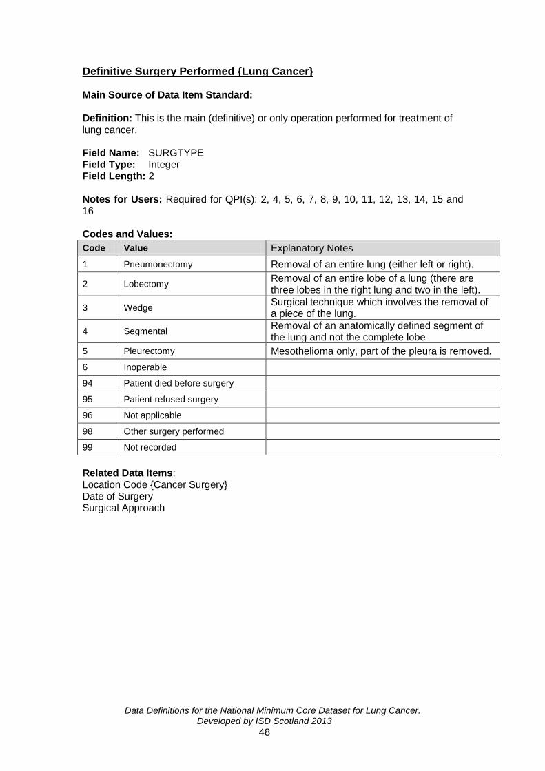
Data Definitions for the National Minimum Core Dataset for Lung Cancer. Developed by ISD Scotland 2013
48
Definitive Surgery Performed {Lung Cancer} Main Source of Data Item Standard: Definition: This is the main (definitive) or only operation performed for treatment of lung cancer. Field Name: SURGTYPE Field Type: Integer Field Length: 2 Notes for Users: Required for QPI(s): 2, 4, 5, 6, 7, 8, 9, 10, 11, 12, 13, 14, 15 and 16 Codes and Values:
Code Value Explanatory Notes
1 Pneumonectomy Removal of an entire lung (either left or right).
2 Lobectomy Removal of an entire lobe of a lung (there are three lobes in the right lung and two in the left).
3 Wedge Surgical technique which involves the removal of a piece of the lung.
4 Segmental Removal of an anatomically defined segment of the lung and not the complete lobe
5 Pleurectomy Mesothelioma only, part of the pleura is removed.
6 Inoperable
94 Patient died before surgery
95 Patient refused surgery
96 Not applicable
98 Other surgery performed
99 Not recorded
Related Data Items: Location Code {Cancer Surgery} Date of Surgery Surgical Approach
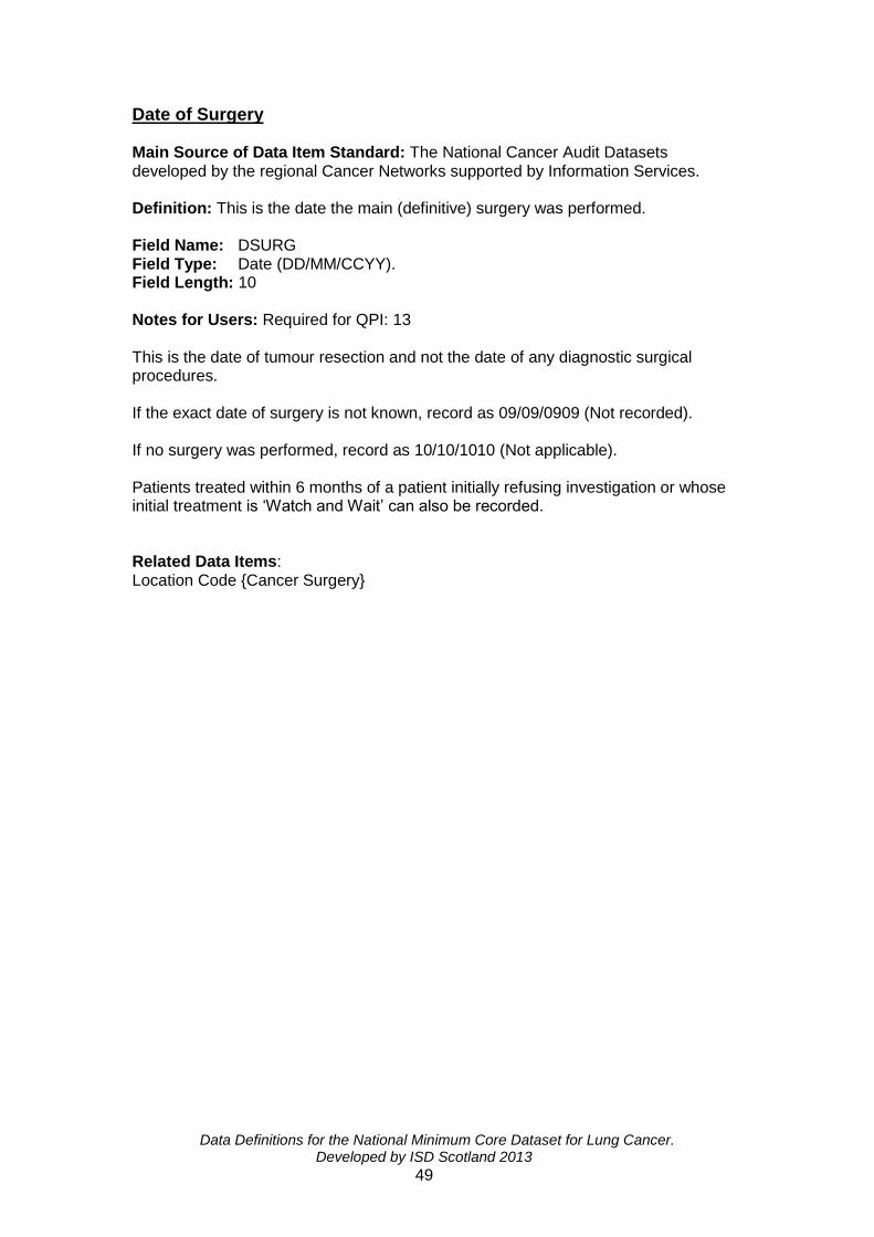
Data Definitions for the National Minimum Core Dataset for Lung Cancer. Developed by ISD Scotland 2013
49
Date of Surgery Main Source of Data Item Standard: The National Cancer Audit Datasets developed by the regional Cancer Networks supported by Information Services. Definition: This is the date the main (definitive) surgery was performed. Field Name: DSURG Field Type: Date (DD/MM/CCYY). Field Length: 10 Notes for Users: Required for QPI: 13 This is the date of tumour resection and not the date of any diagnostic surgical procedures. If the exact date of surgery is not known, record as 09/09/0909 (Not recorded). If no surgery was performed, record as 10/10/1010 (Not applicable). Patients treated within 6 months of a patient initially refusing investigation or whose initial treatment is ‘Watch and Wait’ can also be recorded. Related Data Items: Location Code {Cancer Surgery}

Data Definitions for the National Minimum Core Dataset for Lung Cancer. Developed by ISD Scotland 2013
50
Surgical Approach Main Source of Data Item Standard: The National Cancer Audit Datasets developed by the regional Cancer Networks supported by Information Services. Definition: The type of surgical procedure(s) performed for investigation and/or treatment of Cancer Field Name: APPROACH Field Type: Integer Field length: 2 Notes for Users: Required for analysis purposes Codes and Values:
Code Value Explanatory Notes
1 Open
2 Thorascopic Video assisted
3 Thorascopic - Converted Video assisted
94 Patient died before treatment
95 Patient refused treatment
96 Not applicable
99 Not recorded
Related Data Items: Location Code {Cancer Surgery}

Data Definitions for the National Minimum Core Dataset for Lung Cancer. Developed by ISD Scotland 2013
51
Histological/Cytological Diagnosis Following Surgery {Lung Cancer} Main Source of Data Item Standard: The National Cancer Audit Datasets developed by the Scottish Pathology Network supported by Information Services. Definition: This is the histological/cytological microscopic examination, following surgical resection, of the specimen by a pathologist to determine the presence of malignancy and the classification of the malignant tumour. Field Name: HISTSURG Field Type: Integer Field Length: 2 Notes for Users: Required for QPI(s): 2, 4, 5, 6, 7, 12, 13 If subtype is unknown use code 13 or 21 to record if tumour type is SCLC or NSCLC. Adequate tissue sampling should be undertaken, ensuring appropriate balance of risk to patients, to allow for pathological diagnosis including tumour sub-typing and analysis of predictive markers (NICE 2011 Lung Cancer: The diagnosis and treatment of lung cancer. April 2011. CG121 http://www.nice.org.uk/nicemedia/live/13465/54202/54202.pdf).
If a report is no more specific than “malignant cells” and does not further classify the tumour as carcinoma or other type of malignancy, the histology should be recorded as "other malignancies". Findings reported as “carcinoma, NOS” should also be recorded as "other malignancies. Pathologists often state 'favouring' a certain tumour sub-type and this should be documented as recorded rather than Not Otherwise Specified 'NOS'. Pathology taken within 6 months of a patient initially refusing further investigation or whose initial treatment is ‘Watch and Wait’ can also be recorded.

Data Definitions for the National Minimum Core Dataset for Lung Cancer. Developed by ISD Scotland 2013
52
Codes and Values:
Code Description Explanatory Note
11 Squamous NSCLC Includes all variants
12 Adenocarcinoma NSCLC Includes: acinar, papillary, bronchiolo-alveolar, solid, signet ring cell and mucus cell types or patterns
13 NSCLC, not otherwise specified (NOS)
14 Other specific non-small cell carcinomas
NSCLC Includes: Other neuroendocrine tumours and large cell neuroendocrine carcinomas, salivary-type carcinomas Includes large cell carcinoma and undifferentiated, pleomorphic, sarcomatoid or anaplastic carcinoma
21 Small cell carcinoma (SCLC)
SCLC Includes:
22 Carcinoid tumour Includes typical and atypical carcinoid
31 Combination of non-small cell components
NSCLC Includes adenosquamous carcinoma and other mixed NSCLC-type cases
32 Small cell/non-small cell components
SCLC
41 Other malignancies (including malignancy NOS)
Includes cases reported as ‘carcinoma, NOS’ and metastatic tumours
42 Mesothelioma Unspecified Mesothelioma
43 Epithelioid Mesothelioma Mesothelioma
44 Sarcomatoid/Spindle Cell Mesothelioma
Mesothelioma
45 Biphasic Mesothelioma Mesothelioma
8 Negative histology
95 Patient refused investigation
96 Not applicable e.g. no pathology carried out
99 Not recorded
Related Data Items: Definitive Surgery Performed {Lung Cancer}
Data Definitions for the National Minimum Core Dataset for Lung Cancer. Developed by ISD Scotland 2013
53
Section 4: Pathological Details

Data Definitions for the National Minimum Core Dataset for Lung Cancer. Developed by ISD Scotland 2013
54
TNM Tumour Classification (Pathological) {Lung Cancer} Common name: Pathological TNM Tumour Classification (Lung Cancer) Main Source of Data Item Standard: TNM Classification (TNM Classification of Malignant Tumours, Eighth Edition, UICC, 2017). Definition: A record of the size and extent of the tumour of the lung following resection of the primary cancer. Field Name: PTLUNG Field Type: Characters Field length: 4 Notes for Users: Required for DCE In cases where there are multiple tumours, the tumour with the worst TNM stage should be recorded. If stage is not documented in the pathology report do not deduce from other information and record as ‘not recorded’. To adhere to the stage grouping in the TNM classification, recording the subdivision codes ‘a’ and ‘b’ in the codes and values table is recommended. If the size of the tumour is not specified as pT2a or pT2b then it should be recorded as pT2a. Pathology taken within 6 months of a patient initially refusing further investigation or whose initial treatment is ‘Watch and Wait’ can also be recorded.

Data Definitions for the National Minimum Core Dataset for Lung Cancer. Developed by ISD Scotland 2013
55
Codes and Values:
Code Value Explanatory Notes
pT0 No evidence of primary tumour
pT1mi Minimally invasive adenocarcinoma
Solitary adenocarcinoma (not more than 3cm in greatest dimension)., with a pre-dominantly lepidic pattern and not more than 5mm invasion in greatest dimension in any one focus)
pT1a Tumour ≤ 1cm in greatest dimension1.
pT1b Tumour > 1cm – ≤ 2cm in greatest dimension1.
pT1c Tumour > 2cm but ≤ 3cm in greatest dimension1.
pT2a > 3cm – 4cm in greatest dimension.
pT2b > 4cm – 5cm in greatest dimension.
pT3
Tumour > 5cm but not more than 7cm in greatest dimension or one that directly invades any of the following: parietal pleura, chest wall (including superior sulcus tumours), phrenic nerve, parietal pericardium; or separate tumour nodule(s) in the same lobe as the primary.
pT4
Tumour more than 7cm or of any size that invades any of the following: diaphragm, mediastinum, heart, great vessels, trachea, recurrent laryngeal nerve, oesophagus, vertebral body, carina; separate tumour nodule(s) in a different ipsilateral lobe to that of the primary.
pTX
Primary tumour cannot be assessed, or tumour proven by presence of malignant cells in sputum or bronchial washings but not visualised by imaging or bronchoscopy.
96 Not applicable Not lung cancer
99 Not recorded
Related Data Items: TNM Nodal Classification (Pathological) {Lung Cancer} TNM Metastases Classification (Pathological) {Lung Cancer}

Data Definitions for the National Minimum Core Dataset for Lung Cancer. Developed by ISD Scotland 2013
56
TNM Nodal Classification (Pathological) {Lung Cancer} Common name: Pathological TNM Nodal Classification (Lung Cancer). Main Source of Data Item Standard: TNM Classification (TNM Classification of Malignant Tumours, Eighth Edition, UICC, 2017). Definition: A record of the extent of metastatic spread of the tumour as detected by microscopy. Field Name: PNLUNG Field Type: Characters Field length: 3 Notes for Users: Required for QPI: DCE In cases where there are multiple tumours, the tumour with the worst TNM stage should be recorded. If stage is not documented in the pathology report do not deduce from other information and record as ‘not recorded’. Pathology taken within 6 months of a patient initially refusing further investigation or whose initial treatment is ‘Watch and Wait’ can also be recorded. Codes and Values:
Code Value Explanatory Notes
pN0 No regional lymph nodes metastasis
Histological examination of hilar and mediastinal lymphadenectomy specimen(s) will ordinarily include 6 or more lymph nodes/stations. Three of these nodes/stations should be mediastinal, including the subcarinal nodes and 3 from N1 nodes/stations. Labelling according to the IASLC chart and table of definitions given in the TNM Supplement is desirable. If all the lymph nodes examined are negative, but the number ordinarily examined is not met, classify as pN0.
pN1
Metastasis in ipsilateral peribronchial and/or ipsilateral hilar lymph nodes and intrapulmonary nodes, including involvement by direct extension.
pN2 Metastasis in ipsilateral mediastinal and/or subcarinal lymph node(s).
pN3 Metastasis in contralateral mediastinal, contralateral hilar, ipsilateral or contralateral scalene, or supraclavicular lymph node(s).
pNX Regional lymph nodes cannot be assessed (e.g. previously removed).
96 Not applicable Not lung cancer
99 Not recorded
Related Data Items: TNM Tumour Classification (Pathological) {Lung Cancer} TNM Metastases Classification (Pathological) {Lung Cancer}

Data Definitions for the National Minimum Core Dataset for Lung Cancer. Developed by ISD Scotland 2013
57
TNM Metastases Classification (Pathological) {Lung Cancer} Common name: Pathological TNM Metastases Classification (Lung Cancer).
Main Source of Data Item Standard: TNM Classification (TNM Classification of Malignant Tumours, Eighth Edition, UICC, 2017).
Definition: The extent of metastatic spread of the tumour as detected by
microscopy.
Field Name: PMLUNG Field Type: Characters Field length: 4 Notes for Users: Required for DCE In cases where there are multiple tumours, the tumour with the worst TNM stage should be recorded. If stage is not documented in the pathology report do not deduce from other information and record as ‘not recorded’. To adhere to the stage grouping in the TNM classification, recording the subdivision codes ‘a’ and ‘b’ in the codes and values table is recommended. Pathology taken within 6 months of a patient initially refusing further investigation or whose initial treatment is ‘Watch and Wait’ can also be recorded.
Codes and Values:
Code Value Explanatory Notes
pM0 No distant metastasis (at autopsy only). i.e. pM0 does not exist and is not valid except at autopsy.
pM1 Distant metastasis
pM1a
Separate tumour nodule(s) in a contralateral lobe; tumour with pleural or pericardial nodules or malignant pleural or pericardial effusion
3.
3Most pleural (pericardial) effusions with lung cancer are due to
tumour. In a few patients, multiple microscopic examinations of pleural (pericardial) fluid are negative for tumour, and the fluid is non-bloody and is not an exudate. Where these elements and clinical judgement dictate that the effusion is not related to the tumour, the effusion should be excluded as a staging descriptor.
pM1b Single extrathoracic metastasis in a single organ
This includes involvement of a single non-regional node.
pM1c Multiple extrathoracic metastasis in a single or multiple organs
96 Not applicable Not lung cancer
99 Not recorded e.g. M status not assessed.
Related Data Items: TNM Tumour Classification (Pathological) {Lung Cancer} TNM Nodal Classification (Pathological) {Lung Cancer}

Data Definitions for the National Minimum Core Dataset for Lung Cancer. Developed by ISD Scotland 2013
58
TNM Tumour Classification (Pathological) {Pleural Mesothelioma} Common name: Pathological TNM Tumour stage (Pleural Mesothelioma) Main Source of Data Item Standard: TNM Classification (TNM Classification of Malignant Tumours, Eighth Edition, UICC, 2017). Definition: A record of the size and extent of the tumour following resection of the primary cancer. Field Name: PTMESO Field Type: Characters Field length: 4 Notes for Users: Required for analysis purposes If stage is not documented in the pathology report do not deduce from other information and record as ‘not recorded’. To adhere to the stage grouping in the TNM classification, recording the subdivision codes ‘a’ and ‘b’ in the codes and values table is recommended. Pathology taken within 6 months of a patient initially refusing further investigation or whose initial treatment is ‘Watch and Wait’ can also be recorded. No TNM classification is available for other mesothelioma types.

Data Definitions for the National Minimum Core Dataset for Lung Cancer. Developed by ISD Scotland 2013
59
Codes and Values:
Code Value Explanatory Notes
pT0 No evidence of primary tumour
pT1 Tumour involves ipsilateral parietal pleura, with or without involvement of visceral, mediastinal or diaphragmatic pleura.
pT2
Tumour involves the ipsilateral pleura (parietal or visceral pleura), with at least one of the following:
Invasion of diaphragmatic muscle
Invasion of lung parenchyma.
pT3
Tumour involves ipsilateral pleura (parietal or visceral pleura), with at least one of the following:
Invasion of endothoracic fascia
Invasion into mediastinal fat
Solitary focus of tumour invading soft tissues of the chest wall
Non-transmural involvement of the pericardium
pT4
Tumour involves ipsilateral pleura (parietal or visceral pleura), with at least one of the following:
Chest wall, with or without associated rib destruction (diffuse or multifocal)
Peritoneum (via direct transdiaphragmatic extension)
Contralateral pleura
Mediastinal organs (oesophagus, trachea, heart, great vessels)
Vertebra, neuroforamen, spinal cord
Internal surface of pericardium (transmural invasion with or without a pericardial effusion)
pTX Primary tumour cannot be assessed
96 Not applicable Diagnosis is not mesothelioma
99 Not recorded
Related data items: TNM Nodal Classification (Pathological) {Pleural Mesothelioma} TNM Metastases Classification (Pathological) {Pleural Mesothelioma}

Data Definitions for the National Minimum Core Dataset for Lung Cancer. Developed by ISD Scotland 2013
60
TNM Nodal Classification (Pathological) {Pleural Mesothelioma} Common name: Pathological TNM Nodal stage (Pleural Mesothelioma) Main Source of Data Item Standard: TNM Classification (TNM Classification of Malignant Tumours, Eighth Edition, UICC, 2017) Definition: A record of the extent of regional lymph node metastases. Field Name: PNMESO Field Type: Characters Field length: 3 Notes for Users: Required for analysis purposes. If stage is not documented in the pathology report do not deduce from other information and record as ‘not recorded’. Pathology taken within 6 months of a patient initially refusing further investigation or whose initial treatment is ‘Watch and Wait’ can also be recorded. No TNM classification is available for other mesothelioma types.
Codes and Values:
Code Value Explanatory Notes
pN0 No regional lymph nodes metastasis.
pN1
Metastasis to ipsilateral intrathoracic lymph nodes (includes ipsilateral bronchopulmonary, hilar, subcarinal, paratracheal, aortopulmonary, paraesophageal, peridiaphragmatic, pericardial fat pad, intercostal and internal mammary nodes)
pN2 Metastasis to contralateral intrathoracic lymph nodes. Metastases to ipsilateral or contralateral supraclavicular lymph nodes
pNX Regional lymph nodes cannot be assessed (e.g. previously removed).
96 Not applicable Diagnosis is not mesothelioma
99 Not recorded
Related Data items: TNM Tumour Classification (Pathological) {Pleural Mesothelioma} TNM Metastases Classification (Pathological) {Pleural Mesothelioma}

Data Definitions for the National Minimum Core Dataset for Lung Cancer. Developed by ISD Scotland 2013
61
TNM Metastases Classification (Pathological) {Pleural Mesothelioma} Common name: Pathological TNM Metastases Classification (Pleural Mesothelioma) Main Source of Data Item Standard: TNM Classification (TNM Classification of Malignant Tumours, Eighth Edition, UICC, 2017) Definition: A record of the extent of metastatic spread of the tumour as detected by microscopy. Field Name: PMMESO Field Type: Characters Field length: 3 Notes for Users: Required for analysis purposes. If stage is not documented in the pathology report do not deduce from other information and record as ‘not recorded’. Pathology taken within 6 months of a patient initially refusing further investigation or whose initial treatment is ‘Watch and Wait’ can also be recorded. No TNM classification is available for other mesothelioma types. Codes and values:
Code Value Explanatory Notes
pM1 Distant metastases Distant metastasis microscopically confirmed
96 Not applicable e.g. Diagnosis is not mesothelioma
99 Not recorded M status is not assessed.
Related data items: TNM Tumour Classification (Pathological) {Pleural Mesothelioma} TNM Nodal Classification (Pathological) {Pleural Mesothelioma}

Data Definitions for the National Minimum Core Dataset for Lung Cancer. Developed by ISD Scotland 2013
62
N2 Lymph Node Stations Main Source of Data Item Standard: Definition: A record of the total number of mediastinal lymph node stations examined microscopically after surgical resection, or at previous mediastinoscopy. Field Name: N2NODES Field Type: Integer Field Length: 2 Notes for Users: Required for QPI: 7
The number of nodal stations where at least 1 lymph node has been examined should be recorded. The number of nodal stations examined will be recorded on pathology report of surgical resection and/or mediastinoscopy, with a corresponding number of lymph nodes examined from each station (as detailed below). Clarification should be sought from pathologist where this cannot be ascertained from the report.
Often nodes from paratracheal and subcarinal nodal stations are sampled at mediastinoscopy prior to surgical resection, these should also be recorded.
Mediastinal (N2) nodal stations are defined as:
Superior Mediastinal Nodes:
Station 2R – Right upper paratracheal nodes
Station 2L – Upper left paratracheal nodes
Station 4R – Right lower paratracheal nodes
Station 4L – Left lower paratracheal nodes.
Aortic Nodes
Station 5 – Aorto-pulmonary nodes
Station 6 – Anterior Mediastinal nodes
Inferior Mediastinal nodes
Station 7 – Subcarinal nodes
Station 8 – Paraesophageal nodes
Station 9 – Pulmonary ligt. Nodes
Codes and values:
Code Value Explanatory Notes
1 0 No N2 stations examined.
2 1-2 At least 1 lymph node from 1 or 2 N2 stations examined
3 3 or more At least 1 lymph node from 3 or more N2 stations examined
96 Not applicable No surgical resection performed
99 Not recorded Related data items:

Data Definitions for the National Minimum Core Dataset for Lung Cancer. Developed by ISD Scotland 2013
63
Section 5: Oncology

Data Definitions for the National Minimum Core Dataset for Lung Cancer. Developed by ISD Scotland 2013
64
Radiotherapy Course Type (1-3) Main Source of Data Item Standard: The National Cancer Audit Datasets developed by the regional Cancer Networks supported by Information Services. Definition: The type of course of external beam radiotherapy administered for the treatment of the cancer. Field Name: RADIOTYPE1 RADIOTYPE2 RADIOTYPE3 Field Type: Integer Field length: 2 Notes for Users: Required for QPI(s): 4, 6, 8, 9, 10, 12, 13, 15 and 16 Combined treatments may be administered concurrently/synchronously e.g. chemotherapy and radiotherapy, intra-operative radiotherapy.
Actual treatment should be recorded. Where patients receive radiotherapy with radical intent (either radical radiotherapy or chemoradiotherapy) and treatment is abandoned prior to receiving full dosage, this should be recorded as palliative. For patients undergoing chemoradiotherapy the radiotherapy element should be recorded as code ‘6’ and recorded also in SACT under code ‘5. Patients treated within 6 months of a patient initially refusing investigation or whose initial treatment is ‘Watch and Wait’ can also be recorded.
All treatments given as part of the initial treatment plan
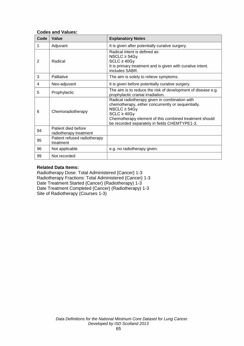
Data Definitions for the National Minimum Core Dataset for Lung Cancer. Developed by ISD Scotland 2013
65
Codes and Values:
Code Value Explanatory Notes
1 Adjuvant It is given after potentially curative surgery.
2 Radical
Radical intent is defined as: NSCLC ≥ 54Gy SCLC ≥ 40Gy It is primary treatment and is given with curative intent. Includes SABR.
3 Palliative The aim is solely to relieve symptoms.
4 Neo-adjuvant It is given before potentially curative surgery.
5 Prophylactic The aim is to reduce the risk of development of disease e.g. prophylactic cranial irradiation.
6 Chemoradiotherapy
Radical radiotherapy given in combination with chemotherapy, either concurrently or sequentially. NSCLC ≥ 54Gy SCLC ≥ 40Gy Chemotherapy element of this combined treatment should be recorded separately in fields CHEMTYPE1-3.
94 Patient died before radiotherapy treatment
95 Patient refused radiotherapy treatment
96 Not applicable e.g. no radiotherapy given.
99 Not recorded
Related Data Items: Radiotherapy Dose: Total Administered {Cancer} 1-3 Radiotherapy Fractions: Total Administered {Cancer} 1-3 Date Treatment Started {Cancer} (Radiotherapy) 1-3 Date Treatment Completed {Cancer} (Radiotherapy) 1-3 Site of Radiotherapy (Courses 1-3)

Data Definitions for the National Minimum Core Dataset for Lung Cancer. Developed by ISD Scotland 2013
66
Stereotactic Ablative Radiotherapy (SABR) Main Source of Data Item Standard: The National Cancer Audit Datasets developed by the regional Cancer Networks supported by Information Services. Definition: An indication of whether or not the patient received stereotactic ablative radiotherapy (SABR).
Field Name: SABR Field Type: Integer Field Length: 2 Notes for Users: Required for QPI(s): 6, 8, 14 Stereotactic ablative radiotherapy (SABR) is a specialised type of radiotherapy which precisely targets the tumour with radiation, whilst lowering the risk of damage to the healthy surrounding tissue. This should also be recorded as code 2, Radical under Radiotherapy Course Type.
Patients treated within 6 months of a patient initially refusing investigation or whose initial treatment is ‘Watch and Wait’ can also be recorded.
All treatments given as part of the initial treatment plan Codes and Values:
Code Value
1 Yes
2 No
95 Patient refused SABR
99 Not recorded
Related Data Items:

Data Definitions for the National Minimum Core Dataset for Lung Cancer. Developed by ISD Scotland 2013
67
Site of Radiotherapy (Courses 1- 3) Main Source of Data Item Standard: The National Cancer Audit Datasets developed by the regional Cancer Networks supported by Information Services Definition: This is the anatomical site(s) where the radiotherapy was given to the patient. Field Name: RADIOSITE1 RADIOSITE2 RADIOSITE3 Field Type: Integer Field length: 2 Notes for Users: Codes and Values:
Code Value Explanatory Notes
1 Chest
2 Brain If Prophylactic Cranial Irradiation (PCI) radiotherapy is given then record site as brain.
3 Bone
94 Patient died before radiotherapy treatment
95 Patient refused radiotherapy treatment
96 Not applicable
98 Other Includes: pleural drain
99 Not recorded
Related Data Items: Radiotherapy Course Type (1-3 Radiotherapy Dose: Total Administered {Cancer} 1-3 Radiotherapy Fractions: Total Administered {Cancer} 1-3 Date Treatment Started {Cancer} (Radiotherapy) 1-3 Date Treatment Completed {Cancer} (Radiotherapy) 1-3

Data Definitions for the National Minimum Core Dataset for Lung Cancer. Developed by ISD Scotland 2013
68
Radiotherapy Dose: Total Administered {Cancer} 1-3 Common name: Total External Beam Radiotherapy Dose Administered {Cancer} Main Source of Data Item Standard: The National Cancer Audit Datasets developed by the regional Cancer Networks supported by Information Services Definition: The Tumour Applied Dose (TAD) actually given (recorded in Gy) between the start and completion dates recorded for the course. Field Name: TOTDOSE1 TOTDOSE2 TOTDOSE3 Field Type: Float nnn.nnn Field length: 7 Notes for Users: Required for QPI: 9 IT systems should ensure that the unit of measurement for values is always clear to users, in whatever medium values are recorded. Dose and fractions using CHART: 5400cGy (54 Gy) in 36 fractions. Dose and fractions using Stereotactic Ablative Radiotherapy (SABR): >5000cGy (50 Gy) in 3-8 fractions. Up to three courses may be recorded. All treatments given as part of the initial treatment plan. If radiotherapy was not given, record ‘888.888’ (Not applicable). If radiotherapy dose is not recorded, record as ‘999.999’ (Not recorded). Codes and Values: Related Data Items: Radiotherapy Course Type (1-3 Radiotherapy Fractions: Total Administered {Cancer} 1-3 Date Treatment Started {Cancer} (Radiotherapy) 1-3 Date Treatment Completed {Cancer} (Radiotherapy) 1-3 Site of Radiotherapy (Courses 1-3)

Data Definitions for the National Minimum Core Dataset for Lung Cancer. Developed by ISD Scotland 2013
69
Radiotherapy Fractions: Total Administered {Cancer} 1-3 Main Source of Data Item Standard: The National Cancer Audit Datasets developed by the regional Cancer Networks supported by Information Services Definition: The number of radiation treatments actually given for any individual course of therapy (described by the start and completion dates of External Beam Radiotherapy). Field Name: FRACTIONS1 FRACTIONS2 FRACTIONS3 Field Type: Float nnnn Field length: 4 Notes for Users: Required for QPI: 9 Dose and fractions using CHART: 5400cGy (54 Gy) in 36 fractions. Dose and fractions using Stereotactic Ablative Radiotherapy (SABR): > 5000cGy (50 Gy) in 3-8 fractions. All treatments given as part of the initial treatment. If radiotherapy has not been given or the patient has refused radiotherapy, record as 1010 (not applicable). If radiotherapy fraction is unknown, record as 9999 (Not recorded). Related Data Items: Radiotherapy Course Type (1-3) Radiotherapy Dose: Total Administered {Cancer} 1-3 Date Treatment Started {Cancer} (Radiotherapy) 1-3 Date Treatment Completed {Cancer} (Radiotherapy) 1-3 Site of Radiotherapy (Courses 1-3)

Data Definitions for the National Minimum Core Dataset for Lung Cancer. Developed by ISD Scotland 2013
70
Date Treatment Started {Cancer} (Radiotherapy) 1-3 Main Source of Data Item Standard: The National Audit Cancer Datasets developed by the regional Cancer Networks supported by Information Services. Definition: The date cancer treatment course commenced. Field Name: RSRTDATE1 RSRTDATE2 RSRTDATE3 Field Type: Date (DD/MM/CCYY) Field length: 10 Notes for Users: This is the first fraction of a course of radiotherapy. Up to three courses may be recorded
For the purposes of national audit, only radiotherapy given as part of the primary treatment plan should be recorded. Palliative radiotherapy to other (metastatic) sites is only recorded if part of the initial treatment plan. If the date radiotherapy started is unknown, record as 09/09/0909 (Not recorded). If radiotherapy has not been given or the patient has refused radiotherapy, record as 10/10/1010 (not applicable). Related Data Items: Radiotherapy Course Type (1-3 Radiotherapy Dose: Total Administered {Cancer} 1-3 Radiotherapy Fractions: Total Administered {Cancer} 1-3 Date Treatment Completed {Cancer} (Radiotherapy) 1-3 Site of Radiotherapy (Courses 1-3)

Data Definitions for the National Minimum Core Dataset for Lung Cancer. Developed by ISD Scotland 2013
71
Date Treatment Completed {Cancer} (Radiotherapy) 1-3 Main Source of Data Item Standard: The National Cancer Audit Datasets developed by the regional Cancer Networks supported by Information Services Definition: The date cancer treatment course ended. Field Name: RCOMPDATE1 RCOMPDATE2 RCOMPDATE3 Field Type: Date (DD/MM/CCYY) Field Length: 10 Notes for Users: Required for QPI: 13 This is the last fraction of a course of radiotherapy. It should be noted this can be the same day as the day the therapy started. If the date treatment completed is unknown, record as 09/09/0909 (Not recorded). If treatment has not been given, record as 10/10/1010 (not applicable). Related Data Item(s): Radiotherapy Course Type (1-3 Radiotherapy Dose: Total Administered {Cancer} 1-3 Radiotherapy Fractions: Total Administered {Cancer} 1-3 Date Treatment Started {Cancer} (Radiotherapy) 1-3 Site of Radiotherapy (Courses 1-3)

Data Definitions for the National Minimum Core Dataset for Lung Cancer. Developed by ISD Scotland 2013
72
Type of Systemic Anti-Cancer Therapy (SACT) 1-3 Main Source of Data Item Standard: The National Cancer Audit Datasets developed by the regional Cancer Networks supported by Information Services.
Definition: The type of course of cytotoxic or biological drugs administered for the treatment of the cancer. Cytotoxic drugs are drugs which destroy cells.
Field Name: CHEMTYPE1, CHEMTYPE2, CHEMTYPE3 Field Type: Integer Field Length: 2
Notes for Users: Required for QPI(s): 4, 10, 11, 12, 13, 15 and 16
Patients may have ongoing systemic therapy both before and after surgery. These patients should be recorded under neo-adjuvant Type. Some patients may have separate completion chemotherapy post-operatively. This may be recorded as two courses neo-adjuvant and adjuvant.
Systemic therapy must be treatment received for initial management and not treatment for recurrence or relapse.
Actual treatment should be recorded. Where patients receive chemoradiotherapy and treatment is abandoned prior to receiving full dosage, this should be recorded as palliative.
For patients undergoing chemoradiotherapy the chemotherapy element should be recorded as code ‘5’ and recorded also in ‘Radiotherapy Course Type) under code ‘6’.
Codes and Values:
Code Value Explanatory Notes
1 Neoadjuvant Therapy given prior to radiotherapy or first definitive surgery to reduce tumour size.
2 Adjuvant Chemotherapy given after surgery within 3 months of surgery
4 Palliative Systemic therapy given for symptom control without curative intent e.g. for patients with metastatic disease at time of diagnosis.
5 Chemoradiotherapy
For curative/radical treatment. Can be sequential or concurrent where radiotherapy = NSCLC ≥ 54Gy or SCLC ≥ 40Gy. Radiotherapy element of this combined treatment should be recorded separately in fields RADIOTYPE1-3.
7 Biological Therapy
94 Patient died before SACT treatment
i.e. Patient who died before receiving planned SACT treatment
95 Patient refused SACT treatment
96 Not applicable e.g. Systemic therapy not given as primary part of therapy.
99 Not recorded
Related Data Items: Systemic Therapy Agent 1-3 {Lung Cancer} Date Treatment Started Systemic Anti-Cancer Therapy (SACT) {Cancer} 1-3 Date Treatment Completed Systemic Anti-Cancer Therapy (SACT) {Cancer} 1-3

Data Definitions for the National Minimum Core Dataset for Lung Cancer. Developed by ISD Scotland 2013
73
Systemic Therapy Agent 1-3 {Lung Cancer} Main Source of Data Item Standard: The National Cancer Audit Datasets developed by the regional Cancer Networks supported by Information Services.
Definition: The type of chemotherapy or biological therapy used either alone or in combination to treat lung cancer.
Field Name: CHEMAGENT1, CHEMAGENT2, CHEMAGENT3 Field Type: Integer Field length: 2
Notes for Users: Required for QPI: 10
Chemotherapy drugs can be given in or outwith the context of a clinical trial.
Up to three courses may be recorded.
If any systemic anti cancer therapy agent is not listed then please contact [email protected] to allocate a code.
Codes and Values:
Code Value Explanatory Notes
1 Cisplatin/Vinorelbine
2 Carboplatin/Vinorelbine
3 Carboplatin/Gemcitabine
4 Gemcitabine
5 Cisplatin/Etoposide
6 Cisplatin/Docetaxel
7 Cisplatin/Gemcitabine
8 Vinorelbine
11 Carboplatin/Etoposide
12 Carboplatin single agent
15 Cisplatin/Pemetrexed
16 Erlotinib
17 Carboplatin/Pemetrexed
18 Carboplatin/paclitaxel
19 Pemetrexed
20 Crizotinib
21 Afatinib
22 Other biological agent
23 Other chemotherapy agent
24 Pembrolizumab
96 Not applicable
99 Not recorded
Related Data Items: Type of Systemic Anti-Cancer Therapy (SACT) 1-3
Data Definitions for the National Minimum Core Dataset for Lung Cancer. Developed by ISD Scotland 2013
74
Date Treatment Started Systemic Anti-Cancer Therapy (SACT) {Cancer} 1-3 Date Treatment Completed Systemic Anti-Cancer Therapy (SACT) {Cancer} 1-3

Data Definitions for the National Minimum Core Dataset for Lung Cancer. Developed by ISD Scotland 2013
75
Date Treatment Started Systemic Anti-Cancer Therapy (SACT) {Cancer} 1-3 Main Source of Data Item Standard: The National Cancer Audit Datasets developed by the regional Cancer Networks supported by Information Services. Definition: The date cancer treatment course commenced. Field Name: CHEMDATE1 CHEMDATE2 CHEMDATE3 Field Type: Date (DD/MM/CCYY) Field length: 10 Notes for Users: This is the first dose of the first cycle of a course of chemotherapy or biological therapy. Up to three courses may be recorded. If the date SACT started is unknown, record as 09/09/0909 (Not recorded). If SACT has not been given or the patient has refused SACT, record as 10/10/1010 (not applicable). Related data items: Type of Systemic Anti-Cancer Therapy (SACT) 1-3 Systemic Therapy Agent 1-3 {Lung Cancer} Date Treatment Completed Systemic Anti-Cancer Therapy (SACT) {Cancer} 1-3

Data Definitions for the National Minimum Core Dataset for Lung Cancer. Developed by ISD Scotland 2013
76
Date Treatment Completed Systemic Anti-Cancer Therapy (SACT) {Cancer} 1-3 Main Source of Data Item Standard: The National Cancer Audit Datasets developed by the regional Cancer Networks supported by Information Services.
Definition: The date cancer treatment course ended. Field Name: CHEMENDATE1 CHEMENDATE2 CHEMENDATE3 Field Type: Date (DD/MM/CCYY) Field length: 10 Notes for Users: Required for QPI: 13 This is the first day of the last cycle of a course of SACT. It should be noted this can be the same day as the day the therapy started. Up to three courses may be recorded. If the date treatment started is unknown, record as 09/09/0909 (Not recorded). If SACT has not been given or the patient has refused SACT, record as 10/10/1010 (Not applicable). Codes and values: Related data items: Type of Systemic Anti-Cancer Therapy (SACT) 1-3 Systemic Therapy Agent 1-3 {Lung Cancer} Date Treatment Started Systemic Anti-Cancer Therapy (SACT) {Cancer} 1-3

Data Definitions for the National Minimum Core Dataset for Lung Cancer. Developed by ISD Scotland 2013
77
Section 6: Clinical Trials

Data Definitions for the National Minimum Core Dataset for Lung Cancer. Developed by ISD Scotland 2013
78
Patient Entered into Clinical Trial Main Source of Data Item Standard: The National Cancer Audit Datasets developed by the regional Cancer Networks supported by Information Services. Definition: An indication of whether or not the patient received treatment within the context of a clinical trial.
Field Name: TRIAL Field Type: Integer Field Length: 2 Notes for Users: Required for QPI(s): 11, 12 This relates only to participation in clinical trials which may be national or international multi-centred trials. The majority of non-commercial multi-centred trials available in Scotland are NCRN badged or equivalent. Some academic and university units may have ongoing local trials which should not be included here. These can be recorded on local trials databases. Codes and Values:
Code Value
1 Yes
2 No
99 Not recorded

Data Definitions for the National Minimum Core Dataset for Lung Cancer. Developed by ISD Scotland 2013
79
Section 7: Death Details

Data Definitions for the National Minimum Core Dataset for Lung Cancer. Developed by ISD Scotland 2013
80
Date of Death Main Source of Data Item Standard: The National Cancer Audit Datasets developed by the regional Cancer Networks supported by Information Services. Definition: This is the certified date of death as recorded by the General Register Office (Scotland) (GRO(S)). Field Name: DOD Field Type: Date (DD/MM/CCYY). Field Length: 10 Notes for Users: Required for QPI: 13 If the exact date is not documented, record as 09/09/0909 (Not recorded). If the patient is alive use the code 10/10/1010 (Not applicable).