lumps and bumps in the spleen case: c10052-12. seven...
TRANSCRIPT

Lumps and Bumps in the Spleen Case: C10052-12. Seven year old, male castrated Wheaton Terrier. Lesion found at necropsy.
Describe the changes: The spleen is moderately enlarged with rounding of margins. Small, white gritty plaques are scattered over the capsular surface. What is the significance of these changes? Nothing! These are incidental findings. This is a congested spleen (attributed to barbiturate euthanasia) and the white plaques are referred to as gamna-gandy bodies which are incidental findings attributed to aging. Case: C12663-01. Ten year old, mixed breed dog. Lesions are found at necropsy.
Describe the changes: The spleen is markedly enlarged with multifocal to coalescing, raised, firm nodules. What are your differentials?
- Nodular hyperplasia (a common finding in older dogs)- what this is - Neoplasia - Granulomatous inflammation

Case: C16438-08. 1 year old male German Shepherd that presented with a fever of unknown origin. The animal was in poor body condition and had neck and lumbosacral pain. Describe the lesion.
Numerous, slightly firm, poorly defined and sometimes coalescing, approx 1.5-2.5 cm in greatest diameter nodules are scattered throughout the spleen. What are some potential etiologies? - Neoplasia - Inflammation (Granulomatous eg. blastomycosis) - Necrosis (infarction due to systemic bacterial disease which in this case was due to infection with Rocky Mountain Spotted Fever (Rickettsia rickettsia) If you see miliary necrosis in the spleen, other good differentials are Tularemia and Yersinia - Autoimmune Disease (Systemic Lupus Erythematosis) could cause severe vasculitis and subsequent infarction What is your Morphologic Diagnosis? acute-subacute splenic infarctions P8362-07. 8-9 week old piglet.
Image is of a dog spleen with multifocal infarcts Describe the changes: The spleen is markedly enlarged. There is a well defined, slightly depressed dark regions. Give a morphologic diagnosis: Splenic Infarct Can you think of an underlying etiology? Infarcts are due to loss of blood supply, which in the spleen is due to torsion or vasculitis. A common cause of vasculitis in pigs in this region is Porcine Circovirus 2. Splenic infacts can also be seen in cases of sepsis as a result of disseminated intravascular coagulation (DIC).

Case: 4654-91: A 9 year old female Afghan Hound with a history of weight loss and a painful left shoulder. Describe the lesion
Large numbers of variably sized (1-3cm) diameter raised, firm nodules randomly located throughout the spleen. What are your differential diagnoses? - Neoplasia (lymphoma, mast cell tumor, in this case osteosarcoma) - Splenic Infarcts (see last case) - Nodular hyperplasia - Granulomatous inflammation (systemic fungal or bacterial diseases) What other areas are commonly affected by this condition (and are important clinically)?
- Osteosarcoma commonly metastasizes to the mammary glands and lungs, doesn’t cross the joints and is usually found “away from the elbow – proximal humerus and towards the knee – distal femur” Case: F4719-03. Nine year old, male castrated DSH cat with a history of chronic vomiting.
Describe the changes: The spleen is diffusely, markedly enlarged and meaty. Differentials? Mast cell tumor (what this was), Lymphoma, Histiocytic sarcoma Note: Meaty spleens are firm (indicating cellular infiltrates versus congestion which are bloody on cut section). How do you explain the vomiting? Animals with mast cells tumors have systemic histamine release which can create gastrointestinal ulceration as well as act directly on the chemoreceptive trigger center (CTZ=vomiting center) in the brain

Case: E6921-90. 5 year old, horse with a history of fever and weight loss.
Describe the changes: The spleen is enlarged and has multiple, coalescing 1-5mm white areas replacing normal splenic architecture. Differentials?
- Neoplasia (lymphosarcoma, mast cell tumor, histiocytic sarcoma) - Nodular hyperplasia - Granulomatous inflammation
This was a mast cell tumor diagnosed with histopathology. Case: C6976-03. 13 year old, male, castrated, Labrador Retriever. Describe the changes: There is a large, firm, multilobulated mass attached to and arising from the spleen. Differentials: -Neoplasia (round cell tumor, sarcoma- chondroid or osteoid in origin)
- This was a spindle cell sarcoma with chondroid differentiation (which explains why it was so firm).

Mediastinal Masses
Case: C392-03. Nine year old, FS Rottweiler with a 5-6 week history of vomiting and diarrhea as well as difficulty breathing, and anorexia. Abdominal radiographs show a soft tissue opacity within the thorax and pleural effusion. See also F5617-13, and C29091-05.
Describe the changes: Within the cranial mediastinum, there is a pale, firm, multilobulated mass. What are some differentials? -thymoma -mesothelioma -lymphoma (which it was in this case) -metastatic carcinoma Case: X2589-87. Four year old goat, found dead in a field. His throat seemed to swell up.
Describe the changes: There is a large, firm, white, multilobular mass within the anterior and dorsal mediastinum. Multiple lymph nodes are also diffusely enlarged. What is the most likely diagnosis? Lymphosarcoma (other differentials include mesothelioma and thymoma)

Case 3059-02. FS, 10 year old Golden Retriever.
www.quizlet.com Describe the changes: The mediastinum is markedly expanded by a poorly demarcated, friable, 14cm in greatest diameter, black-grey mass. Several, smaller, round 0.5-2cm black-grey masses are present within the mediastinal fat and adhered to the surface of the pericardium. Many pinpoint black nodules approximately 5mm in greatest diameter are scattered throughout the pulmonary parenchyma. What is the likely diagnosis? Malignant mediastinal melanoma (Differentials: Hemangiosarcoma, lymphoma or other round cell sarcoma, carcinoma) Case: B23437-98. A 300 kg feedlot steer.
Cornell Veterinary Medicine Describe the lesion. A large firm, to soft tan-white mass fills the cranioventral thorax with extension through the thoracic inlet. The mass is poorly circumscribed with infiltration into the surrounding tissues. On cut surface, the mass consists of variable sized homogeneous white nodules separated by
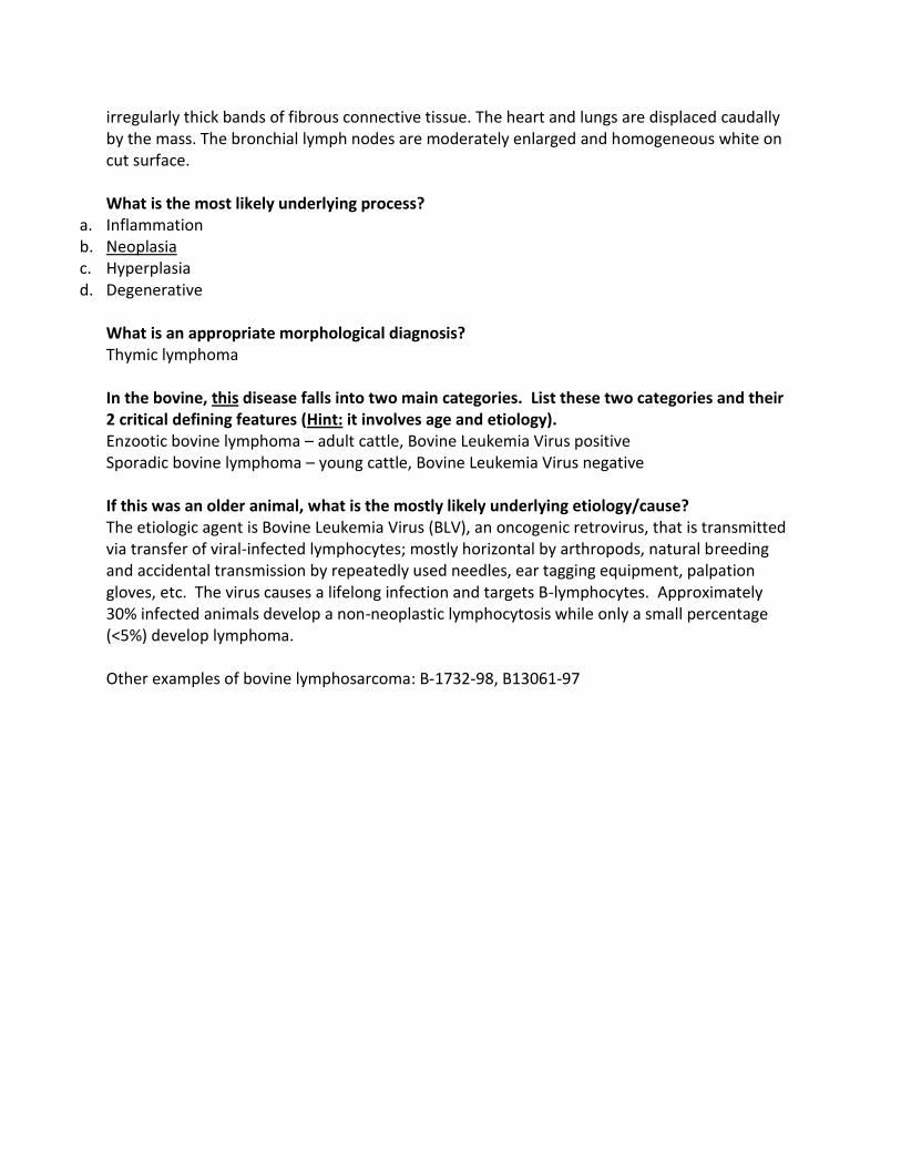
irregularly thick bands of fibrous connective tissue. The heart and lungs are displaced caudally by the mass. The bronchial lymph nodes are moderately enlarged and homogeneous white on cut surface. What is the most likely underlying process?
a. Inflammation b. Neoplasia c. Hyperplasia d. Degenerative
What is an appropriate morphological diagnosis? Thymic lymphoma In the bovine, this disease falls into two main categories. List these two categories and their 2 critical defining features (Hint: it involves age and etiology). Enzootic bovine lymphoma – adult cattle, Bovine Leukemia Virus positive Sporadic bovine lymphoma – young cattle, Bovine Leukemia Virus negative If this was an older animal, what is the mostly likely underlying etiology/cause? The etiologic agent is Bovine Leukemia Virus (BLV), an oncogenic retrovirus, that is transmitted via transfer of viral-infected lymphocytes; mostly horizontal by arthropods, natural breeding and accidental transmission by repeatedly used needles, ear tagging equipment, palpation gloves, etc. The virus causes a lifelong infection and targets B-lymphocytes. Approximately 30% infected animals develop a non-neoplastic lymphocytosis while only a small percentage (<5%) develop lymphoma. Other examples of bovine lymphosarcoma: B-1732-98, B13061-97
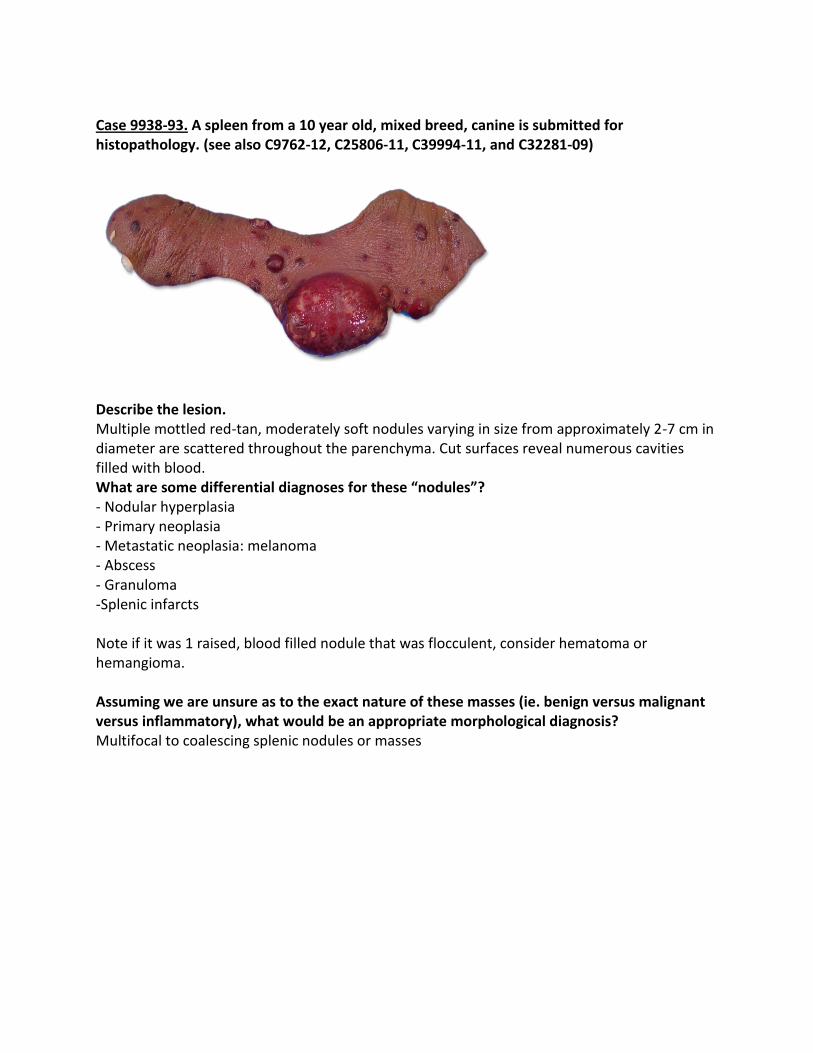
Case 9938-93. A spleen from a 10 year old, mixed breed, canine is submitted for histopathology. (see also C9762-12, C25806-11, C39994-11, and C32281-09)
Describe the lesion. Multiple mottled red-tan, moderately soft nodules varying in size from approximately 2-7 cm in diameter are scattered throughout the parenchyma. Cut surfaces reveal numerous cavities filled with blood. What are some differential diagnoses for these “nodules”? - Nodular hyperplasia - Primary neoplasia - Metastatic neoplasia: melanoma - Abscess - Granuloma -Splenic infarcts Note if it was 1 raised, blood filled nodule that was flocculent, consider hematoma or hemangioma. Assuming we are unsure as to the exact nature of these masses (ie. benign versus malignant versus inflammatory), what would be an appropriate morphological diagnosis? Multifocal to coalescing splenic nodules or masses

Case 31258-04. 9 year old, male, Welsh Springer with a one week history of PU/PD, lethargy and weakness which progressed to pain in all joints and neck pain. Liver and spleen were mottled on ultrasound.
Describe the changes: There is a firm, 9 X 3 x 5cm, firm mass lateral to but not infiltrating the larynx (site of the right submandibular lymph node). The spleen is moderately enlarged, meaty. Scattered within the parenchyma are hundreds of pale, white, slightly raised, firm, 2-10mm nodules. Other lymph nodes are diffusely enlarged. What is the likely diagnosis? This is multicentric lymphoma (metastatic neoplasia and other disseminated round cell tumors would be differentials). Immunohistochemistry for class II MHC and HM47 confirmed diagnosis of a B-Cell lymphoma Other Examples of multicentric lymphoma: F2200-08, F8954-09, F21360-09 Case: B1457-87: Abnormalities were detected at slaughter of a mature, beef cow from Manitoba.
Cornell Veterinary Medicine Describe the lesion. The lymph nodes are enlarged, firm and filled with granular and purulent material.
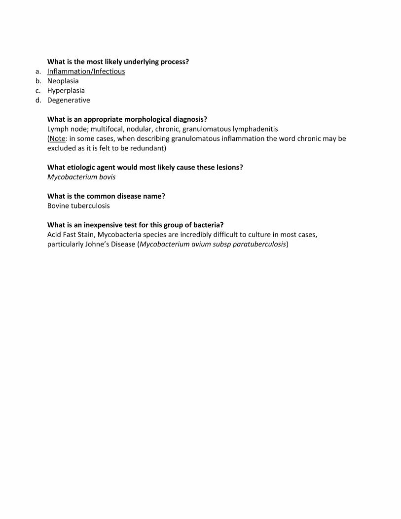
What is the most likely underlying process?
a. Inflammation/Infectious b. Neoplasia c. Hyperplasia d. Degenerative
What is an appropriate morphological diagnosis? Lymph node; multifocal, nodular, chronic, granulomatous lymphadenitis (Note: in some cases, when describing granulomatous inflammation the word chronic may be excluded as it is felt to be redundant) What etiologic agent would most likely cause these lesions? Mycobacterium bovis What is the common disease name? Bovine tuberculosis What is an inexpensive test for this group of bacteria? Acid Fast Stain, Mycobacteria species are incredibly difficult to culture in most cases, particularly Johne’s Disease (Mycobacterium avium subsp paratuberculosis)

Case: B2593-90. Submandibular lymph nodes from a cow.
www.fao.org Describe the changes: The submandibular lymph nodes are enlarged and on cut surface have a thick fibrous capsule and contain caseous material within the center. Based on the location, what might be a likely diagnosis? Actibobacillosis (wooden tongue) caused by Actinomyces bovis Case: A3084-83: Two year old, back yard chicken. Became mopey and died.
Describe the changes: The spleen is greatly enlarged with raised, firm white nodules of varying diameter (few mm up to 1cm). What is the likely etiology? These are typical lesions of avian tuberculosis caused by Mycobacterium avium. Differentials include lymphoid neoplasia due to retroviruses (leukosis/sarcoma group) or herpesvirus (Marek’s disease)

Case: F4026-93. Six month old, male kitten with pyrexia, progressive anorexia and lethargy.
Cornell Veterinary Medicine Describe the changes: The surface of the spleen is covered in tan-white raised plaques 1-3mm in diameter. What is the likely etiology? Feline Infectious Peritonitis (FIP), a coronavirus What clinical pathology findings were found in this cat? Hypergammaglobulinemia! What histologic findings are typical in this condition? Multiple foci of pyogranulomatous inflammation in many organs (e.g. kidney, serosal surface of the gastrointestinal tract, brain, etc.) and vasculitis Case: X4619-92. Beaver found dead.
Image of necrotizing splenitis and hepatitis (http://o.quizlet.com) Describe the changes: Numerous, multifocal white foci are randomly distributed throughout the spleen parenchyma Name three potential differential diagnoses: Francisella tularensis (tularemia) Clostridium piliforme (Tyzzer’s disease) Yersinia enterocolitica or Yersinia pseudotuberculosis Note: all are potentially zoonotic!!

Case: O-2596-98: A 4 year old ewe with a recent history of submandibular and prescapular lymph node enlargement was found dead in the field. Had 2 healthy twins last year. (see also G14070-06)
Describe the lesion. A large, moderately soft and fluctuant oval mass, approximately 8 cm in the largest dimension, is present within the mesentery immediately adjacent to the reticulum. Cut surface of the mass (interpreted as an enlarged mesenteric lymph node) reveal abundant yellow-green caseous exudates within a laminated (“onion ring”) appearance. What is an appropriate morphological diagnosis? Moderate, multifocal, chronic, suppurative lymphadenitis What is the common name for this condition? Caseous lymphadenitis What is the most likely infectious agent that would produce this type of lesion? Corynebacterium pseudotuberculosis A small gram-positive rod and facultative intracellular bacteria that is found on fomites and in soil and manure contaminated with purulent exudates How can we explain location of these masses (hint: how and why did the masses develop in the tissues shown here)? Infection occurs after C. pseudotuberculosis penetrates through unbroken or abraded skin or through mucous membranes. The bacteria proliferate producing slowly enlarging, localized, and non-painful abscess that typically develop either at the point of entry (in the skin) or in the regional lymph nodes (superficial or external form). From there the bacteria spread via the blood or lymphatic system and cause abscessation of internal lymph nodes or organs (visceral or internal form).

6. What does this mean for the flock and suggest methods of control. This is a production limiting disease because infected animals appear normal but harbour the disease and will sometimes lose weight. Transmission is common during shearing with potential to infect the whole flock. Strict hygiene practices during the shearing process decreases the risk of transmission as cull as culling suspect individuals (skinny ewes, animals with big lymph nodes). Testing and culling is possible, but expensive, sometimes not practical and only for highly motivated clients. Vaccination is available, but not very effective. It is much more of a problem in adults than lambs because of the chronicity and method of infection (from a skin wound at shearing or other causes (eg. head butting or ear biting in goats)).