lumbar radiculopathy - files.bridgeport.edu · lumbar radiculopathy. radicular pain often extends...
TRANSCRIPT

Lumbar Radiculopathy
James J. Lehman, DC, MBA, FACODirector
Health Sciences Postgraduate EducationUniversity of Bridgeport

Learning Objectives
Comprehend and practice concepts of “Evidence-based and patient-centered health care” in order to provide high quality patient care.

Learning Objectives
Implement the scientific method and integrate the use of an evaluation protocol practiced by contemporary chiropractic physician specialists in orthopedics and neuromusculoskeletal medicine.

Learning Objectives
Perform neuromusculoskeletal evaluation procedures and record the objective findings in order to make an assessment/diagnosis of lumbar radiculopathy.

“Diagnosis is the key to successful treatment!”

Lumbar Disc Herniation
ICD 9 722.2 Displacement of
intervertebral disc, site unspecified, without myelopathy

Lumbar Radiculopathy
Radicular pain often extends below the knee in the affected dermatome.

Definition of Orthopedic Test
A provocative maneuver (most often) using stretching, compressing, and contracting to duplicate the pain and identify the involved tissues.

Low Back Pain Spinal Pain and Tissue Identification
Neural Nerve root Spinal cord
Zygapophyseal joint Capsule Nerve
Ligament Muscle Osseous

North American Spine Society: Evidence-Based Clinical Guidelines for Multidisciplinary Spine Care
Clinical Guidelines for Diagnosis and Treatment of Lumbar Disc Herniation with Radiculopathy

Disclaimer
This clinical guideline should not be construed as including all proper methods of care or excluding of other acceptable methods of care reasonably directed to obtaining the same results. The ultimate judgment regarding any specific procedure or treatment is to be made by the physician and patient in light of all circumstances presented by the patient and the needs and resources particular to the locality or institution.
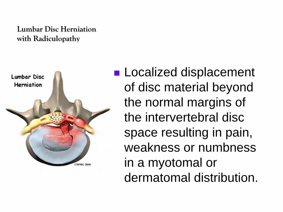
Lumbar Disc Herniation with Radiculopathy
Localized displacement of disc material beyond the normal margins of the intervertebral disc space resulting in pain, weakness or numbness in a myotomal or dermatomal distribution.

Natural History of Lumbar Radiculopathy
The majority of patients will improve independent of treatment. Disc herniations will often shrink/regress over time.

Diagnosis and Imaging
In the assessment of diagnostic tests, both accuracy and the effect of testing on the outcome should be considered.

Accuracy of a Diagnostic Test
Refers to the ability of the examination to detect and characterize pathologic processes.

AccuracySensitivity and Specificity
Sensitivity refers to the proportion of patients with the target disorder who will have a positive test

AccuracySensitivity and Specificity
Specificity refers to the proportion of patients without the target disorder who will have a negative test

AccuracySensitivity and Specificity
Tests that have a high sensitivity and negative test outcomes effectively rule out the disease.

AccuracySensitivity and Specificity
Tests that have a high specificity and positive test outcomes effectively rule in the disease.

Positive Predictive Value (PPV)Negative Predictive Value (NPV)
Performance of a test in a given population can also be stated in terms of positive and negative predictive value, which depends directly on the prevalence of disease in the tested population.

Lumbar Spine Pain
Lower back pain occurs most often between ages 30 and 50
Low Back Pain Fact Sheet. NIH/NINDS
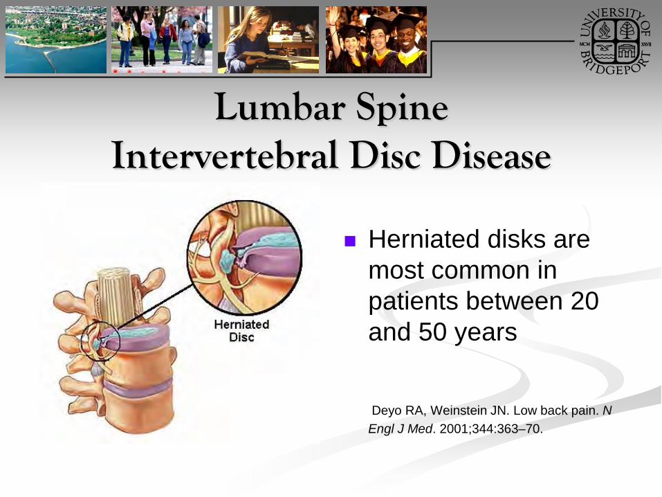
Lumbar SpineIntervertebral Disc Disease
Herniated disks are most common in patients between 20 and 50 years
Deyo RA, Weinstein JN. Low back pain. N Engl J Med. 2001;344:363–70.

Predictive Values and Patient Populations
One of the purposes of a history and physical examination is to increase the prevalence of disease in patients sent for advanced imaging/testing or offered surgery.

Case Presentation
Your patient presents with a posterior lateral herniated lumbar disc at the level of L5-S1, which is located medial to the nerve root of S1. The neurological exam demonstrates motor, sensory and DTR deficits. There are no signs of an upper motor lesion.

Learning Task
Form groups of 3-4 Select spokesperson Write putative SOAP notes for the patient
described in this case presentation. Present and defend your SOAP notes

The gold standard in the diagnosis of lumbar disc herniation is surgery; however, when assessing the validity of subjective complaints or physical examination findings, use of cross-sectional imaging as a gold standard may be considered an acceptable substitute.

Cross-sectional Imaging
Any technique that produces an image in the form of a plane through the body with the structures cut across.
CT MRI PET SPECT scanning Ultrasonography

Computerized Tomography Scan
CT and MRI demonstrate the structure of and blood flow to and from organs,
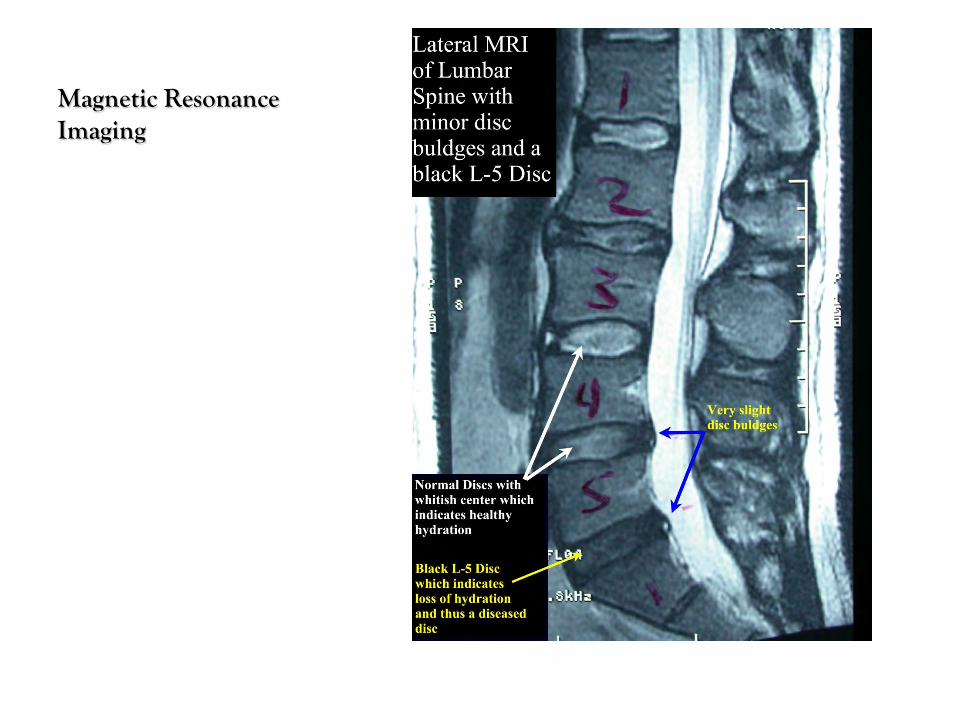
Magnetic Resonance Imaging

Positron Emission Tomography
A PET scan is an imaging test that uses a radioactive substance called a tracer to look for disease in the body.A PET scan shows how organs and tissues are working

Single Photon Emission Computed Tomography (SPECT)
Tomographic imaging of local metabolic and physiological functions in tissues. The image is formed by a computer synthesis of data that is transmitted by single gamma photons emitted by radionuclides administered to the patient.
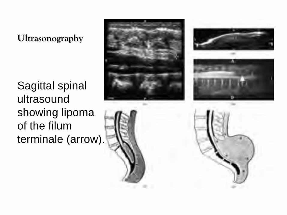
Ultrasonography
Sagittal spinal ultrasound showing lipoma of the filum terminale (arrow).

Discussion
Do you refer every patient that you suspect with a suspected lumbar disc herniation and radiculopathy for surgery and/or a cross-sectional imaging study?
Please explain your protocol and rationale in writing and then verbally.

Diagnosis and Treatment of Lumbar Disc Herniation with Radiculopathy
What history and physical examination findings are consistent with the diagnosis of lumbar disc herniation with radiculopathy?

Physical ExaminationGrade A Recommendation
Motor and Sensory testing
Straight leg raise Lasegue sign Crossed Lasegue Sign
Good evidence for or against recommending intervention

Three-Part Peripheral Nervous System Examination
Sensory Motor Deep Tendon Reflex (DTR)

Straight Leg Raise Test

Lasegue Sign
SLR reproduces the painLower affected lower extremity 15 degrees and pain is eliminatedorFlex knee and hip to 90%Extend kneeSign is present if the pain is reproduced

Crossed Lasegue Sign(Well-Leg-Raising Test)
Straight leg raising and dorsiflexion of the foot are performed on the asymptomatic side of a sciatic patient (radiculopathy)
Pain production in symptomatic lower extremity indicates sign is present.

Jenson Study
Prospective case series calculating the positive predictive value and negative predictive value of sensory and motor abnormalities as signs of the level of a lower lumbar disc herniation.
Jenson OH. The level-diagnosis of a lower disc herniation: the value of sensibility and motor testing. Clin Rheumatol. Dec 1987;6(4):564-569.

Jenson Study
All 52 consecutive patients included in the study had a disc herniation diagnosed by myelogram and confirmed at surgery.
JensonOH. The level-diagnosis of a lower disc herniation: the value of sensibility and motor testing. Clin Rheumatol. Dec 1987;6(4):564-569.

Physical ExaminationSensory testing of dermatomes
Sensory abnormalities found in 54% of patients with herniated disc.L4-5 herniation and L5 dermatome deficitPPV 76% NPV 55%L5-S1 herniation and S1 dermatome deficitPPV 50%NPV 62%
JensonOH. The level-diagnosis of a lower disc herniation: the value of sensibility and motor testing. Clin Rheumatol. Dec 1987;6(4):564-569.
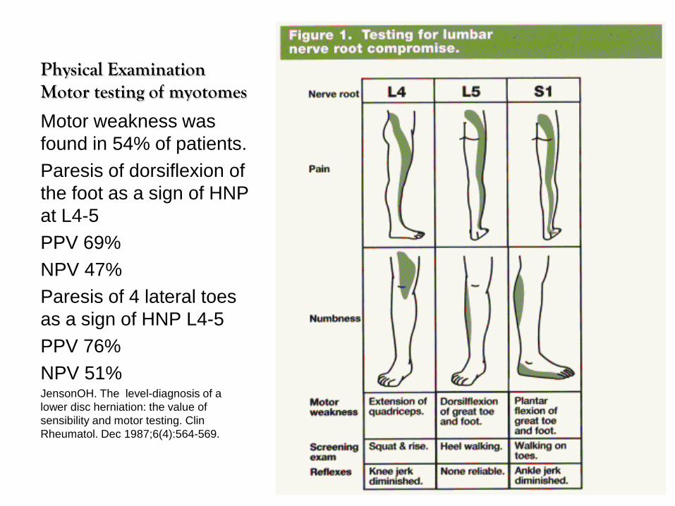
Physical Examination Motor testing of myotomes
Motor weakness was found in 54% of patients.Paresis of dorsiflexion of the foot as a sign of HNP at L4-5PPV 69%NPV 47%Paresis of 4 lateral toes as a sign of HNP L4-5PPV 76%NPV 51%JensonOH. The level-diagnosis of a lower disc herniation: the value of sensibility and motor testing. Clin Rheumatol. Dec 1987;6(4):564-569.

Sensory and Motor Testing
Level 1 diagnostic evidence that sensory and motor testing of a patient with a suspected lumbar disc herniation and radiculopathy can provide specific clues to the level of disc herniation , but are not very sensitive in determining the exact level.
JensonOH. The level-diagnosis of a lower disc herniation: the value of sensibility and motor testing. Clin Rheumatol. Dec 1987;6(4):564-569.

Kortelainen Study
Prospective case series evaluating the reliability of the clinical diagnosis of level of ruptured disc and the utility of lumbar myelography of gaining further information.
All 403 patients had lumbar disc herniation diagnosed by myelogram and confirmed at surgery

Kortelainen Study
L5 pain projection 79% reliable and 86% with extensor hallucis longus (EHL) weakness.
S1 pain projection 56% reliable and raised to 80% with Achilles DTR deficit and raised to 86% with sensory deficit.
Myelography was accurate 90.8% with 3.7% false + rate and 5.5% false - rate

Kortelainen Study
Cough impulse test + with 74% of patients with disc herniation.
A SLR + more often with lower lumbar herniations than upper lumbar spine.
Projected pain localized 93% of cases and most symptom localizing level of herniation.

Kortelainen Study
Achilles reflex was of value in diagnosis of L5-S1 herniation when associated with pain projection and sensory deficit of S1
Patellar reflex had no value in diagnosis of lower lumbar lesions.
EHL weakness due to L4-5 lesion 70% of cases even with S1 pain projection.

Kortelainen Study
Study provides Level 1 diagnostic evidence that physical examination, including subjective and objective findings such as + SLR, sensory and motor testing, in a patient with a suspected lumbar disc herniation and radiculopathy can provide specific clues to the level of disc herniation.
Kortelainen P. et al. Symptoms and signs of sciatica and their relation to the localization of the lumbar disc herniation. Spine (Phila Pa 1976) Jan-Feb 1985;88-92

Poiraudeau Study
A prospective case series including 78 consecutive patients with 43 confirmed cases of lumbar disc herniation (MRI, CT or myelogram), evaluating the reliability, sensitivity, specificity, positive predictive value and negative predictive value for the diagnosis radiculopathy associated with disc herniation (Bell test, hyperextension test, Lasegue and Crossed Leg Signs)

Bell test
This test was performed with the patient in the standing position. The test was positive when the examiner reproduced or exacerbated the usual radicular pain by pressure applied with the thumb between the spinous processes L4 and L5 or between L5 and S1, or in the near corresponding paraspinal area. When the manoeuvre reproduced only lumbar pain, it was considered negative.

Hyperextension Test
This was performed with the patient standing. The trunk was mobilized passively and slowly over the full range of extension with the knees in extension. The test was positive if the sciatica was reproduced or worsened. If the manoeuvre was interrupted because of lumbar pain, it was considered negative.

Lasègue's sign This was investigated with the patient supine.
The leg affected with sciatica was slowly raised passively, with the patient relaxed and the knee in full extension. Elevation was stopped when the patient began to feel pain. The sign was positive only if sciatica was reproduced or exacerbated. If the manoeuvre was interrupted because of lumbar pain or hamstring stiffness, it was considered negative. When the test was positive, the angle of elevation was recorded using a goniometer. No limiting angle was defined.

Crossed Lasègue's sign
This was performed in the same conditions as the Lasegue Sign but the contralateral leg was passively raised. The sign was positive only if sciatica was reproduced or exacerbated. No limiting angle was defined.

Poiraudeau Study Lasegue sign best sensitivity (0.77-0.83) Crossed leg sign best specificity (0.74-
0.89) Positive Predictive Values of all four were
fair (0.55-0.69) Negative Predictive Values were weak to
fair (0.45-0.63) Poiraudeau S, Foltz V, Drape JL, et al. Value of the bell test and the hyperextension
test for diagnosis in sciatica associated with disc herniation: comparison with Lasegue’s sign and the crossed Lasegue’s sign. Rheumatology (Oxford). Apr 2001;40(4):460-466. http://rheumatology.oxfordjournals.org/content/40/4/460.long

History and Physical Examination References
1. Jensen OH. The level-diagnosis of a lower lumbar disc herniation: the value of sensibility and motor testing. Clin Rheumatol. Dec 1987;6(4):564-569.
2. Kortelainen P, Puranen J, Koivisto E, Lahde S. Symptoms and signs of sciatica and their relation to the localization of the lumbar disc herniation. Spine (Phila Pa 1976). Jan-Feb 1985;10(1):88-92.
3. Poiraudeau S, Foltz V, Drape JL, et al. Value of the bell test and the hyperextension test for diagnosis in sciatica associated with disc herniation: comparison with Lasegue’s sign and the crossed Lasegue’s sign. Rheumatology (Oxford). Apr 2001;40(4):460-466.
4. Rabin A, Gerszten PC, Karausky P, Bunker CH, Potter DM, Welch WC. The sensitivity of the seated straight-leg raise test compared with the supine straight-leg raise test in patients presenting with magnetic resonance imaging evidence of lumbar nerve root compression. Arch Phys Med Rehabil. Jul 2007;88(7):840-843.
5. Vucetic N, Svensson O. Physical signs in lumbar disc hernia. Clin Orthop Relat Res. Dec 1996;(333):192-201.
6. Summers B, Mishra V, Jones JM. The flip test: a reappraisal. Spine (Phila Pa 1976). Jul 1 2009;34(15):1585-1589.

History and Physical Examination References
7. Christodoulides AN. Ipsilateral sciatica on femoral nerve stretch test is pathognomonic of an L4/5 disc protrusion. J Bone Joint Surg Br. Jan 1989;71(1):88-89.
8. Majlesi J, Togay H, Unalan H, Toprak S. The sensitivity and specificity of the Slump and the Straight Leg Raising tests in patients with lumbar disc herniation. J Clin Rheumatol. Apr 2008;14(2):87-91.
9. Albeck MJ. A critical assessment of clinical diagnosis of disc herniation in patients with monoradicular sciatica. Acta Neurochirurgica. 1996;138(1):40-44.
10. Jonsson B, Stromqvist B. Symptoms and signs in degeneration of the lumbar spine. A prospective, consecutive study of 300 operated patients. J Bone Joint Surg Br. May 1993;75(3):381-385.
http://www.spine.org/Documents/LumbarDiscHerniation.pdf

Knowledge…
Knowledge enhances awareness, which improves the potential for accurate diagnosis…

“Diagnosis is the key to successful treatment!”
“Diagnosis is the key to successful treatment!”

Suggested Readings Physical Assessment of lower extremity radiculopathy and
sciatica, Available from: http://www.ncbi.nlm.nih.gov/pmc/articles/PMC2647081/pdf/main.pdf
Evaluation and Management of Lumbar Discopathy with Lumbar Radiculopathy, Available from: http://www.theamericanchiropractor.com/articles-distance-learning/5892-evaluation-and-management-of-lumbar-discopathy-with-lumbar-radiculopathy.html
North American Spine Society: Evidence-Based Clinical Guidelines for Multidisciplinary Spine Care. Clinical Guidelines for Diagnosis and Treatment of Lumbar Disc Herniation with Radiculopathy. Available from: http://www.spine.org/Documents/LumbarDiscHerniation.pdf


Clinical Picture
Please describe what type of specialized tests might be indicated with lumbar radiculopathy due to discopathy.

Clinical Picture
What type of range of motion changes would you expect with lumbar radiculopathy due to discopathy?

Minor’s Sign
List will vary with medial vs. lateral discopathy

Clinical Picture
If a patient presented with leg pain below the knee, a level pelvis, and scoliosis, would you suspect discopathy?
Why?

Vanzetti's Sign
In sciatica the pelvis is always horizontal in spite of scoliosis, but in other lesions with scoliosis the pelvis is inclined. (pelvic obliquity)

Antalgic Lean Sign“Antalgia Sign”
Painful discopathy causes listing in order to reduce mechanical nerve root pain.

Antalgic Lean Sign
Lateral disc protrusion produces a contralateral list
Medial disc protrusion produces an ipsilateral list

Antalgia Sign
Medial protrusion presents with antalgic list to the painful side of lesion
Lateral protrusion presents with antalgic list opposite the side of painful lesion
Central disc lesion presents with flexed antalgic list

Well-Leg-RaisingSLR of unaffected limb presents
1. Increased pain with a medial protrusion due to the compression of the nerve root
2. Decreased pain with lateral protrusion due to pulling away of the nerve root from the protrusion

Kemp’s Test
May be performed in either a standing or sitting position
A positive test involves radicular pain

Kemp’s
Oblique bending toward symptomatic side increases pain with a lateral protrusion
Oblique bending away from symptomatic side increases pain with a medial protrusion

Kemp’s TestAssessment
Intervertebral nerve root encroachment
Muscular strain Ligamentous sprain Pericapsular
inflammation

Kemp’s Test
Once again, the opposite side is tested with increased pain with a medial disc protrusion
Remember modus operandi or MO (medial opposite)

Differentiate Lateral Disc from Medial Disc Protrusion
Antalgic lean or antalgia sign
Fajersztajn’s or Well Leg Test
Kemp’s test

Disc InjuriesExtrusion
Disc extrusion is a focal herniation contained by the posterior longitudinal ligament that extends into the spinal canal

Disc InjuriesSequestered or Fragmented
Sequestered disc is a free fragment that has broken off or through the annular peripheral fibers in the vertebral canal (prolapsed)

Lumbar Disc Degeneration
Disc degeneration may remain asymptomatic for years…

MRI
Disc degeneration may be associated with changes within the disc itself, which may produce pain

Degenerative Disc DegenerationMechanical Instability
Disc degeneration may give rise to mechanical instability that renders the spine vulnerable to trauma

Lumbar Discopathy
Once you make the diagnosis of lumbar discopathy, what is your next clinical step?

Consultation with PatientDiscopathy
It is essential that you first make an accurate diagnosis of discopathy and then discuss the diagnosis and treatment with the patient prior to manipulation…

You are the chiropractic physician of the future…
Mastering the diagnosis and treatment of these neuromusculoskeletal conditions will determine your success in school, clinic, and throughout your career as a chiropractic physician.

Lumbar SpondylosisOsseous and Discal Involvement
Degenerative changes in discs and joints
Bony overgrowths or spur formations, which are osteophytes

Osteophytes
Osteophytes located predominantly at the anterior, lateral, and, less commonly, posterior aspects of the superior and inferior margins of vertebral bodies.

Lumbar Spondylosis
Lumbar Osteophytosis
Osteochondrosis Degenerative Joint
Disease Vertebral
Osteophytosis


Lumbar SpondylosisPast teleologically misleading names
Spondylarthropathy Osteoarthritis Spondylitis

Causes of Lumbar Spondylosis
1. “Sprung back” hyperflexion injury2. “Kissing spines” hyperextension injury3. Capsular and ligamentous sprain injuries
“Facet joint degeneration” or “zygapophyseal joint imbrication”

Spondylolysis with Spondylolisthesis
Separation at pars interarticularis
Anterior slippage of superior vertebral body on inferior body

Meyerding’s Classification of Spondylolisthesis
Grade 1 = 0-25% Grade 2 = 26-50% Grade 3 = 51- 75% Grade 4 = 76%-100%

Anterolisthesis
Spondylolisthesis1. Degenerative (L4-L5 level)2. Spondylolysis or Isthmic
spondylolisthesis3. Congenital cause by inadequate
development of the L5-S1 facet complexes

Lumbar Central Canal StenosisStructural Causes
1. Osseous: inferior facet arthrosis
2. Discogenic: central disc herniation
3. Ligamentous: ligamentum flavum buckling in degenerative spinal disease

Lumbar Central Canal Stenosis
Neurogenic claudication with pain upon walking
Feel like legs are “giving way”
Temperature changes and weakness in legs
Night pain Sciatic tension signs are
present

Lateral Spinal Canal Recess Stenosis
Degenerative joint disease
Encroachment of nerve root in canal
Nerve root entrapment

Lateral Spinal Canal Recess Stenosis Neurogenic Pain
Intermittent episodes of pain in the hips, buttocks, or posterior thigh
Pain referred to foot or toes
Sensorial deficits in calf are common

IVD or Space Occupying LesionMilgram’s Test
Positive with either intrathecal or extrathecal pathology
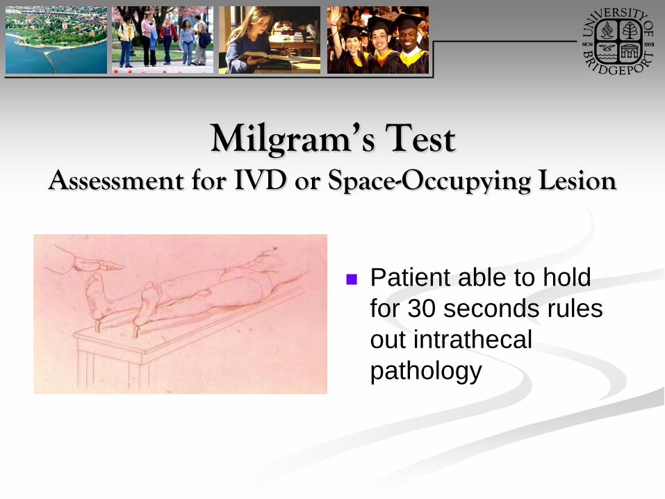
Milgram’s TestAssessment for IVD or Space-Occupying Lesion
Patient able to hold for 30 seconds rules out intrathecal pathology

Positive Milgram’s Test
Indicates intrathecal or extrathecal pathology
The test is positive if the patient experiences low back pain

Intrathecal Pathology
Intrathecal pathology may involve a spinal tumor.

Extrathecal Pathology
Extrathecal pathology may involve a herniated disc or space occupying lesion
Key to Success
“Diagnosis is the key to successful treatment!”

Final Comments
Perform a competent evaluation
Properly assess your patient
Educate your patient Provide high quality
care Be kind…