lower extremity wounds: what is new in saskatchewan •for most dfu, use the lew pathway referral...
TRANSCRIPT

MODULE 6: Diabetic Foot Ulcers
(Approx 30 minutes)

Learning Objectives
Assess severity of diabetic foot ulcers and wound infections
Implement recommended antimicrobial management for infected wounds
Describe the role of debridement and offloading in management of diabetic feet
Use pathway tools to support recommended management of diabetic foot ulcers
At the end of this module, learners will be able to:
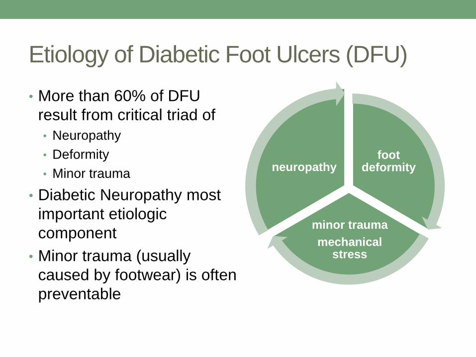
Etiology of Diabetic Foot Ulcers (DFU)
• More than 60% of DFU
result from critical triad of
• Neuropathy
• Deformity
• Minor trauma
• Diabetic Neuropathy most
important etiologic
component
• Minor trauma (usually
caused by footwear) is often
preventable
foot deformity
minor trauma
mechanical stress
neuropathy

Risk factors for DFU
• Diabetic neuropathy
• Peripheral Arterial Disease or other CVD
• Foot deformity
• Past history of foot ulcer
• Prior amputation
• Poor glycemic control
• Smoking
• Diabetic nephropathy – particularly end-stage CKD on
dialysis (4x increased risk of DFU compared to CKD not
on dialysis)

Characteristics of DFU
• Skin ulceration occurs over bony
prominence or on callus
• Ulcer typically deep with distinct
borders
• Surrounding skin atrophic
• Associated diabetic foot complications
– neuropathy, signs of chronic
ischemia/PAD
• Ulceration may be obscured by
• callus
• boggy slough (yellow, green, brown)
• dry eschar (black, brown)
• blister
Ulcer covered by eschar
Ulcer covered
by blister

Relatively innocent-
looking ulcer in callus but
significant subcutaneous
tissue involvement
Suspect underlying
tissue ulceration/necrosis
if adjacent area is purple,
painful, fluctuant, hotter
or colder than
surrounding skin.
Blister formed
under callus and
extending onto
medial foot

Stages of DFU
1. Skin intact; localized erythema over bony prominence, pressure point or callus
2. Shallow wound or ruptured blister
3. Full tissue loss, but no tendon or bone exposure
4. Deep tissue loss with bone, tendon or muscle exposure

Principles of DFU Management
Wound dressings:
• maintain moist wound healing
environment
Offloading:
• redistribution of pressure away
from ulcerated skin
Treat infection:
• oral or systemic
antibiotics – use LEW pathway
antibiotic protocol as a guide
• there is no role for topical
antibiotics if suspicion of infection
Mainstay of
wound healing is
offloading
pressure from
wound. “It is not
what one puts on
a wound that heals
it, but what one
takes off.”

Principles of DFU Management
Debridement:
• done by surgeons and wound care specialists
Improve blood supply:
• endovascular intervention/surgery if large vessel PAD (This
requires assessment by vascular surgery, hence rationale
for specialist referral of all DFU)
Optimal glycemic control:
• blood sugars higher than 11mmol/L impair immune
response and wound healing

Principles of DFU Management

LEW referral for DFU management
• For most DFU, use the LEW Pathway referral form to
make a non-urgent referral to both the homecare team and
a vascular specialist.
• Homecare can initiate appropriate wound care while waiting
for specialist consult.
• Medical leaders for the LEW Pathway recommend that ALL
diabetic foot ulcers are referred to vascular surgery for an
initial assessment to determine if vascular investigations
/interventions are required.
• Once the LEW Pathway is fully established, and there are
adequately trained and experienced nurses throughout the
province, this early specialist referral may not be required
for non-severe DFUs.

LEW Pathway referral form
To open a PDF
of Referral Form-
Lower Extremity
Wound Pathway,
click on the link
in the sidebar.

Diabetic Foot Ulcer
Clinical pearls – suggested by wound care nurses and surgeons:
• >2 cm of skin erythema around a diabetic lower limb ulcer may be indicative of limb-threatening infection (need to admit for IV antibiotics, limb elevation, wound care).
• Presence of pain in a previously insensate foot can be the first and most important indicator of severe infection or underlying osteomyelitis.
• Need to consider other diabetic complications such as CKD before selection and dosing of antibiotics for diabetic foot infections.
• Selection of antibiotic must be guided by previous/recent antibiotics used by patient.
• Poor glycemic control delays ulcer healing.

Signs of Wound Infection in DFU
Increased bacterial burden in wound results in:
• Friable or hyper-granulation tissue in wound bed (raised,
deep/bright red, bleeds readily)
• Increased amount of exudate
• Purulent exudate (greenish or yellow)
• Necrotic slough on wound base
• Odour persistent after wound cleansing
• Increased surrounding skin erythema
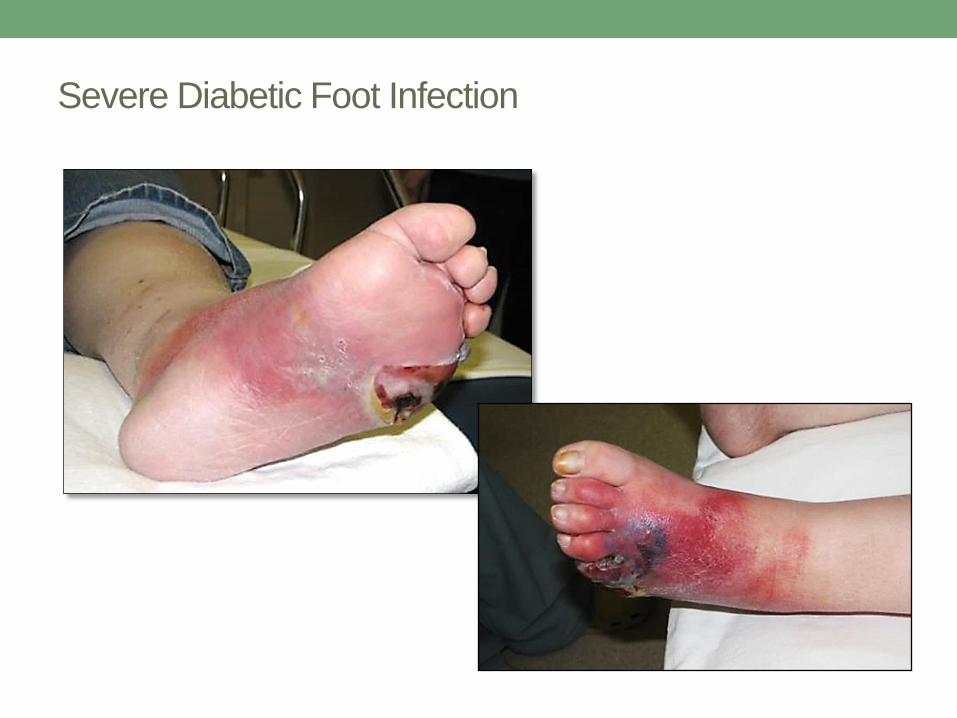
Severe Diabetic Foot Infection
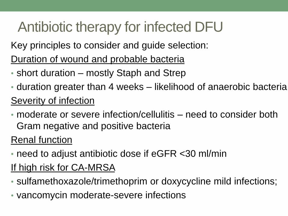
Antibiotic therapy for infected DFU Key principles to consider and guide selection:
Duration of wound and probable bacteria
• short duration – mostly Staph and Strep
• duration greater than 4 weeks – likelihood of anaerobic bacteria
Severity of infection
• moderate or severe infection/cellulitis – need to consider both
Gram negative and positive bacteria
Renal function
• need to adjust antibiotic dose if eGFR <30 ml/min
If high risk for CA-MRSA
• sulfamethoxazole/trimethoprim or doxycycline mild infections;
• vancomycin moderate-severe infections
Antibiotic therapy for infected DFU
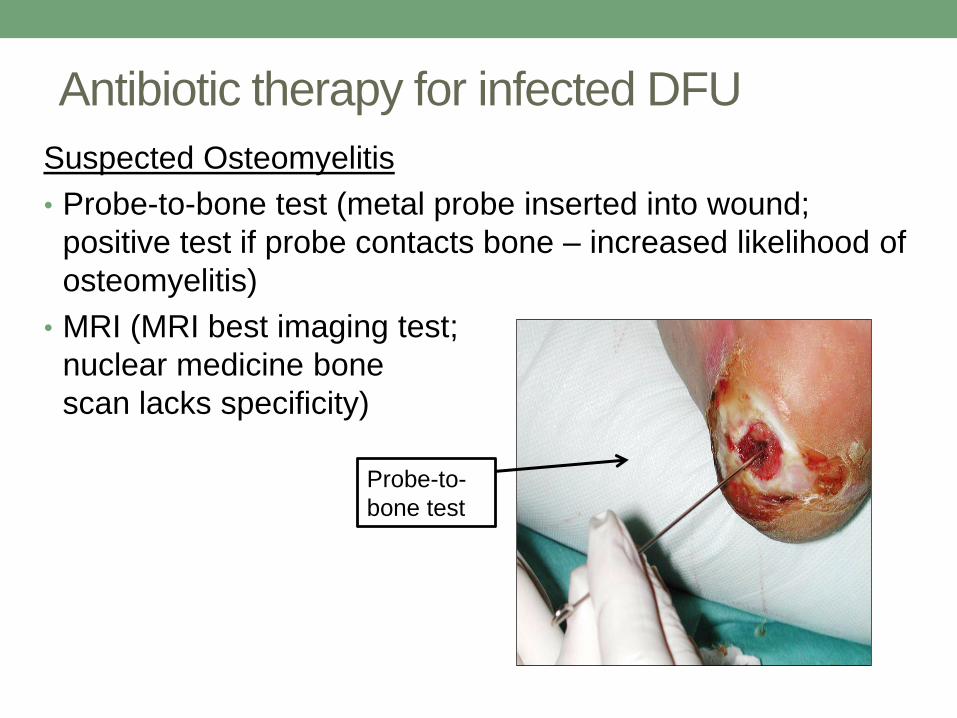
Suspected Osteomyelitis
• Probe-to-bone test (metal probe inserted into wound;
positive test if probe contacts bone – increased likelihood of
osteomyelitis)
• MRI (MRI best imaging test;
nuclear medicine bone
scan lacks specificity)
Probe-to-
bone test
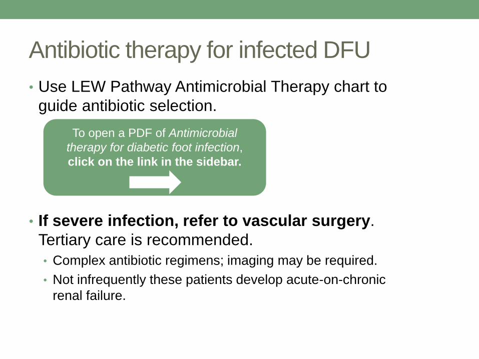
Antibiotic therapy for infected DFU
• Use LEW Pathway Antimicrobial Therapy chart to
guide antibiotic selection.
• If severe infection, refer to vascular surgery.
Tertiary care is recommended.
• Complex antibiotic regimens; imaging may be required.
• Not infrequently these patients develop acute-on-chronic
renal failure.
To open a PDF of Antimicrobial
therapy for diabetic foot infection,
click on the link in the sidebar.


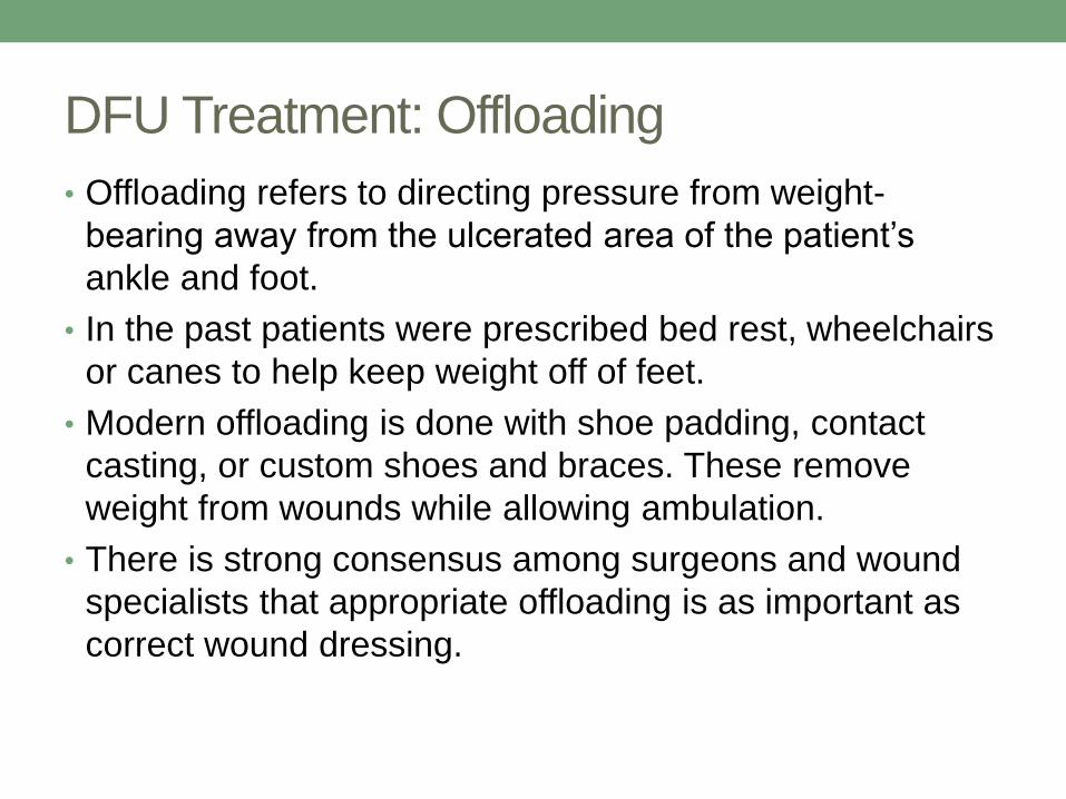
DFU Treatment: Offloading
• Offloading refers to directing pressure from weight-
bearing away from the ulcerated area of the patient’s
ankle and foot.
• In the past patients were prescribed bed rest, wheelchairs
or canes to help keep weight off of feet.
• Modern offloading is done with shoe padding, contact
casting, or custom shoes and braces. These remove
weight from wounds while allowing ambulation.
• There is strong consensus among surgeons and wound
specialists that appropriate offloading is as important as
correct wound dressing.
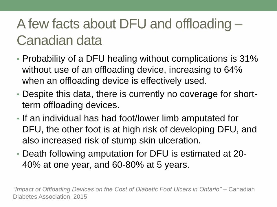
A few facts about DFU and offloading –
Canadian data
• Probability of a DFU healing without complications is 31%
without use of an offloading device, increasing to 64%
when an offloading device is effectively used.
• Despite this data, there is currently no coverage for short-
term offloading devices.
• If an individual has had foot/lower limb amputated for
DFU, the other foot is at high risk of developing DFU, and
also increased risk of stump skin ulceration.
• Death following amputation for DFU is estimated at 20-
40% at one year, and 60-80% at 5 years.
“Impact of Offloading Devices on the Cost of Diabetic Foot Ulcers in Ontario” – Canadian
Diabetes Association, 2015

Short-term offloading
• Customized padding can be placed in
the patient’s shoe; shoe must be deep
enough to accommodate this.
• Consider “healing shoe” or “post-op
shoe” if patient shoes don’t
accommodate padding (for short term
use only)
• Smaller (less than 2 cm), less complex
wounds may heal with short-term
offloading

Coverage for short-term offloading
• No insurance coverage for these products.
• Padding may be supplied by homecare.
• Healing shoes may be provided by homecare but billed to
patient.
• Healing shoes available for purchase from podiatrist,
drugstore, shoe store or medical supply store, or order
on-line.
• Cost starts at around $30.

Longer-term offloading
Total Contact Cast (TCC)
• Provides best wound healing
• Must be removed weekly for wound
assessment and re-applied
• Should be applied by specially
trained cast technician – cost is
covered as applied in hospital
outpatient setting.

Longer-term offloading
Removable Cast/Air Walker
• Most commonly used type of orthoses for
diabetic foot
• Custom fit by orthotist
• Studies show that healing is the same
with walker as with TCC, as long as the
walker is worn consistently
• Because the cast walker is removable, the
patient may not wear it all the time
• If patient cannot feel pain due to neuropathy, there may
be no incentive to comply with offloading
• Need to encourage patient to wear device at all times for
best results
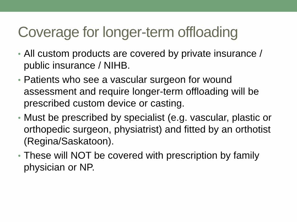
Coverage for longer-term offloading
• All custom products are covered by private insurance /
public insurance / NIHB.
• Patients who see a vascular surgeon for wound
assessment and require longer-term offloading will be
prescribed custom device or casting.
• Must be prescribed by specialist (e.g. vascular, plastic or
orthopedic surgeon, physiatrist) and fitted by an orthotist
(Regina/Saskatoon).
• These will NOT be covered with prescription by family
physician or NP.

DFU: Monitoring and surveillance
• Diabetic foot ulcers can close in 3-4 months with optimal
management.
• To prevent recurrence, regular foot check-up (at every
diabetic review/CDM visit) and foot care are of utmost
importance.
• Encourage patient self-care.
• Encourage patients to pay attention to footwear.
• Primary care providers can play an important role in
connecting patients to community resources for foot care
and diabetes management.

Physician/NP role in supporting DFU patient
• Individuals with DFU tend to possess fewer cognitive
resources than individuals with similar duration DM
without foot ulcer.*
• This is potentially problematic as management of DFU
requires increased demands for self-treatment and
adherence to treatment regimens that may be complex
and of long duration.
• Family physician/nurse practitioner can play an important
role in explaining and promoting adherence; advocating
for patient.
*Reference: Natovich R et al. Cognitive Dysfunction: Part and Parcel of the Diabetic Foot.
Diabetes Care 2016 May; dc 152838. Case control study in Israel – 95 Pts with DFU and 95
controls matched for sex, age, duration DM; Individuals with diabetic foot ulcers had significantly
(P < 0.001) lower cognitive scores than individuals with diabetes without this complication.

References
• Hingorani, Anil et al. The management of diabetic foot: A
clinical practice guideline by the Society for Vascular Surgery in
collaboration with the American Podiatric Medical Association
and the Society for Vascular Medicine. Journal of Vascular
Surgery, 2016: Volume 63 , Issue 2 , 3S - 21S.
• Registered Nurses’ Association of Ontario (2013). Assessment
and Management of Foot Ulcers for People with Diabetes (2nd
ed.). Toronto, ON: Registered Nurses’ Association of Ontario.
• Health Service Executive, Ireland (2009). National best practice
and evidence based guidelines for wound management.
• Bugs and Drugs – www.bugsanddrugs.ca 2016 Anti-infective
Guidelines for Community-acquired Infections

END OF MODULE Proceed to Module 6 Quiz