low dose excellent image quality rapid reconstructionmarwamed.ps/img/cms/pdf folder/hdx...
TRANSCRIPT

Low Dose Excellent Image Quality Rapid Reconstruction

Efficient 3 in 1 Dental X-ray System
CBCT
Panoramic image
> Precise 3-D Anatomical structures - Accurate diagnosis for doctors - Safe implant for patients> Significant reduction in X-ray dose
Cephalometric> Best image quality for diagnosis> Least X-ray exposure for patients
> Anatomic structure> Minimum distortion and artifact
Lateral
TMJ
PA SMV Carpus
Lateral PA Carpus

0.2 Voxel slice 16×8cm
0.2 Voxel slice 16×14.5cm stitch
When running into the same Voxel sizes
FOV 16×8cm: 10 Sec
FOV 16×14.5cm: 30 Sec
Rapid Reconstruction Rapid Visualization
A B HDXWILL
60
3021
Computation times of reconstruction are significantly reduced by the latest GPU technology. It takes much less than competitors.
Low Dose! Excellent Image Quality! Rapid Reconstruction!

Feature
Outstanding Auto-Focusing
Remarkable MAR(Metal Artifact Reduction) algorithm
UFS(Ultra Fast Scan technology)
With MAR
Normal scan
Autofocusing correction
Without MAR
Ultra fast scan
Misaligned result
24 Sec
Stitching: 36 Sec
8 Sec
Stitching: 16 Sec

Diagnosis of TMJ disorder
Bilateral TMJ in a single exposure by wide FOV
Abnormal TMJ image
Accurate 3D volume rendering image
Normal TMJ image
Accurate diagnosis of abnormal TMJ by one shooting.
Clinical Application
Location and angle diagnosis of bilateral mandibular condyle erosion
Diagnosis of TMJ ankylosis & mandibular fossa
DENTRI CT images of 16cm that can check both condyle at once

Clinical Application
Implant diagnosis and shape check-up of anatomical structure
Maximum utilization of three-dimensional measured value of residual bone by one to one actual measurement.
Distance and angle measurement of mental nerve for implant fixation
Planning of implant size and ideal position Maxillary sinus septum

Maxillary sinus septum
Mental foramen
Incisive nerve
Inferior alveolar nerve pathway
Clinical Application
Implant diagnosis and shape check-up of anatomical structure before surgery.
Simultaneous checking of the location of posterior superior alveolar artery and inferior alveolar nerve by one shot.
Acquire the upper and lower jaw structure.
Integrated prosthetic designs and implant planning

Location of horizontally impacted both third molar teeth
Clinical Application
Diagnosis of periodontal pocket and impacted teeth.
Accurate diagnosis of bilateral impacted third molar teeth with just one scanning.
Recognition of relation among buccolingual location of horizontally impacted the third molar tooth, root, and mandibular canalPeriapical lesion

Sinus mucocele
Mucous cyst
Chronic sinusitis spread to orbit
Sinusitis
Clinical Application
Diagnosis of maxillary sinus and sinusitis.
Hypertrophic Mucous Cyst check by high resolution image which is not shown on general radiograph.
Sinus Implant Surgery
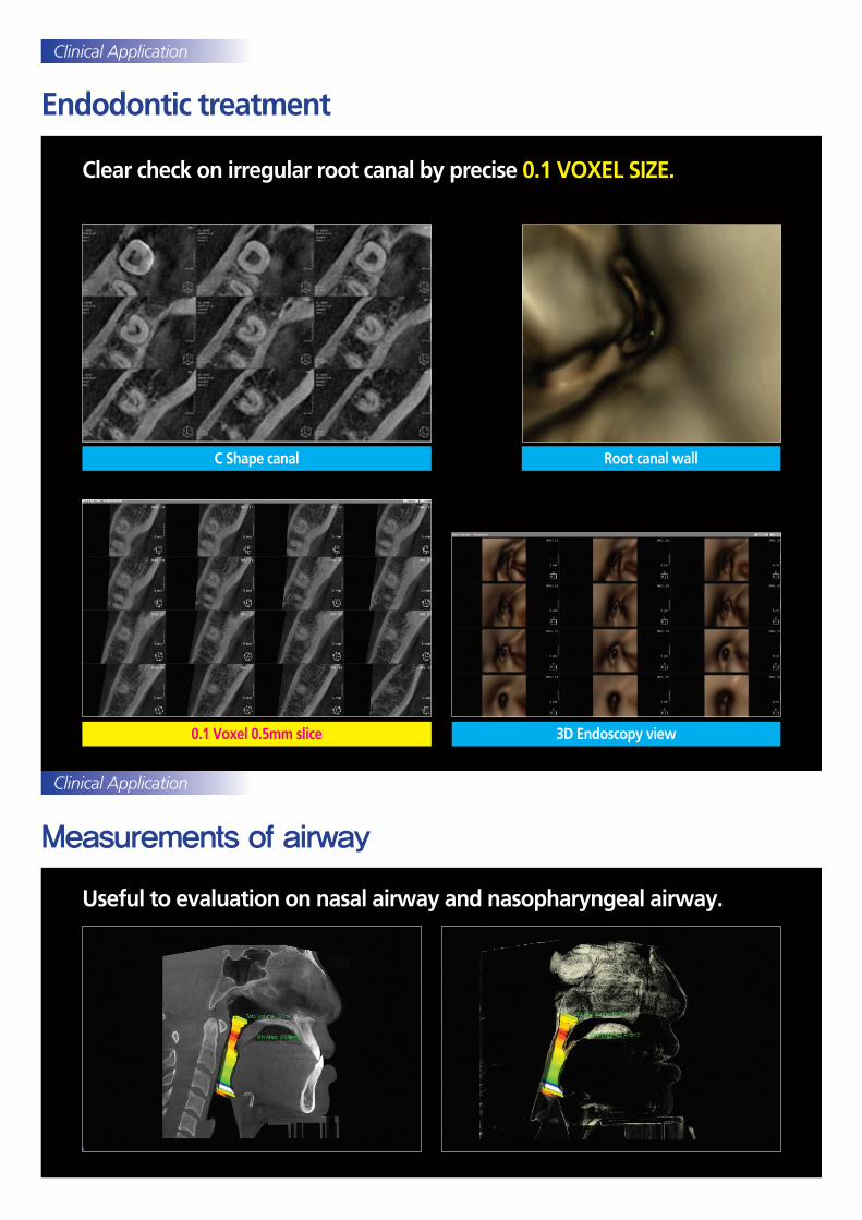
Endodontic treatment
Measurements of airway
Clear check on irregular root canal by precise 0.1 VOXEL SIZE.
Useful to evaluation on nasal airway and nasopharyngeal airway.
0.1 Voxel 0.5mm slice 3D Endoscopy view
Root canal wallC Shape canal
Clinical Application
Clinical Application

Orthodontic treatment
Easy measurement of facial asymmetry by wide FOV 16 × 14.5cm which is necessary to cover the midsagittal plane.
Clinical Application
Location of impacted teeth check
Maxillary asymmetry measurement
3D Cephalometric image without distortion
Bone thickness measurement for miniscrew insertion
Mandibular asymmetry measurement
Asymmetric analysis

Software
Other equipment
Will-Master OnDemand 3D
Patient image management software and 2D viewer
3D viewer
WillCeph
Cephalometric analysis Software(Optional)
Dental unit & chair Intra Oral X-ray With Sensor System
Wireless Portable X-ray
Convenient to Use!Excellent Image Quality!Easy Operation!Reduced Radiation Dose
Star-X Intra Oral X-ray
High quality ImageErgonomic DesignReduced Radiation DoseEasy & Safe operation
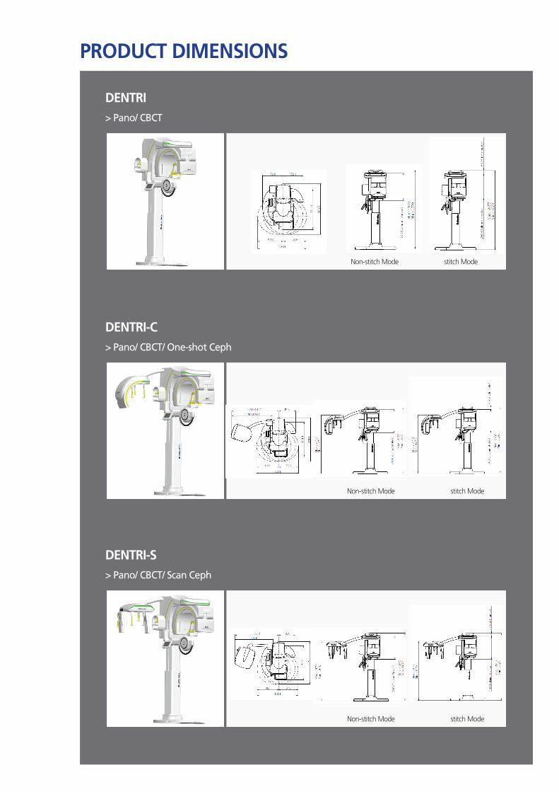
PRODUCT DIMENSIONS
> Pano/ CBCT
> Pano/ CBCT/ One-shot Ceph
> Pano/ CBCT/ Scan Ceph
DENTRI
DENTRI-C
DENTRI-S
Non-stitch Mode stitch Mode
Non-stitch Mode stitch Mode
Non-stitch Mode stitch Mode

SPECIFICATIONS
CN
DE0
3
※ Specifications are subject to change without notice for improvement of product performance.
MODEL PANORAMACEPHALO
CTFOV
ONE-SHOT SCAN 16×14.5 FREE FOV 16×8
DENTRIStitch Mode ● ● ● ● ●
Non-stitch Mode ● ● ● ●
DENTRI-CStitch Mode ● ● ● ● ● ●
Non-stitch Mode ● ● ● ● ●
DENTRI-SStitch Mode ● ● ● ● ● ●
Non-stitch Mode ● ● ● ● ●
X-ray beam Cone beam Patient Position Standing
Tube Voltage 60 kV-110 kV Patient AlignmentVertical Column: electric motion(Optional) Column: electric motion Temple Support: motion
Focal Spot 0.5mm Control Device Alignment: Touch panel Exposure & Image acquisition: PC
Voxel size(CT) 0.10, 0.15, 0.20, 0.25, 0.30
Demensions (W×D×H)
DENTRI: 1201㎜×1361㎜× 2455㎜ DENTRI-C(Moving type): 2006㎜×1361㎜×2455㎜ DENTRI-C(Fix type): 1999㎜×1361㎜×2455 ㎜ DENTRI-S: 1941㎜×1361㎜×2455㎜ (tolerance: ±5%)
Detector Type
CT & Panorama CMOS or aSi
One shot type Cephalo Direct(a-Se, TFT) Install Standing
Scan type Cephalo TDI CCD
System Weight (tolerance: ±10%)
DENTRI: 243 kg DENTRI-C: 270 kg DENTRI-S: 260 kg
Gray Scale
CT & Panorama 14 bits or 16 bits
One shot type Cephalo 14 bits
Scan type Cephalo 16 bits
Scan Time
CT(Pulsed) 8 s, 16 s, 24 s, 36 s
Panorama 14 s and less
One shot type Cephalo 0.5 s, 1.0 s, 1.5 s, 2.0 sField of View(CT) (cm×cm) (Diam.×Height)
Max. 16×14.5(Stitch) 16×8 Free FOV
Scan type Cephalo 8.2 s and less OS Window 7 64 bit
Reconstruction Time (With MAR) less than 40 s Memory 16 G
Sales OfficeTaehwa Bldg, 29, Insa-dong 5-gil, Jongno-gu, Seoul, Korea Tel.82-1544-5735 / Fax. 82-2-2003-8497 www.hdx.co.kr
R&D Center14F, KCC Weltz Valley, 205, Gasan digital 1-ro, Geumcheon-gu, Seoul, Korea www.hdx-will.com
Manufacturing Facility#105, 201, 202, 203, 204, 38, Osongsaengmyeong4-ro, Osong-eup, Heungdeok-gu, Cheongju-si, Chungcheongbuk-do, Korea Tel. 82-43-710-7318 / Fax. 82-43-710-7312