long-term follow-up analysis of percutaneous balloon ... · citation: ezhumalai b, awasthy n,...
TRANSCRIPT
*Corresponding author email: [email protected] Group
Symbiosis www.symbiosisonline.org www.symbiosisonlinepublishing.com
Long-Term Follow-Up Analysis of Percutaneous Balloon Pulmonary Valvuloplasty: Experience of an Indian
Tertiary Care HospitalBabu Ezhumalai1*, Neeraj Awasthy2, Ajit Ananthakrishna3, Santhosh Satheesh3, Balachander Jayaraman3
1Department of Interventional Cardiology, MIOT International, Chennai, India.2Department of Paediatric Cardiology, Max Super Speciality Hospitals, New Delhi, India.
3Department of Cardiology, Jawaharlal Institute of Postgraduate Medical Education and Research, Puducherry, India.
Journal of Clinical Trials in Cardiology Open AccessResearch article
AbstractObjectives: Our objectives were to study the effectiveness and long-term follow-up outcomes of percutaneous Balloon Pulmonary
Valvuloplasty (BPV) performed in adults and children at an Indian tertiary care hospital.
Methods: This retrospective study included patients with more than mild congenital valvular Pulmonary Stenosis (PS) having echocardiographic peak gradient ≥50mmHg, who underwent BPV in a tertiary care hospital in India from 1988 to 2011. Clinical profile, echocardiographic details, procedural data, complications etc were studied. Patients were followed with annual echocardiographic and clinical assessment.
Results: 108 patients including 37 children and 71 adults underwent BPV. Single balloon technique was mostly employed (99.1%). The mean balloon/annulus ratio was 1.22 ± 0.02. The transvalvular peak-to-peak gradient measured immediately before and after valvuloplasty was 103.4 ± 33.9mmHg and 33.9 ± 17.5mmHg respectively (65.1% reduction, p<0.0001). Intervention was successful and partially successful in 100 (92.6%) subjects and 8 (7.4%) subjects respectively, irrespective of age and gender. Less than moderate pulmonary regurgitation occurred in 73 (67.6%) patients. At discharge, peak Doppler gradient was reduced by 74.1% compared to admission value. This was further significantly reduced (25.4%) at short-term follow-up. This reduction in gradient was sustained at mid-term and long-term follow-up. The median duration of follow-up was 15years (range: 2 to 27 years). All patients were asymptomatic at short-term and mid-term follow-up. Two (1.9%) patients who developed restenosis on long-term underwent repeat valvuloplasty.
Conclusions: BPV is a well-tolerated non-surgical treatment modality for congenital valvular PS producing highly effective results irrespective of age or gender. The results are excellent at immediate-term, short-term, mid-term and long-term.
Keywords: Balloon Pulmonary Valvuloplasty; Pulmonary Stenosis; Pulmonary Valve; BPV; PS;
Received: November 10, 2016; Accepted: November 15, 2016; Published: November 30, 2016
*Corresponding author: Babu Ezhumalai, MIOT International, 4/112, Mount Poonamallee Road, Manapakkam, Chennai - 600 089, India, Tel: +91 9818640604; E-mail: [email protected]
IntroductionBalloon Pulmonary Valvuloplasty (BPV) is the earliest
percutaneous balloon dilatation procedure and until now it has remained the most successful of all percutaneous balloon valvuloplasty procedures.
The treatment for critical valvular Pulmonary Stenosis (PS) by surgical valvotomy started in the middle of 20th century [1]. The first attempt to relieve valvular PS was done in the early 1950s by using a ureteral catheter with a wire [2]. After almost three decades, critical PS in a neonate was relieved by rapidly withdrawing an inflated Berman catheter across the severely narrowed Pulmonary Valve (PV) [3]. The first series of BPV was published in 1982 by KAN et al [4]. Since then BPV became the treatment of choice for congenital isolated valvular PS and the
technique of BPV has been carried out successfully till date with only minor modifications like the use of double-balloon technique and Inoue balloon [5,6].
Dysplastic pulmonary valve is usually of familial origin especially Noonan’s syndrome. Some operators have performed BPV in patients with dysplastic pulmonary valve, though the results were not as promising as in valvular PS [7].
The present study elucidates a long term data of quite a large number of patients undergoing balloon pulmonary valvuloplasty in the Indian subcontinent. There is lack of such a literature from this region in the recent past. Moreover this study provides information on balloon; annulus ratio and Pulmonary Regurgitation (PR) following BPV and also compares the results of BPV between adults and children.

Page 2 of 5Citation: Ezhumalai B, Awasthy N, Ananthakrishna A, Satheesh S, Jayaraman B (2016) Long-Term Follow-Up Analysis of Percutaneous Balloon Pulmonary Valvuloplasty: Experience of an Indian Tertiary Care Hospital. J Clin Trial Cardiol 3(1): 1-5.
Long-Term Follow-Up Analysis of Percutaneous Balloon Pulmonary Valvuloplasty: Experience of an Indian Tertiary Care Hospital
Copyright: © 2016 Ezhumalai, et al.
ObjectivesOur objectives were to study the effectiveness and long-
term follow-up outcomes of percutaneous Balloon Pulmonary Valvuloplasty (BPV) performed in adults and children at an Indian tertiary care hospital.Materials and methods
This retrospective, single center, observational study included patients with more than mild congenital valvular PS having echocardiographic peak gradient across Pulmonary Valve (PV) ≥ 50mmHg, who underwent BPV at JIPMER, a tertiary care hospital in India from 1988 to 2011. Ethical committee approval was obtained for this study. The clinical profile, echocardiographic details, procedural data, complications and follow-up data were studied in all these patients.
Antibiotic prophylaxis was administered before starting the procedure. Local anaesthesia was used for adults while general anaesthesia was used for paediatric patients. Vascular access was obtained usually in right femoral vein. Arterial line placed in left femoral artery or radial artery was used for monitoring pressure. All hemodynamic parameters and vitals were monitored continuously throughout the procedure.
Right heart catheterization was done routinely in all patients. Right Ventricle (RV) angiogram was performed using Berman angiographic catheter in lateral view. Moreover, RV angiogram was also useful in identifying infundibular stenosis.
Stenosed PV was crossed using Swan Ganz catheter and baseline transvalvular peak-to-peak pressure gradient was recorded. The size of the balloon chosen was 120% to 140% of the diameter of annulus of PV measured in echocardiography. The balloon was advanced over 0.035” 260cm J tipped Amplatz superstiff guidewire and positioned across the stenotic PV. This balloon was inflated by hand for brief period using diluted contrast (1 in 4 dilution) until the waist produced by the stenotic PV on the balloon disappeared. Post-valvuloplasty transvalvular pressure gradient was again recorded. Repeated balloon inflations were done until satisfactory reduction in transvalvular gradient occurred. Injection metoprolol was administered when there was significant residual gradient because of infundibular spasm. RV angiogram was not routinely performed after valvuloplasty. Unfractionated heparin (50units/kg) was administered after balloon dilatation.
Results of intervention were declared as ‘successful’ when RV to pulmonary artery (PA) peak-to-peak gradient was reduced by 50% or more at cardiac catheterization, and ‘partially successful’ when the gradient was reduced by 30-50%. Before discharge, post-valvuloplasty gradient was assessed echocardiographically. After discharge, patients were followed with annual echocardiographic and clinical assessment.
Statistical analysis was performed using SPSS software (SPSS Inc. Released 2007. SPSS for Windows, Version 16.0. Chicago: SPSS Inc). Categorical variables are expressed as numbers (percentage) while continuous variables with normal
distribution are expressed as ‘Mean±Standard Deviation’. Chi-square Test, Fisher’s exact test and Student’s paired t test were used for analyses. P value <0.05 was considered significant for all tests.Results
Totally 118 subjects were planned for BPV based on echocardiography during the study period. Seven subjects were excluded after RV angiogram due to presence of significant infundibular stenosis in 3 subjects, additional presence of supravalvular narrowing in 2 subjects, and diffuse narrowing of RVOT in 2 subjects. Three patients were excluded from the study because balloon could not be negotiated through very critically stenosed PV and these 3 children underwent surgery. The remaining 108 patients who underwent BPV were included in our study. The baseline characteristics of these patients are mentioned in Table 1. There were 48 (44.4%) males and 60 (55.6%) females (p=0.13; Chi-square test) with a mean age of 17.7±10.5 years (range: 6 months to 50 years). There were 37 (34.3%) children and 71 (65.7%) adults (p<0.0001; Chi-square test). The associated anomalies in these patients are depicted in Table 2.
The mean balloon/annulus ratio in our study was 1.22 ± 0.02 (1.23 ± 0.02 in children vs 1.22 ± 0.02 in adults; p=0.03).
Table 1: Baseline characteristics of 108 patients
Parameter n (%)
Age (mean±SD) 17.7±10.5
Males 48 (44.4%)
Females 60 (55.6%)
Children 37 (34.3%)
Adults 71 (65.7%)
Symptoms ≥ NYHA class III 105 (97.2%)
Echocardiography Peak PV gradient in mmHg (mean±SD) 119.6±35.3
TR (more than moderate) 108 (100%)
RV dysfunction (TAPSE<17mm) 1 (0.9%)
PV annulus in mm (mean±SD) 20.1±0.9Note: n - Number of Patients; SD - Standard Deviation; NYHA - New York Heart Association Classification of Cardiac Symptoms; TR - Tricuspid Regurgitation; RV - Right Ventricle; PV - Pulmonary Valve; TAPSE - Tricuspid Plane Annular Systolic Contraction
Table 2: Associated anomalies in patients who underwent percutaneous balloon pulmonary valvuloplastyAssociated anomalies n (%)Facial dysmorphism 4 (3.7%)Atrial septal defect 3 (2.8%)Mental retardation and deafness 2 (1.9%)Left-sided superior venacava 1 (0.9%)Situs inversus with dextrocardia 1 (0.9%)Note: n - Number of Patients; TR - Tricuspid Regurgitation; RV - Right Ventricle
Page 3 of 5Citation: Ezhumalai B, Awasthy N, Ananthakrishna A, Satheesh S, Jayaraman B (2016) Long-Term Follow-Up Analysis of Percutaneous Balloon Pulmonary Valvuloplasty: Experience of an Indian Tertiary Care Hospital. J Clin Trial Cardiol 3(1): 1-5.
Long-Term Follow-Up Analysis of Percutaneous Balloon Pulmonary Valvuloplasty: Experience of an Indian Tertiary Care Hospital
Copyright: © 2016 Ezhumalai, et al.
Single balloon technique was employed in 107 (99.1%) patients (Table 3), while double balloon technique was used in 1 (0.9%) patient with PV annulus greater than 22mm. Sequential balloon dilatation was done in 29 (26.9%) patients. The number of balloon inflations needed for adequately dilating PV and relieving stenosis in these patients is also depicted in Table 3.
The mean transvalvular peak-to-peak gradient measured before and after BPV were 103.4 ± 33.9mmHg and 33.9 ±17.5mmHg respectively (p<0.0001, Students’ paired t test). The mean of percentage reduction in the gradient was 65.1 ± 11.9%. Overall, intervention was successful in 100 (92.6%) subjects and partially successful in 8 (7.4%) subjects. Statistically there was no significant difference between children and adults, or males and females with respect to the prevalence of successful and partially successful results (Table 4). Infundibular spasm leading to inadequate fall in transvalvular pressure gradient occurred in 31 (26%) patients. On administration of injection metoprolol, there was further 20% reduction in transvalvular pressure gradient immediately in these patients with infundibular spasm. Less than moderate PR could be demonstrated using Doppler in 73 (67.6%) patients with mild PR and mild-to-moderate PR occurring in 59 (54.6%) and 14 (12.9%) patients respectively. Severe PR did not occur in any patient.
In this study, there were 4 (3.7%) patients with dysplastic PV who underwent BPV. All were children with facial dysmorphism. Interventions in these patients with dysplastic PV resulted in success and partial success in 3 patients and 1 patient respectively.
The overall prevalence of complications of BPV in our study was 12.0% with major complications 4 (3.7%) being less common than minor complications 9 (8.3%). Vasovagal syncope 8 (7.4%) was the most common minor complication followed by femoral vein thrombosis 1 (0.9%). Each of the following major complication occurred in 1 (0.9%) patient. Complete heart block during BPV in a patient necessitated temporary pacing but got spontaneously reverted to normal sinus rhythm after a few hours. Transient atrial fibrillation lead to embolic stroke and hemiplegia. The patient who developed cardiac arrest was successfully revived on table. There was one mortality in a child (5 years of age) because of development of suicide RV secondary to balloon dilatation of PV.
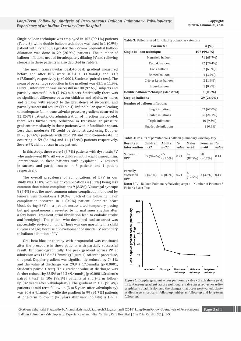
Oral beta-blocker therapy with propranolol was continued after the procedure in those patients with partially successful result. Echocardiographically, the peak gradient across PV at admission was 115.6 ± 34.7mmHg (Figure 1). After the procedure, this peak Doppler gradient was significantly reduced by 74.1% and the value at discharge was 29.9 ± 17.5mmHg (p<0.0001, Student’s paired t test). This gradient value at discharge was further reduced by 25.5% to 22.3 ± 9.4mmHg (p<0.0001, Student’s paired t test) in 106 (98.1%) patients at short-term follow-up (≤2 years after valvuloplasty). The gradient in 103 (95.4%) patients at mid-term follow-up (3 to 5 years after valvuloplasty) was 20.6 ± 9.1mmHg, while the gradient in 99 (91.7%) patients at long-term follow-up (≥6 years after valvuloplasty) is 19.6 ±
Table 4: Results of percutaneous balloon pulmonary valvuloplastyResults of intervention
Childrenn=37
Adultsn=71
*p value
Malesn=48
Femalesn=60
*p value
Successful BPV 35 (94.6%) 65
(91.5%) 0.71 42 (87.5%)
58 (96.7%) 0.14
Partially successful BPV
2 (5.4%) 6 (8.5%) 0.71 6 (12.5%) 2 (3.3%) 0.14
Note: BPV - Balloon Pulmonary Valvuloplasty; n – Number of Patients; * Fisher’s Exact Test
Figure 1: Doppler gradient across pulmonary valve - Graph shows peak instantaneous gradient across pulmonary valve assessed echocardio-graphically at admission and the changes that occur post-valvuloplasty at discharge, short-term follow-up, mid-term follow-up and long-term follow-up.
Table 3: Balloons used for dilating pulmonary stenosis
Parameter n (%)
Single balloon technique 107 (99.1%)
Mansfield balloon 71 (65.7%)
Tyshak balloon 22 (20.4%)
Cook balloon 7 (6.5%)
Scimed balloon 4 (3.7%)
Cribier Letac balloon 2 (1.9%)
Inoue balloon 1 (0.9%)
Double balloon technique (Mansfield) 1 (0.9%)
Step-up balloons 29 (26.9%)
Number of balloon inflations
Single inflation 67 (62.0%)
Double inflations 26 (24.1%)
Triple inflations 10 (9.3%)
Quadruple inflations 1 (0.9%)

Page 4 of 5Citation: Ezhumalai B, Awasthy N, Ananthakrishna A, Satheesh S, Jayaraman B (2016) Long-Term Follow-Up Analysis of Percutaneous Balloon Pulmonary Valvuloplasty: Experience of an Indian Tertiary Care Hospital. J Clin Trial Cardiol 3(1): 1-5.
Long-Term Follow-Up Analysis of Percutaneous Balloon Pulmonary Valvuloplasty: Experience of an Indian Tertiary Care Hospital
Copyright: © 2016 Ezhumalai, et al.
10.4mmHg. The peak Doppler gradient reduced at short-term was sustained at mid-term and long-term follow-ups. There was no statistically significant difference between Doppler gradients of short-term, mid-term and long-term follow-ups (p=0.12, One-way ANOVA test).
The median duration of follow-up in this study was 15years (range: 2 years to 27 years). There was no event of re-hospitalization for heart failure reported in this study. All patients were asymptomatic at short-term and mid-term follow-up. Two (1.9%) patients became symptomatic when they developed significant restenosis (peak Doppler gradient more than 50mmHg) on long-term follow-up, 13 to 18 years after valvuloplasty. Both patients had partially successful result with the initial procedure. Repeat BPV performed in these two patients produced successful results. No patient in this study needed valve replacement for PR neither acutely nor on long-term follow-up.Discussion
In our study, BPV produced equally and highly effective results in both adults and children irrespective of the gender [8-10]. The immediate improvement thus produced persisted on short-term, mid-term and long-term follow-up [11-16].
Though conventionally recommended balloon/annulus ratio for performing BPV is 1.2-1.4, current recommendations suggest a ratio of 1.2-1.25 [17-19]. The overall mean balloon/annulus ratio of 1.22 ± 0.02 in our study is falling within this range. In our study, single balloon technique was predominantly employed than double balloon technique. As per prior studies, the results produced by double balloon technique is comparable to single balloon technique but it is not superior when the balloon/annulus ratios are equivalent [20]. Hence, double balloon technique was used infrequently in our study. The main reason for choosing a particular type of balloon for dilating stenotic PV was its availability at our institute. Initially Mansfield balloon was more available at our institute and it was used quite often. Later Tyshak balloon with properties like low profile and easy trackability became available and was used frequently. Inoue balloon was used in only one patient in our study. The use of Inoue balloon is generally preferred during BPV because it allows easy positioning and stepwise dilatation minimizing the injury to RVOT [6, 21, 22].
In some patients, hypertrophied infundibular muscles can go into spasm post-PBV resulting in significant residual gradient even after adequate dilatation of PV and this resolves gradually over time with regression of infundibular hypertrophy. In these patients, the administration of oral beta-blockers relieved infundibular spasm and lead to sustained improvement of pressure gradient across PV on follow-up. The extreme form of this phenomenon may be ‘suicide RV’ which occurs immediately after BPV because of the following reasons. With sudden release of the high afterload during BPV, the hypercontractile RV may lead to dynamic subvalvular muscular obstruction. The resultant gradient across RVOT in suicide RV may be higher than the pre-BPV gradient value and in the presence of intact atrial septum
the cardiac output may fall profoundly low. Fluid administration combined with intravenous beta-blockers or calcium channel blockers may be helpful in treating this condition.
As stated in literature, BPV produced excellent short-term results in our study [17, 23]. In prior studies, restenosis has been shown to occur in nearly 10% of patients at intermediate-term follow-up [24]. On long-term follow-up, there is minimal chance of 1% to 2% restenosis to occur [13, 14, 24]. In our study, two patients who developed significant restenosis on long-term follow-up were benefitted by repeat balloon dilatation of PV. Hence repeat valvuloplasty is the treatment of choice in the management of restenosis after prior BPV [25]. PR, a dreaded long term complication of BPV, was not observed to have been present in significant severity in our patients to warrant valve replacement.
LimitationsThis was a retrospective study and follow-up data were not
correlated with symptoms of the patients.Conclusions
In conclusion, percutaneous balloon pulmonary valvuloplasty is a well-tolerated and effective non-surgical treatment modality for patients with congenital valvular pulmonary stenosis producing excellent immediate, short-term, mid-term and long-term results. The results of this procedure are equally and highly effective in both adults and children irrespective of the gender. Significant residual gradient after valvuloplasty resolves gradually with regression of infundibular hypertrophy. Less than moderate pulmonary regurgitation develops in most cases, but rarely requires surgical treatment. The incidence of restenosis is truly low. On follow-up, these patients do extremely well. Repeat valvuloplasty is the treatment of choice in the management of restenosis of BPV.
References1. Brock RC. Pulmonary valvulotomy for the relief of congenital stenosis:
report of three cases. Br Med J. 1948;1(4562):1121-1126.
2. Rubio V, Limon Lason R. Treatment of pulmonary stenosis and of tricuspid stenosis using a modified catheter. Proceedings of the Second World Congress on Cardiology. 1954:205.
3. Semb BK, Tjonneland S, Stake G, Aabyholm G. Balloon valvuloplasy of congenital pulmonary valve stenosis with tricuspid valve insufficiency. Cardiovasc Radiol. 1979;2(4):239-241.
4. Kan JS, White RI Jr, Mitchell SE, Gardner TJ. Percutaneous balloon valvuloplasty: A new method for treating congenital pulmonary valve stenosis. N Engl J Med. 1982;307(9):540–542.
5. Al Kasab S, Ribeiro P, Al Zaibag M. Use of double balloon technique for percutaneous balloon pulmonary valvotomy in adults. Br Heart J. 1987;58(2):136-141.
6. Lau KW, Hung JS, Wu JJ, Chern MS, Yeh KH, Fu M. Pulmonary valvuloplasty in adults using the Inoue balloon catheter. Cathet Cardiovasc Diagn. 1993;29(2):99-104.
7. Kan JS, White RI Jr, Mitchell SE, Anderson JH, Gardner TJ. Percutaneous transluminal balloon valvuloplasty for pulmonary valve stenosis. Circulation. 1984;69(3):554-560.

Page 5 of 5Citation: Ezhumalai B, Awasthy N, Ananthakrishna A, Satheesh S, Jayaraman B (2016) Long-Term Follow-Up Analysis of Percutaneous Balloon Pulmonary Valvuloplasty: Experience of an Indian Tertiary Care Hospital. J Clin Trial Cardiol 3(1): 1-5.
Long-Term Follow-Up Analysis of Percutaneous Balloon Pulmonary Valvuloplasty: Experience of an Indian Tertiary Care Hospital
Copyright: © 2016 Ezhumalai, et al.
8. Inglessis I, Landzberg MJ. Interventional catheterization in adult congenital heart disease. Circulation. 2007;115(12):1622–1633.
9. Rao PS. Balloon pulmonary valvuloplasty in children. J Invasive Cardiol. 2005;17(6):323-325.
10. Stanger P, Cassidy SC, Girod DA, Kan JS, Lababidi Z, Shapiro SR. Balloon pulmonary valvuloplasty: results of the Valvuloplasty and Angioplasty of Congenital Anomalies Registry. Am J Cardiol. 1990;65(11):775-783.
11. Cheragh H, ul Hassan M, Hafizullah M, Gul AM. Outcome of balloon pulmonic valvuloplasty with 18 months follow up. J Ayub Med Coll Abbottabad. 2009;21(3):95-99.
12. Silvilairat S, Pongprot Y, Sittiwangkul R, Phornphutkul C. Factors determining immediate and medium-term results after pulmonary balloon valvuloplasty. J Med Assoc Thai. 2006;89(9):1404–1411.
13. Teupe CH, Burger W, Schrader R, Zeiher AM. Late (five to nine years) follow-up after balloon dilatation of valvular pulmonary stenosis in adults. Am J Cardiol. 1997;80(2):240-242.
14. Chen CR, Cheng TO, Huang T, Zhou YL, Chen JY, Huang YG, et al. Percutaneous valvuloplasty for pulmonic stenosis in adolescents and adults. N Engl J Med. 1996;335(1):21-25.
15. Fawzy ME, Award M, Galal O, Shoukri M, Hegazy H, Dunn B, et al. Long-term results of pulmonary balloon valvulotomy in adult patients. J Heart Valve Dis. 2001;10(6):812-818.
16. Lababidi Z, Wu JR. Percutaneous balloon pulmonary valvuloplasty. Am J Cardiol. 1983;52(5):560-562.
17. Rao PS. Influence of balloon size on short-term and long-term results of balloon pulmonary valvuloplasty. Tex Heart Inst J. 1987;14(1):57-61.
18. Radtke W, Keane JL, Fellows KE, Lang P, Lock JE. Percutaneous balloon valvotomy of congenital pulmonary stenosis using oversized balloons. J Am Coll Cardiol. 1986;8(4):909-915.
19. Rao PS. Percutaneous balloon pulmonary valvuloplasty: state of the art. Catheter Cardiovasc Interv. 2007;69(5):747–763.
20. Rao PS, Fawzy ME. Double balloon technique for percutaneous pulmonary valvuloplasty: comparison with single balloon technique. J Intervent Cardiol. 1988;1:257-262.
21. Lin S-C, Hwang J-J, Hsu K-L, Lee C-M, Wang J-K, Tseng C-D, et al. Balloon pulmonary valvuloplasty in adults with congenital valvular pulmonary stenosis. Acta Cardiol Sin. 2004;20:147-153.
22. Bahl VK, Chandra S, Wasir HS. Pulmonary valvuloplasty using Inoue balloon catheter. Int J Cardiol. 1994;45(2):141-143.
23. Rao PS. Balloon dilatation in infants and children with dysplastic pulmonary valves: short-term and intermediate-term results. Am Heart J. 1988;116:1168-1173.
24. Rao PS. Pulmonic valve disease. In: Alpert JS, Dalen JE, Rahimtoola SH, editors. Valvular heart disease. 3rd ed. Philadelphia: Lippincot Williams and Wilkins. 2000:339-76.
25. Rao PS, Galal O, Wilson AD. Feasibility and effectiveness of repeat balloon dilatation of restenosed obstructions following previous balloon valvuloplasty/angioplasty. Am Heart J. 1996;132:403-407.