long non-coding rna linp1 induces tumorigenesis of wilms ... · and therapy of wilms’ tumor. long...
TRANSCRIPT

5691
Abstract. – OBJECTIVE: Recent studies have discovered that long non-coding RNAs (lncRNAs) play an important role in the devel-opment of malignant tumors. The aim of this work was to investigate the exact role of lncRNA LINP1 in the development of Wilms’ tumor and to explore the possible underlying mechanism.
PATIENTS AND METHODS: The expression of lncRNA in non-homologous end joining pathway 1 (LINP1) in tissue samples of Wilms’ tumor was detected by Real Time-quantitative Polymerase Chain Reaction (RT-qPCR). The relationship be-tween the expression of lung cancer associated transcript 1 (LUCAT1) and patients’ overall sur-vival time was analyzed. Subsequent function-al experiments were conducted to identify the changes in biological behaviors of Wilms’ tu-mor cells after the gain or loss of LINP1. More-over, the underlying mechanism of LINP1 func-tion was explored.
RESULTS: QRT-PCR results showed that LINP1 expression level in Wilms’ tumor tissues was significantly higher than that of adjacent tissues. LINP1 expression was negatively asso-ciated with the overall survival time of patients with Wilms’ tumor. Cell growth ability was mark-edly inhibited and promoted after down-regu-lation and overexpression of LINP1 in vitro, re-spectively. Moreover, after the loss and gain of LINP1 in vitro, cell migration and invasion abil-ities were remarkably repressed and promot-ed, respectively. Furthermore, the loss of LINP1 in vitro could significantly decrease the expres-sions of targeted proteins in the Wnt/β-caten-in signaling pathway. However, the expressions of targeted proteins in the Wnt/β-catenin signal-ing pathway were remarkably up-regulated after over-expression of LINP1.
CONCLUSIONS: LINP1 could enhance cell metastasis and proliferation via inducing the Wnt/β-catenin signaling pathway. Our findings might provide a new prospect for the diagnosis and therapy of Wilms’ tumor.
Key Words:Long non-coding RNA, LINP1, Wilms’ tumor,
Wnt/β-catenin signaling pathway.
Introduction
Wilms’ tumor is the most frequent pediatric re-nal cancer, which affects one in 10,000 children annually. The overall survival rate of Wilms’ tu-mor is more than 90%1. When embryonic nephro-genic cells fail to undergo terminal differentiation, Wilms’ tumor may happen. Great advances have been achieved in combination therapy to improve the prognosis of most patients. However, almost 10% of patients with Wilms’ tumor eventually develop metastasis and recurrence, contributing to poor prognosis2,3. Thus, it is crucial to uncov-er the molecular mechanism of the progression of Wilms’ tumors and to identify potential targets to improve the prognosis of this pediatric disease.
As a subtype of non-coding RNA (ncRNA), long noncoding RNAs (lncRNAs) regulate a va-riety of cellular processes and pathways in cancer development. For instance, the expression level of lncRNA-CCHE1 is positively correlated with the malignancy of colorectal carcinoma, which regulates the ERK/COX-2 pathway4. Through regulation of OIP5 expression, lncRNA OIP5-AS1 promotes the proliferation and inhibits the apoptosis of bladder cancer cells5. Activated by ZEB1, lncRNA HCCL5 accelerates cell viability, cell migration, epithelial-mesenchymal transition (EMT), as well as the malignancy of hepatocel-lular carcinoma6. Moreover, the over-expression of lincRNA-p21 represses the proliferation of gas-tric cancer cells, whereas increases cell sensitivi-ty of radiotherapy by regulating the beta-catenin
European Review for Medical and Pharmacological Sciences 2019; 23: 5691-5698
K.-R. ZHU, Q.-F. SUN, Y.-Q. ZHANG
Department of Pediatrics, Jining No. 1 People’s Hospital, Jining, China
Kangru Zhu and Qiufen Sun contributed equally to this work
Corresponding Author: Yanqin Zhang, MM; e-mail: [email protected]
Long non-coding RNA LINP1 induces tumorigenesis of Wilms’ tumor by affecting Wnt/β-catenin signaling pathway

K.-R. Zhu, Q.-F. Sun, Y.-Q. Zhang
5692
signaling pathway7. However, the specific role of lncRNA LINP1 in the progression of Wilms’ tu-mor and the possible underlying molecular mech-anism have not been fully elucidated yet.
In this work, LINP1 was highly expressed in Wilms’ tumor tissues when compared with ad-jacent normal tissues. Moreover, LINP1 signifi-cantly promoted the proliferation and metastasis of Wilms’ tumor in vitro. Previous studies have showed that the Wnt/β-catenin signaling pathway is a fundamental pathway in tumor development. Our findings also demonstrated that LINP1 par-ticipated in the tumorigenesis of Wilms’ tumor by regulating the Wnt/β-catenin signaling pathway.
Patients and Methods
Clinical SamplesTumor samples and adjacent tissues (≥ 5 cm
away from the edge of tumor) were collected from Wilms’ tumor patients (n = 52) who underwent sur-gery at Jining No. 1 People’s Hospital from 2010 to 2012. Written informed consent was obtained from each patient before the operation. All fresh tissues were preserved at -80°C for subsequent use. This study was approved by the Human Research Ethics Committee of the Jining No. 1 People’s Hospital.
Cell CultureCells were first collected and digested from
fresh Wilms’ tumor tissues. All cells were cul-tured in Dulbecco’s Modified Eagle’s Medium (DMEM, Gibco, Grand Island, NY, USA) con-taining 10% fetal bovine serum (FBS, Gibco, Grand Island, NY, USA), and maintained in an incubator with 5% CO2 at 37°C.
Cell TransfectionAfter synthesis, short hairpin RNA (shRNA)
targeting LINP1 (sh-LINP1), lentivirus targeting LINP1 (LINP1) or scrambled oligonucleotides (NC) was cloned into pGLVH1/GFP+Puro vector (GenePharma, Shanghai, China). Then, Wilms’ tumor cells were transfected with sh-LINP1, LINP1 lentivirus (LINP1) and NC according to the instructions. Subsequently, GFP-positive cells were chosen for the following experiments.
RNA Extraction and Real Time-Quantitative Polymerase Chain Reaction (RT-qPCR)
Total RNA in tissues and cells was extracted in strict accordance with the TRIzol reagent (Invit-
rogen, Carlsbad, CA, USA). After that, extracted total RNA was reverse-transcribed to complemen-tary deoxyribose nucleic acids (cDNAs) through the Reverse Transcription Kit (TaKaRa Biotech-nology Co., Ltd., Dalian, China). Primers used for Real Time-quantitative Polymerase Chain Reaction (RT-qPCR) were as follows: LINP1, for-ward: 5′-AGCCGGTCCAGTACACCTTT-3′ and reverse: 5′-GGAAAGCACCGTCTGTTGTT-3′; glyceraldehyde 3-phosphate dehydrogenase (GAPDH), forward: 5′-CCAAAATCAGATGG-GGCAATGCTGG-3′ and reverse: 5′-TGATGG-CATGGACTGTGGTCATTCA-3′. Thermal cycle was as follows: 30 sec at 95°C, 5 sec for a total of 40 cycles at 95°C, 35 sec at 60°C.
Western Blot AnalysisCells were first washed with pre-cooled Phos-
phate-Buffered Saline (PBS; Gibco, Grand Is-land, NY, USA) and lysed with cell lysis solution (RIPA; Beyotime, Shanghai, China). The con-centration of extracted protein was detected by the bicinchoninic acid (BCA) method (Thermo Fisher Scientific, Waltham, MA, USA). After separation, the proteins were transferred onto polyvinylidene difluoride (PVDF) membranes (Millipore, Billerica, MA, USA). Then, the membranes were blocked with Tris-Buffered Sa-line and Tween (TBST; Sigma-Aldrich, St. Lou-is, MO, USA; 25 mM Tris, 140 mM NaCl, and 0.1% Tween 20, pH 7.5) containing 5% skimmed milk for 2 h. Subsequently, the membranes were incubated with primary antibodies of target pro-teins including Wnt3a, β-catenin, C-myc and Survivin (Abcam, Cambridge, MA, USA) in the Wnt/β-catenin signaling pathway and GAPDH (Abcam, Cambridge, MA, USA) at 4°C over-night. After washing with TBST three times (10 min for each), the membranes were incubated with the corresponding secondary antibody at room temperature for 1 h. Finally, immunoreac-tive bands were analyzed by Image J software (NIH, Bethesda, MD, USA).
MTT (3-(4,5-Dimethylthiazol-2-yl)-2,5-Diphenyl Tetrazolium Bromide) Assay
A total of 2×10³ transfected cells were first seed-ed into 96-well plates. Following manufacturer’s protocol, cell proliferation was assessed by Cell Proliferation Reagent Kit I (MTT; Roche, Basel, Switzerland) every 24 h. Absorbance at 490 nm was assessed using an enzyme-linked immuno-sorbent assay (ELISA) reader system (Multiskan Ascent, LabSystems, Helsinki, Finland).
lncRNA LINP1 promotes the progress of Wilms’ tumor
5693
Colony Formation AssayTransfected cells were first seeded into 6-well
plates, followed by culture for 2 weeks. Formed colonies were fixed with methanol for 30 min and stained with 0.5% crystal violet for 5 min. Colo-nies containing more than 50 cells were counted, and the mean number of formed colonies was calculated. The analysis was conducted with Image-Pro Plus 6.0 (Media Cybernetics, Silver Springs, MD, USA).
Wound Healing AssayCells were first seeded into 6-well plates and
incubated overnight. After being scratched with a pipette tip, the cells were cultured in serum-free DMEM. At 48 h, the relative distance was ob-served under a light microscope (Olympus, To-kyo, Japan). Each assay was independently re-peated in triplicate.
Transwell AssayA total of 1×10⁵ cells in serum-free DMEM
were seeded into the upper chamber (Corning Inc., Lowell, MA, USA) of 24-well plates pre-coated with Matrigel Matrix dilution (BD Biosciences, Franklin Lakes, NJ, USA). Meanwhile, the low-er chamber was added with DMEM and FBS. 48 h later, after being wiped by a cotton swab, the top surface of chambers was immersed with 4% paraformaldehyde for 10 min and stained with 1% crystal violet for 30 min. Three fields were ran-domly for each sample, and the number of invad-ing cells was counted under a Leica DMI4000B microscope (Leica Microsystems, Heidelberg, Germany).
Statistical AnalysisGraphPad Prism 5.0 (La Jolla, CA, USA) was
adopted for all statistical analysis. Data were expressed as mean ± SD (Standard Deviation). Student’s t-test and Kaplan-Meier method were utilized when appropriate. p-value < 0.05 was considered statistically significant.
Results
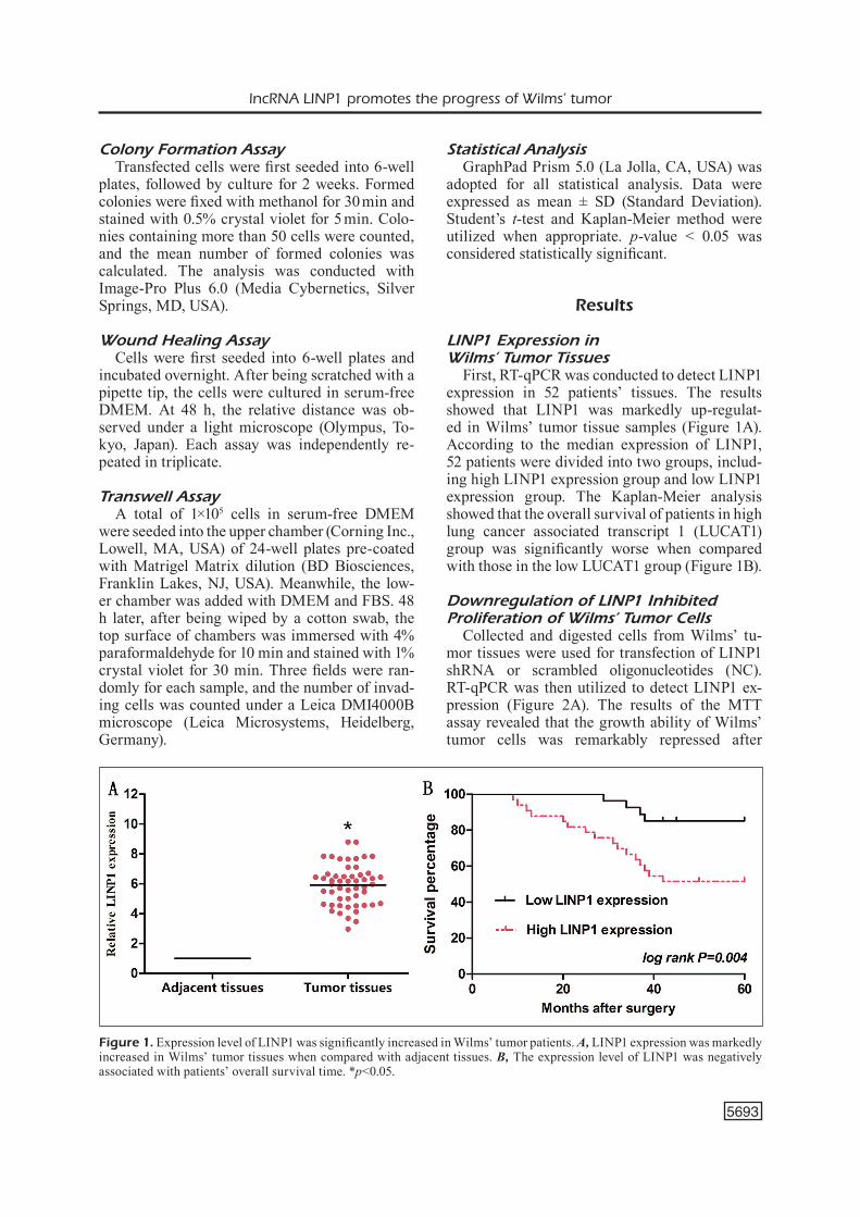
LINP1 Expression in Wilms’ Tumor Tissues
First, RT-qPCR was conducted to detect LINP1 expression in 52 patients’ tissues. The results showed that LINP1 was markedly up-regulat-ed in Wilms’ tumor tissue samples (Figure 1A). According to the median expression of LINP1, 52 patients were divided into two groups, includ-ing high LINP1 expression group and low LINP1 expression group. The Kaplan-Meier analysis showed that the overall survival of patients in high lung cancer associated transcript 1 (LUCAT1) group was significantly worse when compared with those in the low LUCAT1 group (Figure 1B).
Downregulation of LINP1 Inhibited Proliferation of Wilms’ Tumor Cells
Collected and digested cells from Wilms’ tu-mor tissues were used for transfection of LINP1 shRNA or scrambled oligonucleotides (NC). RT-qPCR was then utilized to detect LINP1 ex-pression (Figure 2A). The results of the MTT assay revealed that the growth ability of Wilms’ tumor cells was remarkably repressed after
Figure 1. Expression level of LINP1 was significantly increased in Wilms’ tumor patients. A, LINP1 expression was markedly increased in Wilms’ tumor tissues when compared with adjacent tissues. B, The expression level of LINP1 was negatively associated with patients’ overall survival time. *p<0.05.

K.-R. Zhu, Q.-F. Sun, Y.-Q. Zhang
5694
Figure 2. LINP1 promoted Wilms’ tumor cell proliferation. A, LINP1 expression in Wilms’ tumor cells transfected with LINP1 shRNA (sh-LINP1) or scrambled oligonucleotides (NC) was detected by RT-qPCR. GAPDH was used as an internal control. B, MTT assay revealed that the growth ability of Wilms’ tumor cells was remarkably repressed in the sh-LINP1 group when compared with the NC group. C, Colony formation assay revealed that the number of Wilms’ tumor cell colonies was significantly reduced in the sh-LINP1 group when compared with the NC group (Magnification × 40). D, LINP1 expression in Wilms’ tumor cells transfected with LINP1 lentivirus (LINP1) or scrambled oligonucleotides (NC) was detected by RT-qPCR. GAPDH was used as an internal control. E, MTT assay revealed that the growth ability of Wilms’ tumor cells was markedly enhanced in the LINP1 lentivirus (LINP1) group when compared with the NC group. F, Colony formation assay also revealed that the number of Wilms’ tumor cell colonies was remarkably increased in the LINP1 lentivirus (LINP1) group when compared with the NC group (magnification × 40). The results represented the average of three independent experiments (mean ± standard error of the mean). *p<0.05, compared with control cells.

lncRNA LINP1 promotes the progress of Wilms’ tumor
5695
down-regulation LINP1 (Figure 2B). Colony formation assay also revealed that the number of formed colonies was markedly reduced due to loss of LINP1 in vitro (Figure 2C). To further identify the function of LINP1 in Wilms’ tumor, collected and digested cells from Wilms’ tumor tissues were used for transfection of LINP1 len-tivirus (LINP1) or scrambled oligonucleotides (NC). Similarly, RT-qPCR was utilized to detect LINP1 expression (Figure 2D). The results of the MTT assay revealed that the growth ability of Wilms’ tumor cells was significantly promoted after over-expression of LINP1 (Figure 2E). Fur-thermore, colony formation assay demonstrated that the number of formed colonies was remark-ably increased after over-expression of LINP1 in Wilms’ tumor cells (Figure 2F).
Downregulation of LINP1 Inhibited Migration and Invasion of Wilms’ Tumor Cells
Wound healing assay revealed that the relative migrating distance of Wilms’ tumor cells was markedly repressed after the loss of LINP1 (Fig-ure 3A). Subsequent transwell assay also indicated that the number of invaded cells was remarkably reduced after the loss of LINP1 in Wilms’ tumor cells (Figure 3B). Similarly, after over-expression of LINP1 in vitro, the relative migrating distance of Wilms’ tumor cells was significantly promoted (Figure 3C). In addition, transwell assay illustrat-ed that the number of invaded cells was remark-ably increased after over-expression of LINP1 in Wilms’ tumor cells (Figure 3D).
Interaction Between Wnt/β-Catenin Signaling Pathway and LINP1 in Wilms’ Tumor
To explore the underlying mechanism of LINP1 function in Wilms’ tumor, Western blot assay was conducted to detect the expressions of target proteins in the Wnt/β-catenin signal-ing pathway, including Wnt3a, β-catenin, C-myc and Survivin. The results showed that the protein expression levels of Wnt3a, β-catenin, C-myc and Survivin were significantly down-regulat-ed after the loss of LINP1 (Figure 4A). Howev-er, the expressions of Wnt3a, β-catenin, C-myc and Survivin were markedly up-regulated after over-expression of LINP1 (Figure 4B). These results suggested that LINP1 participated in the regulation of the Wnt/β-catenin signaling path-way, further promoting Wilms’ tumor develop-ment and progression.
Discussion
Numerous studies have proved that ncRNAs participate in a variety of important biological processes, including tumor growth. Previous ev-idence has revealed that several ncRNAs play an important role in the development of Wilms’ tu-mor. For instance, the over-expression of miR-21 and low-expression of PTEN inhibit the prolif-eration and invasion of Wilms’ tumor cells8. By targeting FRS2, miR-613 depresses the prolifer-ation and migration of Wilms’ tumor. This may provide an innovative target for the diagnosis and therapy of Wilms’ tumor9. By antagonizing tu-mor suppressor miR-195, LINC00473 functions as an oncogene in Wilms’ tumor via regulating IKKalpha10.
Non-homologous end joining (NHEJ) is one of the major mechanisms for repairing dam-aged DNA in cancer cells. Previous studies have demonstrated that lncRNA in non-homol-ogous end joining pathway 1 (LINP1) promotes NHEJ-mediated DNA repair in multiple malig-nancies. For example, LINP1 regulates NHEJ signal pathway and promotes DNA damage re-pair in cervical cancer cells, further decreasing ionizing radiation sensitivity11,12. LINP1 func-tions as an oncogene in breast cancer, which fa-cilitates the progression and chemoresistance13. The over-expression of LINP1 enhances the malignant progression of prostate cancer by negatively modulating p5314. However, Zhang et al15 have shown that LINP1 serves as a tumor suppressor in lung cancer by inhibiting EMT. Therefore, we explored the role of LINP1 in Wilms’ tumor. The results showed that LINP1 was significantly up-regulated in Wilms’ tumor tissues. Meanwhile, LINP1 expression was as-sociated with the prognosis of patients. Besides, the proliferation and metastasis of Wilms’ tumor cells were markedly inhibited after the loss of LINP1. However, the proliferation and metas-tasis of Wilms’ tumor cells were remarkably promoted after over-expression of LINP1. The above results indicated that LINP1 promoted tu-morigenesis of Wilms’ tumor and might act as an oncogene.
Wnt proteins mediate diverse processes during embryogenesis by modulating stem cell division and migration. Wnt3a, β-catenin, C-myc and Survivin are target proteins in the Wnt/β-catenin signaling pathway. Li et al16 have suggested that aberrant activation of the Wnt/β-catenin signal-ing pathway plays an important role in regulat-

K.-R. Zhu, Q.-F. Sun, Y.-Q. Zhang
5696
Figure 3. LINP1 promoted Wilms’ tumor cell migration and invasion. A, The relative migrating distance of Wilms’ tumor cells was significantly decreased in the sh-LINP1 group when compared with the NC group (Magnification × 10). B, The transwell assay showed that the number of invaded Wilms’ tumor cells was markedly decreased in the sh-LINP1 group when compared with the NC group (Magnification × 40). C, The relative migrating distance of Wilms’ tumor cells was remarkably increased in the LINP1 lentivirus (LINP1) group when compared with the NC group (Magnification × 10). D, The transwell assay showed that the number of invaded Wilms’ tumor cells was significantly increased in the LINP1 lentivirus (LINP1) group when compared with the NC group (magnification × 40). The results represented the average of three independent experiments (mean ± standard error of the mean). *p<0.05, compared with control cells.

lncRNA LINP1 promotes the progress of Wilms’ tumor
5697
ing the development of several human cancers. For instance, by modulating the Wnt/β-Catenin/Axin2 signaling, c-Myb facilitates the invasion and migration of breast cancer cells. Through the activation of the Wnt/β-catenin signaling inhib-itors (DKK1 and SFRP2), TET1 serves as a tu-mor suppressor in ovarian cancer via inhibiting EMT17. LncRNA CRNDE enhances the prolifer-
ation and chemoresistance of colorectal cancer via modulating the expression of miR-181a-5p, which directly mediates the regulation of the Wnt/β-catenin pathway18. Our work showed that the expressions of target proteins in the Wnt/β-catenin signaling pathway were signifi-cantly down-regulated after the loss of LINP1. However, the protein expression levels of target
Figure 4. Interaction between LINP1 and Wnt/β-catenin signaling pathway. A, Western blot assay revealed that the expres-sions of target proteins in the Wnt/β-catenin signaling pathway were significantly down-regulated in the sh-LINP1 group when compared with the NC group. B, Western blot assay indicated that the expressions of target proteins in the Wnt/β-catenin sig-naling pathway were markedly up-regulated in the LINP1 lentivirus (LINP1) group when compared with the NC group. The results represented the average of three independent experiments. *p<0.05.

K.-R. Zhu, Q.-F. Sun, Y.-Q. Zhang
5698
proteins in the Wnt/β-catenin signaling pathway were remarkably up-regulated after over-expres-sion of LINP1. All the results above suggested that LINP1 might promote tumorigenesis of Wilms’ tumor via regulating the Wnt/β-catenin signaling pathway.
Conclusions
LINP1 enhanced Wilms’ tumor cell prolifera-tion and metastasis by regulating the Wnt/β-cat-enin signaling pathway. Our findings indicat-ed that LINP1 might contribute to therapy for Wilms’ tumor as a candidate target.
Conflict of InterestThe Authors declare that they have no conflict of interests.
References
1) Charlton J, PavasoviC v, PritChard-Jones K. Biomark-ers to detect Wilms tumors in pediatric pa-tients: where are we now? Future Oncol 2015; 11: 2221-2234.
2) Cone eB, dalton ss, van noord M, traCy et, riCe he, routh JC. Biomarkers for Wilms tumor: a sys-tematic review. J Urol 2016; 196: 1530-1535.
3) liu G, ZhanG y, Fu K, hu J, Zhao Z, Fu W, liu G. Meta-analysis of the effect of preoperative che-motherapy on Wilms’ tumor. J BUON 2018; 23: 211-217.
4) GaBallah hh, GaBer ra, elrashidy Ma, elshahat da, haBlus Ma, eBeid aM. Expression of long non-coding RNA CCHE1 in colorectal carcinoma: correlations with clinicopathological fea-tures and ERK/COX-2 pathway. Mol Biol Rep 2018; 10.1007/s11033-018-4521-0.
5) WanG y, shi F, Xia y, Zhao h. LncRNA OIP5-AS1 predicts poor prognosis and regulates cell prolif-eration and apoptosis in bladder cancer. J Cell Biochem 2018; 10.1002/jcb.28024.
6) PenG l, JianG B, yuan X, Qiu y, PenG J, huanG y, ZhanG C, ZhanG y, lin Z, li J, yao W, denG W, ZhanG y, MenG M, Pan X, li C, yin d, Bi X, li G, lin dC. Super-enhancer-associated long noncoding RNA HCCL5 is activated by ZEB1 and promotes the malignancy of hepatocellu-lar carcinoma. Cancer Res 2019; 79: 572-584.
7) Chen l, yuan d, yanG y, ren M. LincRNA-p21 en-hances the sensitivity of radiotherapy for gas-
tric cancer by targeting the beta-catenin sig-naling pathway. J Cell Biochem 2018; 10.1002/jcb.27905.
8) Cui M, liu W, ZhanG l, Guo F, liu y, Chen F, liu t, Ma r, Wu r. Over-expression of miR-21 and lower PTEN levels in Wilms’ tumor with aggressive be-havior. Tohoku J Exp Med 2017; 242: 43-52.
9) WanG hF, ZhanG yy, ZhuanG hW, Xu M. MicroR-NA-613 attenuates the proliferation, migration and invasion of Wilms’ tumor via targeting FRS2. Eur Rev Med Pharmacol Sci 2017; 21: 3360-3369.
10) Zhu s, Fu W, ZhanG l, Fu K, hu J, Jia W, liu G. LINC00473 antagonizes the tumour suppres-sor miR-195 to mediate the pathogenesis of Wilms tu-mour via IKKalpha. Cell Prolif 2018; 51: e12416.
11) ZhanG y, he Q, hu Z, FenG y, Fan l, tanG Z, yuan J, shan W, li C, hu X, tanyi Jl, Fan y, huanG Q, Mon-tone K, danG Cv, ZhanG l. Long noncoding RNA LINP1 regulates repair of DNA double-strand breaks in triple-negative breast cancer. Nat Struct Mol Biol 2016; 23: 522-530.
12) WanG X, liu h, shi l, yu X, Gu y, sun X. LINP1 fa-cilitates DNA damage repair through non-homol-ogous end joining (NHEJ) pathway and subse-quently decreases the sensitivity of cervical can-cer cells to ionizing radiation. Cell Cycle 2018; 17: 439-447.
13) lianG y, li y, sonG X, ZhanG n, sanG y, ZhanG h, liu y, Chen B, Zhao W, WanG l, Guo r, yu Z, yanG Q. Long noncoding RNA LINP1 acts as an oncogene and promotes chemoresistance in breast cancer. Cancer Biol Ther 2018; 19: 120-131.
14) Wu hF, ren lG, Xiao JQ, ZhanG y, Mao XW, Zhou lF. Long non-coding RNA LINP1 pro-motes the malignant progression of prostate cancer by reg-ulating p53. Eur Rev Med Pharmacol Sci 2018; 22: 4467-4476.
15) ZhanG C, hao y, WanG y, Xu J, tenG y, yanG X. TGF-beta/SMAD4-regulated lncRNA-LINP1 in-hibits epithelial-mesenchymal transition in lung cancer. Int J Biol Sci 2018; 14: 1715-1723.
16) li y, Jin K, van Pelt GW, van daM h, yu X, MesKer We, ten diJKe P, Zhou F, ZhanG l. c-Myb enhances breast cancer invasion and metastasis through the Wnt/beta-catenin/Axin2 pathway. Cancer Res 2016; 76: 3364-3375.
17) duan h, yan Z, Chen W, Wu y, han J, Guo h, Qiao J. TET1 inhibits EMT of ovarian cancer cells through activating Wnt/beta-catenin signaling in-hibitors DKK1 and SFRP2. Gynecol Oncol 2017; 147: 408-417.
18) han P, li JW, ZhanG BM, lv JC, li yM, Gu Xy, yu ZW, Jia yh, Bai XF, li l, liu yl, Cui BB. The lncRNA CRNDE promotes colorectal cancer cell prolifer-ation and chemoresistance via miR-181a-5p-me-diated regulation of Wnt/beta-catenin signaling. Mol Cancer 2017; 16: 9.