long covid-19: proposed primary care clinical guidelines
TRANSCRIPT

International Journal of
Environmental Research
and Public Health
Review
Long Covid-19: Proposed Primary Care Clinical Guidelines forDiagnosis and Disease Management
Antoni Sisó-Almirall 1,2,3,*, Pilar Brito-Zerón 4,5,6, Laura Conangla Ferrín 1 , Belchin Kostov 2,3,7,Anna Moragas Moreno 8 , Jordi Mestres 1, Jaume Sellarès 9, Gisela Galindo 10, Ramon Morera 11, Josep Basora 12,Antoni Trilla 13, Manuel Ramos-Casals 4,6 and on behalf of the CAMFiC long COVID-19 Study Group †
�����������������
Citation: Sisó-Almirall, A.;
Brito-Zerón, P.; Conangla Ferrín, L.;
Kostov, B.; Moragas Moreno, A.;
Mestres, J.; Sellarès, J.; Galindo, G.;
Morera, R.; Basora, J.; et al. Long
Covid-19: Proposed Primary Care
Clinical Guidelines for Diagnosis and
Disease Management. Int. J. Environ.
Res. Public Health 2021, 18, 4350.
https://doi.org/10.3390/
ijerph18084350
Academic Editor: Joan Puig-Barberà
Received: 19 February 2021
Accepted: 16 April 2021
Published: 20 April 2021
Publisher’s Note: MDPI stays neutral
with regard to jurisdictional claims in
published maps and institutional affil-
iations.
Copyright: © 2021 by the authors.
Licensee MDPI, Basel, Switzerland.
This article is an open access article
distributed under the terms and
conditions of the Creative Commons
Attribution (CC BY) license (https://
creativecommons.org/licenses/by/
4.0/).
1 Permanent Board of the Catalan Society of Family and Community Medicine (CAMFiC),08009 Barcelona, Spain; [email protected] (L.C.F.); [email protected] (J.M.)
2 Primary Care Centre Les Corts, Consorci d’Atenció Primària de Salut Barcelona Esquerra (CAPSBE),08028 Barcelona, Spain; [email protected]
3 Primary Healthcare Transversal Research Group, Institut d’Investigacions Biomèdiques August Pi iSunyer (IDIBAPS), 08036 Barcelona, Spain
4 Laboratory of Autoimmune Diseases Josep Font, IDIBAPS-CELLEX, 08036 Barcelona, Spain;[email protected] (P.B.-Z.); [email protected] (M.R.-C.)
5 Autoimmune Diseases Unit, Department of Medicine, Hospital CIMA-Sanitas, 08034 Barcelona, Spain6 Department of Autoimmune Diseases, ICMiD, Hospital Clínic, 08036 Barcelona, Spain7 Department of Statistics and Operations Research, Universitat Politècnica de Catalunya (UPC),
08034 Barcelona, Spain8 Jaume I Health Centre, Institut Català de la Salut, Universitat Rovira i Virgili, 43005 Tarragona, Spain;
[email protected] College of Catalan Physicians, 08017 Barcelona, Spain; [email protected] Permanent Board of the Spanish Society of Family and Community Medicine (semFYC),
08009 Barcelona, Spain; [email protected] Board of Spanish Society of Managers of Primary Care (SEDAP), 28026 Madrid, Spain; [email protected] IDIAP Jordi Gol, 08007 Barcelona, Spain; [email protected] Faculty of Medicine and Health Sciences, University of Barcelona, 08036 Barcelona, Spain;
[email protected]* Correspondence: [email protected]† ANNEX. Members of the CAMFiC long COVID-19 Study Group: Alarcón Belmonte I; Also Fontanet A;
Barrot de la Fuente J; Brotons Cuixart C; Burdoy Joaquin E; Caballol Angelats R; Cabré Vila JJ; CanteroGómez FX; Carbonell Abella C; Carrillo Muñoz R; Casasa Plana A; Copetti Fanlo S; Cots Yago JM; DenielRosanas J; Díez-Cascón P; Ferrer-Vidal Cortella D; Fernández Pérez J; Franch Nadal J; Guirado Vila P; Hoyo J;Lozano Fernández JJ; Limón Ramírez E; Llor Vila C; Martin Luján F; Martin Álvarez R; Mas Heredia M;Mascort Roca J; Montero Alia JJ; Moreno Escrivà S; Ortega Vila Y; Perelló Bratescu A; Sans Corrales M;Sequeira Aymar E; Serrano Manzano M; Serrano-Pons J; Solà Gonfaus M; Solanes Cabús M; VeganzonesGuanyabens I; Vilaseca Llobet JM; Villafàfila Ferrero R; Vinyoles Bargalló E.
Abstract: Long COVID-19 may be defined as patients who, four weeks after the diagnosis of SARS-Cov-2 infection, continue to have signs and symptoms not explainable by other causes. The estimatedfrequency is around 10% and signs and symptoms may last for months. The main long-term man-ifestations observed in other coronaviruses (Severe Acute Respiratory Syndrome (SARS), MiddleEast respiratory syndrome (MERS)) are very similar to and have clear clinical parallels with SARS-CoV-2: mainly respiratory, musculoskeletal, and neuropsychiatric. The growing number of patientsworldwide will have an impact on health systems. Therefore, the main objective of these clinicalpractice guidelines is to identify patients with signs and symptoms of long COVID-19 in primarycare through a protocolized diagnostic process that studies possible etiologies and establishes anaccurate differential diagnosis. The guidelines have been developed pragmatically by compilingthe few studies published so far on long COVID-19, editorials and expert opinions, press releases,and the authors’ clinical experience. Patients with long COVID-19 should be managed using struc-tured primary care visits based on the time from diagnosis of SARS-CoV-2 infection. Based on thecurrent limited evidence, disease management of long COVID-19 signs and symptoms will require aholistic, longitudinal follow up in primary care, multidisciplinary rehabilitation services, and theempowerment of affected patient groups.
Int. J. Environ. Res. Public Health 2021, 18, 4350. https://doi.org/10.3390/ijerph18084350 https://www.mdpi.com/journal/ijerph
Int. J. Environ. Res. Public Health 2021, 18, 4350 2 of 20
Keywords: SARS-CoV-2; primary care; long COVID-19
1. Introduction
Severe acute respiratory syndrome 2 coronavirus (SARS-CoV-2), first detected inDecember 2019 in Wuhan, China, is the seventh coronavirus known to infect humansafter the identification, in this century, of SARS and Middle East Respiratory Syndrome(MERS) viruses. The lack of pre-virus immunity has led to an exponential increase ininfected patients worldwide and the pandemic is one of the biggest health challengesfacing humanity in the last 100 years [1,2]. The rapid and unpredictable worldwide spreadof SARS-CoV-2, with most infected people having no or only mild signs and symptoms,appears to have initially been related to cases imported from the first countries affected [3].As of January 13, 2021, there are >90 million confirmed cases worldwide and 2 milliondeaths [4].
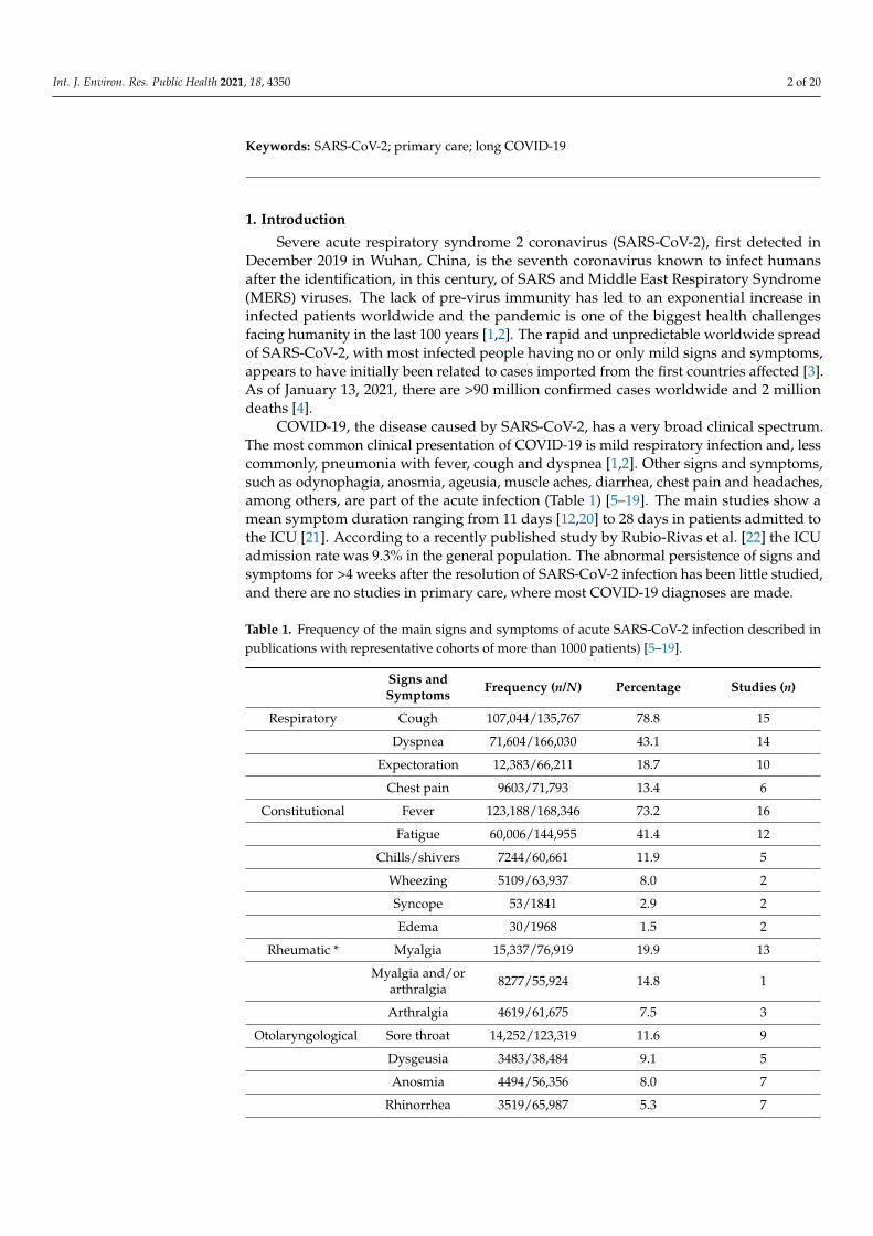
COVID-19, the disease caused by SARS-CoV-2, has a very broad clinical spectrum.The most common clinical presentation of COVID-19 is mild respiratory infection and, lesscommonly, pneumonia with fever, cough and dyspnea [1,2]. Other signs and symptoms,such as odynophagia, anosmia, ageusia, muscle aches, diarrhea, chest pain and headaches,among others, are part of the acute infection (Table 1) [5–19]. The main studies show amean symptom duration ranging from 11 days [12,20] to 28 days in patients admitted tothe ICU [21]. According to a recently published study by Rubio-Rivas et al. [22] the ICUadmission rate was 9.3% in the general population. The abnormal persistence of signs andsymptoms for >4 weeks after the resolution of SARS-CoV-2 infection has been little studied,and there are no studies in primary care, where most COVID-19 diagnoses are made.
Table 1. Frequency of the main signs and symptoms of acute SARS-CoV-2 infection described inpublications with representative cohorts of more than 1000 patients) [5–19].
Signs andSymptoms Frequency (n/N) Percentage Studies (n)
Respiratory Cough 107,044/135,767 78.8 15
Dyspnea 71,604/166,030 43.1 14
Expectoration 12,383/66,211 18.7 10
Chest pain 9603/71,793 13.4 6
Constitutional Fever 123,188/168,346 73.2 16
Fatigue 60,006/144,955 41.4 12
Chills/shivers 7244/60,661 11.9 5
Wheezing 5109/63,937 8.0 2
Syncope 53/1841 2.9 2
Edema 30/1968 1.5 2
Rheumatic * Myalgia 15,337/76,919 19.9 13
Myalgia and/orarthralgia 8277/55,924 14.8 1
Arthralgia 4619/61,675 7.5 3
Otolaryngological Sore throat 14,252/123,319 11.6 9
Dysgeusia 3483/38,484 9.1 5
Anosmia 4494/56,356 8.0 7
Rhinorrhea 3519/65,987 5.3 7

Int. J. Environ. Res. Public Health 2021, 18, 4350 3 of 20
Table 1. Cont.
Signs andSymptoms Frequency (n/N) Percentage Studies (n)
Nasal congestion 2684/55,924 4.8 1
Hemoptysis 660/61,775 1.1 6
Otalgia 631/75,336 0.8 2
Digestivecomplaints Anorexia 4084/19,092 21.4 4
Diarrhea 20,249/153,778 13.2 13
Nausea orvomiting 17,142/136,902 12.5 13
Abdominal pain 7421/69,573 10.7 4
Neurological Confusion/alteredconsciousness 18,434/70,032 26.3 2
Headache 17,734/128,233 13.8 12
Other Conjunctivitis 782/138,724 0.6 5* Among the different categories of signs and symptoms, there is a wide heterogeneity of definition of eachvariable between the different studies, and therefore, the frequencies derived from merging data from differentstudies should be interpreted with caution.
The objective of this document was to develop primary care clinical guidelines forpatients with long COVID-19 signs and symptoms to enable primary care professionals toaddress the health consequences that go beyond acute SARS-CoV-2 infection.
2. Definitions
In the absence of consensus and internationally accepted definitions, we used therecently-proposed definitions in the National Institute for Health and Care Excellence(NICE) working document, based on the effects of COVID-19 at different time points [23]:
- Acute COVID-19 infection: Signs and symptoms of COVID-19 for up to 4 weeks. Wesupport the definition of COVID-19 infection included in the NICE guidelines [23],including people with suspected infection who, in the early phases of the pandemic,did not have access to testing for SARS-CoV-2 infection.
- Ongoing symptomatic COVID-19: Signs and symptoms of COVID-19 from 4 to 12 weeksnot explained by an alternative diagnosis after protocolized study.
- Post-COVID-19 syndrome: Signs and symptoms that develop during or following aninfection consistent with COVID-19, continue for >12 weeks and are not explainedby an alternative diagnosis. The term “syndrome” reflects the concurrence of amultisystem, fluctuating, and often overlapping clusters of signs and symptoms that,in some patients, may follow a relapsing-remitting pattern and that may change overtime and affect any bodily system.
3. Methodology
In the absence of evidence-based clinical practice guidelines (CPGs) for the man-agement of long COVID-19 [24,25], Catalan Society of Family and Community Medicine(CAMFiC) established a working group to develop a CPG, consisting mainly of primarycare professionals (90%), together with specialists in internal medicine, autoimmune dis-eases, infectious disease, epidemiology and statistics.
The CPG focuses on patients with long COVID-19 not requiring hospitalization, whosediagnosis and follow-up has been made in primary care (probably > 80% of affected people).It does not focus on hospitalized patients, whose follow-up and management will be carriedout by the hospital outpatient department.

Int. J. Environ. Res. Public Health 2021, 18, 4350 4 of 20
In these guidelines, we also used the term “Long COVID-19”, which includes bothongoing and post-COVID-19 syndrome according to the NICE definitions [23]. We endorsethe rationale for the terms used by the NICE guidelines, whose intention is to reflect thefact that signs and symptoms occur after the acute infection is ended (but not that theperson has recovered), avoiding time-specific terms like “chronic” or “persistent”.
Signs and symptoms present during the first 4 weeks of infection, worsening orrecurrence of manifestations already present before infection, manifestations arising fromphysical or psychic sequelae reasonably attributable to infection, and post-viral immune-related manifestations not initially present and which may appear once the infection iscured fall outside the scope of the guidelines [26].
We used a pragmatic approach based on the few published studies on SARS-CoV-2,editorials and expert opinions, press releases and the clinical experience of the authors.Academic sources were identified by a systematic search of the PubMed database, through13 January 2021, with the main terms SARS-CoV-2 and COVID-19 in combination withthe secondary terms chronic, persistent, ongoing, long-term, recovery time, and post-viral,along with specific searches for each individual symptom included in the guidelines.
4. Planning of Care for Patients with Long Covid-19
The care of patients with long COVID-19 should be structured in three consecutivevisits according to the time from diagnosis of SARS-CoV-2 infection.
The first primary care visit (V1) of patients with long COVID-19 is essential. Theobjective should be a history and examination and complementary tests to study thepossible underlying causes of long symptoms. We endorse the recommendations suggestedby the NICE guidelines for assessing people with long COVID-19 [23]. The visit shouldbe made from the 4th week after confirmation of the diagnosis of SARS-CoV-2 infectionwith a positive SARS-CoV-2 test (PCR, antigen or antibody) or after the start of signs andsymptoms of COVID-19 in case laboratory test is unavailable (preferably between the 5thand 6th week, depending on availability and resources), and should last at least 30 min,with active support from nurses, including:
Personal background: The medical record may be relevant when analyzing long-termsymptoms. The family physician has the most comprehensive long-term information onthe pre-infection health status.
SARS-CoV-2 infection: Diagnostic confirmation of SARS-CoV-2 infection (date andmicrobiological test), symptoms and approximate onset dates, hospital admission anddischarge dates, maximum oxygen requirements, ICU admission and duration, therapiesreceived, and complications during admission should be recorded. The intensity of eachsymptom may be assessed subjectively on a visual analog scale (VAS: 0–10). The diagnosticalgorithms proposed for each individual symptom in paragraph IV will apply.
Physical examination: A complete physical examination, with measurement of vitalsigns and baseline oxygen saturation, should be made, paying special attention to assessingthe oropharynx and cardiorespiratory system.
Laboratory studies: A basic first visit laboratory study should be made, supplementedaccording to individualized patient criteria (Table 2).
Complementary tests: Lung parenchyma evaluation is essential in all patients withCOVID-19. Chest X-ray in at least the two conventional projections is the conventionaltest, and allows for agile general evaluation, and is usually accessible urgently. However,in primary care, chest ultrasound should be used when possible, as it is very usefulin evaluating pneumonia and its complications (as shown in hospitals) [27] and in thedifferential diagnosis. It is accessible, may be used in outpatients, at home or in nursinghomes, and is useful for the diagnosis and subsequent monitoring. Chest ultrasoundmay show peripheral pulmonary involvement, pulmonary interstitial disease (focal ordiffuse), pleural contact condensations (pneumonic or thromboembolic), pneumothoraxand pleural effusion. Other complementary tests will be determined by the symptomspresented (Table 2).
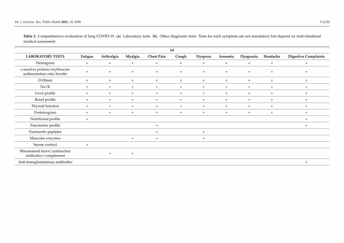
Int. J. Environ. Res. Public Health 2021, 18, 4350 5 of 20
Table 2. Comprehensive evaluation of long COVID-19. (a). Laboratory tests. (b). Other diagnostic tests. Tests for each symptom are not mandatory but depend on individualizedmedical assessment.
(a)
LABORATORY TESTS Fatigue Arthralgia Myalgia Chest Pain Cough Dyspnea Anosmia Dysgeusia Headache Digestive Complaints
Hemogram + + + + + + + + + +
c-reactive protein/erythrocytesedimentation rate/ferritin + + + + + + + + + +
D-Dimer + + + + + + + + + +
Na/K + + + + + + + + + +
Liver profile + + + + + + + + + +
Renal profile + + + + + + + + + +
Thyroid function + + + + + + + + + +
Proteinogram + + + + + + + + + +
Nutritional profile + +
Pancreatric profile + +
Natriuretic peptides + +
Muscular enzymes + + +
Serum cortisol +
Rheumatoid factor/antinuclearantibodies/complement + +
Anti-transglutaminase antibodies +

Int. J. Environ. Res. Public Health 2021, 18, 4350 6 of 20
Table 2. Cont.
(b)
OTHER DIAGNOSTIC TESTS Fatigue Arthralgia Myalgia Chest Pain Cough Dyspnea Anosmia Dysgeusia Headache Digestive Complaints
Vital signs + + + + + + + + + +
Oxygen saturation + + + + + + + + + +
Electrocardiogram + + + + + + + + + +
Chest X-ray/lung ultrasound + + + + + + + + + +
Spirometry + + + +
Chest computed tomography + +
Funduscopy +
Joint ultrasound +
Abdominal ultrasound +
Fecal occult blood +
Digestive endoscopy +

Int. J. Environ. Res. Public Health 2021, 18, 4350 7 of 20
The second visit (V2) should be made from week 8 (preferably between weeks 9 and10, depending on availability and resources). The objective is to evaluate the results of theV1 tests, make a differential diagnosis with other post-COVID-19 situations, and apply thecorresponding diagnostic algorithms to identify potential causes that reasonably explainthe symptoms.
The third visit (V3) should be made from week 12 (between week 13 and week 14 week,depending on availability and resources) to evaluate the evolution of long-term symptomsand re-evaluate possible causes using the corresponding diagnostic algorithms.
5. Assessment of Individual Signs and Symptoms
Primary care evaluations should follow the same principles as normal clinical practicefor the same symptoms: a careful history that considers the medical record and a physicalexamination centered on the reported symptoms.
5.1. Fatigue
Fatigue is one of the most common extra-respiratory symptoms of acute SARS-CoV-2infection (41%, Table 1). Studies estimate the frequency as 35–45% at 4 weeks [28,29],30–77% at 8 weeks [30–32] and 16–55% at 12 weeks post-infection [33,34] (Table 3). Theprofound, prolonged nature of fatigue in some COVID-19 patients shares characteristicswith the chronic fatigue syndrome (CFS) described after other infections, including SARS,MERS and community-acquired pneumonia [24,25]. No association has been reportedbetween the long-term fatigue associated with COVID-19 and other long-term fatiguestates, or with COVID-19 severity, or with any laboratory marker of inflammation andcell turnover or pro-inflammatory molecules. However, women and people with a pre-existing diagnosis of depression/anxiety were over-represented in patients with long-termfatigue [35].
Algorithm S1 summarizes the diagnostic approach for patients with fatigue persisting>4 weeks after SARS-CoV-2 infection. In these patients, V1 should include (File S1):
Specific clinical history: The history should record the onset date and specific ques-tions about fatigue (symptoms and accompanying signs, concomitant psychosocial andemotional factors, related drugs, substance abuse, sleep disorders and exposure to toxins),pre-COVID-19 infection diseases possibly associated with chronic fatigue, organ-specificsequelae resulting from severe COVID-19 infection requiring hospital admission that maycause fatigue, and other current symptoms coexisting with fatigue.
Specific studies: Tests should include specific laboratory tests (chloride, bicarbonate,calcium, phosphate, muscle enzymes, plasma cortisol levels) and spirometry under safeconditions (according to the recommendations given by The Italian Respiratory Society forlung function testing in the context of COVID-19 [36]).
5.2. Arthralgia
Patients with acute SARS-CoV-2 infection may develop arthralgia (7.5%) (Table 1),defined as non-arthritic pain in ≥1 joints without evidence of inflammation (edema, jointpain or heat). It may be accompanied by difficult-to-locate muscle pain (arthromyalgia,musculoskeletal pain). Joint pain may persist in 10–15% of patients at 4 weeks [28,37] and16–27% at 8 weeks [31,37] (Table 3).
Algorithm S2 summarizes the diagnostic approach in patients with joint pain persist-ing >4 weeks after SARS-CoV-2 infection (File S1). In these patients, V1 should include:
Specific clinical history: Record date of onset of joint pain, type of pain (nociceptive,neuropathic or mixed), location, duration, modification with exercise or rest (factors thatrelieve, worsen or trigger it) and response to analgesia. Pre-COVID-19 diseases possiblyassociated with joint pain and current symptoms co-existing with arthralgia (especiallychronic fatigue) should be evaluated.
Specific studies: Tests should include specific laboratory tests (uric acid, proteinogram,antinuclear antibodies [ANA], rheumatoid factor, complement C3 and C4 levels). If joint

Int. J. Environ. Res. Public Health 2021, 18, 4350 8 of 20
inflammation is suspected, joint ultrasound is indicated (or simple radiology if not avail-able). Monitoring of inflammation (synovitis and enthesitis) and peripheral joint damagemay be useful, although there are insufficient data to recommend a specific ultrasoundevaluation system or its periodicity.
5.3. Myalgia
Muscle pain or myalgia may affect ≥1 muscles and is usually benign and self-limiting.Ligaments, tendons, and fasciae may also be involved. In large series, myalgia is reportedin 20% of cases of acute SARS-CoV-2 infection (Table 1) and may persist in 15% of patientsat 4 weeks [29], in 6–13% at 8 weeks [30,31] and in 16% at 12 weeks [34] (Table 3).
Algorithm S3 summarizes the diagnostic approach to myalgia persisting >4 weeksafter SARS-CoV-2 infection (File S1). In these patients, V1 should include:
Specific clinical history: Record the onset date, location, duration, modification withexercise or rest, factors that relieve, worsen or trigger myalgia, response to analgesia,pre-COVID-19 infections possibly associated with myalgia, and other current symptomsco-existing with myalgia (especially chronic fatigue and generalized pain).
Specific studies: The following specific laboratory tests should be added (proteino-gram, creatine kinase, aldolase, lactate dehydrogenase, ANA, rheumatoid factor).
5.4. Chest Pain
Chest pain, defined as any pain located between the diaphragm and the base of theneck, appears in up to 13% of acute SARS-CoV-2 infections (Table 1). Chest pain maypersist in 20% of patients at 4 weeks [28], in 22% at 8 weeks [31] and in 11% at 12 weeks [34](Table 3). No studies have specifically described the characteristics of long-term chestpain in COVID-19. In our clinical experience, a significant percentage of patients refer tohigh central chest pain, a symptom described in a large patient-led survey as “pulmonaryburning,” a kind of burning sensation in the chest, tension and some shortness of breath,especially reported after a dry cough [38].
Algorithm S4 summarizes the diagnostic approach to chest pain persisting >4 weeksafter SARS-CoV-2 infection (File S1). In these patients, V1 should include:
Specific clinical history: Collect the date of onset, location, duration, triggers, modifi-cation with exercise or rest, accompanying symptoms, history of trauma or fall.
Specific studies: The following tests should be added: laboratory tests (troponins andcreatine phosphokinase (CPK) -MB depending on availability), electrocardiogram, chestultrasound (shown to be useful in the differential diagnosis of pleuritic pain to distinguishwhether its origin is in the chest wall or lung surface) [39], and spirometry (under safeconditions); chest computed tomography (CT) evaluation may be considered.
5.5. Cough
Cough occurs in about 80% of acute SARS-CoV-2 infections (Table 1), while long-termcough has been reported in 33–43% of cases at 4 weeks [28,29], in 5–46% at 8 weeks [30–32],and in 2–17% at 12 weeks [33,34] (Table 3).
The diagnostic approach to patients with cough persisting >4 weeks after SARS-CoV-2infection is summarized in Algorithm S5 (File S1). In these patients, V1 should include:
Specific clinical history: Collect the date of onset and characteristics (mostly dry,irritating, nonproductive cough; in productive cough the sputum characteristics should beinvestigated). Possible organ-specific sequelae resulting from severe COVID-19 infectionrequiring hospital admission that may cause chronic coughing, and iatrogenic sequelaerelated to invasive maneuvers (post-intubation orotracheal, post-tracheostomy) shouldbe evaluated. Current symptoms co-existing with cough, especially new-onset fever anddyspnea, and other alarming symptoms should be evaluated.
Specific studies: Spirometry (under safe conditions) is advised.

Int. J. Environ. Res. Public Health 2021, 18, 4350 9 of 20
5.6. Dyspnea
Dyspnea, the feeling of shortness of breath and difficulty in breathing properly, issometimes confused with fatigue, and may be difficult to describe, for sociocultural reasons.Dyspnea is reported in 43% of patients with acute SARS-CoV-2 infection (Table 1). Long-term dyspnea is reported in 11–33% of cases at 4 weeks [28,29,37], in 8–63% of cases at8 weeks [30–32,37], and 14% beyond 12 weeks [33] (Table 3).
Algorithm S6 summarizes the diagnostic approach to patients with dyspnea persisting>4 weeks after SARS-CoV-2 infection (File S1). In these patients, V1 should include:
Specific clinical history: Collect the data of onset and characteristics. It is importantto rule out acute onset and to evaluate an association with increased physical demandor dyspnea at rest and, especially, an association with other symptoms, such as chestpain. The modified Medical Research Council (MRC) dyspnea scale may be administered.Organ-specific sequelae of severe COVID-19 infection requiring hospitalization that maycause dyspnea, invasive maneuvers and techniques carried out during the acute episodeand which may have been an iatrogenic cause of secondary dyspnea, should be ruledout. Current symptoms co-existing with dyspnea, especially new-onset fever, shouldbe assessed.
Specific studies: Additional laboratory tests should be added (troponins and CPK-MB,natriuretic peptides according to availability). Gasometry is recommended if baselineoxygen saturation is persistently decreased without known prior cause, respiratory func-tional tests (simple spirometry and diffusing capacity of carbon monoxide [DLCO]), chestradiology, and six-minute walk test (6MWT). CT or angio-CT should be considered.
Sudden-onset dyspnea (or baseline dyspnea flare-up) usually requires urgent attention,especially if associated with alarming symptoms, paying particular attention to respiratorysuperinfections, pulmonary thromboembolism (especially in patients with a history ofhospitalization and severity), post-COVID-19 heart failure and organizing pneumonia. Latedevelopment of new respiratory symptoms and opacities (>2 weeks after the first symptomsof COVID-19), especially if not detected in previous CT studies, may suggest post-viralorganizing pneumonia (already described in patients with influenza virus infection).
5.7. Anosmia/Dysgeusia
Partial (hyposmia) or complete (anosmia) loss of smell may be temporary or perma-nent, depending on the cause. Almost all patients with anosmia perceive salty, sweet,acid, and bitter substances normally, but do not discriminate flavors, which also dependheavily on smell. Therefore, patients refer to loss of the sense of taste (dysgeusia/ageusia)and do not enjoy food. Viral upper respiratory tract infection is a common cause of olfac-tory dysfunction, in part because the olfactory epithelium is adjacent to the respiratoryepithelium, where many viruses that cause upper respiratory infection replicate, and partlybecause olfactory neurons directly access the environment. These viruses could causeolfactory dysfunction not only through nasal obstruction, but also through transient orpersistent direct damage to the sensory epithelium. Anosmia after viral infection is knownas post-infectious or post-viral olfactory loss. Anosmia and dysgeusia are present in 8–9%of patients with acute SARS-CoV-2 infection (Table 1). Studies show a frequency of 12–56%of long-term anosmia at 4 weeks [28,29,40–43], 2–25% at 8 weeks [30,31,42] and 13–46% at12 weeks [34,44], while for dysgeusia, the rates are 9–50% [28,29,40,43], 1–10% [30,31], and11–31% [34,44], respectively (Table 3).
The diagnostic approach to patients with anosmia/dysgeusia >4 weeks after SARS-CoV-2 infection is summarized in Algorithm S7 (File S1). In these patients, V1 should include:
Specific clinical history: Collect the date of onset and characteristics and rule outprevious disease (especially ENT and neurological).
Specific studies: A specific physical examination including a complete otolaryngologi-cal examination should be made.

Int. J. Environ. Res. Public Health 2021, 18, 4350 10 of 20
5.8. Headache
Headache has been reported in 14% of SARS-CoV-2 infection patients (Table 1).Reports show a frequency of long-term headache of 14% at 4 weeks [28], 9–15% at8 weeks [29,31] and 18% at 12 weeks [33] (Table 3).
Algorithm S8 summarizes the diagnostic approach to patients with headache persist-ing beyond 4 weeks after SARS-CoV-2 infection (File S1). In these patients, V1 should include:
Specific clinical history: Collect the date of onset and the main features. Evaluatemanifestations leading to the suspicion of an underlying organic disease. A prior diagnosisof headache (reported in 50% of patients with long-term headache) [45] or neurological dis-ease, and current symptoms coexisting with headache, especially neurological symptoms,should be evaluated.
Specific studies: The examination should include blood pressure, temporal arteryinspection and palpation in patients aged >50 years, temporomandibular joint examination,cranial palpation (painful spots, paranasal sinus, examination of sensitive points andtriggers) and a complete neurological assessment (level of consciousness and meningogenicsigns, gait, dysmetria, Romberg test, facial asymmetry, funduscopy).
5.9. Digestive Signs and Symptoms
The overall rate of patients with acute SARS-CoV-2 infection with gastrointestinalsymptoms is 34%, including anorexia (21%), diarrhea (13%), nausea or vomiting (12%) andabdominal pain (11%) [46] (Table 1). Diarrhea, the most common gastrointestinal clinicalsign, usually consists of non-severe, non-dehydrating semi-liquid stools [46]. It is unclearwhat percentage of these patients received treatments that produced gastrointestinal sideeffects during the first pandemic wave. Reports show the persistence of diarrhea as3–9% [29,31], anorexia as 8% [31], nausea as 6% [29] and abdominal pain as 3% at 8 weekspost-infection [29] (Table 3).
Table 3. Summary of data from the main studies on symptoms reported as long COVID-19 (>4 w) * [28–34,37,40–44,47].
Signs and Symptoms of Long COVID-19 Weeks after the First Symptom of Acute COVID-19 Infection
4 w 8 w 12 w
Global frequency 13.3% 4.5% 2.3%
Constitutional Fever - 0% [37], 3% [29] -
Chills 5% [28] - -
Fatigue 35% [28], 45% [29] 30% [30], 53% [31], 77% [32] 16% [33], 55% [34]
Rheumaticmanifestations Arthralgia 10% [37], 15% [28] 16% [37], 27% [31] -
Myalgia 15% [29] 6% [31], 13% [30] 16% [34]
Respiratorymanifestations Dyspnea 11% [37], 27% [28], 33% [29] 8% [37], 31% [30], 43% [31],
63% [32] 14% [33]
Chest pain 20% [28] 22% [31] 11% [34]
Cough 33% [29], 43% [28] 5% [30], 18% [31], 46% [32] 2% [33], 17% [34]
Expectoration - 8% [31] 2% [33]
Otolaryngologicalmanifestations Rhinorrhea 28% [28] 12% [29], 15% [31] -
Sore throat 15% [28] 7% [31], 9% [29] -
Anosmia 12% [29], 23% [28], 28% [40], 43%[41], 46% [42], 56% [43] 2% [30], 17% [31], 25% [42] 13% [34], 46% [44]
Dysgeusia 9% [29], 17% [40], 24% [28],50% [43] 1% [30], 10% [31] 11% [34], 31% [44]
Anosmia/Dysgeusia 28% [37], 9% [47] 2% [47], 23% [37] 4% [33]

Int. J. Environ. Res. Public Health 2021, 18, 4350 11 of 20
Table 3. Cont.
Signs and Symptoms of Long COVID-19 Weeks after the First Symptom of Acute COVID-19 Infection
4 w 8 w 12 w
Digestivecomplaints Abdominal pain 15% [28] 3% [29] -
Nausea 10% [28] 6% [29] -
Vomiting 4% [28] - -
Diarrhea - 3% [31], 9% [29] -
Diarrhea orvomiting 17% [37] 11% [37] 31% [33]
Anorexia - 8% [31] -
Weight loss >5% 16% [37] 17% [37] -
Neurologicalmanifestations Headache 14% [28] 9% [31], 15% [29] 18% [33]
Behavioraldisorder - - 27% [34]
Memory loss - - 34% [34]
Sleep disorders - - 31% [34]
Vertigo/dizziness - 6% [31] -
Othermanifestations Dry syndrome - 16% [31] -
Hair loss - - 20% [34]
Conjunctivitis - 16% [31] -
* Unfortunately, there are wide variations in study designs, populations evaluated (unselected, or specifically studied in a particularspecialty or pathology) and in the collection of symptoms (self-report, or medical evaluation with or without scans), and a lack ofstandardized definitions of persistent symptoms, since aggravated prior symptoms or symptoms derived from the aftermath of severebilateral pneumonia may be included.
Algorithm S9 summarizes the diagnostic approach to patients with digestive symp-toms persisting beyond 4 weeks after SARS-CoV-2 infection (File S1). In these patients, V1should include:
Specific clinical history: Collect the date of onset and main features, previous gastroin-testinal disease and former and current treatments.
Specific studies: The following tests should be added: laboratory tests (pancreaticenzymes, anti-transglutaminase tissue immunoglobulin A), determination of occult bloodin feces, abdominal ultrasound, and assess digestive endoscopy, functional studies, andfood intolerance.
5.10. Other Long-Term Signs and Symptoms
A wide range of long-term symptoms is reported (Table 3), including general symp-toms (fever, chills, intolerance to temperature changes), otolaryngologic symptoms (rhinitis,nasal congestion, tinnitus, vertigo, pain, oropharyngeal discomfort), neuropsychologicalsymptoms (confusion or “mental fog”, concentration and sleep disorders, instability), dry-ness or conjunctivitis. As with other stressful life situations or major illnesses, COVID-19can lead to temporary intense hair loss (telogen effluvium) weeks after acute disease. Thereis not sufficient information to propose specific approaches to most of these symptoms,which are heterogeneous, unaccompanied by alterations in complementary tests, andgenerally too nonspecific to be attributed to a specific organic involvement. If there arenormal results in the appropriate diagnostic tests, most could be included in syndromicpresentations related to chronic upper airway involvement (as with other respiratory viralinfections) or central sensitivity syndromes (such as CFS, FM, and SHQM).
Int. J. Environ. Res. Public Health 2021, 18, 4350 12 of 20
6. Diagnostic Approach to Long Covid-19
The main objective of this CPG is to guide the clinical approach to patients withlong COVID-19 in primary care following a protocolized study of the symptoms. Beforeestablishing a probable diagnosis of post-COVID-19 syndrome, a differential diagnosiswith other post-COVID-19 situations should be made once the corresponding diagnosticalgorithms are applied to identify potential causes that may reasonably explain the symptoms.
6.1. Differential Diagnosis
The diagnostic approach to long COVID-19 should start by ruling out processes un-related to SARS-CoV-2 infection. The standard primary care assessment should includethe diagnosis of other, matching pathologies unrelated to the viral infection. In addi-tion, primary care review of the medical record may identify pre-existing pathologies orsymptoms which could be exacerbated after infection, always including the appropriatecomplementary tests to rule out other etiologies.
Other processes directly related to SARS-CoV-2 infection should be investigated as apotential origin of the long-term symptoms.
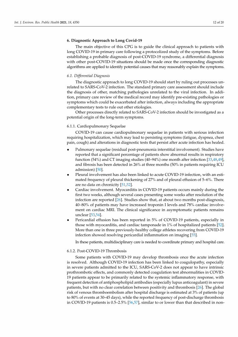
6.1.1. Cardiopulmonary Sequelae
COVID-19 can cause cardiopulmonary sequelae in patients with serious infectionrequiring hospitalization, which may lead to persisting symptoms (fatigue, dyspnea, chestpain, cough) and alterations in diagnostic tests that persist after acute infection has healed.
• Pulmonary sequelae (residual post-pneumonia interstitial involvement). Studies havereported that a significant percentage of patients show abnormal results in respiratoryfunction (54%) and CT imaging studies (40–94%) one month after infection [33,48,49],and fibrosis has been detected in 26% at three months (50% in patients requiring ICUadmission) [50].
• Pleural involvement has also been linked to acute COVID-19 infection, with an esti-mated frequency of pleural thickening of 27% and of pleural effusion of 5–6%. Thereare no data on chronicity [51,52].
• Cardiac involvement. Myocarditis in COVID-19 patients occurs mainly during thefirst two weeks, although several cases presenting some weeks after resolution of theinfection are reported [26]. Studies show that, at about two months post-diagnosis,40–80% of patients may have increased troponin I levels and 78% cardiac involve-ment on cardiac MRI. The clinical significance in asymptomatic patients remainsunclear [53,54].
• Pericardial effusion has been reported in 5% of COVID-19 patients, especially inthose with myocarditis, and cardiac tamponade in 1% of hospitalized patients [52].More than one in three previously-healthy college athletes recovering from COVID-19infection showed resolving pericardial inflammation on imaging [55].
In these patients, multidisciplinary care is needed to coordinate primary and hospital care.
6.1.2. Post-COVID-19 Thrombosis
Some patients with COVID-19 may develop thrombosis once the acute infectionis resolved. Although COVID-19 infection has been linked to coagulopathy, especiallyin severe patients admitted to the ICU, SARS-CoV-2 does not appear to have intrinsicprothrombotic effects, and commonly detected coagulation test abnormalities in COVID-19 patients appear to be primarily related to the systemic inflammatory response, withfrequent detection of antiphospholipid antibodies (especially lupus anticoagulant) in severepatients, but with no clear correlation between positivity and thrombosis [26]. The globalrisk of venous thromboembolism after hospital discharge is estimated at 3% of patients (upto 80% of events at 30–45 days), while the reported frequency of post-discharge thrombosisin COVID-19 patients is 0.5–2.5% [56,57], similar to or lower than that described in non-

Int. J. Environ. Res. Public Health 2021, 18, 4350 13 of 20
COVID-19 populations. If vascular ultrasound is not available in primary care, applicationof the Wells criteria remains useful.
6.1.3. Post-COVID-19 Immune-Mediated Manifestations
Many of the broad spectrum of post-COVID-19 immune-mediated manifestationsmay occur occasionally in the post-infectious phase [26]. Most are very infrequent andhave an autoimmune basis. Despite this, primary care physicians should be aware ofthese manifestations, as early suspicion and guidance are crucial in most cases. The mainones are:
• Arthritis: If there are inflammatory data on the physical examination or ultrasoundscan, post-COVID-19 arthritis should be ruled out. Fewer than 10 cases are reportedworldwide, affecting men, with a mean age of 54 years and a variety of joint presenta-tions (symmetrical polyarthritis, monoarthritis, enthesitis, and psoriatic arthritis), andmainly appear once the infection is resolved [26].
• Myositis: The frequency of elevated creatine kinase levels in acute COVID-19 infectionis approximately 10%, but there are no data on persistence. A review of isolated casesof COVID-19 patients with myositis/rhabdomyolysis showed most cases occurredin adult males with myalgia (in some cases severe) and appeared mainly during thefirst week of COVID-19 infection, with creatine kinase levels >10,000 U/L and renalimpairment [26].
• Pancreatitis: COVID-19 patients with abdominal pain and elevated pancreatic en-zymes diagnosed with acute pancreatitis (mostly females) are reported. The clinicaland epidemiological scenario is broad and includes infants and older patients, pa-tients with clinical symptoms, post-mortem studies, familial cases, and patients withunderlying predisposing factors [26].
• Other manifestations: Skin (perniosis), neurological (encephalitis, Guillain-Barré syn-drome, myelitis), renal (tubulopathies, glomerulonephritis), hematological (idiopathicthrombocytopenic purpura, autoimmune hemolytic anemia), endocrine (thyroiditis,manifesting as clinical symptoms of thyrotoxicosis), and systemic autoimmune (lupus,vasculitis, sarcoidosis, Kawasaki disease) diseases have been reported in COVID-19patients [26].
6.2. Specific Evaluation of Emotional Well-Being and Mental Health
The impact that the pandemic has produced on mental health is still far from beingadequately measured and quantified in primary care. In patients who have contractedCOVID-19, the impact is undeniable, although, etiopathogenically, it is more reasonableto consider it a reactive manifestation after experiencing a highly traumatic situation(with severe implications for the affected person, not only with respect to their ownhealth, but also with deep family, social, and work implications) that do not classify itas a manifestation directly caused by the virus. Despite this, the importance of thesemanifestations requires a specific section, not only because of their enormous impact onthe quality of life of the affected person, but also because of the possible impact they mayhave on their relationship with other manifestations of long COVID-19.
A key function of primary care is not to medicalize these situations, always consider-ing the key role of social determinants such as poverty, discrimination, and social exclusion;mental health and well-being are reinforced by increased social solidarity, informal socialsupport, mutual assistance and other collective or community-based measures. Mostpatients infected by the virus did not required hospital admission and were diagnosed andfollowed in primary care, but isolation leads to reduced physical activity and greater isola-tion, which may be particularly problematic in older adults, due to reduced physical abilityand potential increases in mental health problems, such as anxiety and depression. Personswith previous psychiatric disease who have experienced worsening or decompensationduring COVID-19 disease are especially vulnerable. Isolation during hospital admissioncan also lead to a worse experience of the pandemic disease process. Patients have ex-

Int. J. Environ. Res. Public Health 2021, 18, 4350 14 of 20
perienced new anti-contagion preventive standards, including isolation in their rooms,distancing at stressful vital moments (such as serious illness or a poor prognosis) whichmay sometimes have an impact on the human relationship with the doctor. COVID-19survivors after ICU admission have an increased risk of long-lasting severe functional limi-tations, psychological distress, post-traumatic stress disorder and depression [58]. A recentstudy shows that 36% of patients had altered mental and cognitive questionnaire scores(HADS, TICS, CFQ, PCL-5, IES-R) at three months, although there were no significantdifferences between patients according to COVID-19 severity [59].
6.3. Diagnosis of Post-COVID-19 Syndrome
After ruling out the above-mentioned processes, a tentative diagnosis of ongoingCOVID-19 in the second visit (V2) may be made, enabling the primary care team planfor V3, which will evaluate the evolution (whether symptoms still persist) and reassesspossible causes of the long-term symptoms using the corresponding diagnostic algorithms;if the symptoms persist and no etiology reasonably explains the persisting symptoms andcomplementary tests at V3 are unaltered, the diagnosis of post-COVID-19 syndrome isconfirmed, and a diagnostic approach to some post-COVID-19 syndromic presentationsshould be made:
Long-term fatigue: If fatigue persists as the main symptom, compliance with theclassification criteria for chronic fatigue syndrome should be evaluated. If the criteria aremet, the diagnosis of chronic fatigue syndrome associated with COVID-19 (which willbe confirmed when the criteria are still fulfilled at six months) is made and the protocolof referral to the corresponding multidisciplinary unit is applied. In the case of non-compliance, evaluation of the loss of physical condition related to the pandemic andpsychological factors is recommended, as are adapted guidelines to increase progressiveresistance training, physical activity programs, etc.
Long-term generalized pain: When generalized pain persists as the main symptom,compliance with the classification criteria for chronic generalized pain (CGP) and fibromyal-gia (FM) should be evaluated. If positive, the diagnosis of CGP or FM associated withCOVID-19 is established and the patient should be referred to the pain unit or rheumatol-ogy department, depending on availability or closeness. Other factors (loss of physicalcondition, psychological factors) should be evaluated if not.
Long-term dyspnea: When dyspnea persists as the main symptom, and after ruling outall complementary tests for alternative diseases, the so-called respiratory burn syndrome,or chronic inflammation of the respiratory tract (trachea, bronchi, bronchioles) may behypothesized, and referral to pulmonology or otolaryngology considered.
Long-term anosmia/dysgeusia: In these patients, referral to otolaryngology for follow-up and treatment using specific olfactory training therapies is recommended.
Long-term headache: There are no studies on the characteristics of long-term headachein COVID-19, but it could be included among the primary headaches, which are diagnosedaccording to symptoms in the absence of organic or structural alterations. Ruling out acentral sensitization syndrome, and a possible neurology consultation are recommended.
Long-term digestive signs and symptoms: In cases of chronicity (duration >3 months),ruling out central sensitization syndromes and evaluating referral to the digestive depart-ment are recommended.
7. Limitations and Perspective
The lack of a scientifically accepted definition, together with the limited currently-available scientific evidence and the wide methodological differences among the fewreported studies, make it very difficult to assess the frequency of long COVID-19. Studiesof symptomatic COVID-19 at >4 weeks post-diagnosis have widely-differing methodolo-gies and no overall figure can be suggested, especially in outpatients followed in primarycare. Studies suggest about 10% of COVID-19 patients may have related symptoms be-yond 3 weeks, which persist in some patients for months [24,25], while the UK Office for
Int. J. Environ. Res. Public Health 2021, 18, 4350 15 of 20
National Statistics has estimated that 20% may have symptoms that persist after 5 weeks,and 10% may have symptoms for 12 weeks or longer after acute infection [60]. The largeststudy until now has evaluated symptoms in >4000 people through a mobile application,reporting figures of 13.3% at four weeks, 4.5% at eight weeks and 2.3% at 12 weeks [20]. Incontrast, a recent Chinese study in 1655 patients with COVID-19 who required hospital-ization has shown that 76% of patients reported ≥1 symptom at six months of follow-up(measured using a self-reported symptom questionnaire asking about newly-occurringpersistent symptoms or any symptoms that worsened after COVID-19), of which 63%were associated with fatigue or muscle weakness [61]. This very wide variation in thefrequency of long COVID-19 symptoms between studies is also reported with respectto the individual frequency of the main individual symptoms eight weeks after acuteinfection [28–34,37,40–44,47] (Table 3). Unfortunately, there are wide variations in studydesigns, populations evaluated (unselected, or specifically studied in a particular specialtyor pathology) and symptom identification (self-report, medical evaluation with or withouttests), and there is also the lack of a standardized definition of long-term symptoms, sinceaggravated prior symptoms or symptoms derived from the typical aftermath of severebilateral pneumonia may be included. Progress cannot be made without a minimumconsensus on the definition of the variables to be measured (the timing of the appearanceof long-term symptoms -before, during or after acute infection- should be clearly defined)and the measurement instruments (usually unvalidated, self-reported questionnaires, somemeasured dichotomously, others categorized, others quantified with VAS scales). Anothersignificant limitation, as already seen in some studies [62], is the measurement of the grossfrequency at a specific time point with respect to the total number of patients, allowing theinclusion of people who develop the symptom de novo once the acute infection is resolved.This is very important when assessing symptoms of general involvement (fatigue, chronicpain, headache, memory disturbances...) that may be more closely related to psychologicaland social determinants than to a direct etiopathogenic link with SARS-CoV-2 infection.
Little is known about the etiopathogenic mechanisms responsible for symptom persis-tence in COVID-19. Since long-term manifestations affect various organs and systems, theymay have very diverse etiopathogenic origins, probably driven by genetic predisposition,some pathological mechanisms of the virus, the phenotypic presentation of the diseasein acute infections, and the individual immune response. Studies detecting viral RNApersistence in respiratory and extra-respiratory tissues weeks after acute infection areincreasingly common, although a clear pathogenic link between viral detection and virus-related organ-specific damage is remains unclear [63–67]. Some studies have suggestedpotential immune-mediated mechanisms of damage, e.g., in patients with migraine-likefeatures, activation of the trigeminovascular system by inflammation or direct SARS-CoV-2involvement has been suggested [45], while structural alterations (olfactory cleft and olfac-tory bulb abnormalities on MRI) have been identified in patients presenting with long-termanosmia [68].
By organ, the main long-term manifestations observed in other coronaviruses (SARS,MERS) have very clear pathological parallels with SARS-CoV-2: mainly respiratory, mus-culoskeletal and neuropsychiatric [24,25]. Structuring this wide variety of symptoms andalterations that some COVID-19 patients may present after recovery from the infection intosyndromes that can reasonably group them medically for better management and iden-tification is a challenge. Some opinion articles have suggested differentiated syndromesrelating to some symptoms or groups of prominent symptoms, such as general symptomsaround fatigue, ENT symptoms, severe post-pneumonia respiratory consequences or men-tal health [69] (Figure 1). Among the signs and symptoms reported, fatigue is undeniablythe key symptomatic marker of long COVID-19, both in frequency and the impact onthe quality of life [60], and is linked to a wide variety of concomitant systemic symptoms(diffuse myalgia, asthenia, depression, sleep disturbances . . . ) that are similar to post-SARSand other post-viral syndromes [70,71] and with a syndromic profile overlapping with cen-tral sensitivity syndromes such as CFS and FM. Several epidemiological and socioeconomic

Int. J. Environ. Res. Public Health 2021, 18, 4350 16 of 20
factors directly associated with the pandemic have been implicated in the pathogenesisof these processes, including distress, insomnia, reduced physical activity and changes indiet, and lifestyle related to confinement [24,25]. Various neuropsychological alterationsrelated to memory, sleep quality, or concentration presented by patients with long-COVID-19 are also part of central sensitization syndromes. Although most patient associationsare opposed to long COVID-19 being compared with or assimilated to other syndromescharacterized by a set of subjective symptoms in the absence of objective alterations indiagnostic examinations [60], the overlap of the signs and symptoms they present withprocesses like fibromyalgia, chronic fatigue syndrome, or multiple chemical sensitivity is,medically, incontestable.
Int. J. Environ. Res. Public Health 2021, 18, x 14 of 19
RNA persistence in respiratory and extra-respiratory tissues weeks after acute infection are increasingly common, although a clear pathogenic link between viral detection and virus-related organ-specific damage is remains unclear [63–67]. Some studies have sug-gested potential immune-mediated mechanisms of damage, e.g., in patients with mi-graine-like features, activation of the trigeminovascular system by inflammation or direct SARS-CoV-2 involvement has been suggested [45], while structural alterations (olfactory cleft and olfactory bulb abnormalities on MRI) have been identified in patients presenting with long-term anosmia [68].
By organ, the main long-term manifestations observed in other coronaviruses (SARS, MERS) have very clear pathological parallels with SARS-CoV-2: mainly respira-tory, musculoskeletal and neuropsychiatric [24,25]. Structuring this wide variety of symptoms and alterations that some COVID-19 patients may present after recovery from the infection into syndromes that can reasonably group them medically for better man-agement and identification is a challenge. Some opinion articles have suggested differ-entiated syndromes relating to some symptoms or groups of prominent symptoms, such as general symptoms around fatigue, ENT symptoms, severe post-pneumonia respira-tory consequences or mental health [69] (Figure 1). Among the signs and symptoms re-ported, fatigue is undeniably the key symptomatic marker of long COVID-19, both in frequency and the impact on the quality of life [60], and is linked to a wide variety of concomitant systemic symptoms (diffuse myalgia, asthenia, depression, sleep disturb-ances…) that are similar to post-SARS and other post-viral syndromes [70,71] and with a syndromic profile overlapping with central sensitivity syndromes such as CFS and FM. Several epidemiological and socioeconomic factors directly associated with the pandemic have been implicated in the pathogenesis of these processes, including distress, insom-nia, reduced physical activity and changes in diet, and lifestyle related to confinement [24,25]. Various neuropsychological alterations related to memory, sleep quality, or concentration presented by patients with long-COVID-19 are also part of central sensiti-zation syndromes. Although most patient associations are opposed to long COVID-19 being compared with or assimilated to other syndromes characterized by a set of subjec-tive symptoms in the absence of objective alterations in diagnostic examinations [60], the overlap of the signs and symptoms they present with processes like fibromyalgia, chronic fatigue syndrome, or multiple chemical sensitivity is, medically, incontestable.
Figure 1. A graphic proposal for the multidisciplinary care of patients with long COVID-19 in primary care. Rehab: rehabilitation, ENT: Ear, Nose and Throat, FM: fibromyalgia, CFS: chronic fatigue syndrome, NRL: neurological, Cardiovasc: cardiovascular, Psychol/psych: psychologi-cal/psychiatric.
Mental health
Post-pneumonia
DigestiveCardio
vasc
ENT training Respiratoryrehab
Musculosketalrehab
Psychol/psychcareFM/CFS
ENT symptoms
NRL
Post-COVID-19 syndromes
Referral pathways Enhanced careareas
Autoimmune
Figure 1. A graphic proposal for the multidisciplinary care of patients with long COVID-19 in primarycare. Rehab: rehabilitation, ENT: Ear, Nose and Throat, FM: fibromyalgia, CFS: chronic fatigue syn-drome, NRL: neurological, Cardiovasc: cardiovascular, Psychol/psych: psychological/psychiatric.
Information on factors that may identify populations most at risk of long COVID-19is very scarce. A prepress study suggests an increased risk of long COVID-19 in females,persons aged >70 years, and patients requiring hospitalization during the acute infection,and asthma, without differences between countries or socioeconomic groups. The numberof symptoms during the acute infection also appears to have an influence, as people with≥5 symptoms more often had long-term symptoms. The symptoms with the greatestpredictive power were fatigue, headache, dyspnea, hoarseness and myalgia (in peopleaged ≥70 years: fever, loss of smell and cardiopulmonary comorbidities) [20]. Anotherstudy showed a higher frequency in people aged ≥50 years but no other risk factors [59],and a higher risk of long-term anxiety/depression and fatigue/muscle weakness in womenand older patients [61]. The impact on the quality of life is multidimensional but appearsto be related to the care received according to severity as, in patients who required ICUadmission, the worsened quality of life focused especially on pain and mobility while, innon-hospitalized patients, it focused on anxiety or depression [34].
Not surprisingly, considering the lack of reasonable pathogenic mechanisms involvedin long COVID-19, there is no clear treatment pathway. We endorse the key points formanaging long COVID-19 proposed by the NICE guidelines [23], including giving adviceand information on self-management of symptoms, self-monitoring at home (heart rate,blood pressure, pulse oximetry, sleep surveillance), a central role for multidisciplinaryrehabilitation support (covering physical, psychological and psychiatric aspects) withoccupational therapy, physiotherapy, clinical psychology and psychiatric therapy andrehabilitation medicine (Figure 1).
Int. J. Environ. Res. Public Health 2021, 18, 4350 17 of 20
Unfortunately, the current level of scientific evidence on long COVID-19 is verylow, and the lack of internationally accepted definitions results in such a high level ofheterogeneity that it makes overall analysis and, even more, meta-analysis, very difficult.As strong conclusions and plausible etiopathogenic explanations are not available, themain objective of these guidelines is to arouse scientific interest, thus facilitating studies ofthe pathogenic mechanisms that may aid the early detection and correct management oflong COVID-19 manifestations.
8. Conclusions
Long-term manifestations are increasingly recognized in COVID-19 patients, withsystemic clinical presentations affecting a wide range of organs and systems. However,the natural history of long COVID-19 in primary care remains unknown. The estimated10% of COVID-19 survivors with a long-term course translates into 200,000 people inSpain, with the number increasing daily. Patients, many young and healthy before theirillness, report in the press and on social media worldwide that health professionals rejectthem or treat them as hypochondriacs [24,25,60]. Based on the current limited evidence, asignificant percentage of patients with long COVID-19 are expected to recover without theneed for hospital care, so a holistic, longitudinal primary care follow up will suffice, withthe involvement of multidisciplinary rehabilitation services and the creation of groups ofaffected patients from the same community or territory that harness the potential of theuse of videos and other technologies remotely. Long COVID-19 should be managed jointlywith pre-existing comorbidities and those caused by the infection. We are aware of thelimitations that a first guideline proposal in primary care may have, and that continuousupdates are essential, as some editorials pointed out for the first guidelines publishedby NICE [60,72]. Considering the very limited evidence available at the time of writing,we consider these guidelines as ‘living’ guidelines that will be periodically reviewed andupdated, as most of the keys to ensuring adequate management of long COVID-19 remainto be defined (frequency, natural history, risk factors, prognostic markers, validated tools,interventions) [23].
However, the volume of current and future long COVID-19 patients is so high thatprimary care may not be able to cope with their care, considering current resources andthe protocolized care of post-COVID-19 patients in primary care. The feasibility of theapplication of this CPG would only be possible with the provision of additional, specifichuman resources to serve this large group of patients. In addition, if comprehensivemultidisciplinary care is really to be implemented, it is also essential to provide primarycare centers with additional support staff in the main areas identified (rehabilitation,mental health) trying to avoid fragmented care. Any attempt to add this care to the primarycare portfolio of services with the human resources currently available would result in aresounding and absolute failure.
Supplementary Materials: The following are available online at https://www.mdpi.com/article/10.3390/ijerph18084350/s1, File S1: Algorithms S1–S9.
Author Contributions: Conceptualization, A.S.-A., B.K. and M.R.-C.; methodology, A.S.-A., B.K. andM.R.-C.; software, B.K.; validation, A.S.-A., P.B.-Z., L.C.F., B.K., A.M.M., J.M., J.S., G.G., R.M., J.B., A.T.and M.R.-C.; formal analysis, A.S.-A., P.B.-Z., B.K. and M.R.-C.; investigation, A.S.-A., P.B.-Z., L.C.F.,B.K., A.M.M., J.M., J.S., G.G., R.M., J.B., A.T. and M.R.-C.; writing—original draft preparation, A.S.-A.,B.K. and M.R.-C.; writing—review and editing, A.S.-A., B.K. and M.R.-C.; supervision, A.S.-A., B.K.and M.R.-C.; project administration, A.S.-A., B.K. and M.R.-C. All authors have read and agreed tothe published version of the manuscript.
Funding: This research received no external funding.
Institutional Review Board Statement: Not applicable.
Informed Consent Statement: Not applicable.

Int. J. Environ. Res. Public Health 2021, 18, 4350 18 of 20
Data Availability Statement: No new data were created or analyzed in this study. Data sharing isnot applicable to this article.
Conflicts of Interest: The authors declare no conflict of interest.
References1. Sisó-Almirall, A.; Kostov, B.; Mas-Heredia, M.; Vilanova-Rotllan, S.; Sequeira-Aymar, E.; Sans-Corrales, M.; Sant-Arderiu, E.;
Cayuelas-Redondo, L.; Martínez-Pérez, A.; García-Plana, N.; et al. Prognostic factors in Spanish COVID-19 patients: A case seriesfrom Barcelona. PLoS ONE 2020, 15, e0237960. [CrossRef] [PubMed]
2. Connors, J.M.; Levy, J.H. COVID-19 and its implications for thrombosis and anticoagulation. Blood 2020, 135, 2033–2040.[CrossRef] [PubMed]
3. Rader, B.; Scarpino, S.V.; Nande, A.; Hill, A.L.; Adlam, B.; Reiner, R.C.; Pigott, D.M.; Gutierrez, B.; Zarebski, A.E.; Shrestha, M.;et al. Crowding and the shape of COVID-19 epidemics. Nat. Med. 2020, 26, 1829–1834. [CrossRef] [PubMed]
4. WHO Coronavirus Disease (COVID-19) Dashboard. Available online: https://covid19.who.int/ (accessed on 13 January 2021).5. Casas Rojo, J.M.; Antón Santos, J.M.; Millán Núñez-Cortés, J.; Lumbreras Bermejo, C.; Ramos Rincón, J.M.; Roy-Vallejo, E.;
Artero-Mora, A.; Arnalich-Fernández, F.; García-Bruñén, J.M.; Vargas-Núñez, J.A.; et al. Clinical characteristics of patientshospitalized with COVID-19 in Spain: Results from the SEMI-COVID-19 Network. medRxiv 2020. [CrossRef]
6. Richardson, S.; Hirsch, J.S.; Narasimhan, M.; Crawford, J.M.; McGinn, T.; Davidson, K.W. The Northwell COVID-19 ResearchConsortium. Presenting Characteristics, Comorbidities, and Outcomes among 5700 Patients Hospitalized with COVID-19 in the New YorkCity Area. JAMA 2020, 323, 2052–2059. [CrossRef] [PubMed]
7. Zhang, H.; Liao, Y.-S.; Gong, J.; Liu, J.; Xia, X.; Zhang, H. Clinical characteristics of coronavirus disease (COVID-19) patients withgastrointestinal symptoms: A report of 164 cases. Dig. Liver Dis. 2020, 52, 1076–1079. [CrossRef] [PubMed]
8. Guan, W.J.; Ni, Z.Y.; Hu, Y.; Liang, W.H.; Ou, C.Q.; He, J.X.; Liu, L.; Shan, H.; Lei, C.L.; Hui, D.S.C.; et al. Clinical Characteristicsof Coronavirus Disease 2019 in China. N. Engl. J. Med. 2020, 382, 1708–1720. [CrossRef] [PubMed]
9. Lapostolle, F.; Schneider, E.; Vianu, I.; Dollet, G.; Roche, B.; Berdah, J.; Michel, J.; Goix, L.; Chanzy, E.; Petrovic, T.; et al. Clinicalfeatures of 1487 COVID-19 patients with outpatient management in the Greater Paris: The COVID-call study. Intern. Emerg. Med.2020, 15, 813–817. [CrossRef]
10. Dankwa, E.; Hall, M.; Pritchard, M.; Baillie, J.K.; Carson, G.; Docherty, A.B.; Donnelly, C.A.; Dunning, J.; Fraser, C.; Hardwick, H.;et al. ISARIC COVID-19 Clinical Data Report: 3 September 2020. medRxiv 2020. [CrossRef]
11. WHO. Report of the WHO-China Joint Mission on Coronavirus Disease 2019 (COVID-19). Available online: https://www.who.int/publications/i/item/report-of-the-who-china-joint-mission-on-coronavirus-disease-2019-(covid-19) (accessed on 25January 2021).
12. Lechien, J.R.; Chiesa-Estomba, C.M.; Place, S.; Van Laethem, Y.; Cabaraux, P.; Mat, Q.; Huet, K.; Plzak, J.; Horoi, M.; Hans, S.; et al.Clinical and epidemiological characteristics of 1420 European patients with mild-to-moderate coronavirus disease 2019. J. Intern.Med. 2020, 288, 335–344. [CrossRef]
13. Argenziano, M.G.; Bruce, S.L.; Slater, C.L.; Tiao, J.R.; Baldwin, M.R.; Barr, R.G.; Chang, B.P.; Chau, K.H.; Choi, J.J.; Gavin, N.; et al.Characterization and clinical course of 1000 patients with coronavirus disease 2019 in New York: Retrospective case series. BMJ2020, 369, m1996. [CrossRef] [PubMed]
14. Zhang, X.; Cai, H.; Hu, J.; Lian, J.; Gu, J.; Zhang, S.; Ye, C.; Lu, Y.; Jin, C.; Yu, G.; et al. Epidemiological, clinical characteristics ofcases of SARS-CoV-2 infection with abnormal imaging findings. Int. J. Infect. Dis. 2020, 94, 81–87. [CrossRef] [PubMed]
15. Borobia, A.M.; Carcas, A.J.; Arnalich, F.; Álvarez-Sala, R.; Monserrat-Villatoro, J.; Quintana, M.; Figueira, J.C.; Santos-Olmo,R.M.T.; García-Rodríguez, J.; Martín-Vega, A.; et al. A Cohort of Patients with COVID-19 in a Major Teaching Hospital in Europe.J. Clin. Med. 2020, 9, 1733. [CrossRef] [PubMed]
16. Guan, W.J.; Liang, W.H.; Zhao, Y.; Liang, H.-R.; Chen, Z.-S.; Li, Y.-M.; Liu, X.-Q.; Chen, R.-C.; Tang, C.-L.; Wang, T.; et al.Comorbidity and its impact on 1590 patients with COVID-19 in China: A nationwide analysis. Eur. Respir. J. 2020, 55, 2000547.[CrossRef] [PubMed]
17. Imam, Z.; Odish, F.; Gill, I.; O’Connor, D.; Armstrong, J.; Vanood, A.; Ibironke, O.; Hanna, A.; Ranski, A.; Halalau, A. Older ageand comorbidity are independent mortality predictors in a large cohort of 1305 COVID-19 patients in Michigan, United States. J.Intern. Med. 2020, 288. [CrossRef]
18. Romero-Sánchez, C.M.; Díaz-Maroto, I.; Fernández-Díaz, E.; Sánchez-Larsen, Á.; Layos-Romero, A.; García-García, J.; González,E.; Redondo-Peñas, I.; Perona-Moratalla, A.B.; Del Valle-Pérez, J.A.; et al. Neurologic manifestations in hospitalized patients withCOVID-19: The ALBACOVID registry. Neurology 2020, 95, e1060–e1070. [CrossRef] [PubMed]
19. Zhang, J.; Wang, X.; Jia, X.; Li, J.; Hu, K.; Chen, G.; Wei, J.; Gong, Z.; Zhou, C.; Yu, H.; et al. Risk factors for disease severity,unimprovement, and mortality in COVID-19 patients in Wuhan, China. Clin. Microbiol. Infect. 2020, 26, 767–772. [CrossRef][PubMed]
20. Sudre, C.H.; Lee, K.; Ni Lochlainn, M.; Varsavsky, T.; Murray, B.; Graham, M.S.; Menni, C.; Modat, M.; Bowyer, R.; Nguyen, L.;et al. Symptom clusters in Covid19: A potential clinical prediction tool from the COVID Symptom study app. medRxiv 2020.[CrossRef]
21. Beigel, J.H.; Tomashek, K.M.; Dodd, L.E.; Mehta, A.K.; Zingman, B.S.; Kalil, A.C.; Hohmann, E.; Chu, H.Y.; Luetkemeyer, A.;Kline, S.; et al. Remdesivir for the Treatment of Covid-19–Preliminary Report. N. Engl. J. Med. 2020. [CrossRef]

Int. J. Environ. Res. Public Health 2021, 18, 4350 19 of 20
22. Rubio-Rivas, M.; Corbella, X.; Mora-Luján, J.M.; Loureiro-Amigo, J.; Sampalo, A.L.; Bergua, C.Y.; Atiénzar, P.J.E.; García, L.F.D.;Ferrer, R.G.; Canteli, S.P.; et al. Predicting Clinical Outcome with Phenotypic Clusters in COVID-19 Pneumonia: An Analysis of12,066 Hospitalized Patients from the Spanish Registry SEMI-COVID-19. J. Clin. Med. 2020, 9, 3488. [CrossRef] [PubMed]
23. NICE. COVID-19 Rapid Guideline: Managing the Long-Term Effects of COVID-19. Available online: https://www.nice.org.uk/guidance/ng188 (accessed on 13 January 2021).
24. Rimmer, A. Covid-19: Impact of long term symptoms will be profound, warns BMA. BMJ 2020, 370, m3218. [CrossRef] [PubMed]25. Greenhalgh, T.; Knight, M.; A’Court, C.; Buxton, M.; Husain, L. Management of post-acute covid-19 in primary care. BMJ 2020,
370, m3026. [CrossRef] [PubMed]26. Ramos-Casals, M.; Brito-Zerón, P.; Mariette, X. Systemic and Organ-Specific Immune-Related Manifestations of COVID-19. Nat.
Rev. Rheumatol. 2021, in press.27. Volpicelli, G.; Lamorte, A.; Villén, T. What’s new in lung ultrasound during the COVID-19 pandemic. Intensive Care Med. 2020, 46,
1445–1448. [CrossRef]28. Tenforde, M.W.; Kim, S.S.; Lindsell, C.J.; Billig Rose, E.; Shapiro, N.I.; Files, D.C.; Gibbs, K.W.; Erickson, H.L.; Steingrub, J.S.;
Smithline, H.A.; et al. Symptom Duration and Risk Factors for Delayed Return to Usual Health Among Outpatients withCOVID-19 in a Multistate Health Care Systems Network-United States, March-June 2020. MMWR Morb. Mortal. Wkly Rep. 2020,69, 993–998. [CrossRef] [PubMed]
29. Daher, A.; Balfanz, P.; Cornelissen, C.; Müller, A.; Bergs, I.; Marx, N.; Müller-Wieland, D.; Hartmann, B.; Dreher, M.; Müller, T.Follow up of patients with severe coronavirus disease 2019 (COVID-19): Pulmonary and extrapulmonary disease sequelae. Respir.Med. 2020, 174, 106197. [CrossRef] [PubMed]
30. Rosales-Castillo, A.; Ríos, C.G.D.L.; García, J.D.M. Persistent symptoms after acute COVID-19 infection: Importance of follow-up.Medicina clinica. Med. Clínica 2021, 35–36. [CrossRef] [PubMed]
31. Carfì, A.; Bernabei, R.; Landi, F. Persistent Symptoms in Patients after Acute COVID-19. JAMA 2020. [CrossRef] [PubMed]32. Mandal, S.; Barnett, J.; E Brill, S.; Brown, J.S.; Denneny, E.K.; Hare, S.S.; Heightman, M.; E Hillman, T.; Jacob, J.; Jarvis, H.C.; et al.
‘Long-COVID’: A cross-sectional study of persisting symptoms, biomarker and imaging abnormalities following hospitalisationfor COVID-19. Thorax 2021, 76, 396–398. [CrossRef]
33. Zhao, Y.-M.; Shang, Y.-M.; Song, W.-B.; Li, Q.-Q.; Xie, H.; Xu, Q.-F.; Jia, J.-L.; Li, L.-M.; Mao, H.-L.; Zhou, X.-M.; et al. Follow-upstudy of the pulmonary function and related physiological characteristics of COVID-19 survivors three months after recovery.EClinicalMedicine 2020, 25, 100463. [CrossRef]
34. Garrigues, E.; Janvier, P.; Kherabi, Y.; Le Bot, A.; Hamon, A.; Gouze, H.; Doucet, L.; Berkani, S.; Oliosi, E.; Mallart, E.; et al.Post-discharge persistent symptoms and health-related quality of life after hospitalization for COVID-19. J. Infect. 2020, 81, e4–e6.[CrossRef]
35. Townsend, L.; Dyer, A.H.; Jones, K.; Dunne, J.; Mooney, A.; Gaffney, F.; O’Connor, L.; Leavy, D.; O’Brien, K.; Dowds, J.; et al.Persistent fatigue following SARS-CoV-2 infection is common and independent of severity of initial infection. PLoS ONE 2020, 15,e0240784. [CrossRef]
36. Milanese, M.; Corsico, A.G.; Bellofiore, S.; Carrozzi, L.; Di Marco, F.; Iovene, B.; Richeldi, L.; Sanna, A.; Santus, P.; Schisano, M.;et al. Suggestions for lung function testing in the context of COVID-19. Respir. Med. 2021, 177, 106292. [CrossRef] [PubMed]
37. Carvalho-Schneider, C.; Laurent, E.; Lemaignen, A.; Beaufils, E.; Bourbao-Tournois, C.; Laribi, S.; Flament, T.; Ferreira-Maldent,N.; Bruyère, F.; Stefi, K.; et al. Follow-up of adults with non-critical COVID-19 two months after symptoms’ onset. Clin. Microbiol.Infect. 2020. [CrossRef] [PubMed]
38. Lambert, N.J.; Survivor Corps. COVID-19 “Long Hauler” Symptoms Survey Report. Indiana University School of Medicine,Indianapolis, USA. Available online: https://dig.abclocal.go.com/wls/documents/2020/072720-wls-covid-symptom-study-doc.pdf (accessed on 25 January 2021).
39. Volpicelli, G.; Cardinale, L.; Berchialla, P.; Mussa, A.; Bar, F.; Frascisco, M.F. A comparison of different diagnostic tests in thebedside evaluation of pleuritic pain in the ED. Am. J. Emerg. Med. 2012, 30, 317–324. [CrossRef]
40. Cho, R.H.W.; To, Z.W.H.; Yeung, Z.W.; Tso, E.Y.K.; Fung, K.S.C.; Chau, S.K.Y.; Leung, E.Y.L.; Hui, T.S.C.; Tsang, S.W.C.; Kung,K.N.; et al. COVID-19 Viral Load in the Severity of and Recovery from Olfactory and Gustatory Dysfunction. Laryngoscope 2020,130, 2680–2685. [CrossRef] [PubMed]
41. Gorzkowski, V.; Bevilacqua, S.; Charmillon, A.; Jankowski, R.; Gallet, P.; Rumeau, C.; Nguyen, D.T. Evolution of OlfactoryDisorders in COVID-19 Patients. Laryngoscope 2020, 130, 2667–2673. [CrossRef]
42. Lechien, J.R.; Chiesa-Estomba, C.M.; Beckers, E.; Mustin, V.; Ducarme, M.; Journe, F.; Marchant, A.; Jouffe, L.; Barillari, M.R.;Cammaroto, G.; et al. Prevalence and 6-month recovery of olfactory dysfunction: A multicentre study of 1363 COVID-19 patients.J. Intern. Med. 2021. [CrossRef] [PubMed]
43. Fjaeldstad, A.W. Prolonged complaints of chemosensory loss after COVID-19. Dan. Med. J. 2020, 67, A05200340.44. Brandão Neto, D.; Fornazieri, M.A.; Dib, C.; Di Francesco, R.C.; Doty, R.L.; Voegels, R.L.; Pinna, F.D.R. Chemosensory Dysfunction
in COVID-19: Prevalences, Recovery Rates, and Clinical Associations on a Large Brazilian Sample. Otolaryngol. Neck. Surg. 2020.[CrossRef] [PubMed]
45. Caronna, E.; Ballvé, A.; Llauradó, A.; Gallardo, V.J.; Ariton, D.M.; Lallana, S.; Maza, S.L.; Gadea, M.O.; Quibus, L.; Restrepo, J.L.;et al. Headache: A striking prodromal and persistent symptom, predictive of COVID-19 clinical evolution. Cephalalgia 2020, 40,1410–1421. [CrossRef] [PubMed]
Int. J. Environ. Res. Public Health 2021, 18, 4350 20 of 20
46. Deidda, S.; Tora, L.; Firinu, D.; Del Giacco, S.; Campagna, M.; Meloni, F.; Orrù, G.; Chessa, L.; Carta, M.G.; Melis, A.; et al.Gastrointestinal Coronavirus disease 2019: Epidemiology, clinical features, pathogenesis, prevention and management. ExpertRev. Gastroenterol. Hepatol. 2021, 15, 41–50. [CrossRef] [PubMed]
47. Lv, H.; Zhang, W.; Zhu, Z.; Xiong, Q.; Xiang, R.; Wang, Y.; Shi, W.; Deng, Z.; Xu, Y. Prevalence and recovery time of olfactory andgustatory dysfunctions of hospitalized patients with COVID-19 in Wuhan, China. Int. J. Infect. Dis. 2020. [CrossRef] [PubMed]
48. Frija-Masson, J.; Debray, M.-P.; Gilbert, M.; Lescure, F.-X.; Travert, F.; Borie, R.; Khalil, A.; Crestani, B.; D’Ortho, M.-P.; Bancal, C.Functional characteristics of patients with SARS-CoV-2 pneumonia at 30 days post-infection. Eur. Respir. J. 2020, 56, 2001754.[CrossRef] [PubMed]
49. Mo, X.; Jian, W.; Su, Z.; Chen, M.; Peng, H.; Peng, P.; Lei, C.; Chen, R.; Zhong, N.; Li, S. Abnormal pulmonary function inCOVID-19 patients at time of hospital discharge. Eur. Respir. J. 2020, 55, 2001217. [CrossRef]
50. Van den Borst, B.; Peters, J.B.; Brink, M.; Schoon, Y.; Bleeker-Rovers, C.P.; Schers, H.; Van Hees, H.W.H.; Van Helvoort, H.;Boogaard, M.V.D.; Van Der Hoeven, H.; et al. Comprehensive health assessment three months after recovery from acuteCOVID-19. Clin. Infect. Dis. 2020. [CrossRef] [PubMed]
51. Zhu, J.; Zhong, Z.; Li, H.; Ji, P.; Pang, J.; Li, B.; Zhang, J. CT imaging features of 4121 patients with COVID-19: A meta-analysis. J.Med. Virol. 2020, 92, 891–902. [CrossRef]
52. Bao, C.; Liu, X.; Zhang, H.; Li, Y.; Liu, J. Coronavirus Disease 2019 (COVID-19) CT Findings: A Systematic Review andMeta-analysis. J. Am. Coll. Radiol. 2020, 17, 701–709. [CrossRef]
53. Puntmann, V.O.; Carerj, M.L.; Wieters, I.; Fahim, M.; Arendt, C.; Hoffmann, J.; Shchendrygina, A.; Escher, F.; Vasa-Nicotera, M.;Zeiher, A.M.; et al. Outcomes of Cardiovascular Magnetic Resonance Imaging in Patients Recently Recovered from CoronavirusDisease 2019 (COVID-19). JAMA Cardiol. 2020, 5, 1265. [CrossRef]
54. Yancy, C.W.; Fonarow, G.C. Coronavirus Disease 2019 (COVID-19) and the Heart—Is Heart Failure the Next Chapter? JAMACardiol. 2020, 5, 1216. [CrossRef] [PubMed]
55. Brito, D.; Meester, S.; Yanamala, N.; Patel, H.B.; Balcik, B.J.; Casaclang-Verzosa, G.; Seetharam, K.; Riveros, D.; Beto, R.J.; Balla, S.;et al. High Prevalence of Pericardial Involvement in College Student Athletes Recovering from COVID-19. JACC Cardiovasc.Imaging 2021, 14, 541–555. [CrossRef] [PubMed]
56. Patell, R.; Bogue, T.; Koshy, A.; Bindal, P.; Merrill, M.; Aird, W.C.; Bauer, K.A.; Zwicker, J.I. Post-discharge thrombosis andhemorrhage in patients with COVID-19. Blood 2020, 136, 1342–1346. [CrossRef] [PubMed]
57. Roberts, L.N.; Whyte, M.B.; Georgiou, L.; Giron, G.; Czuprynska, J.; Rea, C.; Vadher, B.; Patel, R.K.; Gee, E.; Arya, R. Postdischargevenous thromboembolism following hospital admission with COVID-19. Blood 2020, 136, 1347–1350. [CrossRef]
58. Hosey, M.M.; Needham, D.M. Survivorship after COVID-19 ICU stay. Nat. Rev. Dis. Prim. 2020, 6, 60. [CrossRef] [PubMed]59. Petersen, M.S.; Kristiansen, M.F.; Hanusson, K.D.; Danielsen, M.E.; Steig, B.Á.; Gaini, S.; Strøm, M.; Weihe, P. Long COVID in the
Faroe Islands—A longitudinal study among non-hospitalized patients. Clin. Infect. Dis. 2020. [CrossRef] [PubMed]60. Gorna, R.; MacDermott, N.; Rayner, C.; O’Hara, M.; Evans, S.; Agyen, L.; Nutland, W.; Rogers, N.; Hastie, C. Long COVID
guidelines need to reflect lived experience. Lancet 2021, 397, 455–457. [CrossRef]61. Huang, C.; Huang, L.; Wang, Y.; Li, X.; Ren, L.; Gu, X.; Kang, L.; Guo, L.; Liu, M.; Zhou, X.; et al. 6-month consequences of
COVID-19 in patients discharged from hospital: A cohort study. Lancet 2021, 397, 220–232. [CrossRef]62. Halpin, S.J.; McIvor, C.; Whyatt, G.; Adams, A.; Harvey, O.; McLean, L.; Walshaw, C.; Kemp, S.; Corrado, J.; Singh, R.; et al.
Postdischarge symptoms and rehabilitation needs in survivors of COVID-19 infection: A cross-sectional evaluation. J. Med. Virol.2021, 93, 1013–1022. [CrossRef]
63. Zapor, M. Persistent Detection and Infectious Potential of SARS-CoV-2 Virus in Clinical Specimens from COVID-19 Patients.Viruses 2020, 12, 1384. [CrossRef]
64. Jamiolkowski, D.; Mühleisen, B.; Müller, S.; A Navarini, A.; Tzankov, A.; Roider, E. SARS-CoV-2 PCR testing of skin for COVID-19diagnostics: A case report. Lancet 2020, 396, 598–599. [CrossRef]
65. Matschke, J.; Lütgehetmann, M.; Hagel, C.; Sperhake, J.P.; Schröder, A.S.; Edler, C.; Mushumba, H.; Fitzek, A.; Allweiss, L.;Dandri, M.; et al. Neuropathology of patients with COVID-19 in Germany: A post-mortem case series. Lancet Neurol. 2020, 19,919–929. [CrossRef]
66. Xiao, F.; Tang, M.; Zheng, X.; Liu, Y.; Li, X.; Shan, H. Evidence for Gastrointestinal Infection of SARS-CoV-2. Gastroenterology 2020,158, 1831–1833.e3. [CrossRef] [PubMed]
67. Lemprière, S. SARS-CoV-2 detected in olfactory neurons. Nat. Rev. Neurol. 2021, 17, 63. [CrossRef] [PubMed]68. Kandemirli, S.G.; Altundag, A.; Yildirim, D.; Sanli, D.E.T.; Saatci, O. Olfactory Bulb MRI and Paranasal Sinus CT Findings in
Persistent COVID-19 Anosmia. Acad. Radiol. 2021, 28, 28–35. [CrossRef] [PubMed]69. Nabavi, N. Long covid: How to define it and how to manage it. BMJ 2020, 370, m3489. [CrossRef] [PubMed]70. Moldofsky, H.; Patcai, J. Chronic widespread musculoskeletal pain, fatigue, depression and disordered sleep in chronic post-SARS
syndrome; a case-controlled study. BMC Neurol. 2011, 11, 37. [CrossRef] [PubMed]71. Brodin, P. Immune determinants of COVID-19 disease presentation and severity. Nat. Med. 2021, 27, 28–33. [CrossRef]72. Sivan, M.; Taylor, S. NICE guideline on long covid. BMJ 2020, 371, m4938. [CrossRef] [PubMed]