london pancreas workshop 2016londonpancreasworkshop.org.uk/lpw_final_abstract_version_2016.pdf ·...
TRANSCRIPT
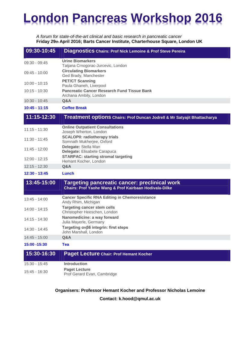
A forum for state-of-the-art clinical and basic research in pancreatic cancer Friday 29th April 2016; Barts Cancer Institute, Charterhouse Square, London UK
09:30-10:45 Diagnostics Chairs: Prof Nick Lemoine & Prof Steve Pereira
09:30 - 09:45 Urine Biomarkers Tatjana Crnogorac-Jurcevic, London
09:45 - 10:00 Circulating Biomarkers Ged Brady, Manchester
10:00 - 10:15 PET/CT Scanning Paula Ghaneh, Liverpool
10:15 - 10:30 Pancreatic Cancer Research Fund Tissue Bank Archana Ambily, London
10:30 - 10:45 Q&A
10:45 - 11:15 Coffee Break
11:15-12:30 Treatment options Chairs: Prof Duncan Jodrell & Mr Satyajit Bhattacharya
11:15 - 11:30 Online Outpatient Consultations Joseph Wherton, London
11:30 - 11:45 SCALOPII: radiotherapy trials Somnath Mukherjee, Oxford
11:45 - 12:00 Delegate: Stella Man Delegate: Elisabete Carapuca
12:00 - 12:15 STARPAC: starting stromal targeting Hemant Kocher, London
12:15 - 12:30 Q&A
12:30 - 13:45 Lunch
13:45-15:00 Targeting pancreatic cancer: preclinical work
Chairs: Prof Yaohe Wang & Prof Kairbaan Hodivala-Dilke
13:45 - 14:00 Cancer Specific RNA Editing in Chemoresistance Andy Rhim, Michigan
14:00 - 14:15 Targeting cancer stem cells Christopher Heeschen, London
14:15 - 14:30 Nanomedicine: a way forward Julia Mayerle, Germany
14:30 - 14:45 Targeting αvβ6 integrin: first steps John Marshall, London
14:45 - 15:00 Q&A
15:00 -15:30 Tea
15:30-16:30 Paget Lecture Chair: Prof Hemant Kocher
15:30 - 15:45 Introduction
15:45 - 16:30 Paget Lecture Prof Gerard Evan, Cambridge
Organisers: Professor Hemant Kocher and Professor Nicholas Lemoine
Contact: [email protected]
London Pancreas Workshop 2016

2
LONDON PANCREAS WORKSHOP 2016
Invited Speakers’ Abstracts
Space for notes

3
LONDON PANCREAS WORKSHOP 2016
Urine Biomarkers in pancreatic cancer
Tatjana Crnogorac-Jurcevic, Barts Cancer Institute, London
Resume: I obtained the MBBS, MD degree at the Medical Faculty, University of Zagreb, Croatia, and my PhD at Imperial College in London, UK. After a postdoctoral experience in molecular biology at CNRS in Toulouse, France and molecular oncology at CRUK in London, I joined Barts Cancer Institute in November 2004.
Keywords: Pancreatic adenocarcinoma, early detection, biomarkers, urine
Abstract: At present, majority of pancreatic adenocarcinoma (PDAC) patients are diagnosed at an advanced stage which results with almost invariably poor prognosis. Early detection of this disease is likely to have an impact on improving survival of PDAC patients. After a proof of concept study that protein signatures of PDAC can be found in urine (1), we have subsequently performed an in-depth LC-MS/MS study and described a panel of three protein biomarkers in urine that were able to differentiate stage I/II patients from healthy individuals with accuracy of >90% (2). In addition, we have recently shown that circulating-free miRNAs can also detect early stage PDAC (3). Both of these biomarkers now need to be validated further in a large, multicentre validation study in both symptomatic and asymptomatic groups with high risk of developing this malignancy.
References:
Debernardi S, Massat NJ, Radon TP, Sangaralingam A, Banissi A, Ennis DP, Dowe T, Chelala C, Pereira SP, Kocher HM, Young BD, Bond-Smith G, Hutchins R, Crnogorac-Jurcevic T. Noninvasive urinary miRNA biomarkers for early detection of pancreatic adenocarcinoma. Am J Cancer Res. 2015 Oct 15;5(11):3455-66.
Radon TP, Massat NJ, Jones R, Alrawashdeh W, Dumartin L, Ennis D, Duffy SW, Kocher HM, Pereira SP, Guarner L(posthumous), Murta-Nascimento C, Real FX, Malats N, Neoptolemos J, Costello E, Greenhalf W, Lemoine NR, Crnogorac-Jurcevic T. Identification of a three-biomarker panel in urine for early detection of pancreatic adenocarcinoma. Clin Cancer Res. 2015, Aug 1;21(15):3512-21.
Weeks ME, Hariharan D, Petronijevic Li, Radon TP, Whiteman HJ, Kocher HM, Timms JF, Lemoine NR, Crnogorac-Jurcevic T. Analysis of the urine proteome in patients with pancreatic ductal adenocarcinoma. Proteomics-Clinical Applications. 2008;2(7-8):1047-1057.

4
LONDON PANCREAS WORKSHOP 2016
Circulating Biomarkers in pancreatic cancer
Ged Brady, Cancer Research UK Manchester Institute, Manchester
Resume: Throughout his career Dr Brady has developed and applied advanced molecular biological methods for analysing basic and clinical biology. In 2011 he joined Clinical & Experimental Pharmacology headed by Professor Dive in order to further develop and apply blood borne oncology biomarkers aimed at benefiting clinical practise and patient outcome.
Keywords: Biomarkers, blood, KRAS, ctDNA, CTCs
Abstract: The challenge to improve outcomes for patients diagnosed with advanced pancreatic cancer remains very real with only small improvements in median survival gained by the use of systemic chemotherapy and little improvement in 5-year survival over the past decades. The advent of next generation sequencing (NGS) of tumor nucleic acids has opened up the possibility of improving outcomes through personalized therapies selected on the basis of tumor genetics. To circumvent problems associated with tumor sampling we have developed and evaluated blood-borne nucleic acids biomarkers for patients diagnosed with advanced pancreatic cancer. Our pipeline encompasses novel approaches for NGS analysis of both circulating tumour DNA (ctDNA) and circulating tumor cells (CTCs) enriched using either epitope dependent (CellSearch) or independent systems (Parsortix)1-3. Here we present data from the analysis of blood samples from 30 patients with pancreatic cancer. For ctDNA analysis we have applied whole genome NGS as well as targeted sequencing of around 600 genes linked to pancreatic cancer. We have also used droplet digital PCR (ddPCR) to evaluate CTC enrichment and establish the sensitivity of ctDNA NGS. Our results demonstrate that both ddPCR and NGS identified KRAS mutations in patient ctDNA with the targeted NGS identifying additional mutations in samples that either harboured or lacked detectable KRAS mutations. CTCs have been identified and ongoing analysis is comparing the epitope dependent and independent CTC outputs with the ctDNA data. In summary, the results establish combined CTC and ctDNA molecular analysis is practical and may be more promising than either approach alone. References:
Rothwell, D.G., Smith, N., Morris, D., Leong, H.S., Li, Y., Hollebecque, A., Ayub, M., Carter, L., Antonello, J., Franklin, L., Miller, C., Blackhall, F., Dive, C., and Brady, G. (2015). Genetic profiling of tumours using both circulating free DNA and circulating tumour cells isolated from the same preserved whole blood sample. Molecular oncology Epub ahead of print.
Girotti, M.R., Gremel, G., Lee, R., Galvani, E., Rothwell, D., Viros, A., Mandal, A.K., Lim, K.H., Saturno, G., Furney, S.J., Baenke, F., Pedersen, M., Rogan, J., Swan, J., Smith, M., Fusi, A., Oudit, D., Dhomen, N., Brady, G., Lorigan, P., Dive, C., and Marais, R. (2016). Application of sequencing, liquid biopsies and patient-derived xenografts for personalized medicine in melanoma. Cancer discovery 6, 286-99.
Chudziak, J., Burt, D.J., Mohan, S., Rothwell, D.G., Mesquita, B., Antonello, J., Dalby, S., Ayub, M., Priest, L., Carter, L., Krebs, M.G., Blackhall, F., Dive, C., and Brady, G. (2016). Clinical evaluation of a novel microfluidic device for epitope-independent enrichment of circulating tumour cells in patients with small cell lung cancer. The Analyst 141, 669-678.

5
LONDON PANCREAS WORKSHOP 2016
PET-CT in pancreatic cancer Paula Ghaneh, University of Liverpool, Liverpool Resume: Professor of Surgery, consultant surgeon, University of Liverpool. Speciality is hepato-pancreato-biliary surgery. Research interests are in pancreatic diseases. Deputy Director of the CR-UK Liverpool Cancer Trials Unit. Member of (i) NCRI upper GI clinical subgroup; (ii) NICE technology appraisal committee. Chief investigator for the ESPAC-5F and PET-PANC studies. Keywords: PET-CT, pancreatic cancer, diagnosis. Abstract: The diagnosis of pancreatic cancer is challenging and patients with pancreatic cancer may be relatively asymptomatic during its early course. The role of imaging is to identify a pancreatic lesion, determine its malignant potential, and assess its resectability. Standard diagnostic practice consists of contrast enhanced multi detector computed tomography (MDCT). endoluminal ultrasound (EUS), magnetic resonance imaging (MRI) for equivocal liver lesions and therapeutic endoscopic retrograde cholangiopancreatography (ERCP) for biliary obstruction. There are up to 10-20% of patients in whom an accurate diagnosis is difficult. Combined positron emission tomography and computed tomography (PET/CT) adds precise anatomic localization to functional data. The use of a hybrid imaging technique such as PET/CT may add further value to the diagnosis and staging of pancreatic cancer. PET/CT may also add value in assessing tumour response to chemotherapy and targeted therapeutics. Novel tracers in PET may also add further value in the diagnosis, staging and management of pancreatic disease. References:
Rijkers AP , Valkema R , Duivenvoorden, van Eijck CHJ. Usefulness of F-18-fluorodeoxyglucose positron emission tomography to confirm suspected pancreatic cancer: A meta-analysis. EJSO 2014; 40: 794-804.
Santhosh S1, Mittal BR, Rana SS, Srinivasan R, Bhattacharya A, Das A, Bhasin D. Metabolic signatures of malignant and non-malignant mass-forming lesions in the periampulla and pancreas in FDG PET/CT scan: an atlas with pathologic correlation. Abdom Imaging. 2015 Jun;40(5):1285-315.
Wu LM, Hu JN, Hua J, Liu MJ, Chen J, Xu JR. Diagnostic value of diffusion-weighted magnetic resonance imaging compared with fluorodeoxyglucose positron emission tomography/computed tomography for pancreaticmalignancy: a meta-analysis using a hierarchical regression model. J Gastroenterol Hepatol. 2012 Jun;27(6):1027-35.

6
LONDON PANCREAS WORKSHOP 2016
Pancreatic Cancer Research Fund Tissue Bank Archana Ambily, Barts Cancer Institute, London Resume: I finished my PhD at King’s College London and then worked with multi-national pharmaceutical company to understand sample acquisition and processing before joining as Tissue Bank Coordinator for Pancreatic Cancer Research Fund Tissue Bank. Keywords: Pancreatic cancer, tissue, blood, urine, saliva Abstract: The National Cancer Institute (USA) Consensus meeting in 2009
highlighted that a defining barrier to conduct translational research in pancreatic cancer is the lack of appropriately collected, clinically and molecularly annotated, and properly stored biologic material. The aim of the Pancreatic Cancer Research Tissue Bank is to develop a unique resource of clinically annotated biological materials from 3 different donor cohorts – 1) Patients with different types of pancreas cancer 2) their relatives/spouse/partner 3) healthy controls that may or may not have benign pancreatic conditions. The Tissue Bank will be backed by a bioinformatics platform to support its data mining facilities. The PCRFTB has established links with several collaborating sites across the UK who will contribute to the sample collection using harmonized standard operating procedures. Such a resource, efficiently and ethically collected, will provide researchers with high quality, relevant materials, helping to raise the standard of pancreas cancer research and facilitating the coordinated translation of scientific findings into the clinical setting.
References:
http://www.bbc.co.uk/news/health-35263463
http://www.bbc.co.uk/news/health-35319296
http://www.telegraph.co.uk/news/health/news/12100947/PICPUB-Scientists-hope-tissue-bank-will-hold-key-to-combat-pancreatic-cancer.html

7
LONDON PANCREAS WORKSHOP 2016
VOCAL: Virtual Online Consultations - Advantages and Limitations Joseph Wherton, Barts and the London School of Medicine and Dentistry, London Resume: Joe Wherton is a Senior Research Fellow at Barts and The London School of Medicine and Dentistry. His research includes interdisciplinary collaborations with academic and healthcare organisations to support the user-centred design of assisted living technologies. He obtained his Psychology BSc at University of Bath and his Psychology PhD at University of York. Keywords: Health Technology, cancer surgery, diabetes, qualitative research, ethnography Abstract: Remote video consultations between clinician and patient are technically possible and increasingly acceptable. They are being introduced in some settings alongside (and occasionally replacing) face-to-face or telephone consultations. There are a number of video communication tools available, such as Skype, that offer potential advantages to patients (who are spared the cost and inconvenience of travel) and the healthcare system (e.g. they may be more cost-effective). The VOCAL study aims to define good practice and inform its implementation in relation to clinician-patient consultations via Skype and similar virtual media. The project is based in two contrasting clinical departments (Diabetes and Cancer Surgery). The research consists of a series of in-depth qualitative studies of interpersonal interaction via Skype (micro-level) embedded in an organisational case study (meso-level) with key informant interviews at national policy level (macro-level). At micro-level, we are using video and screen capture to record what is said and done, in order to investigate the clinician-patient interaction. At meso-level we are exploring the socio-technical microsystem that supports the remote consultation, thereby identifying how organisations can best support the introduction and sustainability of this service model. At the macro-level we are capturing the perspective of national policymakers to understand the national level context of the introduction of virtual consultation in the NHS. This presentation will include preliminary findings from VOCAL to highlight how the technology can influence clinician-patient interaction, and issues to be considered for the routine use of such technology within clinical settings. References: Greenhalgh T, Vijayaraghavan S, Wherton J, Shaw S, et al. Virtual online consultations: advantages and limitations (VOCAL) study. BMJ Open, 2016;6:e009388 doi:10.1136/bmjopen-2015-009388

8
LONDON PANCREAS WORKSHOP 2016
SCALOP-2: A multi-centre randomised study of induction chemotherapy followed by capecitabine (+/- nelfinavir) with high or standard dose radiotherapy for locally advanced non-metastatic pancreatic cancer Somnath Mukherjee, CRUK/MRC Oxford Institute for Radiation Oncology, Oxford. Pradeep Virdee, Sharon Love, Elizabeth Ward, Claire Hamill, Christopher N Hurt, Pippa Corrie, David Sebag-Montefiore, Philip Parsons, Bethan Tranter, Tom Crosby, Catherine Chaytor, Maria Hawkins, Ganesh Radhakrishna, Eric O’Neill, Lisa Durrant, Sue Campbell, Rebecca Wiltshire, Timothy Maughan, John Bridgewater Resume: Senior Clinical Researcher and Honorary Consultant Oncologist at Oxford, Somnath led the SCALOP trial, which established modern pancreatic chemoradiotherapy practice in the UK. His active research membership includes CTRAD, NCRI upper GI CSG and NCRI pancreatic subgroup; ASCO LAPC guideline committee, NICE pancreatic cancer guideline committee and ESTRO pancreatic target volume guideline committee. Keywords: LAPC, gemcitabine, nab-paclitaxel, nelfinavir, radiotherapy Abstract: Chemotherapy followed by consolidation chemoradiotherapy is a treatment option for locally advanced non-metastatic pancreatic cancer (LAPC), but outcome remains poor. Hypoxia and hypovascularity confer radio-resistance; nelfinavir may enhance radio-sensitivity by reducing hypoxia and increasing vascularity. SCALOP-2 will investigate radiotherapy (RT) dose-escalation, addition of nelfinavir, and a combination of both approaches. An incorporated safety run-in will determine the dose of nelfinavir for administration alongside CRT. Eligible patients will receive 3 cycles of induction GEMABX chemotherapy [gemcitabine and nab-paclitaxel] followed by re-staging. Responding/stable patients will receive further treatment in stage 1 (non-randomised) and stage 2 (randomised). Stage 1 investigates 3 dose levels of nelfinavir (750mg, 1000mg, 1250mg bd) alongside capecitabine-based CRT. Up to 12 evaluable patients will be recruited to declare the maximum tolerated dose (MTD) of nelfinavir. Stage 2 is a randomised, 2x2 + 1 factorial design. Following induction chemotherapy, participants will be randomised to 1 cycle of GEMABX followed by either; nelfinavir, capecitabine with 50.4Gy/28# (arm A), capecitabine with 50.4Gy/28# (arm B), nelfinavir, capecitabine with 60Gy/30# (arm C), capecitabine with 60Gy/30# (arm D) or 2 further cycles GEMABX (arm E). Co-primary endpoints are 12-month OS (RT dose question) and PFS (nelfinavir question). Secondary include: toxicity, QoL, treatment compliance, CA19-9 response. 170 patients will be randomised over 4 years. Acknowledgements: Sponsor: The University of Oxford Funder: This work is supported by CRUK [grant number C28958/A17139] Application Title: CRUK/07/040: SCALOP-2. Celgene Limited are providing an educational grant and free nab‐paclitaxel to support the study. Radiotherapy Trials Quality Assurance provided by NCRI Radiotherapy Trials Quality Assurance Team (RTTQA). Trial Management: Oncology Clinical Trials Office & Statistical Input: Centre for Statistics in Medicine both part of Oxford Clinical Trials Research Unit (OCTRU), a UKCRC and NCRI registered Clinical Trials Unit. All participating sites and patients.
9
LONDON PANCREAS WORKSHOP 2016
Modifying oncolytic viral therapy for pancreatic cancer Stella Man, Barts Cancer Institute, London Authors: Y.K. Stella Man, Constantia Pantelidou, Alfonso Blázquez-Moreno, Lynda Coughlan, John Marshall, Hemant Kocher, Gunnel Halldén Abstract: Targeting avb6 -integrin expressing pancreatic cancer cells with oncolytic adenoviral mutants expressing a FMDV peptide (Ad5-FMDV): Diagnosis of pancreatic ductal adenocarcinomas (PDAC) often occurs too late for effective treatment due to non-specific presentation of symptoms. Novel detection methods and improved therapies are urgently needed. More than 60% of pancreatic cancers express avb6 integrin receptors, a molecule that is absent from healthy epithelia and thus an ideal pancreatic cancer cell target. We engineered an adenoviral (Ad5) mutant by incorporating a high affinity avb6 peptide (FMDV) into the fibre-knob domain for enhanced and specific cancer cell uptake (Ad5-FMDV). We demonstrate that Ad5-FMDV virus can replicate and kill pancreatic cancer cells with enhanced efficacy compared to wild-type Ad5. To investigate the efficacy of our viruses more effectively, three-dimensional organotypic cultures consisting of pancreatic cancer cells and pancreatic stellate cells were created. Our data demonstrate that this is a more suitable method for optimising viral administration protocols than traditional cell culture. We are currently developing a protocol to radiolabel Ad5-FMDV to enable the creation of an in-vivo diagnostic tool. Early preliminary data suggest labelling methods that retain the virus functionality and activity. Our modified oncolytic virus, Ad∆∆ (Ad5 with the E1ACR2-domain and E1B19K-gene deleted) previously demonstrated superior efficacy and therapeutic index in preclinical models of pancreatic cancer compared to earlier prototype mutants. Viral propagation of Ad∆∆ was prevented in normal cells. Ad∆∆ combined with the retargeting moiety (FMDV peptide) is currently under construction to incorporate the desirable effects into a single highly selective and potent oncolytic mutant. We anticipate that by combining the oncolytic activity of Ad∆∆, the specific uptake in pancreatic cancer cells through avb6 integrin, and radioactive labelling, the new mutant enables monitoring of targeted cells by imaging and destruction of cancer cells by oncolysis.
10
LONDON PANCREAS WORKSHOP 2016
Developing drug combinations to co-target pancreatic cancer and its supporting stroma Elisabete Carapuca, Barts Cancer Institute, London Authors: Elisabete F Carapuça, Emilios Gemenetzidis, Christine Feig, Tashinga E Bapiro, Michael D Williams, Abigail S Wilson, Francesca R Delvecchio, Prabhu Arumugam, Richard P Grose, Nicholas R Lemoine, Frances M. Richards, Hemant M Kocher Abstract: PDAC is characterised by an intense desmoplastic stroma, due to the activation of pancreatic stellate cells (PSC), which are responsible for the increased synthesis and secretion of several connective tissue components such as fibronectin and collagen type I. Abundant desmoplasia and poor tissue perfusion of PDAC tumours limit the access of therapies to neoplastic pancreatic cells and blunt treatment efficacy, and that, by co-targeting stroma and cancer cells could be a potential effective therapy against PDAC progression. Treatment of Organotypic Cultures (OT) and the KPC mouse (LSL-KrasG12D/+; LSL-Trp53R172H/+; Pdx-1-Cre) was performed to mimic human chemotherapy regimen cycles with All-Trans Retinoic Acid (ATRA), targeting PSC, and the chemotherapeutic agent gemcitabine, targeting pancreatic cancer cells (PCC). In the OT model, ATRA alone or in combination with gemcitabine resulted in reduced PCC and PSC invasion, reduced proliferation and increased apoptosis of PCC. Stellate cells activation state was altered by ATRA and by the combination of gemcitabine and ATRA, as indicated by a significant reduction in deposition of the extra-cellular matrix substrates fibronectin and collagen type I, implying stromal re-modelling. This effect was mediated by disrupting various signalling cascades (Wnt, hedgehog, retinoid, EMT and FGF) in cancer as well as stellate cells, effecting various epithelial cellular functions. While tumour angiogenesis is commonly viewed as having a pro-tumourigenic effect, we here show that the expanded vasculature permitted the decrease of hypoxia, which in turn resulted in smaller tumours in mice treated with combination therapy. Stromal co-targeting (ATRA) alongside chemotherapy (gemcitabine) is a potential clinical strategy, as it could dampen multiple signalling cascades in the tumour-stroma cross-talk, rather than ablating stroma or targeting a single pathway, therefore, altering a number of key cellular processes in tumour progression.

11
LONDON PANCREAS WORKSHOP 2016
STAR_PAC: A Phase 1B study repurposing ATRA as stromal targeting agent along with gemcitabine and nabPaclitaxel for pancreatic cancer
Hemant Kocher, Barts and Brighton CECM, Barts Cancer Institute, Queen Mary University of London; Barts and the London HPB Centre, Royal London Hospital, Barts Health NHS Trust. David J Propper, Sara Alabaf, Kelly Mousa,
Bernard North, Vasia Papoutsaki, Donna-Michele Smith, Archana Ambily, Fieke Froeling, Bristi Basu, Roopinder Gillmore, Brian Davidson, Debashis Sarker, Duncan Jodrell, Nandita DeSouza, Peter Sasieni Resume: My research directions include investigating the role of cytoskeletal proteins in pancreatic cancer progression, invasion and metastasis; investigating the role of pancreatic stellate cells; developing organotypic pancreatic cancer models and developing novel clinical trials and biomarkers inspired by the laboratory results. My clinical interests include epidemiology in patients with HPB disorders, innovative surgical techniques, risk-assessment and quality improvement.
Keywords: Pancreatic cancer, ATRA, Gemcitabine, nab-Paclitaxel, Bayesian CRM method Abstract: Pancreatic cancer (PDAC) is the fourth-highest cancer killer worldwide and is responsible for 6% of cancer deaths. Around 80% of patients are diagnosed at a late stage when cancer has spread and surgical removal is no longer possible. At present there are no treatments available which will shrink the tumour to enable surgical removal. A main factor in the lack of treatment options for patients is that pancreatic cancer is surrounded by a thick scar tissue called the stroma, which forms a barrier to prevent chemotherapy from entering and shrinking the tumour. Research carried out in laboratories has shown that a derivative of Vitamin A, All-Trans Retinoic Acid (ATRA), may have the ability to break down this stroma allowing chemotherapy to reach the cancer. STARPAC will test the combination of ATRA with two chemotherapy drugs; Gemcitabine and Nab-Paclitaxel in patients with locally advanced or metastatic pancreatic cancer. There are two parts to the study; the first will test different doses of the drugs on around 24 patients to find the highest dose patients can take without too many side effects. The second part will test this dose on around 10 patients to find the dose that will produce the desired effect with limited side-effects. Patients will take ATRA for up to 6 cycles and chemotherapy until their cancer worsens and will be followed-up for 12 months. The study will also explore the ability of a type of scan, DW-MRI, to detect changes in the cancer. Patients can also opt to donate additional tumour samples and normal cell samples. Funding: MRC DPFS Scheme, Celgene IIT programme
References:
http://public.ukcrn.org.uk/search/StudyDetail.aspx?StudyID=20118
Gastroenterology 2013. 145(5):1121-32. PMID: 23891972.
J Pathol. 2013 PMID: 23359139
12
LONDON PANCREAS WORKSHOP 2016
Novel roles for de-aminating enzymes in pancreatic cancer. Andrew D. Rhim, University of Michigan, USA Resume: I am Assistant Professor of Internal Medicine-Gastroenterology at the University of Michigan Medical School. I was the William Osler MD Fellow in Medicine and Instructor of Medicine at the Perleman School of Medicine at the University of Pennsylvania. I completed my medical training, residency in internal medicine, clinical fellowship and research fellowship in genetics at the University of Pennsylvania. My research is focused on understanding the key molecular and cellular events that occur when precancerous lesions transform into cancer. Abstract: One of the hallmarks of cancer is the generation of vast intratumoral phenotypic heterogeneity. The generation of multiple subpopulations of cancer cells that can react to various stressors in divergent ways represents a key mechanism whereby cancer, and certainly pancreatic cancer, can persist despite exposure to various treatments. The mechanism for this, however, has not been thoroughly described. Here, I will discuss our ongoing work in understanding the role for endogenous deaminating enzymes in cancer. Deaminating enzymes are not typically expressed in pancreas epithelia, but during pancreatitis and early pancreatic cancer, enzymes such as ADAR1 and APOBEC3A and B are upregulated. Because these enzymes edit specific DNA and RNA sequences, de novo expression and function of these enzymes may play a vital role in the creation of new mutations and clones as well as the modulation of phenotype within subclones through alteration of mRNA transcripts. Here, we will describe our work defining the role that these enzymes may play in shaping pancreatic cancer biology, especially through the generation of intratumoral phenotypic heterogeneity, through the use of deep sequencing, new bioinformatic approaches and genetically engineered mouse models.
References:
Roberts NJ, Norris AL, Petersen GM, Bondy ML, Brand R, Gallinger S, Kurtz RC, Olson SH, Rustgi AK, Schwartz AG, Stoffel E, Syngal S, Zogopoulos G, Ali SZ, Axilbund J, Chaffee KG, Chen YC, Cote ML, Childs EJ, Douville C, Goes FS, Herman JM, Iacobuzio-Donahue C, Kramer M, Makohon-Moore A, McCombie RW, McMahon KW, Niknafs N, Parla J, Pirooznia M, Potash JB, Rhim AD, Smith AL, Wang Y, Wolfgang CL, Wood LD, Zandi PP, Goggins M, Karchin R, Eshleman JR, Papadopoulos N, Kinzler KW, Vogelstein B, Hruban RH, Klein AP. Whole Genome Sequencing Defines the Genetic Heterogeneity of Familial Pancreatic Cancer. Cancer Discov. 2016 Feb;6(2):166-75.
Rhim AD, Oberstein PE, Thomas DH, Mirek ET, Palermo CF, Sastra SA, Dekleva EN, Saunders T, Becerra CP, Tattersall IW, Westphalen CB, Kitajewski J, Fernandez-Barrena MG, Fernandez-Zapico ME, Iacobuzio-Donahue C, Olive KP, Stanger BZ. Stromal elements act to restrain, rather than support, pancreatic ductal adenocarcinoma. Cancer Cell. 2014 Jun 16;25(6):735-47
Rhim AD, Thege FI, Santana SM, Lannin TB, Saha TN, Tsai S, Maggs LR, Kochman ML, Ginsberg GG, Lieb JG, Chandrasekhara V, Drebin JA, Ahmad N, Yang YX, Kirby BJ, Stanger BZ. Detection of circulating pancreas epithelial cells in patients with pancreatic cystic lesions. Gastroenterology. 2014 Mar;146(3):647-51.

13
LONDON PANCREAS WORKSHOP 2016
Targeting pancreatic cancer stem cells Christopher Heeschen, Barts Cancer Institute, London Resume: We have provided conclusive evidence, down to the single cell, that cancer stem cells represent the root of pancreatic cancer by giving rise to all differentiated progenies within each subclone. These cells are essential for the metastatic behaviour and, due to their inherent chemoresistance, represent an important source for disease relapse. Keywords: Cancer stem cells, macrophages, tumour microenvironment Abstract: Pancreatic cancer is still a devastating diagnosis. The almost uniform occurrence of disease relapse can be attributed at least in part to the existence of pancreatic cancer stem cells and their distinct molecular features. Specifically, we have shown that
pancreatic cancer is hierarchical organised and cancer stem cells as the root of this disease drive tumour progression, metastasis and chemoresistance.
pharmacological or genetic elimination of pancreatic cancer stem cells results in increased survival in preclinical mouse models.
cancer stem cells bear a distinct metabolic phenotype with limited plasticity that is amendable to therapeutic targeting. Circulating cancer stem cells are suitable for metabolic profiling and may be used for precision medicine approaches.
the pancreatic tumour microenvironment acts as a cancer stem cell-promoting entity further enhancing stemness and chemoresistance of cancer stem cells.
Building on these findings and newly developed technology in our lab, we are now systematically determining, which common key regulators, intrinsic or extrinsic, control the process of stemness in pancreatic cancer. We are developing means to most efficiently target identified key regulators as part of a multimodal treatment strategy, which should aid in improving treatment response and preventing relapse. References:
Hermann PC, Huber SL, Herrler T, Aicher A, Ellwart JW, Guba M, Bruns CJ, Heeschen C. Distinct populations of cancer stem cells determine tumor growth and metastatic activity in human pancreatic cancer. Cell Stem Cell. 2007 Sep 13;1(3):313-23.
Miranda-Lorenzo I, Dorado J, Lonardo E, Alcala S, Serrano AG, Clausell-Tormos J, Cioffi M, Megias D, Zagorac S, Balic A, Hidalgo M, Erkan M, Kleeff J, Scarpa A, Sainz B Jr, Heeschen C. Intracellular autofluorescence: a biomarker for epithelial cancer stem cells. Nat Methods. 2014 Nov;11(11):1161-9.
Sancho P, Burgos-Ramos E, Tavera A, Bou Kheir T, Jagust P, Schoenhals M, Barneda D, Sellers K, Campos-Olivas R, Graña O, Viera CR, Yuneva M, Sainz B Jr, Heeschen C. Pancreatic cancer stem cells are characterized by a distinct metabolism with limited metabolic plasticity. Cell Metab. 2015 Oct 6;22(4):590-605.

14
LONDON PANCREAS WORKSHOP 2016
Nanomedicine in pancreatic cancer – a way forward?
Julia Mayerle, Ernst-Moritz-Arndt-University, Greifswald, Germany. Julia Mayerle, Markus M. Lerch, Ujjwal Mukund Mahajan Resume: The research of Prof. Lerch and Prof. Mayerle has been continuously funded by the German Research Foundation (DFG), the German Cancer Aid (DKH), the Federal Ministry of Education and Research (BMBF) and others. Over 5 years they have established a Pancreas Centre for Northern Germany which recruits to EUROPAC-1, ESPAC-3 and other clinical trials. They have a longstanding expertise in preclinical pancreas research and translational projects on pancreatic disorders. Abstract: Pancreatic ductal adenocarcinoma (PDAC) is one of the most aggressive malignancies and projected to be the second leading cause of cancer related death by 2030. Despite extensive knowledge therapeutic options remain temporary and ineffective. One plausible explanation for the futile response to therapy is an insufficient and nonspecific delivery of anticancer drugs to the tumour site and the lack of companion diagnostics to predict therapy response. Cancer cells overexpress underglycosylated MUC1 (uMUC1). These molecules extend 100-200 nm above the surface, making uMUC1 an accessible target for targeted delivery of therapeutic probes. Owing to its specificity, selectivity and better tolerability with minimal side effects, siRNAs provide the basis for development of selective anti-cancer therapy compared with the relative promiscuity of small-molecular inhibitors. Superparamagnetic iron oxide nanoparticles (SPIONs) are of potential use for targeted drug delivery and biomedical imaging applications. Previously we established superparamagnetic iron oxide nanoparticles (SPIONs) coupled with siRNA directed against the cell cycle specific serine-threonine-kinase, Polo-like kinase-1 (siPLK1-StAv-SPIONs) to serve a dual purpose for delivery of siPLK1 to the tumour and for noninvasive assessment of efficiency of delivery in vivo by imaging of tumour response through the aid of SPIONs. siPLK1-StAv-SPIONs were designed and synthesized as theranostics to function via a membrane translocation peptide (MPAP) as well as a tumor selective peptide (EPPT1) to increase endosomal escape and tumour specificity, respectively. In a syngenic orthotopic as well as an endogenous cancer model tumour specific silencing of PLK1 halted tumour growth, marked by a decrease in tumour cell proliferation and an increase in apoptosis. siPLK1-StAv-SPIONs treatment represents an important step toward the application of siRNAs as cancer therapeutic agents by providing a new imaging strategy and overcoming systemic dose limitations of small molecular inhibitors.
References:
Leppkes M, Maueröder C, Hirth S, Nowecki S, Günther C, Billmeier U, Paulus S, Biermann M, Munoz LE, Hoffmann M, Wildner D, Croxford AL, Waisman A, Mowen K, Jenne DE, Krenn V, Mayerle J, Lerch MM, Schett G, Wirtz S, Neurath MF, Herrmann M, Becker C. Externalized decondensed neutrophil chromatin occludes pancreatic ducts and drives pancreatitis. Nat Commun. 2016 Mar 11;7:10973.
Seufferlein T, Mayerle J. Pancreatic cancer in 2015: Precision medicine in pancreatic cancer - fact or fiction? Nat Rev Gastroenterol Hepatol. 2016;13(2):74-5.
Mayerle J, Beyer G, Simon P, Dickson EJ, Carter RC, Duthie F, Lerch MM, McKay CJ. Prospective cohort study comparing transient EUS guided elastography to EUS-FNA for the diagnosis of solid pancreatic mass lesions. Pancreatology. 2016 Jan-Feb;16(1):110-4.
15
LONDON PANCREAS WORKSHOP 2016
The integrin v6 is a promising therapeutic target for treating PDAC John Marshall, Barts Cancer Institute, London. S Vallath, C Reader, J Morton, O Sansom, TRJ Evans, A Biankin, HM Kocher, J.F. Marshall Resume: For over 25 years I have studied the biology of tumour invasion with a particular interest in the roles of the adhesion molecules expressed on the cell surface that mediate this process. Our group concentrates on the study of integrins that are the principal family of adhesion molecules that mediate interaction between cells and the extracellular matrix (ECM).
Abstract: The integrin v6 is not usually expressed on adult cells unless they are
undergoing tissue remodeling, as v6 activates TGF, the cytokine that
orchestrates wound healing. We find that v6 is not expressed by normal
pancreas but is upregulated on over 90% of PDAC. In vitro experiments show v6 promotes cell survival, adhesion, migration and invasion through Matrigel.
Treatment of xenografts of human PDAC cell line CfPac1 with v6-blocking antibody 264RAD stopped tumour growth. In combination with Gemcitabine 264RAD shrank established (100mm3) tumours significantly (p<0.001). In separate studies we treated novel transgenic mice (Pdx1-Cre; LSL-KRasG12D/+; LSL-Dusp6-
/-) with Gemcitabine or 264RAD+Gemcitabine once they had developed PDAC. The Gemcitabine alone group all died within 17 days but the combination therapy group lived significantly longer, the last animal dying at 59 days. Analysis shows that the 264RAD-treated tumours had significantly reduced pErk and Ki67 suggesting reduced growth signaling and proliferation. Additionally, reductions seen in endomucin and SMA protein expression suggested reductions in angiogenesis and myofibroblast activity in the microenvironment. This may be due
to reduced TGF signaling since pSMAD3 and nuclear SMAD4 levels were also suppressed. Thus 264RAD suppressed PDAC development probably by targeting both the pancreatic cancer cells and the microenvironment. These studies have resulted in the funding of a Phase I trial for 264RAD therapy of PDAC patients, sponsored by Cancer Research UK in collaboration with AstraZeneca-Medimmune. References:
Slack RJ, Hafeji M, Rogers R, Ludbrook SB, Marshall JF, Flint DJ, Pyne S, Denyer JC. Pharmacological Characterization of the αvβ6 Integrin Binding and Internalization Kinetics of the Foot-and-Mouth Disease Virus Derived Peptide A20FMDV2. Pharmacology. 2016;97(3-4):114-25.
Moore KM, Thomas GJ, Duffy SW, Warwick J, Gabe R, Chou P, Ellis IO, Green AR, Haider S, Brouilette K, Saha A, Vallath S, Bowen R, Chelala C, Eccles D, Tapper WJ, Thompson AM, Quinlan P, Jordan L, Gillett C, Brentnall A, Violette S, Weinreb PH, Kendrew J, Barry ST, Hart IR, Jones JL, Marshall JF. Therapeutic targeting of integrin αvβ6 in breast cancer. J Natl Cancer Inst. 2014 Jun 28;106
John AE, Luckett JC, Tatler AL, Awais RO, Desai A, Habgood A, Ludbrook S, Blanchard AD, Perkins AC, Jenkins RG, Marshall JF. Preclinical SPECT/CT imaging of αvβ6 integrins for molecular stratification of idiopathic pulmonary fibrosis. J Nucl Med. 2013 Dec;54(12):2146-52.

16
LONDON PANCREAS WORKSHOP 2016
PAGET LECTURE
Killing cancers by targeting their engines, not their drivers Gerard Evan, Department of Biochemistry, Cambridge University, Cambridge Resume: Gerard Evan received his BA in Biochemistry from the University of Oxford and his Ph.D. in Molecular Immunology from the MRC Laboratory of Molecular Biology at the University of Cambridge. He was then a Medical Research Council Post-Doctoral Fellow and, later, a Science and Engineering Research Council Fellow in the laboratory of J. Michael Bishop at the University of California, San Francisco (UCSF). He then returned to the UK to become an Assistant Member of the Cambridge Branch of the Ludwig Institute for Cancer Research and a Research Fellow of Downing College, University of Cambridge. In 1988 he joined the Imperial Cancer Research Fund (ICRF) Laboratories in London as a Senior Scientist (1988-90) and then Principal Scientist from 1990-1999. In 1996 he was awarded the Royal Society’s Napier Professor of Cancer Research. He was appointed to the Gerson and Barbara Bass Bakar Distinguished Professor of Cancer Biology at the University of California, San Francisco in 1999 and in 2009 moved to the UK to take up the Sir William Dunn chair of Biochemistry in the University of Cambridge. Gerard is a member of EMBO, a Fellow of the UK Academy of Medical Sciences and the Royal Society.
Abstract: Cancers are genetically complex, rogue somatic clones driven by mutations that disrupt the self-regulating intra- and extracellular networks that restrain untoward growth, proliferation, survival and invasion. Because of their pivotal importance in tissue ontogeny and homeostasis, such networks have evolved to be highly robust and possessed of remarkable plasticity and ability to self-organize and self-correct. In great part, such robustness is a consequence of high levels of functional redundancy in biological signaling networks. However, while this confers great stability on tissue architecture and design, it also underlies the capacity of cells and tissues to adapt and/or evolve in response to pharmacological perturbation and is the underlying cause of the high rate of relapse in patients treated with targeted anti-cancer drugs. Our approach is to identify essential non-redundant signaling nodes (engines) that serve as the common hubs for the diverse inputs of upstream oncogenic drivers, in effect serving as GO/NO-GO switches that decide whether oncogenic signals are transduced. Examples of such common engines are Ras (integrates outputs of RTKs), Myc (transcriptionally integrates outputs of intracellular kinases), E2F (integrates outputs of cell cycle CDKs) and p53 (monitors network integrity, flux and dysfunction). Although each of these engines is pharmacologically intractable at present, it is possible to use novel switchable mouse genetics to explore the therapeutic efficacy and side effects of reversibly inhibiting such engines. Using in vivo models of lung and pancreatic adenocarcinoma, we show how targeting Myc is highly effective at driving regression established cancers, in part by triggering death of tumor cells and rapid collapse of the tumour stroma, while inflicting minimal collateral damage on normal tissues. Unexpectedly, our data also uncover a hitherto unexpected role for Myc in regulating higher order, tissue-specific regenerative programs, including potent modulation of innate and adaptive tumour

17
LONDON PANCREAS WORKSHOP 2016
immunity, so paving the way for combining Myc-targeting therapies with those that disrupt immune blockade.
von Eyss B, Jaenicke LA, Kortlever RM, Royla N, Wiese KE, Letschert S, McDuffus LA, Sauer M, Rosenwald A, Evan GI, Kempa S, Eilers M. A MYC-Driven Change in Mitochondrial Dynamics Limits YAP/TAZ Function in Mammary Epithelial Cells and Breast Cancer. Cancer Cell. 2015 Dec 14;28(6):743-57
Harajly M, Zalzali H, Nawaz Z, Ghayad SE, Ghamloush F, Basma H, Zainedin S, Rabeh W, Jabbour M, Tawil A, Badro DA, Evan GI, Saab R. p53 Restoration in Induction and Maintenance of Senescence: Differential Effects in Premalignant and Malignant Tumor Cells. Mol Cell Biol. 2015 Nov 23;36(3):438-51.
Annibali D, Whitfield JR, Favuzzi E, Jauset T, Serrano E, Cuartas I, Redondo-Campos S, Folch G, Gonzàlez-Juncà A, Sodir NM, Massó-Vallés D, Beaulieu ME, Swigart LB, Mc Gee MM, Somma MP, Nasi S, Seoane J, Evan GI, Soucek L. Myc inhibition is effective against glioma and reveals a role for Myc in proficient mitosis. Nat Commun. 2014 Aug 18;5:4632.

18
LONDON PANCREAS WORKSHOP 2016

19
LONDON PANCREAS WORKSHOP 2016
Chemosensitisation of PDAC cell lines by the VCP inhibitor NMS-873 Yuliana Astuti, Imperial College London, London Authors: Yuliana Astuti, Euan Stronach, Edward W Curry, Hani Gabra, Harpreet S Wasan, Elaina N Maginn Abstract: Pancreatic ductal adenocarcinoma (PDAC) is often characterized by defective DNA damage repair (DDR), which can be associated with resistance to DNA damaging chemotherapies. We carried out an siRNA screen assessing the potential of 639 DDR genes as therapeutic and chemosensitisation targets for PDAC. We identified several ubiquitin proteasome pathway regulators, including valosin-containing protein/p97 (VCP), whose silencing decreased cell viability and increased caspase 3/7 activities in the PANC-1 cell line. Pharmacological inhibition of VCP, with 5µM NMS-873, resulted in an increased sensitivity of PDAC cell lines PANC-1, MiaPaCa, and ASPC-1 to cisplatin-induced apoptosis. Mechanistic studies to understand the anti-cancer activity of VCP inhibition found that NMS-873 treatment decreased DNA double strand break (DSB) repair via the non-homologous end joining (NHEJ) and homologous recombination (HR) pathways: in PANC-1 cells treated with 5µM NMS-873, cisplatin-induced Rad51 foci formation were reduced (14.1% vs 4.5%), while reporter-based competency assays indicated a 33% decrease in NHEJ under these conditions. NMS-873 induced HR downregulation was found to be associated with levels of Rad51 protein, but not BRCA2 or RPA, in PANC-1 cells. Furthermore, this decrease in Rad51 protein level was rescued with the addition of proteasome inhibitor MG312, suggesting that VCP. In summary, we showed that VCP is a potential therapeutic target to improve the efficacy of DNA damaging treatments in PDAC, and suggest that it may regulate proteasomal degradation of Rad51.

20
LONDON PANCREAS WORKSHOP 2016
PI3K/mTOR/MEK-targeted therapies in pancreatic and ovarian cancers Rajpal Burmi, Imperial College London, London Authors: Rajpal S. Burmi, Euan A. Stronach, Hani Gabra, Elaina N. Maginn, Harpreet S. Wasan Abstract: Pancreatic and ovarian cancers represent aggressive malignancies where resistance to chemotherapy is commonplace. Frequent mutations and amplifications found in these tumours can result in aberrant PI3K/mTOR/MEK signalling, promoting tumour progression. Our study assessed the effect of co-targeting these pathways as a therapeutic strategy. The anti-cancer activity of the dual PI3K/mTOR inhibitor PF5212384 was evaluated in pancreatic ductal adenocarcinoma (PDAC) cell lines (BxPC-3, MIA-PaCa-2, Panc05.04, PANC-1) and ovarian cancer cell lines (PEA1, PEA2). PF5212384 treatment resulted in a significant induction of casapase-3/7 activity when co-treated with the MEK inhibitor PD325901, compared to single agent treatment, where in PDAC cell lines this was irrespective of KRAS status. PF5212384-treated PDAC cell lines also demonstrated non-caspase dependent decrease in cell viability which was attributed to autophagy and cell cycle arrest. Reverse Phase Protein Array (RPPA) analysis of BxPC-3, MIA-Pa-Ca-2 and PEA2 cells co-treated with PF5212384 and PD325901 identified MCL-1 and PDCD4 expression as significantly decreased, while BIM expression significantly increased. Furthermore, siRNA/overexpression studies confirmed the significance of these proteins in PF5212384/PD325901- co-treatment-induced apoptosis, hence their roles in PI3K/mTOR/MEK inhibition efficacy as an anti-cancer strategy. Our data highlights the potential clinical benefit of combination PI3K/mTOR/MEK-targeted therapies in pancreatic and ovarian cancers, and identifies MCL-1, PDCD4 and BIM as key regulators of this response.
21
LONDON PANCREAS WORKSHOP 2016
The role of image guided sampling in the management of solid, cystic and mixed lesions affecting the pancreas Deepak Hariharan, Royal London Hospital, Barts Health NHS Trust, London Authors: Deepak Hariharan, Hiba Junaid, Ian Renfrew, Deborah Low, Tim Fotheringham, Satyajit Bhattacharya, Ajit Abraham, Robert Hutchins, Joanne ChinAleong, Hemant M Kocher Abstract: Pancreatic lesions are solid, cystic or mixed in nature. They are often evaluated by image (computerised tomography or ultrasound) guided biopsy (IGB) to confirm diagnosis and plan treatment. This single centre prospective study aimed to determine the accuracy of IGB for all types of pancreatic lesions. Patient demographics, nature of lesion, date and results of IGB with cyto-pathological results were extracted from a prospectively maintained database. The cyto-pathological results were then compared to the gold standard: final surgical outcome or one year follow-up. Test characteristics including sensitivity, specificity, positive predictive value (PPV), negative predictive value (NPV) and diagnostic accuracy were determined. Over a 52 month period, a total of 174 patients were evaluated with IGB. Of these 90% were solid (n=157), 7% cystic (n=12) and 3% mixed (n=5). The overall sensitivity for IGB across all lesions affecting the pancreas was 88% specificity of 90%, PPV of 98%, NPV 61% and a diagnostic accuracy of 88%. The false negative rate for IGB was 10.2%. IGB is a valid method of sampling solid lesions pancreatic lesions with good diagnostic accuracy. Clinicians should however be aware of the false negative rate when counselling patients.

22
LONDON PANCREAS WORKSHOP 2016
The role of EUS sampling in the management of all lesions affecting the pancreas Deepak Hariharan, Royal London Hospital, Barts Health NHS Trust, London Authors: Deepak Hariharan, Hiba Junaid, Ian Renfrew, Deborah Low, Tim Fotheringham, Satyajit Bhattacharya, Ajit Abraham, Robert Hutchins, Joanne ChinAleong, Hemant M Kocher Abstract: Tumours affecting the pancreas are solid, cystic or mixed in nature and endoscopic ultrasound (EUS) along with fine needle aspiration cytology (FNAC) aids diagnosis and helps formulate treatment. This single centre prospective study aimed to determine the accuracy of EUS/FNAC for all types of pancreatic lesions. Patient demographics, nature of lesion, date and results of EUS/FNAC with cyto-pathological results were extracted from a prospectively maintained database. The cyto-pathological results were then compared to the gold standard: final surgical outcome or one year follow-up. Test characteristics including sensitivity, specificity, positive predictive value (PPV), negative predictive value (NPV) and diagnostic accuracy were determined. Over a 52 month period, a total of 170 patients were evaluated with EUS/FNAC of these 50% cystic (n=84), 40% were solid (n=69), and 10% mixed (n=17). The overall sensitivity for EUS/FNAC across all lesions affecting the pancreas was 80% with specificity of 96%, PPV of 90%, NPV of 90% and a diagnostic accuracy of 90%. The false negative rate for EUS/FNAC was 6.4. Differences in accuracy depended on type of pancreatic lesion. EUS/FNAC is a valid method of sampling pancreatic lesions with good diagnostic accuracy. Clinicians should be aware of the false negative rate when counselling patients.

23
LONDON PANCREAS WORKSHOP 2016
In vitro models of drug screening for pancreatic cancer
Hoyin Lam, Kings College London, London Authors: Hoyin Lam, Debashis Sarker, Claire Wells Abstract: Pancreatic ductal adenocarcinoma (PDAC) is an aggressive metastatic disease with limited treatment availability. Recent findings have demonstrated that the stroma in the tumour microenvironment of PDAC contributes towards tumour growth, metastasis and resistance to therapy. Although in vivo and patient derived xenograft models for PDAC have been established, these methods are not suitable for drug screening. In order to facilitate the development of novel therapeutics against PDAC, we identified a suitable cell line model and developed a 3D high content invasion assay with pancreatic stellate cells and collagen matrix for in vitro screening. Here we have characterised the phenotypical behaviour of PDAC cell lines in 2.5D and cellular invasion in our 3D high content invasion assay to identify the representative cell line used for future drug screening. Furthermore, we have demonstrated the translation of the invasive behaviour of our cell line model from the 3D invasion assay into the organotypic assay. Our findings suggest that our approach provides a tool for characterising, drug screening and predicting PDAC behaviour in organotypic assays.
24
LONDON PANCREAS WORKSHOP 2016
Targeting DNA damage response pathways to modulate pancreatic ductal adenocarcinoma sensitivity to DNA damaging agents. Elaina Maginn, Imperial College London, London Authors: Elaina N Maginn, Hani Gabra, Euan A Stronach, Harpreet S Wasan Abstract: The DNA damage response (DDR) is a core signaling pathway dysregulated in pancreatic ductal adenocarcinoma (PDAC), and this is associated with chemoresistance. We have previously demonstrated reversal of cisplatin resistance in PDAC cell lines following inhibition of DNA-PKcs, a component of the non-homologous end joining pathway (NHEJ; repairs DNA double-strand breaks (DSB)). Here, we demonstrate that cisplatin is most effective in PDAC cells (Panc-1, MIA-PaCa-2, AsPc-1) when NHEJ and nucleotide excision repair (NER; removes drug-DNA adducts) or base excision repair (BER; repairs single-strand breaks (SSB)) pathways are simultaneously inhibited. Immunofluorescent microscopy (IFM) suggested induction of SSBs following cisplatin treatment of PDAC cells, and consistent with this, ERCC1 upregulation (NER mediator) was observed. While NER inhibition alone did not affect cisplatin response, a striking increase in apoptosis was observed when both NER/NHEJ were inhibited, prior to cisplatin treatment. IFM identified the basis of this as the conversion of cisplatin-induced SSBs into DSBs, following NER inhibition. Cisplatin response was similarly enhanced when NHEJ/BER were concurrently inhibited. In contrast, HR inhibition did not affect response to cisplatin and NER/BER inhibitors. Across all cell lines, simultaneous NHEJ/NER inhibition increased the apoptotic response to all DNA damaging agents tested, but did not affect Taxol response. Our data suggests that resistance of PDAC to DNA damaging agents is underpinned by sequentially compensatory DDR pathways. Specifically, we identify NHEJ and NER/BER as key pathways driving resistance. Assessment of core regulators of these pathways as targets for the development of novel PDAC therapeutic strategies is underway.

25
LONDON PANCREAS WORKSHOP 2016
Abraham, Ajit 21, 22 Junaid, Hiba 21, 22
Alabaf, Sara 11 Kocher, Hemant 9, 10, 11, 15, 21, 22
Ambily, Archana 6, 11 Lam, Hoyin 23
Arumugam, Prabhu 10 Lemoine, Nicholas 10
Astuti, Yuliana 19 Lerch, Markus 14
Bapiro, Tashinga 10 Love, Sharon 8
Basu, Bristi 11 Low, Deborah 21, 22
Bhattacharya, Satyajit 21, 22 Maginn, Elaina 19, 20, 24
Biankin, A 15 Man, Stella 9
Blázquez-Moreno, Alfonso 9 Marshall, John 9, 15
Brady, Ged 4 Maughan, Timothy 8
Bridgewater, John 8 Mayerle, Julia 14
Burmi, Rajpal S 20 Morton, J 15
Campbell, Sue 8 Mousa, Kelly 11
Carapuca, Elisabete 10 Mukherjee, Somnath 8
Chaytor, Catherine 8 Mukund Mahajan, Ujjwal 14
ChinAleong, Joanne 21, 22 North, Bernard 11
Corrie, Pippa 8 O'Neill, Eric 8
Coughlan, Lynda 9 Pantelidou, Constantia 9
Crnogorac-Jurcevic, Tatjana 3 Papoutsaki, Vasia 11
Crosby, Tom 8 Parsons, Philip 8
Curry, Edward 19 Propper, David 11
Davidson, Brian 11 Radhakrishna, Ganesh 8
Delvecchio, Francesca 10 Reader, Claire 15
DeSouza, Nandita 11 Renfrew, Ian 21, 22
Durrant, Lisa 8 Rhim, Andrew 12
Evan, Gerard 16 Richards, Frances 10
Evans, TRJ 15 Sansom, O 15
Feig, Christine 10 Sarker, Debashis 11, 23
Fotheringham, Tim 21 Sasieni, Peter 11
Froeling, Fieke 11 Sebag-Montefiore, David 8
Gabra, Hani 19, 20, 24 Smith, Donna-Michele 11
Gemenetzidis, Emilios 10 Stronach, Euan 19, 20, 24
Ghaneh, Paula 5 Tranter, Bethan 8
Gillmore, Roopinder 11 Vallath, Sabari 15
Grose, Richard 10 Virdee, Pradeep 8
Halldén, Gunnel 9 Ward, Elizabeth 8
Hamill, Claire 8 Wasan, Harpreet S 19, 20, 24
Hariharan, Deepak 21, 22 Wells, Claire 23
Hawkins, Maria 8 Wherton, Joseph 7
Heeschen, Christopher 13 Williams, Michael D 10
Hurt, Christopher 8 Wilson, Abigail S 10
Hutchins, Robert 21, 22 Wiltshire, Rebecca 8
Jodrell, Duncan 11

26
LONDON PANCREAS WORKSHOP 2016
Gold Supporter
Silver Supporters
Bronze Supporter
Charity Supporters
Journal Partner
Hosts