liver/sds.docx · web viewanalysis of iron storage proteins in chicken liver comparison with...
TRANSCRIPT

Analysis of Iron Storage Proteins in Chicken Liver Comparison with Different Fowls by Mass Spectroscopy and
SDS PAGE
Submitted byIIIrd year B.Sc (B.Z.C)
141501 to 141515
Principal InvestigatorSri Ch.Venkateswarlu
Head of the departmentDepartment of Zoology
Co–InvestigatorDr.K.Siva Prasad, Lecturer
Department of zoology
PARVATHANENI BRAHMAYYASIDDHARTHA COLLEGE OF ARTS & SCIENCE, VIJAYAWADA
(An Autonomous college in the jurisdiction of Krishna University)Re-accredited at the level ‘A’ by the NAAC
Department of Zoology
July 2016

Acknowledgement
I like to express my deep and sincere gratitude to Dr.M.V.N. Padma Rao, Principal, Parvathaneni Brahmayya Siddhartha College of Arts & Science, Vijayawada for providing all facilities and their encouragement for the successful conduct of this research work. I am also grateful to my colleagues especially the faculty members in the Dept. of Zoology for their sincere co- operation and help throughout my project work. I remember with special mention the help rendered to me by Dr.K.Siva Prasad, my collegue and the Co-investigator of this project work.
I place on record my deep sense of gratitude to Academy for the financial assistance.
I express my sincere thanks to Mr.D Samuel Sparjan Babu, Senior Research Fellow, Indian Institute of Oil Palm Research ,Pedavegi, for his assistance in the project planning and for his valuable suggestions in various stages of my project work.
I express my gratitude to all my students who assisted me in my fieldwork and collection of specimens from the study area of the project work.
It is with deep sense of love and gratitude that I recall the encouragement, support and constant help so generously expressed by my family.
My Special thanks to the staff of Zoology Department for typing, photocopying and binding work of this project report.
Above all, I thank God Almighty for His Abundance of Grace showered throughout this research work.

CERTIFICATE This is to certify that the dissertation entitled “Analysis of Iron Storage Proteins in Chicken Liver Comparison with Different Fowls by Mass Spectroscopy and SDS PAGE” is bonafide project report carried out by final BZC students (141501-141515) submitted to the Department of Zoology P.B. SIDDHARTHA COLLEGE OF ARTS & SCIENCE, Vijayawada for the partial fulfillment of the Bachelor of Science during the year 2016-2017.
Project Guide Head of the Department
Signature of External Examinor
CANDIDATE’S DECLARATION
I hereby declare that the project workshop on “Analysis of Iron Storage Proteins in
Chicken Liver Comparison with Different Fowls by Mass Spectroscopy and SDS PAGE”
is a genuine record of research work done by me as the Principal Investigator,
Sri Ch.Venkateswarlu, Head of the department of Zoology and Dr.K.Siva
Prasad as Co-investigator and the work presented in this report has not been
submitted earlier.
Date: III BZC Students

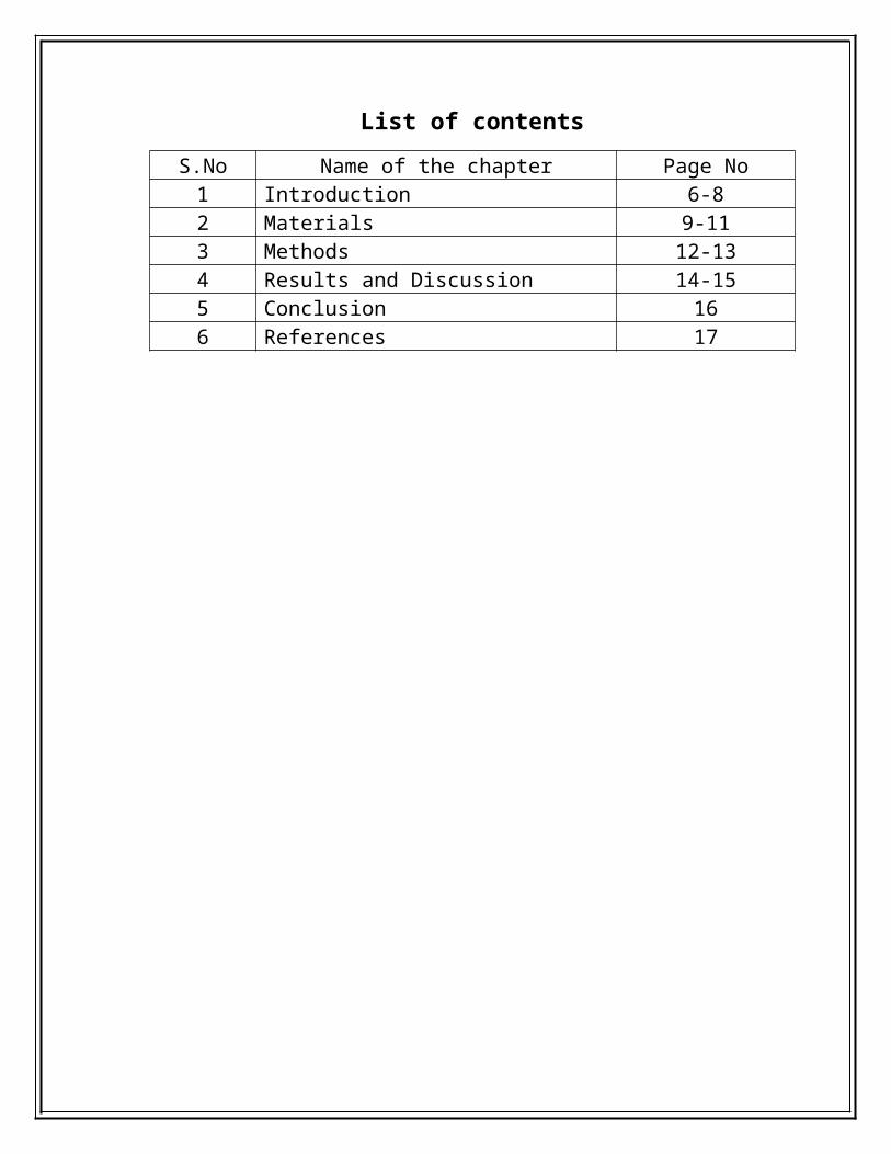
List of contents
S.No Name of the chapter Page No1 Introduction 6-82 Materials 9-113 Methods 12-134 Results and Discussion 14-155 Conclusion 166 References 17
Analysis of Iron Storage Proteins in Chicken Liver Comparison with Different Fowls by Mass Spectroscopy and
SDS PAGE
Introduction:
The present experiment was undertaken to elucidate the phylogenetic
analysis and also to find out various type of proteins present in liver of four
different types of fowls like Broilers and layers. The main difference between
layers and broiler are. An egg laying poultry is called egger or layer whereas
broilers are reared for obtaining meat[1]. So a layer should be able to produce more
number of large sized eggs, without growing too much. On the other hand, a
broiler should yield more meat and hence should be able to grow well [9]. The
housing, nutritional and environmental requirements of broilers are different from
layers. Broilers are fed with vitamin-rich supplementary feed for good growth rate
and better feed efficiency. The ratio for broilers is protein rich with adequate fat.
The layers on the other hand show two distinct phases in their life i.e. the growing
period and laying period[1]. During the growing period, they need enough space.
Over-crowding tends to suppress their growth. Feed is given in a restricted and
calculated manner. During the laying period, the layers need enough space and
adequate lighting. Feed with vitamins, minerals and micronutrients influence the
hatching of eggs[4]. Layers are provided with fibrous cheaper diets using
agricultural by-products.
Poultry meat is an important source of high quality proteins, minerals and
vitamins to balance the human diet. Specially developed breeds of chicken meat
(broiler) are now available with the ability of quick growth and high feed
conversion efficiency [10]. Poultry manure has high fertilizer value and can be used
for increasing yield of all crops. Broiler farming has several advantages: firstly the
initial investment is low, and there is a faster[3] return from the investment. The
rearing period is 6 to 7 weeks only and more number of flocks can be taken in the
same shed and also the amount of protein obtained from a broiler is high when
compared to layer[8].
So, to know the amount of protein and the type of proteins present in
different varieties we prefer SDS PAGE analysis of the chicken liver sample [7]. It
is a very common method for separating proteins by electrophoresis uses a
discontinuous polyacrylamide gel as a support medium and sodium dodecyl sulfate
(SDS) to denature the proteins. The method is called sodium dodecyl sulfate
polyacrylamide gel electrophoresis (SDS-PAGE). SDS (also called lauryl sulfate)
is an anionic detergent[5]. That is when dissolved its molecules have a net negative
charge within a wide pH range. The negative charges on SDS destroy most of the

complex structure of proteins, and are strongly attracted toward an anode
(positively-charged electrode) in an electric field[2]. If proteins of known mass are
run simultaneously with the unknowns, the relationship between retardation factor
(Rf) and mass can be plotted, and the masses of unknown proteins estimated.
Protein separation by SDS-PAGE can be used to estimate relative molecular
mass, to determine the relative abundance of major proteins in a sample, and to
determine the distribution of proteins among fractions[2]. The purity of protein
samples can be assessed and the progress of a fractionation or purification
procedure can be followed. Different staining methods can be used to detect rare
proteins and to learn something about their biochemical properties. Here we used
spectrophotometric analysis to know the concentration of the protein sample.
In the present study, we investigated the proteomes in chicken liver different
fowl’s viz. Natural fowl male and female, broilers and layers in order to identify
biologically important proteins.

Materials: Power pack
SDS PAGE apparatus
Eppendorf tubes
Centrifuge
Scalpels
Mortar and pestle
Micro pipettes and tips

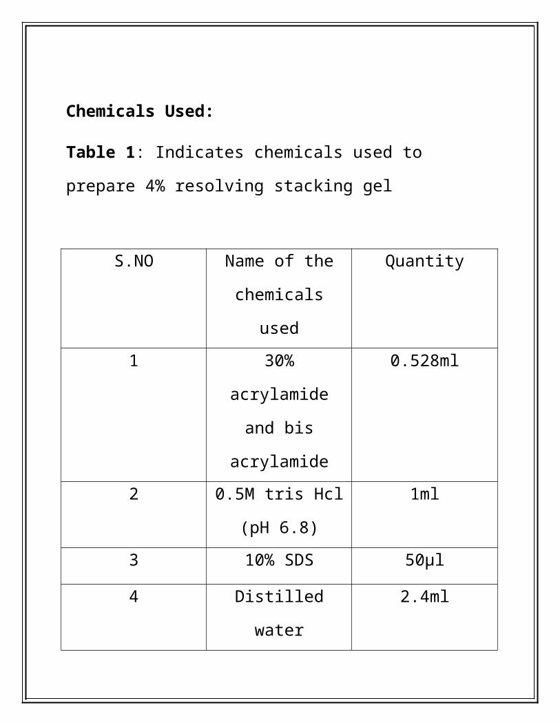
Chemicals Used:
Table 1: Indicates chemicals used to prepare 4% resolving
stacking gel
S.NO Name of the
chemicals used
Quantity
1 30% acrylamide and
bis acrylamide
0.528ml
2 0.5M tris Hcl (pH
6.8)
1ml
3 10% SDS 50µl
4 Distilled water 2.4ml
5 TEMED 4µl
6 10% APS 20µl

Table 2: indicates the use of chemical for preparing 10%
resolving separating gel
S.NO Name of the
chemicals used
Quantity
1 30% acrylamide
and bis acrylamide
3.2 ml
2 0.5M tris Hcl (pH
6.8)
2ml
3 10% SDS 80µl
4 Distilled water 2.68ml
5 TEMED 4µl
6 10% APS 40µl

Table 3:Indicates the use of chemical for staining solution
S.NO Composition For 1Lit
1 Acetic acid 100(ml)
2 CBB 60(mg)
3 Distilled water 900(ml)
Table: 4. Indicates the chemicals used for destining solution
S.NO Component For 1Lit
1 Methanol 100(ml)
2 Acetic Acid 250(ml)
3 Distilled Water 650(ml)
Methodology
Tissue collection (Fig-1)
Four different types of fowl tissues were collected from Vijayawada (i.e.,
broilers, layers and natural fowl male and natural fowl female). The liver tissue
were collected and rinsed with distilled water to remove bound particles and other
impurities if any. Then the tissue is cut into small pieces which are considered as a
primary sample for the experiment.
Protein extraction:
1. About 200mg of tissue from each fowl is taken, homogenized with the help
of mortar and pestle using equal amount of 0.5M Tris-Hcl buffer (pH 7.5)
and 10% SDS buffer.
2. The resulting homogenates were centrifuged at 15000rpm for 30 minutes and
then the supernatants were collected into the fresh autoclaved vials
3. Again centrifuge the whole content by adding 0.5M Tris- Hcl at 15000rpm
for 30 minutes
4. Dissolve the obtained pellet in Tris-Hcl, wait until the pellet gets dissolved
and then process the pellet to SDS PAGE analysis to obtain the type of
protein’s present in the sample.
Lowry’s Method Procedure:

1. 0.2 ml of BSA working standard in 5 test tubes and make up to 1ml
using distilled watetr.
2. The test tube with 1 ml distilled water serve as blank.
3. Add 4.5 ml of Reagent I and incubate for 10 minutes.
4. After incubation add 0.5 ml of reagent II and incubate for 30 minutes
5. Measure the absorbance at 660 nm and plot the standard graph.
6. Estimate the amount of protein present in the given sample from the
standard graph.
Protein profiling using SDS PAGE:
Electrophoresis was carried out in a discontinuous SDS PAGE system using
10% acrylamide gel for resolving, 4% acrylamide gel for stalking. SDS PAGE was
performed at constant voltage of 50 Volts.
Staning and Destaining of SDS PAGE:
1. in the 500mL of the storage solution for at least 1 hr. The gel should return to
its original dimensions during 1. After electrophoresis, the gel is washed off
the glass plates with 500 mL of the gel-fixing solution and soaked in that
solution for 1hr. The purpose of this step is to gently remove the gel from the
plate and begin washing the SDS-containing gel buffers out of the gel.
2. 2. Cover the gel with 500mL of the gel-washing
solution(Staining/Coomassive), and continue to fix the proteins in the gel by

incubating overnight at room temperature on a rocking table with gentle
agitation. The gel should be covered during this process to avoid
contamination and to prevent the evaporation of the solution. At the end of the
time, remove the solution by aspiration.
3. 3. Cover the gel with 400mL of the Coomassie stain. Stain the gel at room
temperature for 3 to 4 hr with gentle agitation. The Coomassie stain is removed
by aspiration after staining.
4. 4. Cover the gel with 250mL of the destaning solution and allow the gel to
destain with gentle agitation. The destain solution should be changed several
times, removing it at each change by aspiration. Continue the destaining until
the protein bands are seen without background staining of the gel.
5. Equilibrate the gel this process.
6. Store the gel in the storage solution as needed.
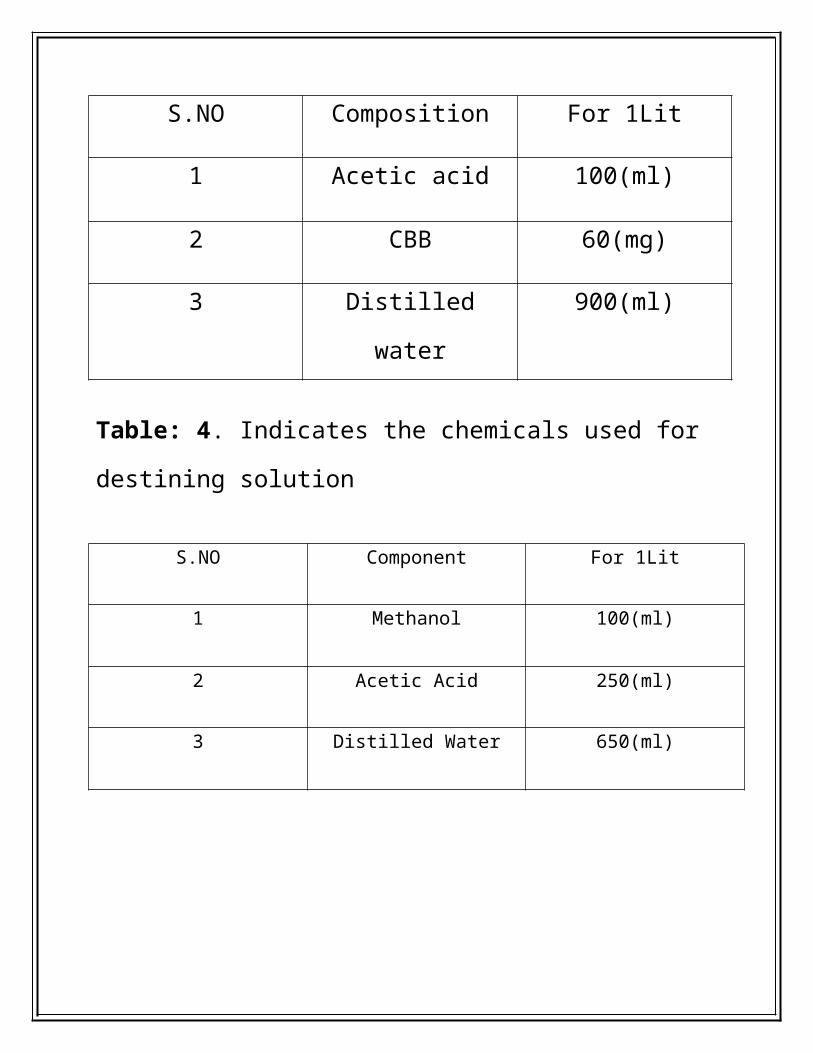
Fig-1: Chicken liver samples
Fig-2: SDS PAGE

Fig-3: SDS PAGE
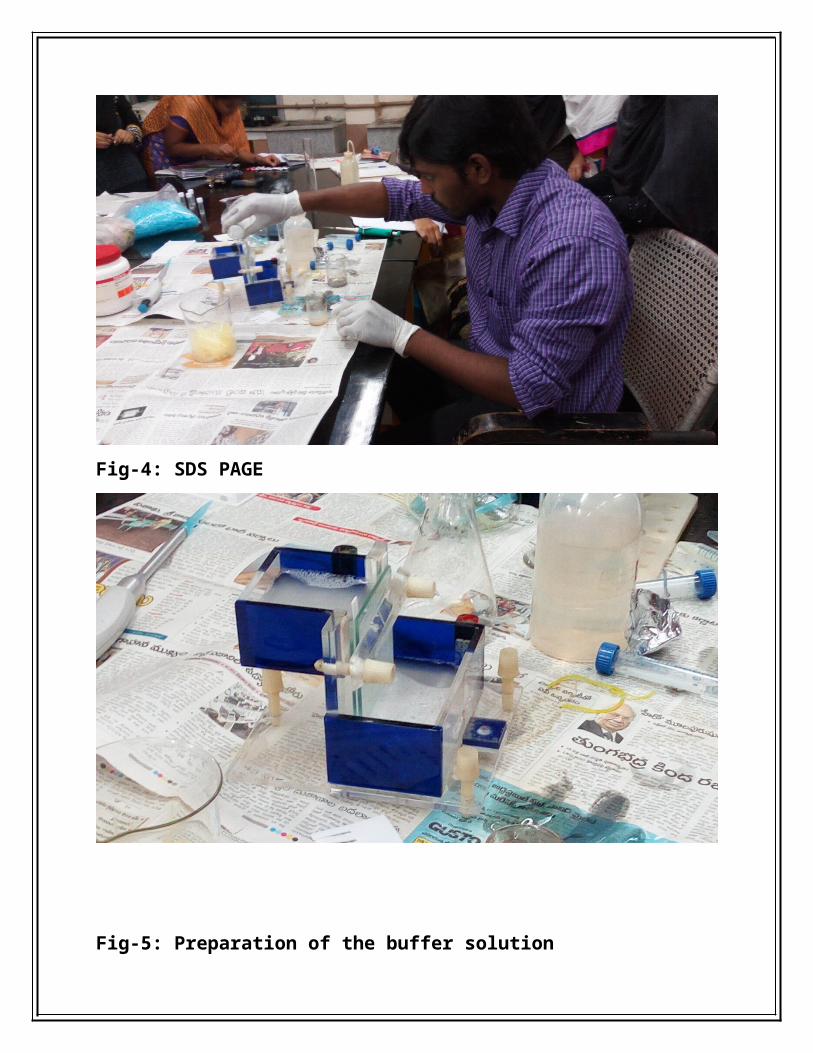
Fig-4: SDS PAGE

Fig-5: Preparation of the buffer solution
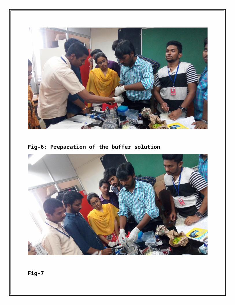
Fig-6: Preparation of the buffer solution
Fig-7
Fig- 8

Fig-9
Fig-10

Results: Estimation of protein concentration
Estimation of protein concentration by using Lowry method:
NF M layers B NFM B20
0.1
0.2
0.3
0.4
0.5
0.6
0.7
0.469
0.371
0.604000000000001
0.346
0.545
Series1
From the above graph we can say that the concentration of Natural fowl female
was less i.e.0.346 and the protein concentration of broiler is obtained high.

In the presence of SDS PAGE banding pattern of chicken liver obtained from four
different sources of fowls. It showed a very distinct polymorphism in
electrophoretic pattern. A total of 10 polypeptide bands were observed from four
different sources. A few bands that was observed in one variety is absent in other.
Fig 1: It shows various bands obtained after SDS PAGE analysis
P1- Broilers (male) P2 Natural fowl (male) P3 layer (female)
P4-Natural fowl (female)
From the obtained results we say that the cross breeding can be
done between P1 and P4
i.e. highly evolved genotypes can be done by which we can
increase the protein content.
Conclusion:Electrophoretic examination of the different Chicken liver proteins had
revealed that there is genetic diversity among different genotypes which was
observed through image processing [6]. So, we can recommend the genetically
diversified species for further breeding programs to obtain a better Fowls with
more protein content .Which can be done with the help of crossbreeding or gene
transfer techniques.
References:1. Díez J. M., Agapito M. T., Recio J. M.1985. Isolation and purification
of duck liver ferritin. Rev. Esp. Fisiol. 41:341–344.Mete A.,
2. van Zeeland Y. R. A., Vaandrager A. B., van Dijk J. E., Marx J. J.
M.,Dorrestein G. M. 2005. Partial purification and characterization of
ferritin from the liver and intestinal mucosa of chickens, turtledoves
and mynahs. Avian Pathol.34:430–434.
3. Passaniti A., Roth T. F. 1989. Purification of chicken liver ferritin by
two novel methods and structural comparison with horse spleen
ferritin. Biochem. J.258:413–419.
4. Santos Benito F. F., Martin Mateo M. C.1983. Isolation, purification
and characterization of spleen ferritin of Gallus domesticus L. Comp.
Biochem. Physiol. B74:643–645
5. Stevens P. W., Dodgson J. B., Engel J. D.1987. Structure and
expression of the chicken ferritin H subunit gene. Mol. Cell.
Biol. 7:1751–1758.
6. Abe T., Kinda T., Takano Y., Chikazawa S., Higuchi M., Kawasaki N.,
Orino K.,
Watanabe K.2006. Relationship between body iron stores and diquat
toxicity in male Fisher-344 rats. Biometals 19:651–657.
7. Shevchenko A, Wilm M, Vorm O, et al. Mass spectrometric
sequencing of proteins silver-stained polyacrylamide gels. Anal Chem.
1996;68(5):850–8. [PubMed]

8. Schuck S, Honsho M, Ekroos K, Shevchenko A, Simons K. Resistance
of cell membranes to different detergents. Proc Natl Acad Sci U S A.
2003;100(10):5795–800. [PMC free article] [PubMed]
9. Perdew GH, Schaup HW, Selivonchick DP. The use of a zwitterionic
detergent in two-dimensional gel electrophoresis of trout liver
microsomes. Anal Biochem. 1983;135 (2):453–5. [PubMed]