liver cirrhosis. liver cirrhosis define cirrhosis. recognize the types of cirrhosis. recognize...
TRANSCRIPT

LIVER CIRRHOSIS

Liver cirrhosis
Define Cirrhosis. Recognize the types of cirrhosis. Recognize the major causes and the
pathogenetic mechanisms leading to cirrhosis.
Describe the pathological findings in cirrhotic livers.
Cirrhosis
Cirrhosis is among the top 10 causes of death in the Western world. The chief worldwide contributors are alcohol abuse and viral hepatitis. Other causes include biliary disease, and iron overload. Cirrhosis is the end-stage of chronic liver disease

Cirrhosis
Cirrhosis is defined by three characteristics1)Fibrosis in the form of delicate bands or
broad scars/septa 2)Nodules containing regenerating
hepatocytes encircled by fibrosis, with diameters varying from very small (<3 mm, micronodules) to large (several centimeters, macronodules)
3)Disruption of the architecture of the entire liver
features of cirrhosis
Vascular architecture is reorganized by
the parenchymal damage and scarring, with the formation of abnormal interconnections between vascular inflow and hepatic vein outflow channels.
Fibrosis is the key feature of progressive damage to the liver. Once cirrhosis has developed, reversal is thought to be rare.

Classifiation of cirrhosis
The classification is based on the underlying etiology.
Many forms of cirrhosis (particularly alcoholic cirrhosis) are initially micronodular, but there is a tendency for nodules to increase in size with time.
Classifiation of cirrhosis based on causes
Alcoholic liver disease 60% to 70%
Viral hepatitis 10% Biliary diseases 5% to
10% Primary hemochromatosis 5% Wilson disease Rare α1-Antitrypsin deficiency Rare Cryptogenic cirrhosis 10% to
15%
Classifiation of cirrhosis
Infrequent types of cirrhosis also include the cirrhosis developing in infants and children
with galactosemia and tyrosinosis drug-induced cirrhosis. Severe fibrosis can occur in the setting of
cardiac disease (sometimes called "cardiac cirrhosis,").
In some cases there is no cause and these are referred to as cryptogenic cirrhosis.
Once cirrhosis is established, it is usually impossible to establish an etiologic diagnosis on morphologic grounds alone
Pathogenesis of cirrhosis
The pathogenetic processes in cirrhosis are progressive fibrosis and reorganization of the vascular microarchitecture of the liver
In the normal liver, interstitial collagens (types I and III) are concentrated in portal tracts and around central veins. The type IV collagen(reticulin) is in the space of Disse.

Pathogenesis of cirrhosis
In cirrhosis, types I and III collagen are deposited in the lobule, creating delicate or broad septal tracts.
There is loss of fenestrations in the sinusoidal endothelial cells (capillarization of sinusoids, that is the sinusoidal space comes to resemble a capillary rather than a channel for exchange of solutes between hepatocytes and plasma).
Pathogenesis of cirrhosis
The major source of excess collagen in cirrhosis is the perisinusoidal stellate cells ( Ito cells), which lie in the space of Disse. Although normally functioning as vitamin A fat-storing cells, during the development of cirrhosis they become activated and transform into myofibroblast-like cells.

Pathogenesis of cirrhosis
Collagen synthesis is stimulated by Chronic inflammation, with production of
inflammatory cytokines. Cytokine production by activated
endogenous cells (Kupffer cells, endothelial cells, hepatocytes, and bile duct epithelial cells).
Disruption of the normal extracellular matrix.
Direct stimulation of stellate cells by toxins

The cirrhotic patient may develop jaundice and even hepatic failure’

Clinical Features
All forms of cirrhosis may be clinically silent.
When symptomatic they lead to nonspecific clinical manifestations: anorexia, weight loss, weakness, osteoporosis, and, in advanced disease, frank debilitation.
Incipient or overt hepatic failure may develop.

Clinical Features
The ultimate mechanism of most cirrhotic deaths is
(1) progressive liver failure , (2) a complication related to portal
hypertension, or (3) the development of
hepatocellular carcinoma

Downloaded from: Robbins & Cotran Pathologic Basis of Disease (on 9 March 2006 01:41 PM)© 2005 Elsevier
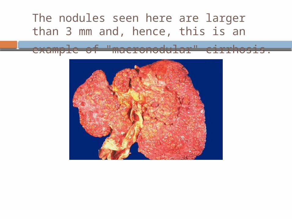
The nodules seen here are larger than 3 mm and, hence, this is an example of
"macronodular" cirrhosis.
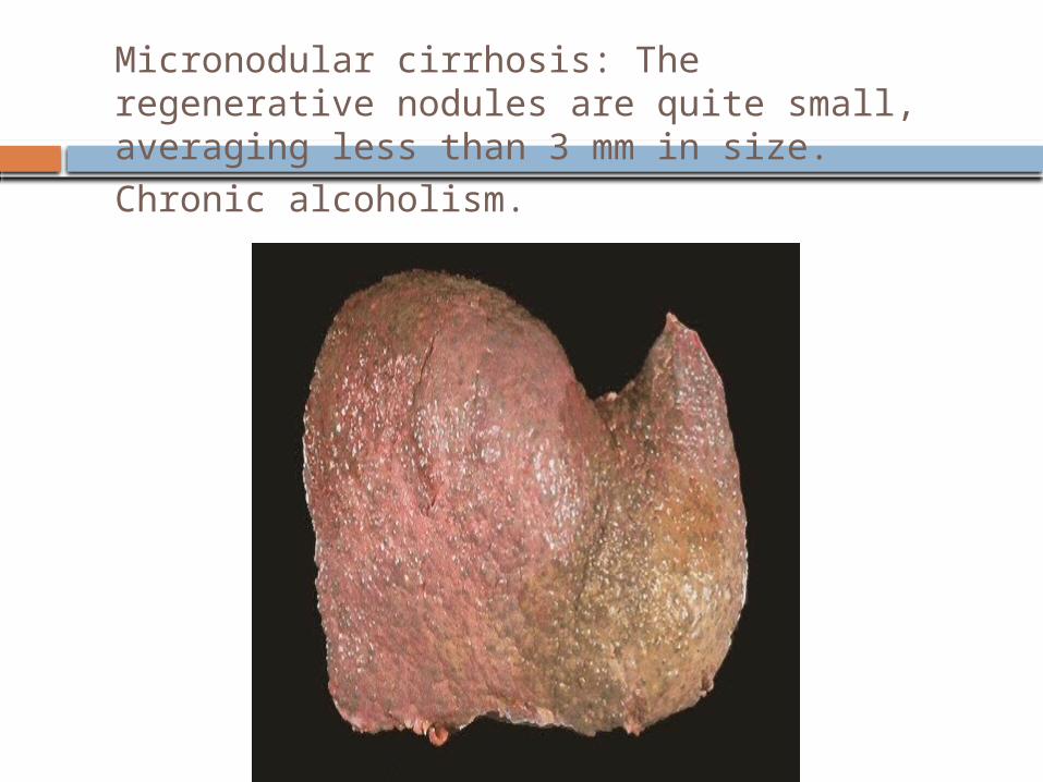
Micronodular cirrhosis: The regenerative nodules are quite small, averaging less than 3 mm in size. Chronic alcoholism.
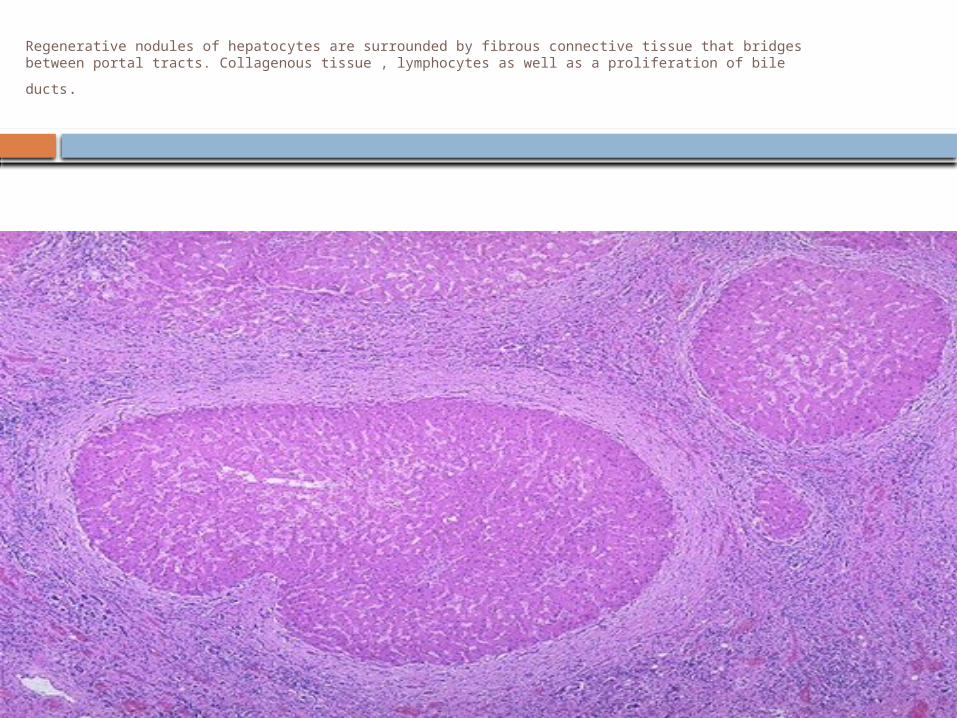
Regenerative nodules of hepatocytes are surrounded by fibrous connective tissue that bridges
between portal tracts. Collagenous tissue , lymphocytes as well as a proliferation of bile ducts.
Chronic Hepatitis, morphology
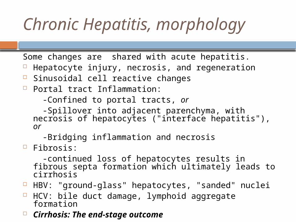
Some changes are shared with acute hepatitis. Hepatocyte injury, necrosis, and regeneration Sinusoidal cell reactive changes Portal tract Inflammation: -Confined to portal tracts, or -Spillover into adjacent parenchyma, with necrosis of
hepatocytes ("interface hepatitis"), or -Bridging inflammation and necrosis Fibrosis: -continued loss of hepatocytes results in fibrous septa
formation which ultimately leads to cirrhosis HBV: "ground-glass" hepatocytes, "sanded" nuclei HCV: bile duct damage, lymphoid aggregate formation Cirrhosis: The end-stage outcome

Downloaded from: Robbins & Cotran Pathologic Basis of Disease (on 9 March 2006 01:41 PM)© 2005 Elsevier

Downloaded from: Robbins & Cotran Pathologic Basis of Disease (on 9 March 2006 01:41 PM)© 2005 Elsevier

Viral hepatitis C which is at a high stage with extensive fibrosis and progression to macronodular cirrhosis, as evidenced by the
large regenerative nodule at the center right.
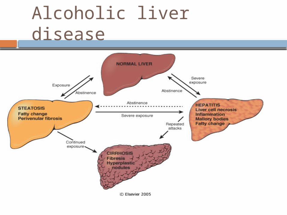
Alcoholic liver disease
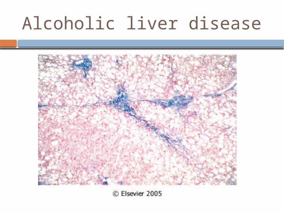
Alcoholic liver disease
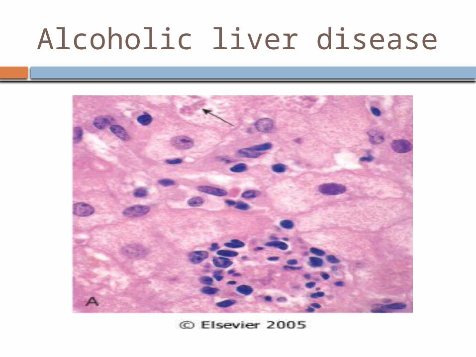
Alcoholic liver disease

Alcoholic liver disease

Alcoholic liver disease