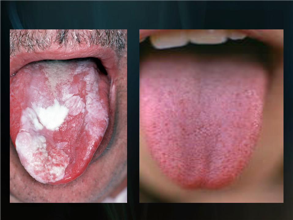
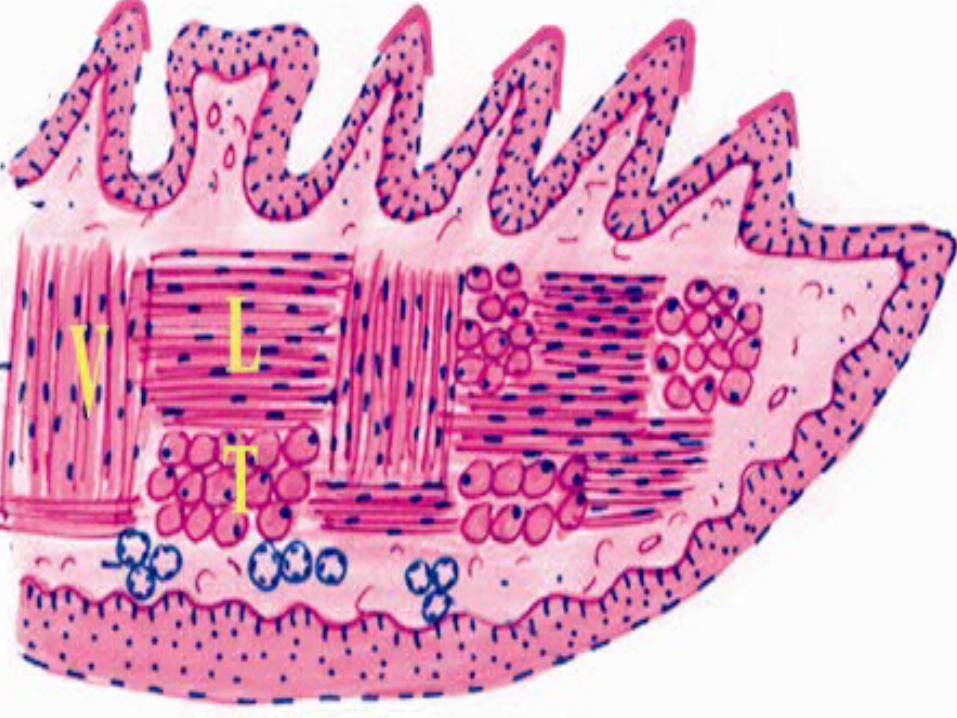
lip&tongue
TRANSCRIPT

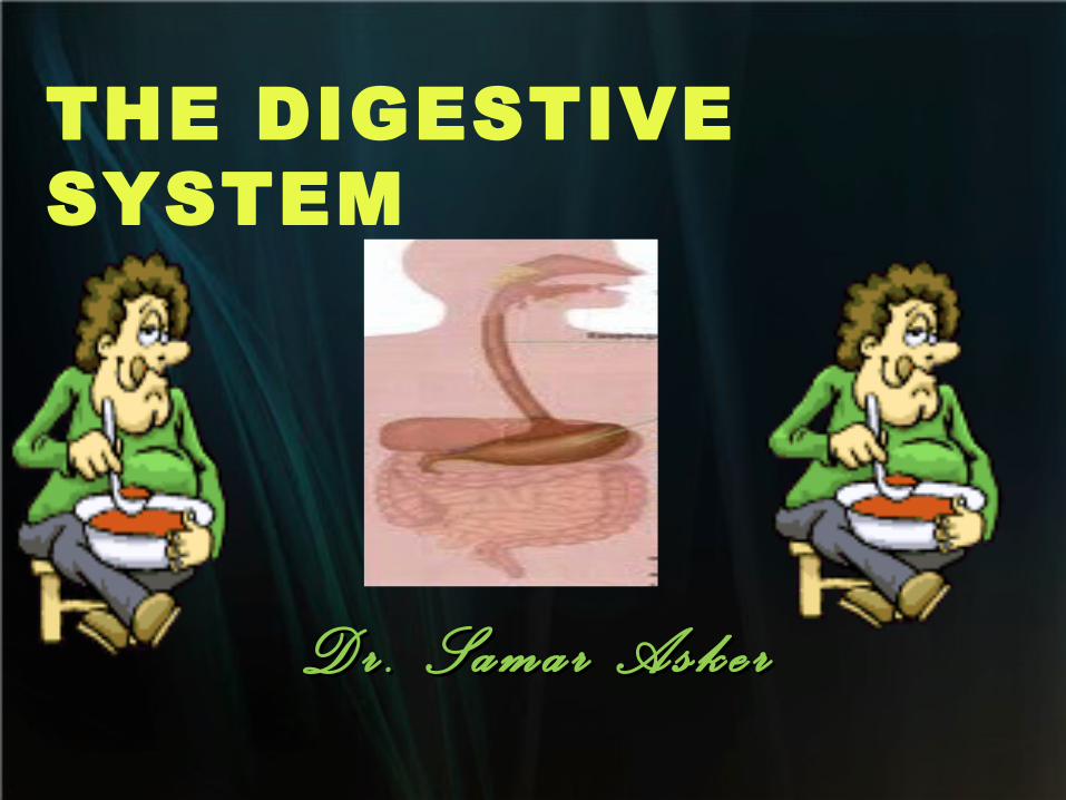
THE DIGESTIVE SYSTEM
D r . S a m a r A s k e rD r . S a m a r A s k e r

The oral cavity represent a common entrance for respiratory &digestive tracts

The oral cavity is bounded anterioly by lip, posterioly by pharynx, superioly by palate inferioly by tongue, and laterally by cheeks.

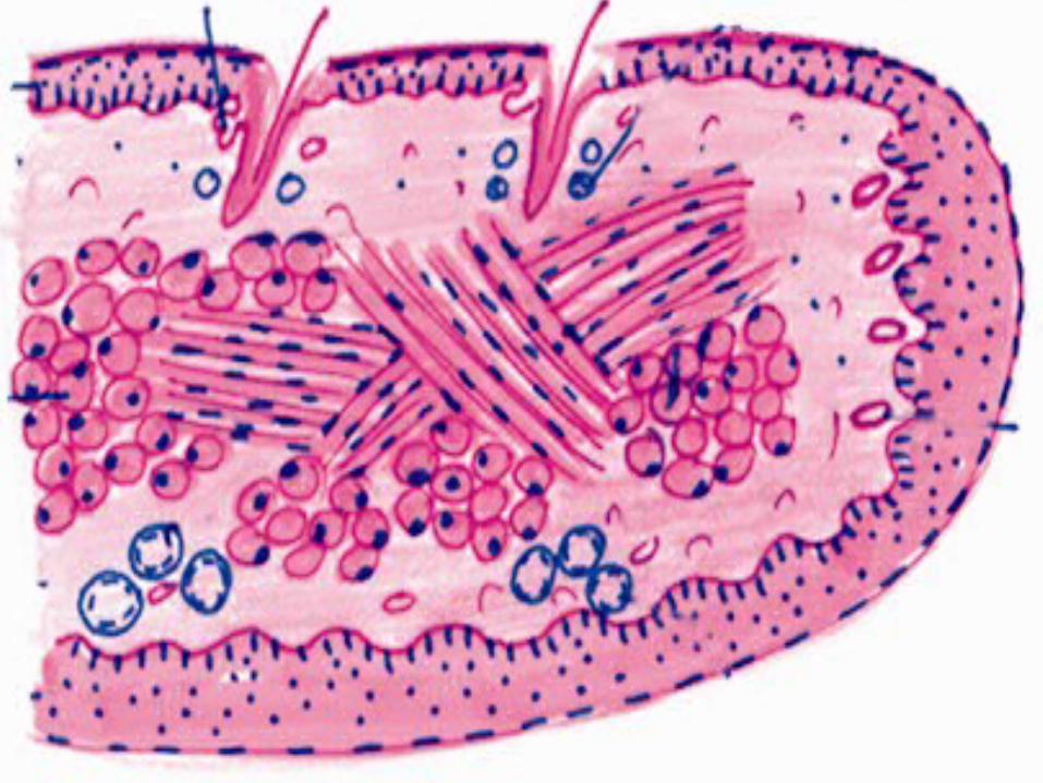
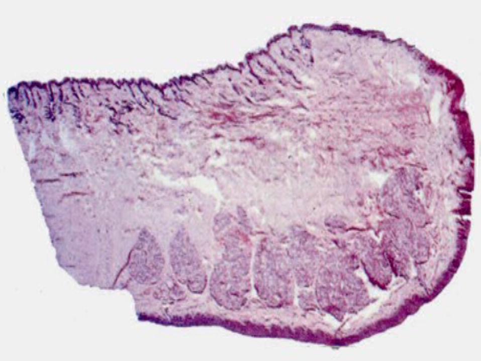
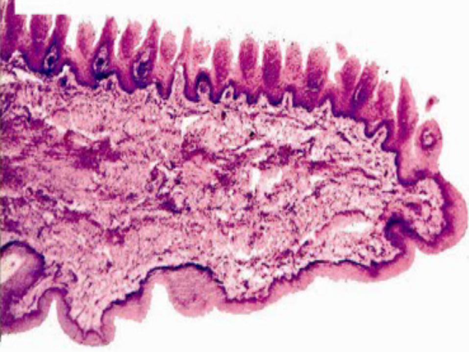
The lip The main bulk is formed of bundles of skeletal muscles (orbicularis oris muscle)
Arranged in different directions.
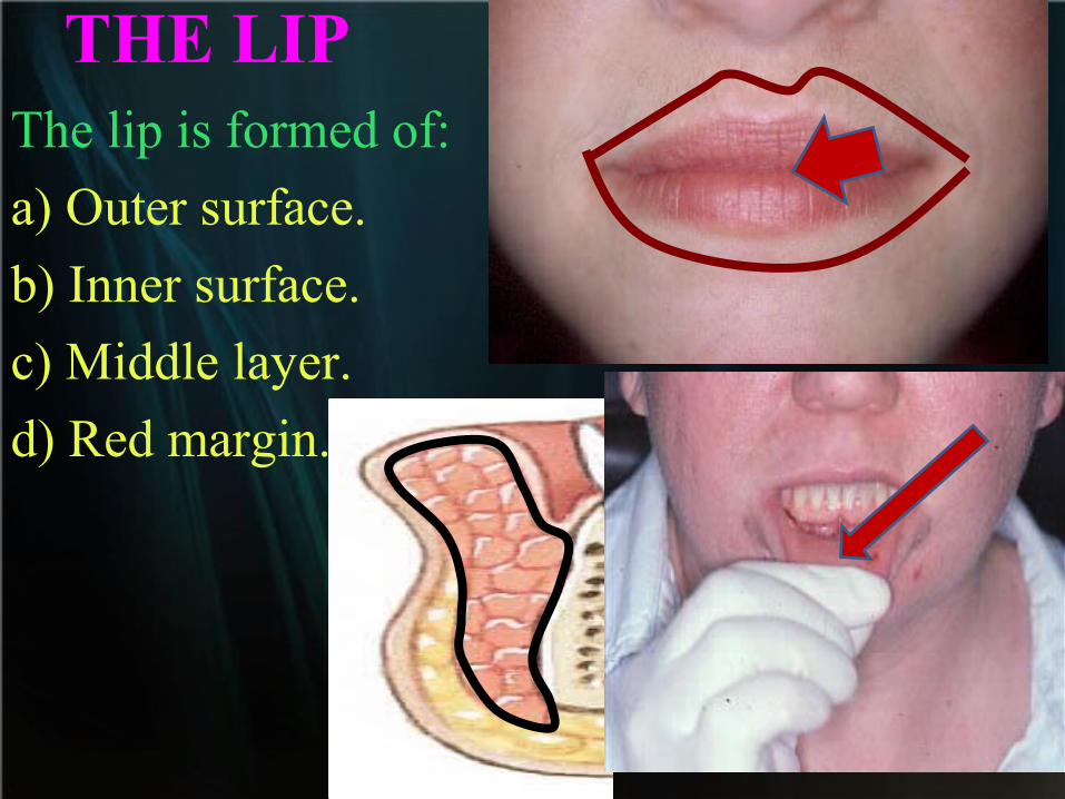
THE LIPThe lip is formed of:
a) Outer surface.
b) Inner surface.
c) Middle layer.
d) Red margin.

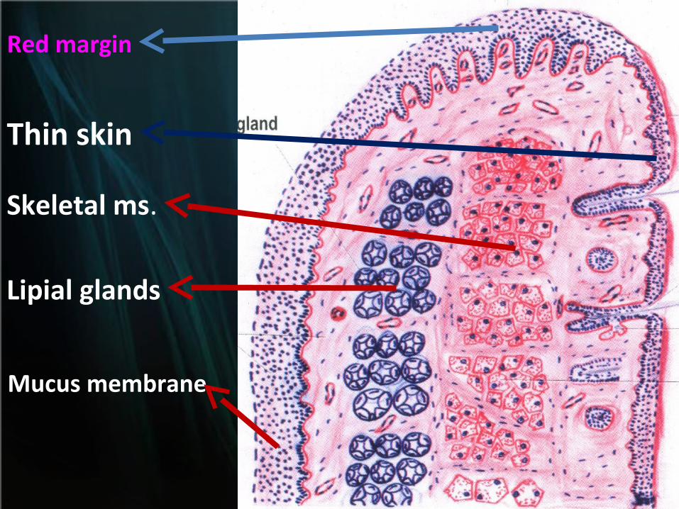
(A) Outer surface:
Thin skinEpidermis: keratinized
stratified squamous epithelium.
Dermis: C.T. rich in hair follicles, sweat glands and sebaceous glands.

(B) Inner surface: Lined by Thick, transparent and wet mucous membrane formed of :
Epithelium: non kretanized stratified squamous
C.T. Corium
contains mucous glands called labial gland (accessory gland ).
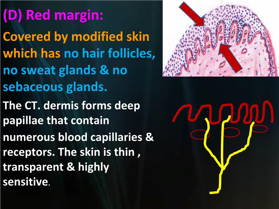
(D) Red margin: Covered by modified skin which has no hair follicles, no sweat glands & no sebaceous glands.The CT. dermis forms deep papillae that contain numerous blood capillaries & receptors. The skin is thin , transparent & highly sensitive.
Thin skin
Red margin
Skeletal ms.
Lipial glands
Mucus membrane
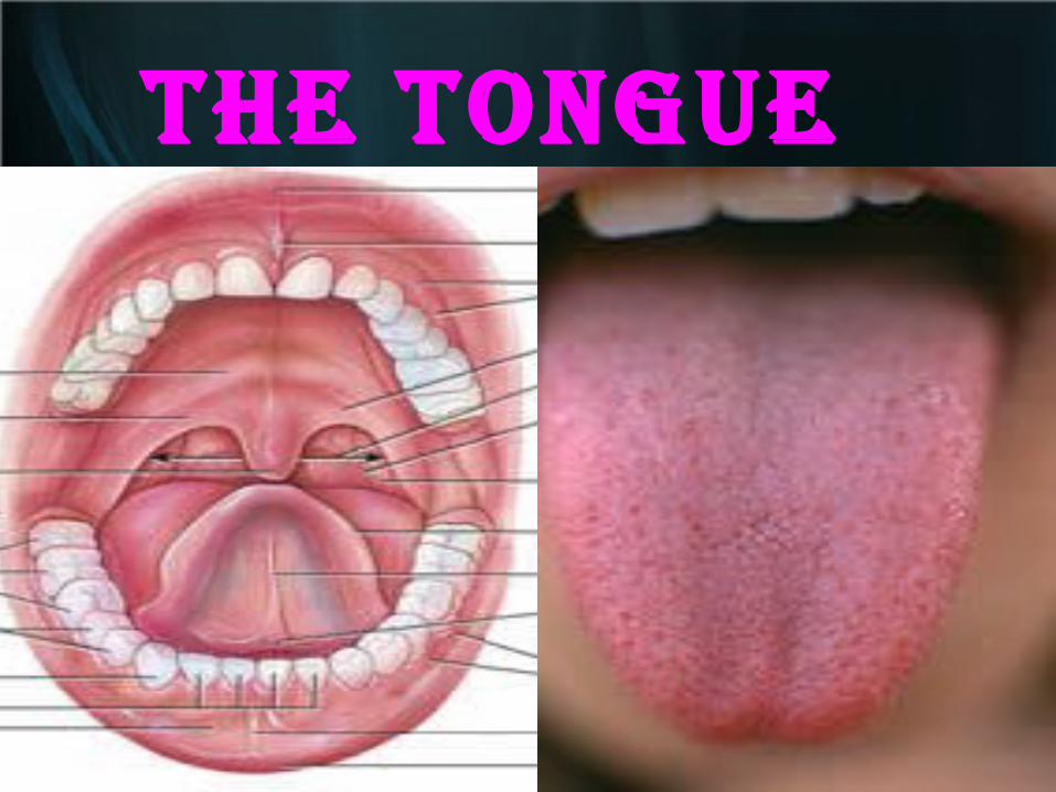
THE TONGUE
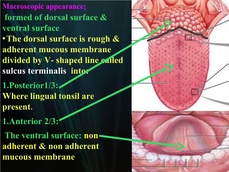
Macroscopic appearance:
formed of dorsal surface & ventral surface•The dorsal surface is rough & adherent mucous membrane divided by V- shaped line called sulcus terminalis into:
1.Posterior1/3: Where lingual tonsil are present.
1.Anterior 2/3:
The ventral surface: non adherent & non adherent mucous membrane
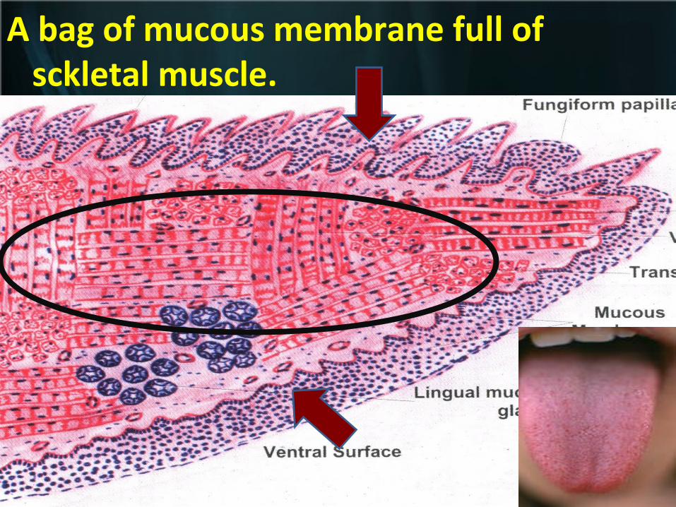
A bag of mucous membrane full of sckletal muscle.
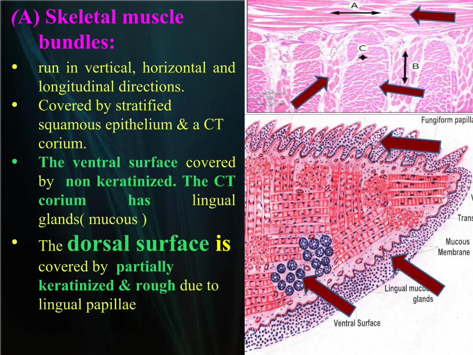
(A) Skeletal muscle bundles:
• run in vertical, horizontal and longitudinal directions.
• Covered by stratified squamous epithelium & a CT corium.
• The ventral surface covered by non keratinized. The CT corium has lingual glands( mucous )
• The dorsal surface is covered by partially keratinized & rough due to lingual papillae
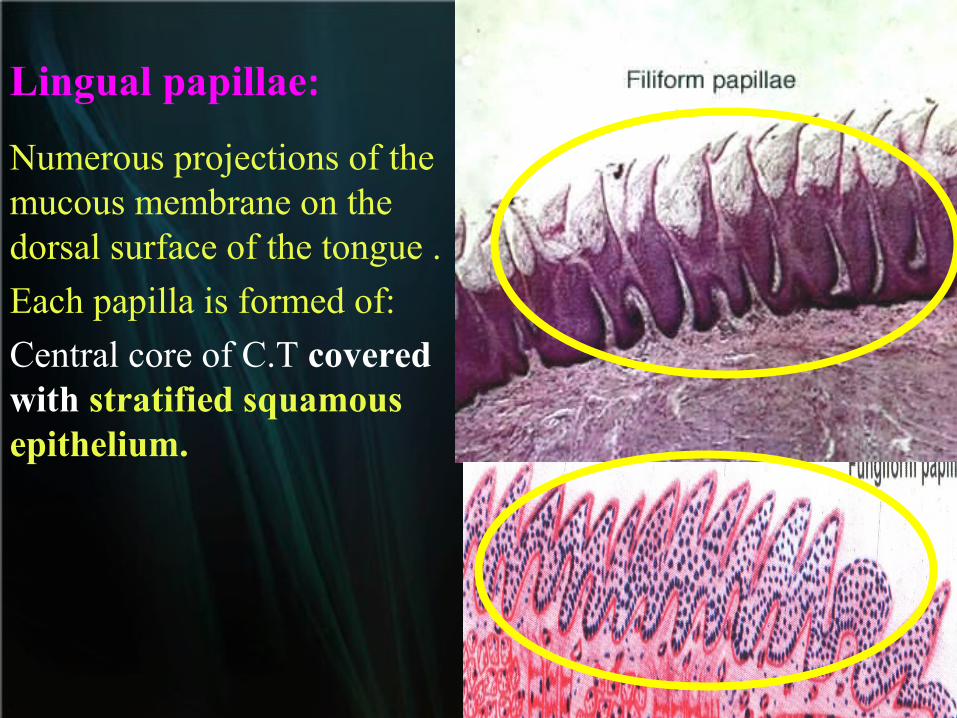
Lingual papillae:
Numerous projections of the mucous membrane on the dorsal surface of the tongue .
Each papilla is formed of:
Central core of C.T covered with stratified squamous epithelium.
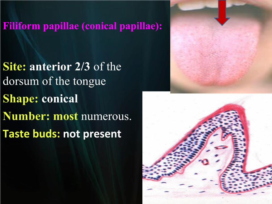
Filiform papillae (conical papillae):
Site: anterior 2/3 of the dorsum of the tongue
Shape: conical
Number: most numerous.
Taste buds: not present

Fungiform papillae:
Site: anterior2/3 particularly on the tip and lateral sides scattered between conical papillae.
Shape: basal narrow neck and rounded top.
Number : less numerous than conical papillae. Taste buds: few.
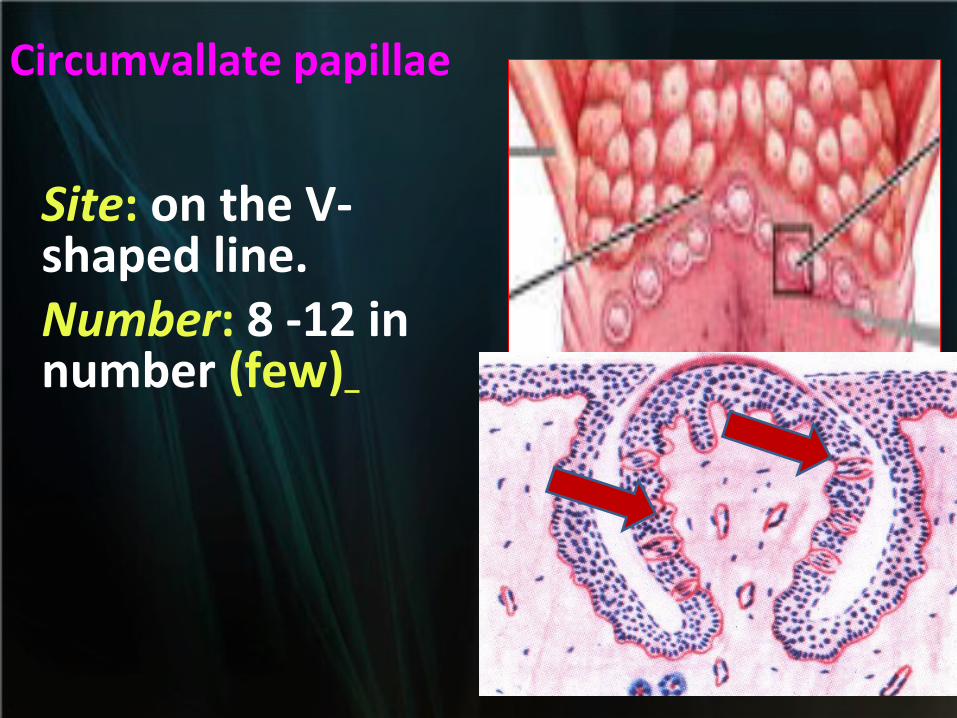
Circumvallate papillae
Site: on the V- shaped line.Number: 8 -12 in number (few)
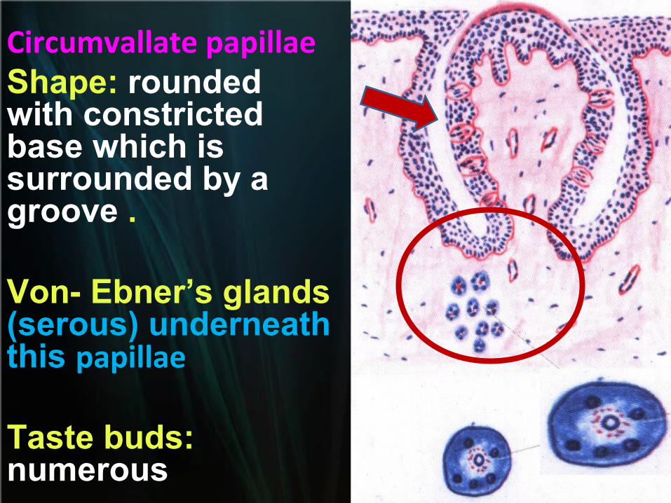
Circumvallate papillaeShape: rounded with constricted base which is surrounded by a groove .
Von- Ebner’s glands (serous) underneath this papillae
Taste buds: numerous

Foliate papillaeSite:
lateral margin of the dorsal surface the tongue of rabbit.
Shape:
parallel broad projections of CT covered by epithelium.
Number:
rabbit tongue but not in human
Taste buds: numerous
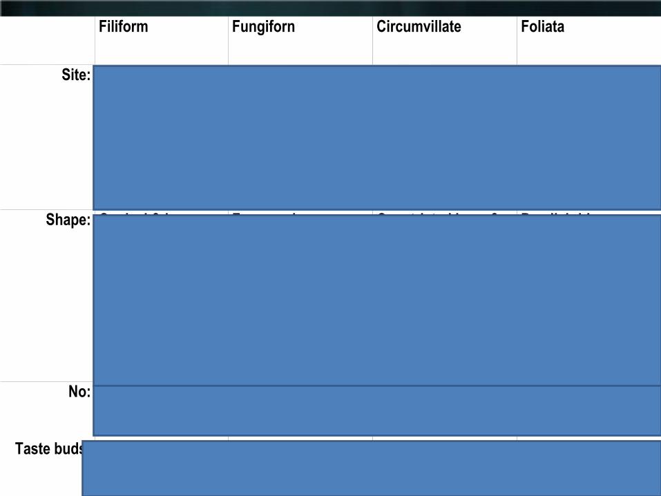
Filiform Fungiforn Circumvillate Foliata
Site: Ant 2/3 of dorsal surface
At tip & lateral sides On V shaped line Lateral margin of post. 1/3 of tongue of rabbit
Shape: Conical & long .(broad base & pointed tip)
Fungus shape narrow base & rounded top
Constricted base & broad top
Parallel ridges
No: Numerous Less in No 8-12 in number
Taste buds: Abscent + ve few (at top) numerous at sides numerous at sides

Taste budsNeuroepithelium responsible for taste sensation.
Sites: on mucous membranes of:Soft palate.Pharynx.Epiglottis.
Professor Kikunae Ikeda
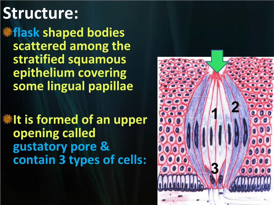
Structure:flask shaped bodies scattered among the stratified squamous epithelium covering some lingual papillae
It is formed of an upper opening called gustatory pore & contain 3 types of cells:
1 2
3
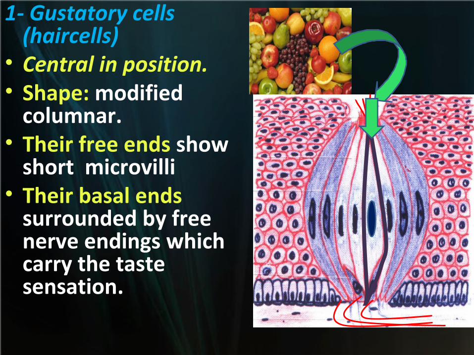
1- Gustatory cells (haircells)
• Central in position.• Shape: modified
columnar.• Their free ends show
short microvilli• Their basal ends
surrounded by free nerve endings which carry the taste sensation.

2- Sustentacular cells:
• Peripheral in position & supporting cells
3- Basal cells: • Basally located &
act as stem cells.
practical
MCQ

The free red margin of the lip contains :
a. Hair follicles.
b. Sweat glands.
c. Sebaceous glands.
d. Numerous receptors.

The mucous membrane covering the inner surface of the lip :
a. Is formed of stratified squamous keratinized epithelium.
b. Contains sebaceous glands.
c. Contains sweat glands.
d. Is thicker than the skin of the outer surface of the lip.
e. Has no glands.
The lip:
a.Its inner surface is covered by mucous membrane.
b.Its outer surface is covered by thick skin.
c.Smooth muscles run in different directions.
d.Labial glands are mainly serous.
Taste buds are :
a.Neuroepithelial structures.
b.Formed of 3 types of cells.
c.Situated between the cells of stratified squamous epithelium.
d.In contact with fine nerve fibres that carry taste sensations.
e.All of the above.•
Gustatory cells of taste buds:
a.Are modified pyramidal cells.
b.Have dark cytoplasm.
c.Act as stem cells for regeneration of other cells.
d.Their basal parts are surrounded by nerve fibres.
•
The skin covering the outer surface of the lip
a.Contains no hair follicles or sebaceous glands.
b.Contains no sweat glands.
c.Is thicker than the inner surface of the lip.
d.All of the above.
e.None of the above.

Mitotic figures are common in the cells of :
a. Basal cell layer.
b. Spinous layer.
c. Horny layer.
d. Clear layer.
e. None of the above.

The cells of granular layer contain :
a. Melanin pigments.
b. Carotene pigments.
c. Eleidin granules.
d. Basophilic keratohyaline granules.
e. Acidophilic keratohyaline granules.
Berbick granules are present in : a. Melanophores.
b. Merkel's cells.
c. Langerhans cells.
d. Stratum lucidum.
e. Meissner's corpuscles