liposomes and skin: from drug delivery to model membranes · 3. liposomes as skin drug delivery...
TRANSCRIPT

e u r o p e a n j o u r n a l o f p h a r m a c e u t i c a l s c i e n c e s 3 4 ( 2 0 0 8 ) 203–222
avai lab le at www.sc iencedi rec t .com
journa l homepage: www.e lsev ier .com/ locate /e jps
Review
Liposomes and skin: From drug delivery tomodel membranes
G.M. El Maghrabya,b, B.W. Barryc, A.C. Williamsd,∗
a College of Pharmacy, King Saud University, Riyadh 11451, P.O. Box 2457, Saudi Arabiab College of Pharmacy, University of Tanta, Tanta, Egyptc Drug Delivery Group, School of Pharmacy, University of Bradford, Bradford BD7 1DP, UKd School of Pharmacy, University of Reading, Whiteknights,P.O. Box 224, Reading, Berkshire RG6 6AD, UK
a r t i c l e i n f o
Article history:
Received 10 March 2008
Received in revised form
18 April 2008
Accepted 2 May 2008
Published on line 8 May 2008
Keywords:
Liposomes
Transdermal
Animal skin
Skin lipid liposomes
Ultradeformable vesicles
Vesicles
a b s t r a c t
The early eighties saw the introduction of liposomes as skin drug delivery systems, ini-
tially promoted primarily for localised effects with minimal systemic delivery. Subsequently,
a novel ultradeformable vesicular system (termed “Transfersomes” by the inventors) was
reported for transdermal delivery with an efficiency similar to subcutaneous injection.
Further research illustrated that the mechanisms of liposome action depended on the appli-
cation regime and the vesicle composition and morphology.
Ethical, health and supply problems with human skin have encouraged researchers to
use skin models. Traditional models involved polymer membranes and animal tissue, but
whilst of value for release studies, such models are not always good mimics for the com-
plex human skin barrier, particularly with respect to the stratum corneal intercellular lipid
domains. These lipids have a multiply bilayered organization, a composition and organiza-
tion somewhat similar to liposomes. Consequently researchers have used vesicles as skin
model membranes. Early work first employed phospholipid liposomes and tested their inter-
actions with skin penetration enhancers, typically using thermal analysis and spectroscopic
analyses. Another approach probed how incorporation of compounds into liposomes led
to the loss of entrapped markers, analogous to “fluidization” of stratum corneum lipids
on treatment with a penetration enhancer. Subsequently scientists employed liposomes
formulated with skin lipids in these types of studies.
Following a brief description of the nature of the skin barrier to transdermal drug deliv-
ery and the use of liposomes in drug delivery through skin, this article critically reviews
the relevance of using different types of vesicles as a model for human skin in permeation
enhancement studies, concentrating primarily on liposomes after briefly surveying older
models. The validity of different types of liposome is considered and traditional skin mod-
els are compared to vesicular model membranes for their precision and accuracy as skin
membrane mimics.
© 2008 Elsevier B.V. All rights reserved.
∗ Corresponding author. Tel.: +44 1183786196; fax: +44 1183786562.E-mail addresses: [email protected] (G.M. El Maghraby), b.w.b
(A.C. Williams).0928-0987/$ – see front matter © 2008 Elsevier B.V. All rights reserved.doi:10.1016/j.ejps.2008.05.002
[email protected] (B.W. Barry), [email protected]
204 e u r o p e a n j o u r n a l o f p h a r m a c e u t i c a l s c i e n c e s 3 4 ( 2 0 0 8 ) 203–222
Contents
1. Introduction . . . . . . . . . . . . . . . . . . . . . . . . . . . . . . . . . . . . . . . . . . . . . . . . . . . . . . . . . . . . . . . . . . . . . . . . . . . . . . . . . . . . . . . . . . . . . . . . . . . . . . . . . . . . . . . . . . 2042. Skin structure . . . . . . . . . . . . . . . . . . . . . . . . . . . . . . . . . . . . . . . . . . . . . . . . . . . . . . . . . . . . . . . . . . . . . . . . . . . . . . . . . . . . . . . . . . . . . . . . . . . . . . . . . . . . . . . . 2043. Liposomes as skin drug delivery systems. . . . . . . . . . . . . . . . . . . . . . . . . . . . . . . . . . . . . . . . . . . . . . . . . . . . . . . . . . . . . . . . . . . . . . . . . . . . . . . . . . . 206
3.1. Localising effects . . . . . . . . . . . . . . . . . . . . . . . . . . . . . . . . . . . . . . . . . . . . . . . . . . . . . . . . . . . . . . . . . . . . . . . . . . . . . . . . . . . . . . . . . . . . . . . . . . . . . . 2063.2. Targeted delivery to skin appendages. . . . . . . . . . . . . . . . . . . . . . . . . . . . . . . . . . . . . . . . . . . . . . . . . . . . . . . . . . . . . . . . . . . . . . . . . . . . . . . . 2073.3. Improved transdermal delivery . . . . . . . . . . . . . . . . . . . . . . . . . . . . . . . . . . . . . . . . . . . . . . . . . . . . . . . . . . . . . . . . . . . . . . . . . . . . . . . . . . . . . . 207
4. Mechanisms of action of liposomes as skin drug delivery systems . . . . . . . . . . . . . . . . . . . . . . . . . . . . . . . . . . . . . . . . . . . . . . . . . . . . . . . 2084.1. Free drug mechanism (see Fig. 3 at A). . . . . . . . . . . . . . . . . . . . . . . . . . . . . . . . . . . . . . . . . . . . . . . . . . . . . . . . . . . . . . . . . . . . . . . . . . . . . . . . 2084.2. Penetration enhancing mechanism (see Fig. 3 at B) . . . . . . . . . . . . . . . . . . . . . . . . . . . . . . . . . . . . . . . . . . . . . . . . . . . . . . . . . . . . . . . . . 2104.3. Vesicle adsorption to and/or fusion with the stratum corneum (see Fig. 3 at C) . . . . . . . . . . . . . . . . . . . . . . . . . . . . . . . . . . . 2114.4. Intact vesicular skin penetration mechanism (see Fig. 3 at D) . . . . . . . . . . . . . . . . . . . . . . . . . . . . . . . . . . . . . . . . . . . . . . . . . . . . . . 211
4.4.1. Traditional liposomes . . . . . . . . . . . . . . . . . . . . . . . . . . . . . . . . . . . . . . . . . . . . . . . . . . . . . . . . . . . . . . . . . . . . . . . . . . . . . . . . . . . . . . . . 2114.4.2. Ultradeformable vesicles . . . . . . . . . . . . . . . . . . . . . . . . . . . . . . . . . . . . . . . . . . . . . . . . . . . . . . . . . . . . . . . . . . . . . . . . . . . . . . . . . . . . 211
4.5. Transappendageal penetration (see Fig. 3 at E) . . . . . . . . . . . . . . . . . . . . . . . . . . . . . . . . . . . . . . . . . . . . . . . . . . . . . . . . . . . . . . . . . . . . . . 2125. Liposomes as skin model membranes . . . . . . . . . . . . . . . . . . . . . . . . . . . . . . . . . . . . . . . . . . . . . . . . . . . . . . . . . . . . . . . . . . . . . . . . . . . . . . . . . . . . . . 213
5.1. Traditional models . . . . . . . . . . . . . . . . . . . . . . . . . . . . . . . . . . . . . . . . . . . . . . . . . . . . . . . . . . . . . . . . . . . . . . . . . . . . . . . . . . . . . . . . . . . . . . . . . . . . 2135.2. Liposomal models . . . . . . . . . . . . . . . . . . . . . . . . . . . . . . . . . . . . . . . . . . . . . . . . . . . . . . . . . . . . . . . . . . . . . . . . . . . . . . . . . . . . . . . . . . . . . . . . . . . . . 215
. . . . .
. . . . .
6. Conclusion. . . . . . . . . . . . . . . . . . . . . . . . . . . . . . . . . . . . . . . . . . . . . . . . .References . . . . . . . . . . . . . . . . . . . . . . . . . . . . . . . . . . . . . . . . . . . . . . . . .
1. Introduction
Transdermal drug delivery offers many advantages over othertraditional routes of administration. Unfortunately, the bar-rier nature of the skin presents a significant obstacle formost drugs to be delivered into and through it (Barry, 1983).Accordingly, researchers are investigating various strategiesto overcome these barrier properties. Ideally, these investiga-tions should employ human skin. However, samples of humanskin of sufficient size and quality for penetration experimentsare not readily accessible to most investigators and in anycase are only available in limited amounts. Thus, many modelshave been explored to replace human tissue and this in itselfis an active area of research. Liposomes have been claimedto serve both uses; they can improve transdermal drug deliv-ery and can be used as a model for the skin membrane. Thisreview deals with the potential of liposomes as a skin drugdelivery system and on their use as a skin model.
2. Skin structure
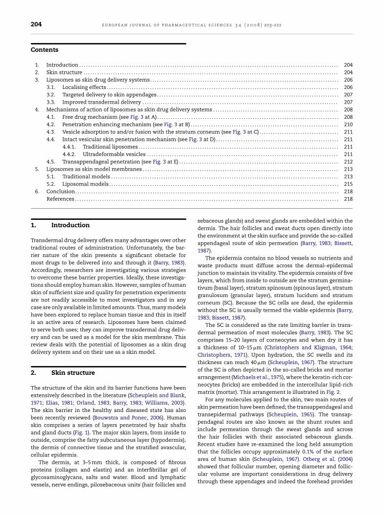
The structure of the skin and its barrier functions have beenextensively described in the literature (Scheuplein and Blank,1971; Elias, 1981; Orland, 1983; Barry, 1983; Williams, 2003).The skin barrier in the healthy and diseased state has alsobeen recently reviewed (Bouwstra and Ponec, 2006). Humanskin comprises a series of layers penetrated by hair shaftsand gland ducts (Fig. 1). The major skin layers, from inside tooutside, comprise the fatty subcutaneous layer (hypodermis),the dermis of connective tissue and the stratified avascular,cellular epidermis.
The dermis, at 3–5 mm thick, is composed of fibrousproteins (collagen and elastin) and an interfibrillar gel ofglycosaminoglycans, salts and water. Blood and lymphaticvessels, nerve endings, pilosebaceous units (hair follicles and
. . . . . . . . . . . . . . . . . . . . . . . . . . . . . . . . . . . . . . . . . . . . . . . . . . . . . . . . . . . . . . 218
. . . . . . . . . . . . . . . . . . . . . . . . . . . . . . . . . . . . . . . . . . . . . . . . . . . . . . . . . . . . . . 218
sebaceous glands) and sweat glands are embedded within thedermis. The hair follicles and sweat ducts open directly intothe environment at the skin surface and provide the so-calledappendageal route of skin permeation (Barry, 1983; Bissett,1987).
The epidermis contains no blood vessels so nutrients andwaste products must diffuse across the dermal–epidermaljunction to maintain its vitality. The epidermis consists of fivelayers, which from inside to outside are the stratum germina-tivum (basal layer), stratum spinosum (spinous layer), stratumgranulosum (granular layer), stratum lucidum and stratumcorneum (SC). Because the SC cells are dead, the epidermiswithout the SC is usually termed the viable epidermis (Barry,1983; Bissett, 1987).
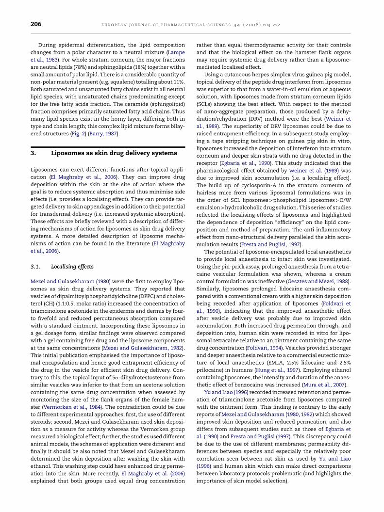
The SC is considered as the rate limiting barrier in trans-dermal permeation of most molecules (Barry, 1983). The SCcomprises 15–20 layers of corneocytes and when dry it hasa thickness of 10–15 �m (Christophers and Kligman, 1964;Christophers, 1971). Upon hydration, the SC swells and itsthickness can reach 40 �m (Scheuplein, 1967). The structureof the SC is often depicted in the so-called bricks and mortararrangement (Michaels et al., 1975), where the keratin-rich cor-neocytes (bricks) are embedded in the intercellular lipid-richmatrix (mortar). This arrangement is illustrated in Fig. 2.
For any molecules applied to the skin, two main routes ofskin permeation have been defined; the transappendageal andtransepidermal pathways (Scheuplein, 1965). The transap-pendageal routes are also known as the shunt routes andinclude permeation through the sweat glands and acrossthe hair follicles with their associated sebaceous glands.Recent studies have re-examined the long held assumptionthat the follicles occupy approximately 0.1% of the surface
area of human skin (Scheuplein, 1967). Otberg et al. (2004)showed that follicular number, opening diameter and follic-ular volume are important considerations in drug deliverythrough these appendages and indeed the forehead provides
e u r o p e a n j o u r n a l o f p h a r m a c e u t i c a l s c i e n c e s 3 4 ( 2 0 0 8 ) 203–222 205
F hroua
11lh0v
wsFotaspal
Fit
ig. 1 – A diagrammatical representation of a cross-section tppendages (from Williams, 2003, with permission).
3.7 mm2/cm2 as the follicular infundibula, i.e. approximately3.7% of the surface area of the forehead is available as fol-icles. Interestingly, the same study also showed that theistorically held view of the follicles providing approximately.1% of the surface area of the stratum corneum appears to bealid for forearm skin.
The transepidermal pathway can be defined as the path-ay where compounds permeate across the intact, unbroken
tratum corneum. This pathway contains two micropathways.irst, the intercellular route, which is a continuous but tortu-us way through the intercellular lipid domains and secondly,he transcellular pathway through the keratinocytes, thencross the intercellular lipids (Fig. 2) (Barry, 1991). As can be
een from Fig. 2, the transcellular pathway requires not onlyartitioning into and diffusion through the keratin bricks butlso into and across the intercellular lipids. Thus, the intercel-ular lipids play a major role in the barrier nature of the SC.ig. 2 – Diagram of the brick and mortar model of the stratum contercellular domains showing the major stratum corneum lipidhrough intact stratum corneum; the transcellular or the tortuou
gh human skin showing the different cell layers and
The role of lipids in the barrier function has been investi-gated thorough permeation studies employing lipid extractedSC. Skin permeability to water was significantly increasedafter removing skin lipids (Scheuplein and Blank, 1971).Further, the permeability of the skin from different bodysites can be related to total lipid content (Elias, 1981).Accordingly, the intercellular pathway is widely regarded asthe main route of permeation of most compounds despitethe relatively small surface area available for this route(Albery and Hadgraft, 1979; Guy and Hadgraft, 1989); nat-urally all molecules traverse by a combination of all threeroutes, the relative importance of which will vary depend-ing on the molecules physico-chemical characteristics. As
the lipid domains offer the primary barrier to permeationof most drugs, extensive research is being conducted tounderstand the composition and organization of these struc-tures.rneum with a simplified lamellar organization ofs. Also shown are the possible drug permeation pathwayss intercellular pathways (after Elias, 1981; Barry, 1991).
u t i c
206 e u r o p e a n j o u r n a l o f p h a r m a c eDuring epidermal differentiation, the lipid compositionchanges from a polar character to a neutral mixture (Lampeet al., 1983). For whole stratum corneum, the major fractionsare neutral lipids (78%) and sphingolipids (18%) together with asmall amount of polar lipid. There is a considerable quantity ofnon-polar material present (e.g. squalene) totalling about 11%.Both saturated and unsaturated fatty chains exist in all neutrallipid species, with unsaturated chains predominating exceptfor the free fatty acids fraction. The ceramide (sphingolipid)fraction comprises primarily saturated fatty acid chains. Thusmany lipid species exist in the horny layer, differing both intype and chain length; this complex lipid mixture forms bilay-ered structures (Fig. 2) (Barry, 1987).
3. Liposomes as skin drug delivery systems
Liposomes can exert different functions after topical appli-cation (El Maghraby et al., 2006). They can improve drugdeposition within the skin at the site of action where thegoal is to reduce systemic absorption and thus minimise sideeffects (i.e. provides a localising effect). They can provide tar-geted delivery to skin appendages in addition to their potentialfor transdermal delivery (i.e. increased systemic absorption).These effects are briefly reviewed with a description of differ-ing mechanisms of action for liposomes as skin drug deliverysystems. A more detailed description of liposome mecha-nisms of action can be found in the literature (El Maghrabyet al., 2006).
3.1. Localising effects
Mezei and Gulasekharam (1980) were the first to employ lipo-somes as skin drug delivery systems. They reported thatvesicles of dipalmitoylphosphatidylcholine (DPPC) and choles-terol (CH) (1.1:0.5, molar ratio) increased the concentration oftriamcinolone acetonide in the epidermis and dermis by four-to fivefold and reduced percutaneous absorption comparedwith a standard ointment. Incorporating these liposomes ina gel dosage form, similar findings were observed comparedwith a gel containing free drug and the liposome componentsat the same concentrations (Mezei and Gulasekharam, 1982).This initial publication emphasised the importance of liposo-mal encapsulation and hence good entrapment efficiency ofthe drug in the vesicle for efficient skin drug delivery. Con-trary to this, the topical input of 5�-dihydrotestosterone fromsimilar vesicles was inferior to that from an acetone solutioncontaining the same drug concentration when assessed bymonitoring the size of the flank organs of the female ham-ster (Vermorken et al., 1984). The contradiction could be dueto different experimental approaches; first, the use of differentsteroids; second, Mezei and Gulasekharam used skin deposi-tion as a measure for activity whereas the Vermorken groupmeasured a biological effect; further, the studies used differentanimal models, the schemes of application were different andfinally it should be also noted that Mezei and Gulasekharam
determined the skin deposition after washing the skin withethanol. This washing step could have enhanced drug perme-ation into the skin. More recently, El Maghraby et al. (2006)explained that both groups used equal drug concentrationa l s c i e n c e s 3 4 ( 2 0 0 8 ) 203–222
rather than equal thermodynamic activity for their controlsand that the biological effect on the hamster flank organsmay require systemic drug delivery rather than a liposome-mediated localised effect.
Using a cutaneous herpes simplex virus guinea pig model,topical delivery of the peptide drug interferon from liposomeswas superior to that from a water-in-oil emulsion or aqueoussolution, with liposomes made from stratum corneum lipids(SCLs) showing the best effect. With respect to the methodof nano-aggregate preparation, those produced by a dehy-dration/rehydration (DRV) method were the best (Weiner etal., 1989). The superiority of DRV liposomes could be due toraised entrapment efficiency. In a subsequent study employ-ing a tape stripping technique on guinea pig skin in vitro,liposomes increased the deposition of interferon into stratumcorneum and deeper skin strata with no drug detected in thereceptor (Egbaria et al., 1990). This study indicated that thepharmacological effect obtained by Weiner et al. (1989) wasdue to improved skin accumulation (i.e. a localising effect).The build up of cyclosporin-A in the stratum corneum ofhairless mice from various liposomal formulations was inthe order of SCL liposomes > phospholipid liposomes > O/Wemulsion > hydroalcoholic drug solution. This series of studiesreflected the localising effects of liposomes and highlightedthe dependence of deposition “efficiency” on the lipid com-position and method of preparation. The anti-inflammatoryeffect from nano-structural delivery paralleled the skin accu-mulation results (Fresta and Puglisi, 1997).
The potential of liposome-encapsulated local anaestheticsto provide local anaesthesia to intact skin was investigated.Using the pin-prick assay, prolonged anaesthesia from a tetra-caine vesicular formulation was shown, whereas a creamcontrol formulation was ineffective (Gesztes and Mezei, 1988).Similarly, liposomes prolonged lidocaine anaesthesia com-pared with a conventional cream with a higher skin depositionbeing recorded after application of liposomes (Foldvari etal., 1990), indicating that the improved anaesthetic effectafter vesicle delivery was probably due to improved skinaccumulation. Both increased drug permeation through, anddeposition into, human skin were recorded in vitro for lipo-somal tetracaine relative to an ointment containing the samedrug concentration (Foldvari, 1994). Vesicles provided strongerand deeper anaesthesia relative to a commercial eutectic mix-ture of local anaesthetics (EMLA, 2.5% lidocaine and 2.5%prilocaine) in humans (Hung et al., 1997). Employing ethanolcontaining liposomes, the intensity and duration of the anaes-thetic effect of benzocaine was increased (Mura et al., 2007).
Yu and Liao (1996) recorded increased retention and perme-ation of triamcinolone acetonide from liposomes comparedwith the ointment form. This finding is contrary to the earlyreports of Mezei and Gulasekharam (1980, 1982) which showedimproved skin deposition and reduced permeation, and alsodiffers from subsequent studies such as those of Egbaria etal. (1990) and Fresta and Puglisi (1997). This discrepancy couldbe due to the use of different membranes; permeability dif-ferences between species and especially the relatively poor
correlation seen between rat skin as used by Yu and Liao(1996) and human skin which can make direct comparisonsbetween laboratory protocols problematic (and highlights theimportance of skin model selection).
t i c a
oidtstvtSaih((
3
Slpc(
blsowiwtwl(ptdi
mraw1(tmim
aiadtdlde
e u r o p e a n j o u r n a l o f p h a r m a c e u
The above studies all employed traditional liposomes madef phospholipids or skin lipids. Planas et al. (1992) reported an
mproved anaesthetic effect of lidocaine and tetracaine whenelivered from ultradeformable vesicles. It is important to notehat the authors applied the tested formulations under occlu-ion for 25 min, although this application protocol is contraryo the recommended “open” application for ultradeformableesicles. Ultradeformable vesicles produced enhanced anaes-hesia compared with drug solution or traditional liposomes.urprisingly, topically applied ultradeformable vesicles gener-ted an effect equivalent to that created after subcutaneousnjection of the same formulation. Using heat-separateduman abdominal epidermal membranes, El Maghraby et al.
2001a) recorded improved skin deposition of 5-fluorouracil5-FU) from a similar vesicular formulation.
.2. Targeted delivery to skin appendages
everal workers have studied the potential of such vesicu-ar structures for targeting the appendages, especially to theilosebaceous units (hair follicles with their associated seba-eous glands). This area was extensively reviewed by Lauer1999) with an update by El Maghraby et al. (2006).
Carboxyfluorescein was selectively targeted into the pilose-aceous units of the hamster ears after application of
iposomes. Liposomes showed better targeting than aqueousolutions even when these solutions contained 10% ethanolr 0.05% sodium lauryl sulphate, or when propylene glycolas the donor vehicle (Lieb et al., 1992). The deposition of �-
nterferon into the skin of humans, hairless mice and hamsteras greater from liposomes compared to an aqueous solu-
ion. The greatest accumulation was seen in hamster skin,hich has the highest follicular density suggesting the fol-
icular pathway as a route for drug deposition from liposomesDu Plessis et al., 1992). Significant amounts of cimetidine werelaced into the pilosebaceous glands and other skin strata ofhe Syrian male hamster ear, after topical application of therug in 50% aqueous ethanol, nonionic surfactant vesicles, or
n phospholipid liposomes (Lieb et al., 1994).Contrary to the above reports, neither liposomes nor mixed
icelles provided any advantage over an ethanolic gel withegard to follicular delivery of isotretinoin. This finding wasttributed to the highly lipophilic nature of the drug whichould intrinsically target the sebaceous gland (Tschan et al.,
997). The results were later explained by El Maghraby et al.2006) on the basis that ethanol can enhance follicular deliveryhrough partial solubilisation of the sebum or softening of the
aterial in the duct. They also added that, whilst these find-ngs could suggest a positive effect of liposomes and mixed
icelles, they were only as effective as the ethanolic gel.Vesicular preparations were superior in the treatment of
cne vulgaris compared to conventional preparations includ-ng alcoholic lotions (Skalko et al., 1992). This was considereds strong evidence that vesicles can effectively target drugelivery to skin appendages (El Maghraby et al., 2006). Recently,he vitro permeation through hamster flank skin and in vivo
eposition in hamster ear demonstrated the potentials ofiquid-state liposomes and surfactant vesicles for successfulelivery of finasteride to the pilosebaceous unit (Tabbakhiant al., 2006).
l s c i e n c e s 3 4 ( 2 0 0 8 ) 203–222 207
3.3. Improved transdermal delivery
Although the majority of reports dealing with standard lipo-somes concentrate on improved drug deposition into skinand its appendages, some early sources cited improved trans-dermal delivery from these nano-aggregates. After finitedose applications to hairless mouse skin, Ganesan et al.(1984) reported that, for lipophilic drugs, greater amountswere delivered from vesicles compared to aqueous solution.Liposome-encapsulated antibodies were distributed rapidlyinto the deep cutaneous regions of piglet skin with a clearlyraised percutaneous absorption compared to aqueous solu-tions (Artman et al., 1990a,b). Fresta and Puglisi (1996) foundthat vesicles of unsaturated phospholipid (fluid liposomes),produced high percutaneous absorption and tissue distribu-tion rather than skin accumulation. Employing a human skingraft model, a liposomal formulation of phosphatidylcholine(PC) and CH augmented the uptake of �-interferon into the epi-dermis of viable human skin compared with aqueous solution(Short et al., 1996).
Whilst researchers were reporting mainly localised orrarely transdermal effects of liposomes, Cevc and Blume (1992)claimed that certain types of lipid vesicles (ultradeformablevesicles) can penetrate intact to the deep layers of the skinand may progress far enough to reach the systemic circula-tion, but they must be applied under non-occlusive conditions.The superiority of ultradeformable vesicles over “standard”liposomes for transdermal drug delivery was shown, andthe importance of open (i.e. non-occluded) application wasemphasised; however, a deviation from this protocol can befound (Planas et al., 1992) where an improved anaestheticeffect was reported after occluded treatment with anaestheticultradeformable vesicles. Transdermal immunization withlarge proteins by means of ultradeformable vesicles has alsobeen reported (Paul et al., 1995). Further, ultradeformable vesi-cles improved the regio-specificity and the biological activityof the corticosteroids in vivo. The effect was dose-dependentand it was concluded that this carrier can target the drug intothe viable skin and, when used in a higher dose, can distributethe medicament throughout the body (Cevc et al., 1997).
Ultradeformable vesicles provided arachidonic acid-induced oedema suppression equivalent to a lotion containingfive times the drug concentration of that in ultradeformablevesicles, after 0.5 h. Subsequently, after 2 h, the Transfersomeformulation was more efficacious than the lotion. Whenstandard nano-carriers (PC, cholesterol) were evaluated, nooedema suppression was found after 0.5 h. After 2 h, however,liposomes produced a measurable suppression which wasabout one-third that of ultradeformable vesicles and abouthalf that of the lotion (with five times more drug). The authorsstated that the late effect of the vesicle formulation arosefrom free drug permeation following its release from lipo-somes (Cevc et al., 1997). El Maghraby et al. (2006) rejected thisexplanation and stated that vesicles should have providedone-fifth of the efficacy of the lotion (containing free drug) forthis explanation to be valid, unless there is some penetration
enhancing effect for such liposomes. Further, successfulsystemic delivery of insulin by ultradeformable vesicles hasbeen reported from in vivo mice and human studies. Theefficiency of the formulation was comparable to that obtained
u t i c
208 e u r o p e a n j o u r n a l o f p h a r m a c eafter SC injection of the same preparation but with a longerlag time. This lag time may be required for vesicle skin pene-tration (Cevc et al., 1995, 1998). It is noteworthy that, in the 10years since these reports, an ultradeformable insulin deliverysystem has not come to market which raises a question overthe efficacy of these nano-aggregates as transdermal deliverysystems.
In a series of studies involving an optimized experimen-tal design, El Maghraby et al. (1999) investigated estradiol skindelivery from a variety of liposomes. The experiments probedoptimized ultradeformable vesicles (El Maghraby et al., 2000a)relative to standard liposomes. The ultradeformable formula-tions included PC with sodium cholate, PC with Span 80 andPC with Tween 80. The standard nano-carriers encompassedpure PC vesicles (non-rigid), PC with CH (membrane stabi-lized liposomes), and two rigid vesicles of DPPC and DPPC/CH.The studies employed low dose open application of the for-mulations to human epidermal membranes hydrated by an“open hydration” protocol that maintained the transepider-mal water gradient. The results indicated that all types ofliposomes improved both estradiol deposition into and per-meation through the epidermis compared with the saturatedaqueous control. The ultradeformable vesicles were betterthan the standard liposomes with respect to transepidermaldrug flux only.
The presence of a surfactant increases the elasticity ofthe lipid bilayers (El Maghraby et al., 2004). Accordingly, itwas concluded that flexible liposomes are more efficient intransdermal drug delivery (El Maghraby et al., 2006). It wassuggested that such surfactants (edge activators) can impartdeformability to the nano-carriers, which allows for improvedtransdermal drug delivery (Cevc, 1996). The incorporation ofethanol in lipid vesicles is an alternative approach to fluidizethe lipid membrane and thus enhance drug provision (Touitou
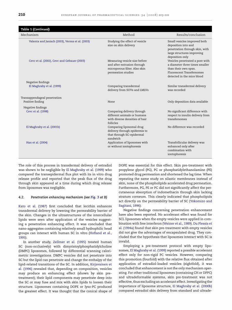
et al., 2000). Ethanol containing vesicles (termed “ethosomes”by the inventors) improved the transdermal delivery of mela-tonin, an anti-jet lag agent with poor skin permeation andlong lag time (Dubey et al., 2007). Also, flexible surfactantFig. 3 – Possible mechanisms of action of liposomes as skin drugpenetration enhancing process of liposome components, (C) indicorneum (SC) and (D) illustrates intact vesicle penetration into orfrom El Maghraby et al., 2006).
a l s c i e n c e s 3 4 ( 2 0 0 8 ) 203–222
vesicles showed higher efficiency compared to rigid vesicles(Honeywell-Nguyen et al., 2003a).
Successful topical delivery of low molecular weight hep-arin was reported after incorporation into surface chargedflexible vesicles made of lipids with Tween 80. These vesi-cles were termed flexosomes and the cationic structureswere the most efficient (Song and Kim, 2006). The Span80-based ultradeformable vesicles, initially optimized by ElMaghraby et al. (2000a), have been recently employed fortopical immunization. The results suggested that hepatitis Bloaded ultradeformable vesicles are able to provide a positiveimmune response (D. Mishra et al., 2006; V. Mishra et al., 2006;Mishra et al., 2007).
A new type of fusogenic vesicles (vesosomes) have beenintroduced for topical immunization. In these, the tetanustoxoid was incorporated into cationic liposomes made ofPC, dioleoyl phosphatidylethanolamine (DOPE) and dioleoyltrimethyl ammonium propane (DOTAP). These vesicles weresubsequently entrapped in an interdigitated lipid bilayerto provide vesicles within a vesicle (vesosomes). Theseaggregates were claimed to offer a promising system for tran-scutaneous immunization (D. Mishra et al., 2006; V. Mishra etal., 2006).
4. Mechanisms of action of liposomes asskin drug delivery systems
Alternative mechanisms have been suggested (El Maghrabyet al., 2006) for liposomes acting as skin drug delivery sys-tems (Fig. 3). In the subsequent sections we will considerthe proposed different mechanisms, illustrating both positiveand negative findings. Examples of studies investigating thesemechanisms are presented in Table 1 .
4.1. Free drug mechanism (see Fig. 3 at A)
According to this process, the drug permeates the skin inde-pendently after exiting from the vesicles (Ganesan et al., 1984).
delivery systems. (A) is the free drug mechanism, (B) is thecates vesicle adsorption to and/or fusion with the stratuminto and through the intact skin (not to scale) (modified

e u r o p e a n j o u r n a l o f p h a r m a c e u t i c a l s c i e n c e s 3 4 ( 2 0 0 8 ) 203–222 209
Table 1 – Examples of studies investigating the mechanisms of action of liposomes as skin drug delivery systems
Mechanism Method Results/conclusion
Free drug mechanismPositive findings None
Negative findingsEl Maghraby et al. (1999) Comparing transdermal flux
plots with in vitro releaseprofiles
Peak flux appeared at a timeduring which drug release wasnegligible
Penetration enhancementPositive findings
Kato et al. (1987) Application of drug in lecithinsolution in propylene glycol
Lecithin enhances the deliveryby lowering the skinpermeability barrier
Hofland et al. (1995) Freeze fracture electronmicroscopy and X-rayscattering studies, performedafter dipping human SC in aliposome
Ultrastructure changes in theintercellular lipids indicatingpenetration enhancement
Zellmer et al. (1995) DSC studies of treating the SCwith liposomes
Change in the enthalpy of SClipid-related transitions
Yokomizo and Sagitani (1996) Skin pre-treatment with PG, PCor PE
Promoted drug permeation andshortened lag time
El Maghraby et al. (1999) PC liposome pre-treatment andpermeation studies frommedicated liposomes
Permeation increased byfourfold after pre-treatmentand eightfold from medicatedliposomes
Negative findingsWeiner et al. (1989) Application of drug with empty
vesiclesNo accelerant effect
Du Plessis et al. (1994a) Skin pre-treatment with emptyvesicles
No accelerant effect
El Maghraby et al. (2000b) Delivery from liposomes orfrom lipid solution
Lipid solution was not efficient
Vesicle adsorption to or fusion with the SCPositive findings
Kirjavainen et al. (1996) SC pre-treatment withliposomes and interactions ofvesicles with SCLL
Skin surface adhesion andfusion or mixing with SC lipidmatrix
El Maghraby et al. (1999), Kirjavainen et al. (1999b) Drug partitioning into the SC Improved partitioning ofvarious drugs
Negative findings None
Intact vesicular skin penetrationTraditional liposomes
Positive findingsFoldvari et al. (1990) Application of liposomes
containing electron densemarker
Electron micrographs withintact liposomes in the dermis
Egbaria et al. (1990), Fresta and Puglisi (1996) Skin deposition of the duallabelled PL or SCL liposomecomponents
The ratio of radiolabelledcomponents of liposomes wasmaintained constantthroughout the skin strata
Negative findingsDu Plessis et al. (1994b) Monitoring effect of vesicle
size on drug skin depositionHigher deposition fromintermediate size and notsmall size liposomes
Zellmer et al. (1995), Korting et al. (1995) Confocal laser scanning orelectron microscopy afterapplication of liposomes
No evidence of intact carrierpenetration
TransfersomesPositive findings
Cevc and Blume (1992) Monitoring the fate of theapplied Transfersomes
Recovery of somelipid-associated radioactivityfrom the liver
210 e u r o p e a n j o u r n a l o f p h a r m a c e u t i c a l s c i e n c e s 3 4 ( 2 0 0 8 ) 203–222
Table 1 (Continued)
Mechanism Method Results/conclusion
Valenta and Janisch (2003), Verma et al. (2003) Studying the effect of vesiclesize on skin delivery
Small vesicles improved bothdeposition into andpenetration through skin, withlarge structures improvingdeposition only
Cevc et al. (2002), Cevc and Gebauer (2003) Measuring vesicle size beforeand after extrusion throughmicroporous filter. Also skinpermeation studies
Vesicles penetrated a pore witha diameter three times smallerthan their own span.Fluorescent Transfersomesdetected in the mice blood
Negative findingsEl Maghraby et al. (1999) Comparing transdermal
delivery from SUVs and LMLVsSimilar transdermal deliverywas recorded
Transappendageal penetrationPositive finding None Only deposition data available
Negative findingsCevc et al. (1998) Comparing delivery through
different animals or humanswith diverse densities of hairfollicles
No significant difference withrespect to insulin delivery fromtransfersomes
El Maghraby et al. (2001b) Comparing liposomal drugdelivery through epidermis tothat through SC–epidermalsandwich
No difference was recorded
Han et al. (2004) Application of liposomes withitho
Transfollicular delivery was
or wThe role of this process in transdermal delivery of estradiolwas shown to be negligible by El Maghraby et al. (1999) whocompared the transepidermal flux plot with its in vitro drugrelease profile and reported that the peak flux of the drugthrough skin appeared at a time during which drug releasefrom liposomes was negligible.
4.2. Penetration enhancing mechanism (see Fig. 3 at B)
Kato et al. (1987) first concluded that lecithin enhancestransdermal delivery by lowering the permeability barrier ofthe skin. Changes in the ultrastructures of the intercellularlipids were seen after application of the vesicles suggest-ing a penetration enhancing effect. It was concluded thatnano-aggregates containing relatively small hydrophilic headgroups can interact with human SC in vitro (Hofland et al.,1995).
In another study, Zellmer et al. (1995) treated humanSC (non-occlusively) with dimyristoylphosphatidylcholine(DMPC) liposomes, followed by differential scanning calori-metric investigations. DMPC vesicles did not penetrate intoSC but the lipid can penetrate and change the enthalpy of thelipid-related transitions of the SC. In addition, Kirjavainen etal. (1996) revealed that, depending on composition, vesiclesmay produce an enhancing effect (shown by skin pre-
treatment); their lipid components may penetrate deep intothe SC or may fuse and mix with skin lipids to loosen theirstructure. Liposomes containing DOPE or lyso-PC producedthe greatest effect. It was thought that the conical shape ofut iontophoresis enhanced only aftercombination withiontophoresis
DOPE was essential for this effect. Skin pre-treatment withpropylene glycol (PG), PC or phosphatidylethanolamine (PE)promoted drug permeation and shortened the lag time. Whenrepeating the same study on silastic membranes instead ofskin, none of the phospholipids accelerated drug permeation.Furthermore, PG, PE or PC did not significantly affect the per-cutaneous absorption of indomethacin through skin lackingstratum corneum. This clearly indicated that phospholipidsact directly on the permeability barrier of SC (Yokomizo andSagitani, 1996).
Negative findings concerning penetration enhancementhave also been reported. No accelerant effect was found forSCL liposomes when the empty vesicles were applied in com-bination with free interferon (Weiner et al., 1989). Du Plessis etal. (1994a) found that skin pre-treatment with empty vesiclesdid not give the advantages of encapsulated drug. They con-cluded that the hypothesis that liposomes interact with SC isinvalid.
Employing a pre-treatment protocol with empty lipo-somes, El Maghraby et al. (1999) reported a possible acceleranteffect only for non-rigid PC vesicles. However, comparingthis promotion (fourfold) with the relative flux obtained afterapplication of estradiol-loaded vesicles (eightfold), it wasconcluded that enhancement is not the only mechanism oper-ating. For other traditional liposomes (containing CH or DPPC)
and ultradeformable systems, skin pre-treatment was noteffective, thus excluding an accelerant effect. Investigating theimportance of liposome structure, El Maghraby et al. (2000b)compared estradiol skin delivery from standard and ultrade-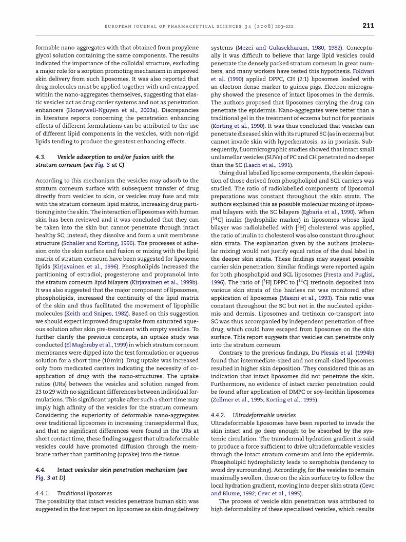
t i c a
fgiasdwteieol
4s
AsdwtsbhssmlptIpomwofcmsoar2miCoasvb
4F
4Ts
e u r o p e a n j o u r n a l o f p h a r m a c e u
ormable nano-aggregates with that obtained from propylenelycol solution containing the same components. The resultsndicated the importance of the colloidal structure, excludingmajor role for a sorption promoting mechanism in improved
kin delivery from such liposomes. It was also reported thatrug molecules must be applied together with and entrappedithin the nano-aggregates themselves, suggesting that elas-
ic vesicles act as drug carrier systems and not as penetrationnhancers (Honeywell-Nguyen et al., 2003a). Discrepanciesn literature reports concerning the penetration enhancingffects of different formulations can be attributed to the usef different lipid components in the vesicles, with non-rigid
ipids tending to produce the greatest enhancing effects.
.3. Vesicle adsorption to and/or fusion with thetratum corneum (see Fig. 3 at C)
ccording to this mechanism the vesicles may adsorb to thetratum corneum surface with subsequent transfer of drugirectly from vesicles to skin, or vesicles may fuse and mixith the stratum corneum lipid matrix, increasing drug parti-
ioning into the skin. The interaction of liposomes with humankin has been reviewed and it was concluded that they cane taken into the skin but cannot penetrate through intactealthy SC; instead, they dissolve and form a unit membranetructure (Schaller and Korting, 1996). The processes of adhe-ion onto the skin surface and fusion or mixing with the lipidatrix of stratum corneum have been suggested for liposome
ipids (Kirjavainen et al., 1996). Phospholipids increased theartitioning of estradiol, progesterone and propranolol intohe stratum corneum lipid bilayers (Kirjavainen et al., 1999b).t was also suggested that the major component of liposomes,hospholipids, increased the continuity of the lipid matrixf the skin and thus facilitated the movement of lipophilicolecules (Keith and Snipes, 1982). Based on this suggestione should expect improved drug uptake from saturated aque-us solution after skin pre-treatment with empty vesicles. Tourther clarify the previous concepts, an uptake study wasonducted (El Maghraby et al., 1999) in which stratum corneumembranes were dipped into the test formulation or aqueous
olution for a short time (10 min). Drug uptake was increasednly from medicated carriers indicating the necessity of co-pplication of drug with the nano-structures. The uptakeatios (URs) between the vesicles and solution ranged from3 to 29 with no significant differences between individual for-ulations. This significant uptake after such a short time may
mply high affinity of the vesicles for the stratum corneum.onsidering the superiority of deformable nano-aggregatesver traditional liposomes in increasing transepidermal flux,nd that no significant differences were found in the URs athort contact time, these finding suggest that ultradeformableesicles could have promoted diffusion through the mem-rane rather than partitioning (uptake) into the tissue.
.4. Intact vesicular skin penetration mechanism (seeig. 3 at D)
.4.1. Traditional liposomeshe possibility that intact vesicles penetrate human skin wasuggested in the first report on liposomes as skin drug delivery
l s c i e n c e s 3 4 ( 2 0 0 8 ) 203–222 211
systems (Mezei and Gulasekharam, 1980, 1982). Conceptu-ally it was difficult to believe that large lipid vesicles couldpenetrate the densely packed stratum corneum in great num-bers, and many workers have tested this hypothesis. Foldvariet al. (1990) applied DPPC, CH (2:1) liposomes loaded withan electron dense marker to guinea pigs. Electron microgra-phy showed the presence of intact liposomes in the dermis.The authors proposed that liposomes carrying the drug canpenetrate the epidermis. Nano-aggregates were better than atraditional gel in the treatment of eczema but not for psoriasis(Korting et al., 1990). It was thus concluded that vesicles canpenetrate diseased skin with its ruptured SC (as in eczema) butcannot invade skin with hyperkeratosis, as in psoriasis. Sub-sequently, fluormicrographic studies showed that intact smallunilamellar vesicles (SUVs) of PC and CH penetrated no deeperthan the SC (Lasch et al., 1991).
Using dual labelled liposome components, the skin deposi-tion of those derived from phospholipid and SCL carriers wasstudied. The ratio of radiolabelled components of liposomalpreparations was constant throughout the skin strata. Theauthors explained this as possible molecular mixing of liposo-mal bilayers with the SC bilayers (Egbaria et al., 1990). When[14C] inulin (hydrophilic marker) in liposomes whose lipidbilayer was radiolabelled with [3H] cholesterol was applied,the ratio of inulin to cholesterol was also constant throughoutskin strata. The explanation given by the authors (molecu-lar mixing) would not justify equal ratios of the dual label inthe deeper skin strata. These findings may suggest possiblecarrier skin penetration. Similar findings were reported againfor both phospholipid and SCL liposomes (Fresta and Puglisi,1996). The ratio of [3H] DPPC to [14C] tretinoin deposited intovarious skin strata of the hairless rat was monitored afterapplication of liposomes (Masini et al., 1993). This ratio wasconstant throughout the SC but not in the nucleated epider-mis and dermis. Liposomes and tretinoin co-transport intoSC was thus accompanied by independent penetration of freedrug, which could have escaped from liposomes on the skinsurface. This report suggests that vesicles can penetrate onlyinto the stratum corneum.
Contrary to the previous findings, Du Plessis et al. (1994b)found that intermediate-sized and not small-sized liposomesresulted in higher skin deposition. They considered this as anindication that intact liposomes did not penetrate the skin.Furthermore, no evidence of intact carrier penetration couldbe found after application of DMPC or soy-lecithin liposomes(Zellmer et al., 1995; Korting et al., 1995).
4.4.2. Ultradeformable vesiclesUltradeformable liposomes have been reported to invade theskin intact and go deep enough to be absorbed by the sys-temic circulation. The transdermal hydration gradient is saidto produce a force sufficient to drive ultradeformable vesiclesthrough the intact stratum corneum and into the epidermis.Phospholipid hydrophilicity leads to xerophobia (tendency toavoid dry surrounding). Accordingly, for the vesicles to remainmaximally swollen, those on the skin surface try to follow the
local hydration gradient, moving into deeper skin strata (Cevcand Blume, 1992; Cevc et al., 1995).The process of vesicle skin penetration was attributed tohigh deformability of these specialised vesicles, which results

212 e u r o p e a n j o u r n a l o f p h a r m a c e u t i c
Fig. 4 – Hydration gradient and deformability-drivenmovement of Transfersomes through small pores (after Cevc
et al., 1996).from “edge activator” molecules (e.g. surfactants) accumulat-ing at the site of high stress due to their raised propensityfor greatly curved structures (Fig. 4). This rearrangement wasclaimed to reduce the energy required for deformation; thestress is reportedly produced upon drying of the vesicleswhich, being flexible, can follow the transdermal hydrationgradient (Cevc et al., 1995).
It is difficult to believe that the presence of the so callededge activators in the vesicles can result in vesicle deforma-bility so that they can penetrate into intact skin with its densestratum corneum lipid packing and which contain very small“pores” relative to that of the vesicle diameter. To test thissupposition, the epidermal permeation of estradiol from largemultilamellar vesicles (LMLVs, at least 557 nm in diameter)was compared with that obtained from smaller entities of amean size of 124–138 nm (SUVs). The concept was to inves-tigate the possibility that intact nano-aggregates penetratethrough skin, assuming that this infiltration is a function ofthe vesicle size (El Maghraby et al., 1999). The SUVs are lessthan the maximum dimension reported to enter skin andthe minimum size of LMLVs is above the maximum volumewhich can invade skin (Cevc et al., 1995). SUVs were as effec-tive as LMLVs, a finding which suggests that intact vesicles donot permeate through human epidermal membrane in vitro(El Maghraby et al., 1999). However, reduction of vesicle sizeimproved drug deposition into deeper strata and penetrationthrough skin, with large structures improving deposition only(Valenta and Janisch, 2003; Verma et al., 2003).
Trotta et al. (2002) incorporated dipotassium glycyrrhiz-inate (DG), an amphiphilic anti-inflammatory drug, intoliposomes. They reported that the agent increased the elas-ticity of the entities. Measuring the vesicle size before andafter extrusion through a microporous filter, elastic particleswere capable of penetrating a pore with a diameter threetimes smaller than their own span. However, they were ableto show only improved skin deposition of DG, not improvedflux. The extrusion results may suggest vesicle elasticity butdo not demonstrate skin penetration. Indeed the “pores” in thestratum corneum lipid matrix are at least 10 times smallerthan the ultradeformable vesicle diameter (which generallyexceeds 100 nm). Also, the largest pores on the skin surface
are provided by the shunts (hair follicles, sweat ducts) whichplay no major role in liposomal transdermal drug penetration(see below). In similar studies, the size of ultradeformable vesi-cles was unchanged after extrusion through semi-permeablea l s c i e n c e s 3 4 ( 2 0 0 8 ) 203–222
membrane barriers. The authors reported the presence ofthe carriers in mice blood after topical application of fluores-cent labelled ultradeformable vesicles. Noteworthy, the sizeof these vesicles was similar to that of the starting liposomesuspension. This was taken as a clear evidence for vesicle inva-sion into and through skin (Cevc et al., 2002; Cevc and Gebauer,2003). However, it should be noted that a vesicular structuremay form spontaneously after absorption of the componentsas monomers.
Fast delivery of intact elastic vesicles into the SC wasrecorded and was thought to be via channel-like regionsin the SC. Again, elastic vesicles were superior to rigidnano-aggregates with non-occlusive application being best(Honeywell-Nguyen et al., 2003b). The transport of vesiclecomponents and a model drug into human skin was moni-tored in vivo. Only elastic vesicle material can rapidly enter theSC reaching almost the SC-viable epidermal junction. How-ever, the distribution profile of the drug in the lower SC layerswas different to that of the vesicle material. This suggests thatonce the elastic vesicles partition into the SC, the drug releasesfrom the carrier (Honeywell-Nguyen et al., 2004; Honeywell-Nguyen and Bouwstra, 2005).
In light of the above reports, it appears that some vesiclesmay penetrate intact to some extent into healthy skin. Ques-tions remain as to how deep into the skin strata intact carriersmove, and if indeed intact structures can carry their payloadthrough the entire tissue.
4.5. Transappendageal penetration (see Fig. 3 at E)
Occlusive application and full skin hydration is supposedlydetrimental for transdermal drug delivery from ultrade-formable vesicles. This effect was attributed to inhibition ofthe transdermal hydration gradient, which is believed to bethe driving force for vesicle-skin penetration (Cevc et al., 1995).Another possible explanation is that over-hydration of the skincan swell the corneocytes and thus close or at least minimisethe size of shunts that may play a role in liposomal skin deliv-ery.
Electron microscopy indicated that liposomes up to 600 nmdiameter can penetrate through skin but those of 1000 nm ormore remain interiorised in the SC. Deposition was higherin hairy guinea pigs but, with regard to penetration thoughskin, no difference could be found between hairless andhairy guinea pigs. Despite this finding, it was concluded thatinvasion is mainly along the hair sheath (Schramlova et al.,1997). However, these findings can reflect only delivery into,rather than through, the hair follicles. Also, vesicular deliv-ery through shunts was excluded on the basis that there wereno significant variations between different animals or humanswith diverse densities of hair follicles, with regard to the Trans-fersomal input of insulin (Cevc et al., 1998).
A novel in vitro technique using human abdominal skinwas developed to explore the role of appendageal transporton liposomal skin delivery of estradiol. The study monitoredvesicular delivery through epidermis and compared this with
penetration through a sandwich of SC and epidermis. As theorifices of these shunts occupy only a small fraction of thetotal skin surface area, there was a negligible chance that theshunts in the two membranes would superimpose. It was thus
t i c a
as2plwe
mc(r
5
Tat
5
Sttippt
tfvdoNtgcodhAsm
fshweeHtotgePa
e u r o p e a n j o u r n a l o f p h a r m a c e u
ssumed that the top layer of SC would block most of thehunts available in the bottom membrane (El Maghraby et al.,001b). From this study, it was concluded that the shunt routeslayed a minor role in estradiol transdermal delivery from
iposomes. Also, the transfollicular delivery from liposomesas enhanced only after combination with iontophoresis (Han
t al., 2004).In summary, it appears that the shunt routes play no
ajor role in liposomal transdermal delivery. However, vesi-le penetration into but not necessarily through hair folliclesi.e. targeting) is clearly demonstrated in numerous literatureeports.
. Liposomes as skin model membranes
able 2 presents a summary of the specifications, advantagesnd limitations of alternative skin models commonly used inransdermal drug delivery research.
.1. Traditional models
kin penetration studies play an essential role in the selec-ion of drugs for dermal or transdermal application. Therefore,he choice of predictive in vitro penetration models is highlymportant. Ideally, human skin should be used to evaluateenetration properties of candidate drugs. However, ethicalroblems, religious restrictions and limited availability madehe use of human skin difficult for most investigators.
Whilst animal skin provides an alternative to human skin,here are clear differences in dermal absorption between dif-erent animals. These differences arise from physiologicalariations but can be compounded by researchers employingifferent percutaneous penetration measurement method-logies (e.g. Scheuplein, 1978; Panchagnula et al., 1997).umerous animal models have been suggested as a substi-
ute for human skin, including primate, porcine, mouse, rat,uinea pig and snake skins. Anatomical investigations showomparable characteristics between porcine ear skin and thatf human skin with respect to the stratum coreneal and epi-ermal thickness as well as the follicular structure and theair density (Wester and Maibach, 1989; Jacobi et al., 2007).mong the rodents, rat skin showed the closest anatomicalimilarity to that of human skin with mouse skin revealingarkedly different features (Wester and Maibach, 1989).The presence of fur may be considered as an important dif-
erence between experimental animals and humans and soeveral hairless species have also been used. These includeairless mice and guinea pigs, with athymic nude mice tohich human skin has been grafted and scared tissue also
mployed as furless models (Reifenrath et al., 1984; Rougiert al., 1987; Bogen et al., 1992; Simon and Maibach, 1998).owever, it is important to note that percutaneous absorp-
ion through animal skin may differ significantly from thatbtained with human tissue. In general, drug absorptionhrough rat skin can be one to three orders of magnitude
reater than in humans, depending on permeant properties,xperimental methods, and exposure site (Bartek et al., 1972;oet et al., 2000). Comparing in vivo absorption of testosterone,fivefold difference between percutaneous absorption in rab-l s c i e n c e s 3 4 ( 2 0 0 8 ) 203–222 213
bits and humans was found, and the rank order of the amountabsorbed was rabbit > rat > pig > human (Bartek et al., 1972).Percutaneous absorption in monkeys has been reported tocompare well to that determined in man (Wester and Maibach,1975; Wester et al., 1980). Other studies have suggested thatabsorption through pig skin may most closely predict humandermal absorption (Reifenrath et al., 1984; Lavker et al., 1991).Several different strains of pig have been employed; Reifenrathet al. (1984) compared the absorption of different chemicalsthrough weanling Yorkshire pig and human skin. In gen-eral, the correlation between these two species was good.Of the tested chemicals, caffeine and parathion showed thelargest difference between human and pig absorption. Caf-feine absorption was over twofold greater in humans than pigtissue whereas parathion exhibited a twofold greater absorp-tion through pig skin than human skin. These results weresimilar to the earlier finding reported by Bartek et al. (1972).Unfortunately, pigs and monkeys are less readily available andare more expensive research models than smaller laboratoryanimals.
The studies outlined above demonstrated two importantissues. The first is that differences between animal modelsand human skin absorption are permeant specific, makinggeneral predictive conclusions a difficult task. The secondissue is that the most readily available and easy to handleanimals show less correlation to human percutaneous absorp-tion than the less readily available and more difficult to houseresearch animals such as pigs and monkeys.
Researchers have employed reconstructed human skinmodels as another option. These models can be very usefulif the penetration barrier of the skin equivalents is similar tothat of human tissue and such skin equivalents have been sug-gested for use in penetration studies (Kriwet and Parenteau,1996). In a very elegant study, Schmook et al. (2001) comparedthe penetration properties of human, pig and rat skin with theGraftskinTM LSETM (living skin equivalent) and the SkinethicTM
HRE (human reconstructed epidermis) models using drugswith widely varying polarity. In agreement with publisheddata, pig skin appeared as the most suitable model for humanskin: the fluxes through the skin and concentrations in theskin were of the same order of magnitude for both tissues,with differences of at most two- or fourfold, respectively.GraftskinTM LSETM provided an adequate barrier to salicylicacid, but was very permeable for the more hydrophobic com-pounds. It was thus concluded that the available reconstitutedskin models could not be regarded as generally useful for invitro penetration studies.
Reconstructed human epidermis has been also employedto investigate the mechanism of action of liposomes as skindrug delivery systems. After application of large unilamellarliposomes (mainly made from soybean PC) to human epider-mis reconstituted in vitro, electron microscopic investigationsrevealed the presence of dose-dependent alterations to themorphology of both the stratum corneum and the viable partof the epidermis. Shrunken lipid droplets were found betweenthe corneocytes. In addition, the corneocytes of various layers
of the stratum corneum and the keratinocytes of the upperlayer of the living epidermis showed lipid deposition. Thisfinding indicates a possible penetration enhancing mecha-nism of liposome components (Korting et al., 1995), and is
214 e u r o p e a n j o u r n a l o f p h a r m a c e u t i c a l s c i e n c e s 3 4 ( 2 0 0 8 ) 203–222
Table 2 – Summary of the specifications, advantages and limitations of various models of human skin employed intransdermal drug delivery research
Model Specification/advantages Limitations
Animal modelsMonkeys
Wester and Maibach (1975), Wester et al. (1980) Percutaneous absorptioncompares well to human skin
Restricted use and high cost
PigsReifenrath et al. (1984), Lavker et al. (1991), Jacobi et al. (2007) The ear showed anatomical
similarities to human skinwith respect to SC andepidermal thickness, follicularstructure and the hair density.Permeability is close tohuman skin
Difficult to obtain and highcost
RodentsBartek et al. (1972), Simon and Maibach (1998), Poet et al. (2000) Relatively cheap and readily
available. Rat may be the bestrodent model. The problem offur can be eliminated usinghairless animals
More permeable than humanskin
Reconstructed skin modelsReconstructed epidermis
Netzlaff et al. (2005) Close to human skin withrespect to general structure,and biochemical features.Useful in toxicity studies.More consistent inpermeability
Relatively weak barrier natureespecially for lipophilic drugs
Reconstructed full thickness skinNakamura et al. (1990) The presence of dermis may
eliminate the weak barriernature to lipophilic drugs
Absence of the vascularnetwork may provide falsebarrier nature
Lamellar matrixMoghimi et al. (1996a,b,c) DSC and X-ray revealed good
structural correlation with theSC lipids. Permeation studiesshowed good barrier nature
The lipid composition may bedissimilar to SC lipids. Needfurther investigations withvarious drugs
Liposomal modelsPhospholipid liposomes
Rolland et al. (1991), Bonina et al. (1994), El Maghraby et al. (2005) Used mainly to study themechanism of action of skinpenetration enhancers. DPPCis the most widely used. ItsTm can be easily measured asit has a narrow mainendothermic peak
Lipid arrangement is differentfrom the of the SC lipidlamellae
Provided misleading resultswith enhancers having twohydrogen-bonding sites in theanti position
SCL liposomesKim et al. (1993), El Maghraby et al. (2005) Successfully used to probe the
mode of action of skinenhancers. Have the samelipid arrangement as SC lipids
Represent the intercellularroute only
Yoneto et al. (1995, 1996), Kirjavainen et al. (1996, 1999a) The effect of agents on therelease of markers suggestedthe effects of these agents onskin
Cellular protein is absent
ProteoliposomesLopez et al. (1996) Comprise a mixture of lipids
and proteins and was used tostudy effect of surfactant
Requires further investigation

t i c a
st(mttr
r(icuoccrp
edTfmhtsti2
smdfatmHlwwAts
smp2asrstSqmcs
e u r o p e a n j o u r n a l o f p h a r m a c e u
imilar to our data obtained after investigating the penetra-ion enhancing effect of PC vesicles using human skin in vitroEl Maghraby et al., 1999). However, it should be noted that
ost reconstructed epidermal membranes are leakier thanhe normal human epidermis and thus offer greater poten-ial for vesicle penetration, providing an exaggerated effectelative to normal human skin.
The use of reconstructed human epidermis has beenecently reviewed and it was concluded that some modelsSkinEthic®, EpiSkin®, and EpiDerm®) are close to human skinn some aspects; their general structure, composition and bio-hemical features are similar to human skin and so they areseful in toxicity studies. The models also have the advantagef being more consistent in permeability and responsiveness,ompared to many human skin samples obtained from surgi-al procedures. Unfortunately, their major limitation is theirelatively weak barrier nature which makes them far moreermeable than excised human skin (Netzlaff et al., 2005).
A tighter, easily maintained reproducible organotypicpidermal culture model was developed, employing rat epi-ermal keratinocytes grown for 3 weeks on a collagen gel.his model exhibited normal stratum corneum structural and
unctional properties (Pasonen-Seppanen et al., 2001). The per-eability of this model to drugs was comparable to that of
uman epidermal cadaver membranes with an average of onlywofold higher permeability (Suhonen et al., 2003). This modelhowed only minor differences in the lipid composition andhermal phase behaviour compared to human skin explain-ng the minor differences in permeability (Pappinen et al.,008).
It is useful to note that the above studies evaluated recon-tructed human epidermal models, which were shown to beore permeable than human skin particularly for lipophilic
rugs. This stimulated researchers to consider reconstructedull thickness models as more suitable models (Nakamura etl., 1990). The presence of dermal tissue may add some barriero lipophilic drug permeation thus mitigating the higher per-
eability of epidermal model especially for lipophilic drugs.owever, reconstructed or cultured full thickness skin in vitro
acks the vascular network present in vivo. This vascular net-ork should minimise the barrier role of the dermis as itill clear any molecule crossing the dermoepidermal junction.ccordingly, the use of epidermal membranes in vitro tends
o provide a more representative model mimicking the in vivoituation.
Accepting that the intercellular lipid domains are respon-ible for the barrier nature of the SC, a simple lamellaresomorphic structure (matrix) was prepared and used to
robe the barrier nature of the SC. This matrix comprised0% cholesterol, 25% water and 55% a mixture of fattycids and their salts. Thermal analysis and X-ray diffractiontudies revealed that the matrix had a good structural cor-elation with the SC. In addition, release and permeationtudies conducted using estradiol and 5-fluorouracil indicatedhat the matrix could provide a good barrier model for the
C intercellular pathway (Moghimi et al., 1996a,b,c). Subse-uently, more refined models have been proposed containingore physiologically relevant lipid combinations, includingeramides, or using a combination of lipids extracted fromkin.
l s c i e n c e s 3 4 ( 2 0 0 8 ) 203–222 215
5.2. Liposomal models
Since SC intercellular lipids form bilayers, liposomes havebeen proposed as models for skin membranes. Simple phos-pholipid liposomes comprising DPPC were initially used beforeWertz et al. (1986) prepared liposomes from a lipid mixtureapproximating the lipid composition of the SC lipids (40%ceramides, 25% cholesterol, 25% palmitic acid and 10% choles-terol sulphate), termed SCL liposomes. These structures wereused mainly to investigate the mechanisms of enhanced skindrug delivery. They were also used to investigate the possibleoxidant or antioxidant effect of certain materials. This sectionwill summarise these applications.
Thermal analysis (differential scanning calorimetry, DSC)has been successfully used to probe the mechanisms ofaction of skin penetration enhancers. The originators usedhuman stratum corneum and tested a variety of penetrationenhancers with different lipophilicities (Goodman and Barry,1983, 1985, 1986, 1988; Barry, 1987). Based on the DSC resultsas well as permeation and partitioning data, Barry (1987) pro-posed four possible mechanisms of action of skin penetrationenhancers:
(1) Disruption of the organization of the intercellular lipidsof the SC increasing the fluidity and thus permitting eas-ier drug permeation through the less rigid environment.Lipophilic enhancers may act primarily via this mecha-nism.
(2) Many accelerants also interact with intracellular protein.The exceptions were Azone and oleic acid; however thesewere most effective as enhancers when dissolved in apolar co-solvent such as propylene glycol (PG), which itselfinteracts with protein. Although drug flux can increasevia lipid interaction alone, once the lipid barrier weak-ens, the protein-filled cells may still provide a significantdiffusional resistance. Thus an enhancer which affectsboth lipid and protein domains could be more potent.Intracellular drug transport could be increased by a sol-vating action of enhancers on the protein helices. Thismechanism encompasses the displacement of boundprotein–water, the expansion of protein structure, and thecompetition with permeants for hydrogen-bonding sites.
(3) The diffusional resistance of the intracellular contentsalters markedly with skin hydration—water itself is quitea potent penetration enhancer. The reason for this is thatin the fully hydrated skin, the intracellular regions will bemore fluid and water will compete for drug-binding sites,lowering the diffusional barrier.
(4) Small polar enhancers such as dimethylsulphoxide(DMSO) and its analogues, the pyrrolidones and PG mayaccumulate in both intercellular and protein regions of thetissue. The presence of these powerful solvents may thenincrease drug partitioning into the skin, yielding increasedfluxes.
The action of a penetration enhancer has been related toits partition coefficient. Small polar enhancers may partitionpreferentially at low concentrations into the protein region ofthe stratum corneum. At high concentrations they could also
216 e u r o p e a n j o u r n a l o f p h a r m a c e u t i c a l s c i e n c e s 3 4 ( 2 0 0 8 ) 203–222
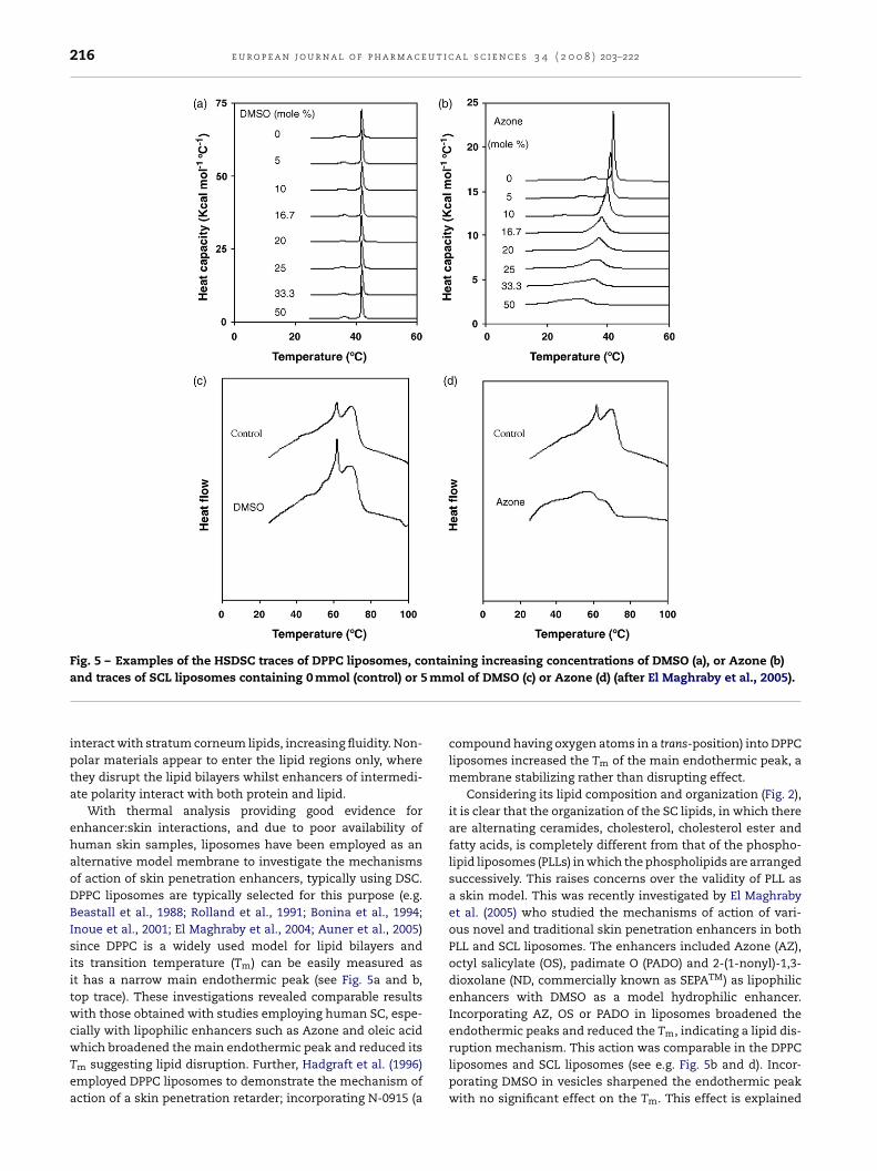
Fig. 5 – Examples of the HSDSC traces of DPPC liposomes, containing increasing concentrations of DMSO (a), or Azone (b)mm
and traces of SCL liposomes containing 0 mmol (control) or 5interact with stratum corneum lipids, increasing fluidity. Non-polar materials appear to enter the lipid regions only, wherethey disrupt the lipid bilayers whilst enhancers of intermedi-ate polarity interact with both protein and lipid.
With thermal analysis providing good evidence forenhancer:skin interactions, and due to poor availability ofhuman skin samples, liposomes have been employed as analternative model membrane to investigate the mechanismsof action of skin penetration enhancers, typically using DSC.DPPC liposomes are typically selected for this purpose (e.g.Beastall et al., 1988; Rolland et al., 1991; Bonina et al., 1994;Inoue et al., 2001; El Maghraby et al., 2004; Auner et al., 2005)since DPPC is a widely used model for lipid bilayers andits transition temperature (Tm) can be easily measured asit has a narrow main endothermic peak (see Fig. 5a and b,top trace). These investigations revealed comparable resultswith those obtained with studies employing human SC, espe-cially with lipophilic enhancers such as Azone and oleic acid
which broadened the main endothermic peak and reduced itsTm suggesting lipid disruption. Further, Hadgraft et al. (1996)employed DPPC liposomes to demonstrate the mechanism ofaction of a skin penetration retarder; incorporating N-0915 (aol of DMSO (c) or Azone (d) (after El Maghraby et al., 2005).
compound having oxygen atoms in a trans-position) into DPPCliposomes increased the Tm of the main endothermic peak, amembrane stabilizing rather than disrupting effect.
Considering its lipid composition and organization (Fig. 2),it is clear that the organization of the SC lipids, in which thereare alternating ceramides, cholesterol, cholesterol ester andfatty acids, is completely different from that of the phospho-lipid liposomes (PLLs) in which the phospholipids are arrangedsuccessively. This raises concerns over the validity of PLL asa skin model. This was recently investigated by El Maghrabyet al. (2005) who studied the mechanisms of action of vari-ous novel and traditional skin penetration enhancers in bothPLL and SCL liposomes. The enhancers included Azone (AZ),octyl salicylate (OS), padimate O (PADO) and 2-(1-nonyl)-1,3-dioxolane (ND, commercially known as SEPATM) as lipophilicenhancers with DMSO as a model hydrophilic enhancer.Incorporating AZ, OS or PADO in liposomes broadened theendothermic peaks and reduced the Tm, indicating a lipid dis-
ruption mechanism. This action was comparable in the DPPCliposomes and SCL liposomes (see e.g. Fig. 5b and d). Incor-porating DMSO in vesicles sharpened the endothermic peakwith no significant effect on the Tm. This effect is explained
e u r o p e a n j o u r n a l o f p h a r m a c e u t i c a l s c i e n c e s 3 4 ( 2 0 0 8 ) 203–222 217
F2
bswEtlaahirbSNrtwToite
m(ssscaedpe
lIebomamtlsft
Fig. 7 – Examples of the HSDSC traces of DPPC liposomes,containing increasing concentrations of ND (a) and traces ofSCL liposomes containing 0 mmol (control) or 5 mmol of ND
ig. 6 – Chemical structure of the enhancer-(1-nonyl)-1,3-dioxolane (ND).
y increased hydration of the bilayers and the formation of aolvation shell around the head groups. Once again the effectas comparable in both PLL and SCL liposomes (Fig. 5a and c;
l Maghraby et al., 2005). For ND, a dioxolane derivative (Fig. 6),he results were rather surprising. Incorporation into DPPCiposomes increased the Tm of the endothermic peak (Fig. 7a),n effect which is consistent with classifying the compound aspenetration retarder; as a dioxolane derivative, ND can formydrogen bonds with each successive PL molecule. However, it
s difficult to classify ND as a penetration retarder when otheresearchers have reported an enhancing effect, shown alsoy thermal and spectroscopic analysis of ND-treated humanC (Diani et al., 1995; Morganti et al., 1999). IncorporatingD in SCL liposomes broadened the endothermic peaks and
educed the Tm, indicating membrane disruption and a pene-ration enhancing effect (El Maghraby et al., 2005) in agreementith the work done on human skin (Morganti et al., 1999).his study thus shows a limitation in using PLL as a modelf human skin and highlighted the importance of consider-
ng the lipid composition and the structural organization ofhe model membrane before making general conclusions ofnhancer effects.
Elsewhere SCL liposomes were successfully used as aodel to investigate the mechanism of action of enhancers
Kim et al., 1993). Employing a fluorescent probe and lipo-omal marker release studies, SCL liposomes were used totudy the mechanism of action of 1-alkyl-2-pyrrolidones askin penetration enhancers (Yoneto et al., 1995, 1996). Theompounds increased the fluidity of the vesicular membranend increased the rate of release of entrapped markers. Theseffects indicate penetration enhancement via a membraneisruption mechanism. Interestingly these effects were com-arable with those obtained using hairless mouse skin (Yonetot al., 1995).
In a series of investigations, liposomes prepared frominolenic acid, DPPC, cholesterol and ceramide III or ceramideV have been used as skin model membranes to study theffect of UV radiation on the skin. In addition, the redoxehaviour of materials such as ascorbic acid and hyaluronann skin upon exposure to UV radiation was studied using theseodels. They concluded that ascorbic acid was degraded afterpro-oxidative effect and its incorporation in the topical for-ulations as an antioxidant may be deleterious. They added
hat considering human skin and its constant exposure to UV
ight and oxygen and an increased pool of iron in irradiatedkin, incorporating hyaluronan or its fragments in cosmeticormulations or sunscreens could be helpful for tissue pro-ection (Trommer et al., 2001, 2002, 2003). The same model(b) (after El Maghraby et al., 2005).
has been successfully used for screening a variety of com-pounds as antioxidants after topical application in an attemptto develop new compounds useful for skin protection againstUV radiation (Trommer and Neubert, 2005).
SCL liposomes have also been used as a model mem-brane to investigate the mechanism of action of phospholipidvesicles as skin drug delivery systems (Kirjavainen et al.,1996). These researchers loaded calcein into SCL liposomesand incubated them with phospholipid vesicles before mon-itoring the effect of the later on calcein release from the
SCL liposomes. Treatment with liposomes containing dio-leylphosphatidylethanolamine (DOPE) or lyso-PC increasedcalcein release indicating destabilisation of the SCL liposomalmembrane. These results supported those obtained after a
u t i c
r
218 e u r o p e a n j o u r n a l o f p h a r m a c e
pre-treatment study conducted on human skin which showedenhanced permeation of the fluorescent marker followingpre-treatment with liposomes containing DOPE or lyso-PC(Kirjavainen et al., 1996). This study indicates the potentialvalue of SCL liposomes as a skin mimic and it was concludedthat liposomes containing DOPE can fuse and mix with theskin lipids to loosen their structure providing a penetrationenhancing effect.
In subsequent studies, liposome–skin interactions andtheir effects on skin permeation of drugs were probed in vitro(Kirjavainen et al., 1999a), employing both human skin andSCL liposomes. The SC penetration of a lipophilic fluorescentprobe was deeper from PC liposomes containing 32% ethanolcompared with ethanol-free liposomes. The penetration pat-tern from DOPE containing liposomes was not affected byethanol. However, addition of ethanol increased the mixingof both PC and DOPE liposomes with the SCL liposomes. Inaddition, ethanol containing liposomes (both types) showed adestabilising effect on the skin lipid liposomes as evidencedby increased calcein release compared with control (contain-ing the same concentration of ethanol). This indicated thatliposomes could have a penetration enhancing effect. Theimproved effects seen with co-use of ethanol might resultfrom the solvent in liposomes providing a looser structurewhich facilitates the transfer of liposome components intoskin, producing the destabilising effect on skin lipids. Thisstudy again shows the potential value of SCL liposomes as askin model.
The effects of phospholipids on fluidity of SC lipid bilay-ers and drug partitioning into them was evaluated, employingthe SCL liposomes as a model for human stratum corneum(Kirjavainen et al., 1999b). The study revealed that differentphospholipids have varied effects on drug partitioning intothe SC lipid bilayers. Thus, incorporating egg-PC, soya-PCor DOPE into skin lipid liposomes increased the partition-ing of drugs into these SC liposomes whereas distearyl-PCdid not change this partitioning. It was suggested that fluidstate phospholipids can disrupt the rigid structure of the skinlipids, thus increasing drug partitioning into the lipid phase.However, gel state phospholipids produce only minor or noeffects. It was concluded that the phospholipid-improvedskin permeation of drugs may be at least partially due toincreased drug partitioning. These results are in good agree-ment with those obtained after a partitioning study employinghuman stratum corneum in vitro (El Maghraby et al., 1999)which further indicated the value of SCL liposomes as skinmodels.
A mixture of lipids and proteins, extracted from pig SCusing a chloroform/methanol mixture, was successfully usedto prepare liposomes (Lopez et al., 1996). These vesicles weretermed “proteoliposomes” and were employed as skin mod-els for studying the interactions of surfactants with skin. Theauthors monitored alterations in the release of an entrappedfluorescent marker from these liposomes after addition ofsurfactant, as a measure for the interaction of surfactantswith the lipid membranes. The study compared proteolipo-
somes with PC vesicles. The results revealed similar changesin bilayer permeability in the presence of the same amounts ofsurfactants, but different release kinetics were evident (Lopezet al., 1996).a l s c i e n c e s 3 4 ( 2 0 0 8 ) 203–222
6. Conclusion
Liposomes offer potential value in dermal and transdermaldrug delivery and recent advances and modifications appearto have generated increased therapeutic potential. Alterationin their composition and structure results in vesicles withtailored properties. Flexible and ultradeformable liposomesare one such advance with claims of enhanced transdermaldrug delivery to efficiencies comparable with subcutaneousadministration. Claims such as non-invasive transcutaneousvaccination using these structures would be a significant clin-ical advance but the mechanisms of action of these vesiclesas skin drug delivery systems require further clarification.Indeed, most literature reports have generated data from ani-mal models and it is well known that animal skin permeabilitydiffers to that in humans (typically more permeable). Accord-ingly, confirmatory investigations on human skin are stillrequired.
The use of liposomes as models for skin membranes pro-vide another important application but it should be noted thatthe simple phospholipids-based liposomes may result in mis-leading conclusions. Those having a lipid composition similarto that of SC lipids can at least partially represent the SC mem-brane.
e f e r e n c e s
Albery, W.J., Hadgraft, J., 1979. Percutaneous absorption: in vitroexperiments. J. Pharm. Pharmacol. 31, 140–147.
Artman, C., Roding, J., Ghyczy, M., Prazel, H.G., 1990a. Liposomesfrom soya phospholipids as percutaneous drug carriers:qualitative in vivo investigations with antibody-loadedliposomes. Arzneium. Forsch. Drug Res. 40 (II), 1363–1365.
Artman, C., Roding, J., Ghyczy, M., Prazel, H.G., 1990b. Liposomesfrom soya phospholipids as percutaneous drug carriers:quantitative in vivo investigations with radioactivity labelledliposomes. Arzneium. Forsch. Drug Res. 40 (II), 1365–1368.
Auner, B.G., O‘Neill, M.A.A., Valenta, C., Hadgraft, J., 2005.Interaction of phloretin and 6-ketocholestanol withDPPC-liposomes as phospholipid model membranes. Int. J.Pharm. 294, 149–155.
Barry, B.W., 1983. Dermatological Formulations: PercutaneousAbsorption. Marcel Dekker, New York/Basel.
Barry, B.W., 1987. Mode of action of penetration enhancers inhuman skin. J. Control. Release 6, 85–97.
Barry, B.W., 1991. Lipid protein partitioning theory of skinpenetration enhancement. J. Control. Release 15, 237–248.
Bartek, M.J., LaBudde, J.A., Maibach, H.I., 1972. Skin permeabilityin vivo: comparison in rat, rabbit, pig and man. J. Invest.Dermatol. 58, 114–123.
Beastall, J.C., Hadgraft, J., Washington, C., 1988. Mechanism ofaction of Azone as a percutaneous penetration enhancer: lipidbilayer fluidity and transition temperature effects. Int. J.Pharm. 43, 207–213.
Bissett, D.L., 1987. Anatomy and biochemistry of the skin. In:Kydonieus, A.F., Berner, B. (Eds.), Transdermal Delivery of
Drugs, vol. 1. CRC Press, Inc., Boca Raton, pp. 29–42.Bogen, K.T., Colston Jr., B.W., Machicao, L.K., 1992. Dermalabsorption of dilute aqueous chloroform, trichloroethylene,and tetrachloroethylene in hairless guinea pigs. Fundam.Appl. Toxicol. 18, 30–39.

t i c a
B
B
C
C
C
C
C
C
C
C
C
C
D
D
D
D
D
E
E
E
e u r o p e a n j o u r n a l o f p h a r m a c e u
onina, F.P., Castelli, F., Montenegro, L., Mazzone, G., 1994.Partitioning and differential scanning calorimetry studies ofN-alkyllactam ester dermal prodrugs of indomethacin. Int. J.Pharm. 106, 115–123.
ouwstra, J.A., Ponec, M., 2006. The skin barrier in healthy anddiseased state. Biochim. Biophys. Acta 1758, 2080–2095.
evc, G., 1996. Transfersomes, liposomes and other lipidsuspensions on the skin: permeation enhancement, vesiclepenetration, and transdermal drug delivery. Crit. Rev. Ther.Drug Carrier Syst. 13 (3–4), 257–388.
evc, G., Blume, G., 1992. Lipid vesicles penetrate into intact skinowing to the transdermal osmotic gradients and hydrationforce. Biochim. Biophys. Acta 1104, 226–232.
evc, G., Gebauer, D., 2003. Hydration-driven transport ofdeformable lipid vesicles through fine pores and skin barrier.Biophys. J. 84, 1010–1024.
evc, G., Schatzlein, A., Blume, G., 1995. Transdermal drugcarriers: basic properties, optimization and transfer efficiencyin the case of epicutaneously applied peptides. J. Control.Release 36, 3–16.
evc, G., Blume, G., Schatzlein, A., 1997. Transfersomes-mediatedtransepidermal delivery improves the regio-specificity andbiological activity of corticosteroids in vivo. J. Control. Release45, 211–226.
evc, G., Schatzlein, A., Richardsen, H., 2002. Ultradeformablelipid vesicles can penetrate skin and other semi-permeablemembrane barriers unfragmented. Evidence from doublelabel CLSM experiments and direct size measurement.Biochim. Biophys. Acta 1564, 21–30.
evc, G., Blume, G., Schatzlein, A., Gebauer, D., Paul, A., 1996. Theskin: a pathway for systemic treatment with patches andlipid-based agent carriers. Adv. Drug Deliv. Rev. 18, 349–378.
evc, G., Gebauer, D., Stieber, J., Schatzlein, A., Blume, G., 1998.Ultraflexible vesicles, Transfersomes, have an extremely lowpore penetration resistance and transport therapeuticamounts of insulin across the intact mammalian skin.Biochim. Biophys. Acta 1368, 201–215.
hristophers, E., 1971. Cellular architecture of the stratumcorneum. J. Invest. Dermatol. 56, 165–169.
hristophers, E., Kligman, A.M., 1964. Visualization of cell layersof the stratum corneum. J. Invest. Dermatol. 42, 407–409.
iani, A.R., Shull, K.L., Zaya, M.J., Brunden, M.N., 1995. Thepenetration enhancer SEPATM augments stimulation of scalphair growth by topical minoxidil in the balding stumptailmacaque. Skin Pharmacol. 8 (5), 221–228.
u Plessis, J., Egbaria, K., Ramachandran, C., Weiner, N.D., 1992.Topical delivery of liposomally encapsulatedgamma-interferon. Antiviral Res. 18, 259–265.
u Plessis, J., Weiner, N., Muller, D.G., 1994a. The influence of invivo treatment of skin with liposomes on the topicalabsorption of a hydrophilic and a hydrophobic drug in vitro.Int. J. Pharm. 103, R1–R5.
u Plessis, J., Ramachandran, C., Weiner, N., Muller, D.G., 1994b.The influence of particle size of liposomes on the dispositionof drug into skin. Int. J. Pharm. 103, 277–282.
ubey, V., Mishra, D., Jain, N.K., 2007. Melatonin loaded ethanolicliposomes: physicochemical characterization and enhancedtransdermal delivery. Eur. J. Pharm. Biopharm. 67, 398–405.
gbaria, K., Ramachandran, C., Kittayanond, D., Weiner, N., 1990.Topical delivery of liposomally encapsulated interferonevaluated by in vitro diffusion studies. Antimicrob. AgentsChemother. 34 (1), 107–110.
l Maghraby, G.M., Williams, A.C., Barry, B.W., 1999. Skin deliveryof oestradiol from deformable and traditional liposomes:
mechanistic studies. J. Pharm. Pharmacol. 51, 1123–1134.l Maghraby, G.M., Williams, A.C., Barry, B.W., 2000a. Oestradiolskin delivery from ultradeformable liposomes: refinement ofsurfactant concentration. Int. J. Pharm. 196, 63–74.
l s c i e n c e s 3 4 ( 2 0 0 8 ) 203–222 219
El Maghraby, G.M., Williams, A.C., Barry, B.W., 2000b. Skin deliveryof oestradiol from lipid vesicles: importance of liposomestructure. Int. J. Pharm. 204, 159–169.
El Maghraby, G.M., Williams, A.C., Barry, B.W., 2001a. Skin deliveryof 5-fluorouracil from ultradeformable and standardliposomes in vitro. J. Pharm. Pharmacol. 53, 1069–1077.
El Maghraby, G.M., Williams, A.C., Barry, B.W., 2001b. Skinhydration and possible shunt route penetration in controlledskin delivery of estradiol from ultradeformable and standardliposomes in vitro. J. Pharm. Pharmacol. 53, 1311–1322.
El Maghraby, G.M., Williams, A.C., Barry, B.W., 2004. Interactionsof surfactants (edge activators) and skin penetrationenhancers with liposomes. Int. J. Pharm. 276, 143–161.
El Maghraby, G.M.M., Campbell, M., Finnin, B., 2005. Mechanismof action of novel skin penetration enhancers: phospholipidversus skin lipid liposomes. Int. J. Pharm. 305, 90–104.
El Maghraby, G.M., Williams, A.C., Barry, B.W., 2006. Can drugbearing liposomes penetrate intact skin? J. Pharm. Pharmacol.58, 415–429.
Elias, P.M., 1981. Epidermal lipids; membranes and keratinization.Int. J. Dermatol. 20, 1–9.
Foldvari, M., 1994. In vitro cutaneous and percutaneous deliveryand in vivo efficacy of tetracaine from liposomal andconventional vehicles. Pharm. Res. 11 (11), 1593–1598.
Foldvari, M., Gesztes, A., Mezei, M., 1990. Dermal drug delivery byliposome encapsulation: clinical and electron microscopicstudies. J. Microencapsul. 7 (4), 479–489.
Fresta, M., Puglisi, G., 1996. Application of liposomes as potentialcutaneous drug delivery systems. In vitro and in vivoinvestigation with radioactivity labelled vesicles. J. DrugTarget 4, 95–101.
Fresta, M., Puglisi, G., 1997. Corticosteroid dermal delivery withskin-lipid liposomes. J. Control. Release 44, 141–151.
Ganesan, M.G., Weiner, N.D., Flynn, G.L., Ho, N.F.H., 1984.Influence of liposomal drug entrapment on percutaneousabsorption. Int. J. Pharm. 20, 139–154.
Gesztes, A., Mezei, M., 1988. Topical anaesthesia of skin byliposome-encapsulated tetracaine. Anesth. Analg. 67,1079–1081.
Goodman, M., Barry, B.W., 1983. Differential scanning calorimetryof human stratum corneum: effect of penetration enhancersAzone and DMSO. Anal. Proc. 26, 397.
Goodman, M., Barry, B.W., 1985. Differential scanning calorimetry(DSC) of human stratum corneum: effect of azone. J. Pharm.Pharmacol. 37, 80P.
Goodman, M., Barry, B.W., 1986. Action of skin permeationenhancers azone, oleic acid and decylmethyl sulphoxide:permeation and DSC studies. J. Pharm. Pharmacol. 38, 71P.
Goodman, M., Barry, B.W., 1988. Action of penetration enhancerson human skin as assessed by permeation of model drugs5-fluorouracil and estradiol. I. Infinite dose technique. J.Invest. Dermatol. 91 (4), 323–327.
Guy, R.H., Hadgraft, J., 1989. Mathematical models ofpercutaneous absorption. In: Bronaugh, R.L., Maibach, H.I.(Eds.), Percutaneous Absorption.Mechanisms-Methodology-Drug Delivery, 2nd edition. MarcelDekker Inc., New York, pp. 13–28.
Han, I., Kim, M., Kim, J., 2004. Enhanced transfollicular delivery ofadriamycin with a liposome and iontophoresis. Exp.Dermatol. 13 (2), 86–92.
Hadgraft, J., Peck, J., Williams, D.G., Pugh, W.J., Allan, G., 1996.Mechanism of action of skin penetration enhancers/retarders:Azone and analogues. Int. J. Pharm. 141, 17–25.
Hofland, H.E.J., Bouwstra, J.A., Bodde, H.E., Spies, F., Junginger,
H.E., 1995. Interaction between liposomes and humanstratum corneum in vitro: freeze fracture electronmicroscopical visualization and small angle X-ray scatteringstudies. Br. J. Dermatol. 132, 853–866.
u t i c
220 e u r o p e a n j o u r n a l o f p h a r m a c eHoneywell-Nguyen, P.L., Arenja, S., Bouwstra, J.A., 2003a. Skinpenetration and mechanisms of action in the delivery of theD2-agonist rotigotine from surfactant-based elastic vesicleformulations. Pharm. Res. 20 (10), 1619–1625.
Honeywell-Nguyen, P.L., Bouwstra, J.A., 2005. Vesicles as a tool fortransdermal and dermal delivery. Drug Discov. Today:Technol. 2 (1), 67–74.
Honeywell-Nguyen, P.L., Gooris, G.S., Bouwstra, J.A., 2004.Quantitative assessment of the transport of elastic and rigidvesicle components and a model drug from these vesicleformulations into human skin in vivo. J. Invest. Dermatol. 123(5), 902–910.
Honeywell-Nguyen, P.L., Wouter Groenink, H.W., de Graaff, A.M.,Bouwstra, J.A., 2003b. The in vivo transport of elastic vesiclesinto human skin: effects of occlusion, volume and duration ofapplication. J. Control. Release 90 (2), 243–255.
Hung, O.R., Comeau, L., Riley, M.R., Tan, S., Whynot, S., Mezei, M.,1997. Comparative topical anaesthesia of EMLA andliposome-encapsulated tetracaine. Can. J. Anaesth. 44,707–711.
Inoue, T., Yanagihara, S., Misono, Y., Suzuki, M., 2001. Effect offatty acids on the phase behaviour of hydrateddipalmitoylphosphatidylcholine bilayer: saturated versusunsaturated fatty acids. Chem. Phys. Lipids 109, 117–133.
Jacobi, U., Kaiser, M., Toll, R., Mangelsdorf, S., Audring, H., Otberg,N., Sterry, W., 2007. Lademann Porcine ear skin: an in vitromodel. Skin Res. Technol. 13, 19–24.
Kato, A., Ishibashi, Y., Miyake, Y., 1987. Effect of egg yolk lecithinon transdermal delivery of bunazosin hydrochloride. J. Pharm.Pharmacol. 39, 399–400.
Keith, A.D., Snipes, W., 1982. Phospholipids as moisturizingagents. In: Frost, P., Horovitz, S. (Eds.), Principles of Cosmeticsfor the Dermatologist. The C.V. Mosby Company, St. Louis, pp.59–69.
Kim, C.K., Hong, M.S., Kim, Y.B., Han, S.K., 1993. Effect ofpenetration enhancers (pyrrolidone derivatives) onmultilamellar liposomes of stratum corneum lipid: a study byUV spectroscopy and differential scanning calorimetry. Int. J.Pharm. 95, 43–50.
Kirjavainen, M., Urtti, A., Jaaskelainen, I., Suhonen, T.M., Paronen,P., Koskela, R.V., Kiesvaara, J., Monkkonen, J., 1996. Interactionof liposomes with human skin in vitro—the influence of lipidcomposition and structure. Biochim. Biophys. Acta 1304,179–189.
Kirjavainen, M., Urtti, A., Koskela, R.V., Kiesvaara, J., Monkkonen,J., 1999a. Liposome-skin interactions and their effects on theskin permeation of drugs. Eur. J. Pharm. Sci. 7, 279–286.
Kirjavainen, M., Monkkonen, J., Saukkosaari, M., Koskela, R.V.,Kiesvaara, J., Urtti, A., 1999b. Phospholipids affect stratumcorneum lipid bilayer fluidity and drug partitioning into thebilayers. J. Control. Release 58, 207–214.
Korting, H.C., Stolz, W., Schmid, M.H., Maierhofer, G., 1995.Interaction of liposomes with human epidermisreconstructed in vitro. Br. J. Dermatol. 132, 571–579.
Korting, H.C., Zeinicke, H., Schafer, K.M., Falco, O.B., 1990.Liposome encapsulation improves efficacy of betamethasonedipropionate in atopic eczema but not in psoriasis vulgaris.Eur. J. Clin. Pharmacol. 39, 349–351.
Kriwet, K., Parenteau, N.L., 1996. In vitro skin models. Cosmet.Toiletries 111, 93–102.
Lampe, M.A., Williams, M.L., Elias, P.M., 1983. Human epidermallipids: characterization and modulations duringdifferentiation. J. Lipid Res. 24, 131.
Lasch, J., Laub, R., Wohlrab, W., 1991. How deep do intact
liposomes penetrate into human skin? J. Control. Release 18,55–58.Lauer, A.C., 1999. Percutaneous drug delivery to the hair follicle.In: Bronaugh, R.L., Maibach, H.I. (Eds.), Percutaneous
a l s c i e n c e s 3 4 ( 2 0 0 8 ) 203–222
Absorption Drugs-Cosmetics-Mechanisms-Methodology, 3rdedition. Marcel Dekker, Inc., New York/Basel, pp. 427–449.
Lavker, R.M., Dong, G., Zheng, P.S., Murphy, G.F., 1991. Hairlessmicropig skin. A novel model for studies of cutaneous biology.Am. J. Pathol. 138, 687–697.
Lieb, L.M., Flynn, G., Weiner, N., 1994. Follicular (pilosebaceousunit) deposition and pharmacological behavior of cimetidineas a function of formulation. Pharm. Res. 11 (10), 1419–1423.
Lieb, L.M., Ramachandran, C., Egbaria, K., Weiner, N., 1992.Topical delivery enhancement with multilamellar liposomesinto pilosebaceous units. I. In vitro evaluation usingfluorescent techniques with hamster ear model. J. Invest.Dermatol. 99 (1), 108–113.
Lopez, O., de la Maza, A., Coderch, L., Parra, J.L., 1996. Formationand characterisation of liposomes from lipid/proteic materialextracted from pig stratum corneum. J. Am. Oil Chem. Soc. 73,443–448.
Masini, V., Bonte, F., Meybeck, A., Wepierre, J., 1993. Cutaneousbioavailability in hairless rats of tretinoin liposomes or gel. J.Pharm. Sci. 82 (1), 17–21.
Mezei, M., Gulasekharam, V., 1980. Liposomes—a selective drugdelivery system for topical route of administration. 1. Lotiondosage form. Life Sci. 26, 1473–1477.
Mezei, M., Gulasekharam, V., 1982. Liposomes—a selective drugdelivery system for topical route of administration: gel dosageform. J. Pharm. Pharmacol. 34, 473–474.
Michaels, A.S., Chanderasekaran, S.K., Shaw, J.E., 1975. Drugpermeation through human skin: theory and in vitroexperimental measurement. Am. Inst. Chem. Eng. J. 21,985–996.
Mishra, D., Dubey, V., Asthana, A., Saraf, D.K., Jain, N.K., 2006.Elastic liposomes mediated transcutaneous immunizationagainst Hepatitis B. Vaccine 24, 4847–4855.
Mishra, D., Mishra, P.K., Dubey, V., Dabadghao, S., Jain, N.K., 2007.Evaluation of uptake and generation of immune response bymurine dendritic cells pulsed with hepatitis B surfaceantigen-loaded elastic liposomes. Vaccine 25, 6939–6944.
Mishra, V., Mahor, S., Rawat, A., Dubey, P., Gupta, P.N., Singh, P.,Vyas, S.P., 2006. Development of novel fusogenic vesosomesfor transcutaneous immunization. Vaccine 24, 5559–5570.
Moghimi, H.R., Williams, A.C., Barry, B.W., 1996a. A lamellarmatrix model for stratum corneum intercellular lipids. I.Characterisation and comparison with stratum corneumintercellular structure. Int. J. Pharm. 131, 103–115.
Moghimi, H.R., Williams, A.C., Barry, B.W., 1996b. A lamellarmatrix model for stratum corneum intercellular lipids. II.Effect of geometry of the stratum corneum on permeation ofmodel drugs 5-fluorouracil and oestradiol. Int. J. Pharm. 131,117–129.
Moghimi, H.R., Williams, A.C., Barry, B.W., 1996c. A lamellarmatrix model for stratum corneum intercellular lipids. III.Effect of terpene penetration enhancers on the release of5-fluorouracil and oestradiol from the matrix. Int. J. Pharm.145, 37–47.
Morganti, F., Bramanti, E., Solaro, R., Benedetti, E., Chiellini, E.,Nannipieri, E., Narducci, P., Krauser, S.F., Samour, C.M., 1999.Thermal and spectroscopic characterization of interactionsbetween 2-nonyl-1,3-dioxolane and stratum corneumcomponents. J. Bioactive Compatible Polym. 14, 162–177.
Mura, P., Maestrelli, F., Rodriguez, L.M.G., Michelacci, I.,Ghelardini, C., Rabasco, A.M., 2007. Development,characterization and in vivo evaluation of benzocaine-loadedliposomes. Eur. J. Pharm. Biopharm. 67, 86–95.
Nakamura, M., Rikimaru, T., Yano, T., Moore, K.G., Pula, P.J.,Schofield, B.H., 1990. Full-thickness human skin explants fortesting the toxicity of topically applied chemicals. J. Invest.Dermatol. 95, 325–332.

t i c a
N
O
O
P
P
P
P
P
P
R
R
R
S
S
S
S
S
S
e u r o p e a n j o u r n a l o f p h a r m a c e u
etzlaff, F., Lehr, C.M., Wertz, P.W., Schaefer, U.F., 2005. Thehuman epidermis models EpiSkin®, SkinEthic®, andEpiDerm®: an evaluation of morphology and their suitabilityfor testing phototoxicity, irritancy, corrosivity and substancetransport. Eur. J. Pharm. Biopharm. 60, 167–178.
rland, G.F., 1983. Structure of the skin. In: Goldsmith, L.A. (Ed.),Biochemistry and Physiology of the Skin, vol. 1. OxfordUniversity Press, pp. 3–63.
tberg, N., Richter, H., Schaefer, H., Blume-Peytavi, U., Sterry, W.,Lademann, J., 2004. Variations of hair follicle size anddistribution in different body sites. J. Invest. Dermatol. 122,14–19.
anchagnula, R., Stemmer, K., Ritschel, W.A., 1997. Animalmodels for transdermal drug delivery. Methods Find Exp. Clin.Pharmacol. 19, 335–341.
appinen, S., Hermansson, M., Kuntsche, J., Somerharju, P.,Wertz, P., Urtti, A., Suhonen, M., 2008. Comparison of ratepidermal keratinocyte organotypic culture (ROC) with intacthuman skin: lipid composition and thermal phase behaviourof the stratum corneum. Biochim. Biophys. Acta 1778, 824–834.
asonen-Seppanen, S., Suhonen, T.M., Kirjavainen, M., Suihko, E.,Urtti, A., Miettinen, M., Hyttinen, M., Tammi, M., Tammi, R.,2001. Vitamin C enhances differentiation of a continuouskeratinocyte cell line (REK) into epidermis with normalstratum corneum ultrastructure and functional permeabilitybarrier. Histochem. Cell. Biol. 116, 287–297.
aul, A., Cevc, G., Bachhawat, B.K., 1995. Transdermalimmunization with large proteins by means ofultradeformable drug carriers. Eur. J. Immunol. 25, 3521–3524.
lanas, M.E., Gonzalez, P., Rodriguez, L., Sanchez, S., Cevc, G.,1992. Noninvasive percutaneous induction of topicalanalgesia by a new type of drug carrier, and prolongation oflocal pain insensitivity by anesthetic liposomes. Anesth.Analg. 75, 615–621.
oet, T.S., Thrall, K.D., Corley, R.A., Hui, X., Edwards, J.A., Weitz,K.K., Maibach, H.I., Wester, R.C., 2000. Utility of real timebreath analysis and physiologically based pharmacokineticmodelling to determine the percutaneous absorption ofmethyl chloroform in rats and humans. Toxicol. Sci. 54, 42–51.
eifenrath, W.G., Chellquist, E.M., Shipwash, E.A., Jederberg,W.W., 1984. Evaluation of animal models for predicting skinpenetration in man. Fundam. Appl. Toxicol. 4, S224–S230.
olland, A., Brzokewicz, A., Shroot, B., Jamoulle, J.C., 1991. Effectof penetration enhancers on the phase transition ofmultilamellar liposomes of dipalmitoylphosphatidylcholine.A study by differential scanning calorimetry. Int. J. Pharm. 76,217–224.
ougier, A., Lotte, C., Maibach, H.I., 1987. The hairless rat: arelevant animal model to predict in vivo percutaneousabsorption in humans. J. Invest. Dermatol. 88, 577–581.
challer, M., Korting, H.C., 1996. Interaction of liposomes withhuman skin: the role of the stratum corneum. Adv. Drug Deliv.Rev. 18, 303–309.
cheuplein, R.J., 1965. Mechanisms of percutaneous absorption. I.Routes of penetration and the influence of solubility. J. Invest.Dermatol. 45, 334–346.
cheuplein, R.J., 1967. Mechanisms of percutaneous absorption.II. Transient diffusion and the relative importance of variousroutes of skin penetration. J. Invest. Dermatol. 48, 79–88.
cheuplein, R.J., Blank, I.H., 1971. Permeability of the skin.Physiol. Rev. 51, 702–747.
cheuplein, R.J., 1978. Permeability of the skin: a review of majorconcepts. Curr. Probl. Dermatol. 7, 172–186.
chmook, F.P., Meingassner, J.G., Billich, A., 2001. Comparison ofhuman skin or epidermis models with human and animalskin in in-vitro percutaneous absorption. Int. J. Pharm. 215,51–56.
l s c i e n c e s 3 4 ( 2 0 0 8 ) 203–222 221
Schramlova, J., Blazek, K., Bartackova, M., Otova, B., Mardesicova,L., Zizkovsky, V., Hulinska, D., 1997. Electron microscopicdemonstration of the penetration of liposomes through skin.Folia Biol. 43 (4), 165–169.
Short, S.M., Paasch, B.D., Turner, J.H., Weiner, N., Daugherty, A.L.,Mrsny, R.J., 1996. Percutaneous absorption ofbiologically-active interferon-gamma in a human skingraft-nude mouse model. Pharm. Res. 13 (7), 1020–1027.
Simon, G.A., Maibach, H.I., 1998. Relevance of hairless mouse asan experimental model of percutaneous penetration in man.Skin Pharmacol. Appl. Skin. Physiol. 11, 80–86.
Skalko, N., Cajkovac, M., Jalsenjak, I., 1992. Liposomes withclindamycin hydrochloride in therapy of acne vulgaris. Int. J.Pharm. 85, 97–101.
Song, Y.K., Kim, C.K., 2006. Topical delivery oflow-molecular-weight heparin with surface-charged flexibleliposomes. Biomaterials 27, 271–280.
Suhonen, T.M., Pasonen-Seppanen, S., Kirjavainen, M., Tammi,M., Tammi, R., Urtti, A., 2003. Epidermal cell culture modelderived from rat keratinocytes with permeabilitycharacteristics comparable to human cadaver skin. Eur. J.Pharm. Sci. 20, 107–113.
Tabbakhian, M., Tavakoli, N., Jaafari, M.R., Daneshamouz, S.,2006. Enhancement of follicular delivery of finasteride byliposomes and niosomes. 1. In vitro permeation and in vivodeposition studies using hamster flank and ear models. Int. J.Pharm. 323, 1–10.
Touitou, E., Godin, B., Weiss, C., 2000. Enhanced delivery of drugsinto and across the skin by ethosomal carriers. Drug Dev. Res.50, 406–415.
Trommer, H., Neubert, R.H.H., 2005. Screening for newantioxidative compounds for topical administration usingskin lipid model system. J. Pharm. Pharmaceut. Sci. 8, 494–506.
Trommer, H., Wagner, J., Graener, H., Neubert, R.H.H., 2001. Theexamination of skin lipid model systems stressed byultraviolet irradiation in the presence of transition metal ions.Eur. J. Pharm. Biopharm. 51, 207–214.
Trommer, H., Bottcher, R., Poppl, A., Hoentsch, J., Wartewig, S.,Neubert, R.H.H., 2002. Role of ascorbic acid in stratumcorneum lipid models exposed to UV irradiation. Pharm. Res.19, 982–990.
Trommer, H., Wartewig, S., Bottcher, R., Poppl, A., Hoentsch, J.,Ozegowski, J.H., Neubert, R.H.H., 2003. The effects ofhyaluronan and its fragments on lipid models exposed to UVirradiation. Int. J. Pharm. 254, 223–234.
Trotta, M., Peira, E., Debernardi, F., Gallarate, M., 2002. Elasticliposomes for skin delivery of dipotassium glycyrrhizinate.Int. J. Pharm. 241, 319–327.
Tschan, T., Steffen, H., Supersaxo, A., 1997. Sebaceous-glanddeposition of isotretinoin after topical application: an in vitrostudy using human facial skin. Skin Pharmacol. 10,126–134.
Valenta, C., Janisch, M., 2003. Permeation of cyproterone acetatethrough pig skin from different vehicles with phospholipids.Int. J. Pharm. 258, 133–139.
Verma, D.D., Verma, S., Blume, G., Fahr, A., 2003. Particle size ofliposomes influences dermal delivery of substance into skin.Int. J. Pharm. 258, 141–151.
Vermorken, A.J.M., Hukkelhoven, M.W.A.C., Vermeesch-Marksiag,A.M.G., Goos, C.M.A.A., Wirtz, P., Ziegenmeyer, J., 1984. Theuse of liposomes in topical application of steroids. J. Pharm.Pharmacol. 36, 334–336.
Weiner, N., Williams, N., Birch, G., Ramachandran, C., Shipman,
C.J.R., Flynn, G., 1989. Topical delivery of liposomallyencapsulated interferon evaluated in cutaneous herpesguinea pig model. Antimicrob. Agents Chemother. 33 (8),1217–1221.
u t i c
different liposome formulations through rat skin in vitro. Int.
222 e u r o p e a n j o u r n a l o f p h a r m a c e
Wester, R.C., Maibach, H.I., 1975. Percutaneous absorption in therhesus monkey compared with man. Toxicol. Appl.Pharmacol. 32, 394–398.
Wester, R.C., Maibach, H.I., 1989. In vivo methods forpercutaneous absorption measurements. In: Bronaugh, R.L.,Maibach, H.I. (Eds.), Percutaneous Absorption:Mechanisms-Methodology-Drug Delivery, 2nd edition. MarcelDekker Inc., New York, pp. 215–237.
Wester, R.C., Noonan, P.K., Maibach, H.I., 1980. Variations inpercutaneous absorption of testosterone in the rhesusmonkey due to anatomic site of application and frequency ofapplication. Arch. Dermatol. Res. 267, 229–235.
Wertz, P.W., Abraham, W., Landmann, L., Downing, D.T., 1986.Preparation of liposomes from stratum corneum lipids. J.Invest. Dermatol. 87, 582–584.
Williams, A.C., 2003. Transdermal and Topical Drug Delivery;from Theory to Clinical Practice. Pharmaceutical Press,London.
Yokomizo, Y., Sagitani, H., 1996. Effects of phospholipids on thepercutaneous penetration of indomethacin through the
a l s c i e n c e s 3 4 ( 2 0 0 8 ) 203–222
dorsal skin of the guinea pig, in vitro. 2. The effects ofhydrophobic group in the phospholipids and acomparison with general enhancers. J. Control. Release 42,37–46.
Yoneto, K., Li, S.K., Ghanem, A.H., Crommelin, D.J., Higuchi, W.I.,1995. A mechanistic study of the effects of the1-alkyl-2-pyrrolidones on bilayer permeability of the stratumcorneum lipid liposomes: a comparison with hairless mousestudies. J. Pharm. Sci. 84, 853–861.
Yoneto, K., Li, S.K., Higuchi, W.I., Jiskoot, W., Herron, J.N., 1996.Fluorescent probe studies of the interactions of1-alkyl-2-pyrrolidones with stratum corneum lipid liposomes.J. Pharm. Sci. 85 (5), 511–517.
Yu, H.Y., Liao, H.M., 1996. Triamcinolone permeation from
J. Pharm. 127, 1–7.Zellmer, S., Pfeil, W., Lasch, J., 1995. Interaction of
phosphatidylcholine liposomes with the human stratumcorneum. Biochim. Biophys. Acta 1237, 176–182.