lipid droplet accumulation is associated with an increase
TRANSCRIPT
The American Journal of Pathology, Vol. 182, No. 3, March 2013
ajp.amjpathol.org
CARDIOVASCULAR, PULMONARY, AND RENAL PATHOLOGY
Lipid Droplet Accumulation Is Associated with an Increasein Hyperglycemia-Induced Renal Damage
Prevention by Liver X ReceptorsEva Kiss,*y Bettina Kränzlin,z Katja Wagenblab,* Mahnaz Bonrouhi,* Joachim Thiery,x Elisabeth Gröne,* Viola Nordström,*Daniel Teupser,{ Norbert Gretz,z Ernst Malle,k and Hermann-Josef Gröne*
From the Department of Cellular and Molecular Pathology,* German Cancer Research Center, Heidelberg, Germany; the Institute of Anatomyand Cell Biology,y and the Medical Research Center, Klinikum Mannheim,z University of Heidelberg, Heidelberg, Germany; the Institute of LaboratoryMedicine,x Clinical Chemistry and Molecular Diagnostics, University of Leipzig, Leipzig, Germany; the Institute of Laboratory Medicine,{
Ludwig-Maximilians-University Munich, Munich, Germany; and the Institute of Molecular Biology and Biochemistry,k Center for Molecular Medicine,Medical University of Graz, Graz, Austria
Accepted for publication
C
P
h
November 19, 2012.
Address correspondence toHermann-Josef Gröne, M.D.,or Eva Kiss, M.D., Departmentof Cellular and MolecularPathology, German CancerResearch Center (DKFZ), ImNeuenheimer Feld 280, D-69120Heidelberg, Germany. E-mail:[email protected] or [email protected].
opyright ª 2013 American Society for Inve
ublished by Elsevier Inc. All rights reserved
ttp://dx.doi.org/10.1016/j.ajpath.2012.11.033
Dyslipidemia is a frequent component of the metabolic disorder of diabetic patients contributing toorgan damage. Herein, in low-density lipoprotein receptoredeficient hyperlipidemic and streptozotozin-induced diabetic mice, hyperglycemia and hyperlipidemia acted reciprocally, accentuating renal injuryand altering renal function. In hyperglycemic-hyperlipidemic kidneys, the accumulation of Tip47-positivelipid droplets in glomeruli, tubular epithelia, and macrophages was accompanied by the concomitantpresence of the oxidative stress markers xanthine oxidoreductase and nitrotyrosine, findings that couldalso be evidenced in renal biopsy samples of diabetic patients. As liver X receptors (LXRa,b) regulategenes linked to lipid and carbohydrate homeostasis and inhibit inflammatory gene expression inmacrophages, the effects of systemic and macrophage-specific LXR activation were analyzed on renaldamage in hyperlipidemic-hyperglycemic mice. LXR stimulation by GW3965 up-regulated genes involvedin cholesterol efflux and down-regulated proinflammatory/profibrotic cytokines, inhibiting the patho-morphology of diabetic nephropathy, renal lipid accumulation, and improving renal function. Xanthineoxidoreductase and nitrotyrosine levels were reduced. In macrophages, GW3965 or LXRa overexpressionsignificantly suppressed glycated or acetylated low-density lipoproteineinduced cytokines and reactiveoxygen species. Specifically, in mice, transgenic expression of LXRa in macrophages significantlyameliorated hyperlipidemic-hyperglycemic nephropathy. The results demonstrate the presence of lipiddropleteinduced oxidative mechanisms and the pathophysiologic role of macrophages in diabetickidneys and indicate the potent regulatory role of LXRs in preventing renal damage in diabetes.(Am J Pathol 2013, 182: 727e741; http://dx.doi.org/10.1016/j.ajpath.2012.11.033)
The work is supported by DFG (German Research Foundation) and SFB938 (H.J.G. and S.P.).
Altered lipoprotein metabolism and intracellular accumulationof unsaturated free fatty acids, cholesteryl esters, and advancedlipoxidation/glyoxylation end products can accelerate thedevelopment and progression of glomerular and tubulointer-stitial injury in patients with diabetes mellitus.1,2 Advancedlipoxidation/glyoxylation end products have been shownto induce expression of chemotactic factors [eg, monocytechemoattractant protein-1 (MCP-1)] and adhesion molecules(eg, intercellular adhesion molecule 1) on endothelial, mesan-gial, and tubular epithelial cells, with consequent migrationof monocytes/macrophages into the kidney.3e5 Growing
stigative Pathology.
.
evidence indicates that macrophages accumulate in diabetickidneys and contribute to the substantial inflammation andfibrosis that can be observed in diabetic nephropathy.6
Macrophages are cells at the intersection between lipidmetabolism and inflammation.7 Excess lipids, through severalsignaling pathways (eg, NF-kB), can activate macrophages,turn them into foam cells, and increase their production ofinflammatory mediators.8,9 Specifically, data indicate

Kiss et al
intracellular lipid droplets as functionally active organelleswith roles in cell signaling, regulation of lipid metabolism, andsynthesis and secretion of inflammatory mediators10; reactiveoxygen species (ROS), cytokines/chemokines (eg, IL-1b andMCP-1), and growth factors (eg, platelet-derived growth factorand transforming growth factorb1) thenwill induce profibroticresponses leading to organ loss.11e13
Liver X receptors (LXRs) a and b are ligand-activatednuclear receptors. Both isoforms are expressed at high levelsin macrophages.14 The renal expression of LXRs was foundto be reduced in animal models of type 1 diabetes comparedwith healthy animals.15
After ligandbinding, LXRs formheterodimerswith retinoidX receptors and regulate transcription of genes involved inlipid/glucose metabolism and inflammation. Natural ligandsof LXRs are derived from the oxidative metabolism ofcholesterol (oxysterols), potentially implying that disorders oflipidmetabolism influence the transcriptional activity ofLXRsand may modulate inflammation.14,16 Previous studies haveshown that LXRs control cholesterol efflux in macrophagesinhibiting foam cell formation17 and reduce lipopolysaccha-ride-induced expression of inflammatory genes (eg, iNOS,COX2, IL-6). Furthermore, LXRs can prevent the develop-ment of atherosclerosis in different rodent models.18,19
Recently, we reported pronounced anti-inflammatory andantifibrotic effects of LXR activation in chronic renal allograftdysfunction and pointed to the substantial contribution ofLXR-modulated inflammatory activity of macrophages inachieving these effects.20
We sought to evaluate the effects of LXR activation bya specific synthetic agonist, GW3965, on the developmentof diabetic nephropathy and to dissect the contribution ofmacrophage LXRa. A mouse model of hyperlipidemia-aggravated hyperglycemia was taken as an animal model tomimic a metabolic state regularly present in patients withdiabetes mellitus. To gain insight into the role of macro-phages, mice specifically expressing LXRa in macrophageswere studied. The concomitant presence of the oxidativestress markers xanthine oxidoreductase (XOR) and nitro-tyrosine with tail-interacting protein of 47 kDa (Tip47)-positive lipid droplets could be shown in glomerular cells,tubular epithelia, and macrophages in kidneys of thesehyperlipidemic-hyperglycemic mice and in renal biopsysamples of diabetic patients. These data, in congruence withthe increased production of ROS and inflammatory/fibroticmediators by lipid-loaded macrophages and tubularepithelial cells, provide insight into the mechanisms ofintracellular lipid accumulationemediated renal lesions,which can be effectively regulated by LXRs. The resultsdemonstrate that LXR activation can prevent the develop-ment of hyperlipidemia/hyperglycemia-induced renallesions by coordinated modulation of lipid metabolism andinflammation. The remarkable potency of LXRa inmacrophages to protect kidneys from diabetic injuryconfirms the importance of the macrophage population inthis renal disease and marks LXRs as in vivo relevant
728
modulators and therapeutic targets of macrophage functionsin diabetic nephropathy.
Materials and Methods
Experimental Design
LDLR�/� mice (C57BL/6 background; The Jackson Labo-ratory, Bar Harbor, ME) were crossed with mice with trans-genic expression of LXRa in macrophages (C57BL/6background, termed mLXRa-tg)21 to generate LDLR�/�
mLXRa-tg animals. Mice were initially maintained on a pel-leted rodent chow diet, and at 7 weeks of age they wererandomly assigned to one of the four diets: chowdiet,Westerndiet (WD), chow þ streptozotocin (STZ), and WD þ STZ.The experimental animal groups were as follows:
LDLR�/�, LDLR�/� with GW3965, and LDLR�/� mLXRa-tg (Table 1). In addition, mLXRa-tg mice and C57BL/6wild-type (WT) mice were evaluated as control animals andare listed in Supplemental Table S1.Rodent chow diet contained 4% fat, 24% protein, and
4.5% crude fiber (8604 Teklad rodent diet; Harlan Labora-tories, Indianapolis, IN). The WD contained 21% fat and0.15% cholesterol (TD 88137 Teklad custom research diet;Harlan Laboratories). The synthetic LXR agonist GW3965(Sigma-Aldrich, Schnelldorf, Germany, and partly synthe-sized according to the method of Collins et al22) was mixedinto rodent chow diet or WD, and mice received 20 mg/kgbody weight per day. The diets were fed for 20 weeks.Hyperglycemia was induced by 40 mg/kg body weight i.p.injection of the islet toxin STZ for 5 consecutive days at7 and 9 weeks of age. The non-STZ groups were injectedwith citrate buffer as controls. All the animals had freeaccess to tap water. Animal experiments were performedaccording to German laws on animal protection.
Histologic/Morphometric Analysis
Mice were sacrificed by cervical dislocation, and wholeanimals were perfused with PBS (pH 7.4) via the leftventricle. Kidneys were cut into 1-mm coronal slices andwere fixed in 4% formaldehyde in PBS or zinc solution forhistologic and immunhistologic analyses and in Karnov-sky`s solution [2% paraformaldehyde and 2.5% glutaralde-hyde in 0.2 mol/L sodium cacodylate buffer (pH 7.2)] forelectron microscopy. In addition, tissue slices were snapfrozen in liquid nitrogen and were stored at �80�C.Morphometric analysis was performed using a semi-
automatic image analyzing system (Leica Q600 Qwin; LeicaMicrosystems, Cambridge, UK). Mesangial matrix increasewas determined on PAS-stained 3-mm kidney slices (50glomeruli per slice) by the point-counting method. Resultswere expressed as a fraction of glomerular surface area.23
Interstitial scarred area was quantified in Masson`s trichro-meestained kidney slices. Ten randomly selected fields(100�) of cortex and outer medulla were evaluated. Results
ajp.amjpathol.org - The American Journal of Pathology

Table 1 Biochemical Data in the Experimental Groups
Group BW (g)Kidney/BWratio (%) Plasma glucose (mg/dL)
Plasma cholesterol(mg/dL)
Plasmatriglyceride(mg/dL)
1. LDLR�/�
1a. Chow (n Z 6) 30.8 � 1.5 0.59 � 0.01 196.7 � 8.2 456.5 � 52.2 192.7 � 15.71b. WD (n Z 6) 39.1 � 1.9* vs 1a 0.62 � 0.06 229.8 � 17.3 1075.0 � 128.8y vs 1a 277.3 � 92.51c. Chow þ STZ(n Z 8)
27.1 � 0.6* vs 1a,y vs 1b 0.68 � 0.02y
vs 1a410.7 � 55.9y vs 1a, 1b 602.2 � 59.9 205.0 � 20.3
1d. WD þ STZ(n Z 6)
24.1 � 0.9* vs 1a, 1b 0.83 � 0.10vs 1a
403.6 � 55.6y vs 1a, 1b 1514.0 � 182.7y vs 1a, 1c 377.0 � 103.7*vs 1a
2. LDLR�/� þ GW39652a. Chow (n Z 4) 31.4 � 0.6 0.60 � 0.01 234.8 � 11.5 438.5 � 21.7 468.3 � 45.6y
vs 1a2b. WD (n Z 6) 31.6 � 1.3* vs 1b 0.57 � 0.02 233.7 � 15.9 971.2 � 268.6* vs 2a 279.4 � 75.82c. Chow þ STZ(n Z 5)
24.9 � 1.2* vs 2a,y vs 2b 0.69 � 0.03 492.0 � 71.7* vs 2a, 2b 445.5 � 52.1 346.8 � 90.4
2d. WD þ STZ(n Z 6)
25.5 � 1.2y vs 2a,* vs 2b 0.59 � 0.03 403.0 � 64.9* vs 2a, 2b 1205.0 � 275.0y vs 2a, 2c 361.7 � 52.1
3. LDLR�/� mLXRa-tg3a. Chow (n Z 6) 29.4 � 0.9 0.58 � 0.04 212.3 � 6.9 296.7 � 22.6* vs 1a,y
vs 2a130.2 � 8.8y
vs 1a, 2a3b. WD (n Z 6) 34.2 � 1.3* vs 3a 0.63 � 0.03 207.3 � 18.5 395.8 � 64.1* vs 1b 133.8 � 19.93c. Chow þ STZ(n Z 7)
22.2 � 1.2y vs 3a, 3b, 1c 0.77 � 0.07 585.5 � 95.7y vs 3a, 3b 426.0 � 23.4y vs 3a 173.4 � 29.4
3d. WD þ STZ(n Z 7)
25.4 � 1.2* vs 3a,y vs 3b 0.78 � 0.04*vs 3a
617.2 � 12.6y vs3a, 3b, 1d,* vs 2d
1496.0 � 235.0y vs 3a,*vs 3b, 3c
345.3 � 86.7
Data are given as means � SEM.*P < 0.05.yP < 0.01.BW, body weight.
LXRs in Diabetic Renal Damage
were expressed as a percentage of the total tubulointerstitialarea, obtained after exclusion of glomeruli.23
Foam cells were evaluated in 50 glomeruli according tothe following classification: 0 (no foam cells), 0.5 to 1(<25%), 1 (26% to 50%), 2 (51% to 75%), and 3 (>75% ofthe glomerular convolute). A final score was calculated asthe sum of indices obtained by multiplication of thepercentage of glomeruli with a respective degree of injurywith the degree of injury (the percentage of glomeruli with0.5 was multiplied by 0.5, that of degree 1 � 1, that ofdegree 2 � 2, and that of degree 3 � 3).23 To judge the renalaccumulation of neutral fat, frozen slices were stained withoil red O.
Electron Microscopy
The thickness of the glomerular basement membrane (GBM)was estimated using the orthogonal intercept method.24,25
Shortly, a subsample of the area of glomerular profiles(three glomeruli of a representative kidney slice per group)was photographed in a systematic independent manner,covering approximately 30% of the total profile area. Asystematic line grid was superimposed over the electronmicroscopy micrographs. Where the grid lines transected theendothelial surface of the GBM, measurements were made of
The American Journal of Pathology - ajp.amjpathol.org
the shortest distance between the endothelial plasmamembrane and the plasma membrane of the podocyte footprocesses, and a harmonic mean GBM thickness was calcu-lated as described.24,25
Immunohistochemical and ImmunocytochemicalAnalysis
Immunohistochemical staining was done on sections ofparaffin-embedded kidney samples. The following antibodieswere used: rat anti-mouse monoclonal antibodies against F4/80 (Serotec, Oxford, UK), Mac-2/galectin-3 (Acris Anti-bodies GmbH, Hiddenhausen, Germany), CD3 (Santa CruzBiotechnology, Santa Cruz, CA), polyclonal rabbit anti-mouse WT1 (C-19; Santa Cruz Biotechnology), and desmin(GeneTex Inc., San Antonio, TX) and mouse ascites fluidagainst a-smooth muscle actin (a-SMA; Sigma-Aldrich, StLouis, MO). An alkaline phosphatase/antiealkaline phos-phatase detection system was applied for the immune stain-ings (Dako, Hamburg, Germany). Control experiments wereperformed by omitting the primary antibody.
Positive glomerular cells were counted in �50 glomeruliand were given as the mean per glomerular slice; interstitialpositive cells were counted in 20 high-power fields (�40) of
729

Kiss et al
cortex and outer medulla and were recorded as mean perhigh-power field.
Polyclonal guinea pig antibodies recognizing Tip47, per-ilipin, and XOR purchased from Progen Biotechnik GmbH(Heidelberg, Germany), and rabbit antibody for nitrotyrosineepitopes (Millipore, Temecula, CA) were used on formalin-fixed mouse kidney slices and human kidney biopsy samplesas well as formalin-fixed mouse peritoneal macrophages (4%paraformaldehyde; 5 minutes). The avidin-biotin complexdetection method was applied. Staining intensities wereevaluated semiquantitatively in whole mouse kidney slices orin 100 randomly chosen macrophages: 0, no staining; 1, mildstaining; 2, moderate staining; and 3, strong staining, anda score was calculated as described for the evaluation of foamcells.
Isolation of Peritoneal Macrophages and Generation ofBone MarroweDerived Macrophages
Peritoneal macrophages were collected with 10 mL of RPMI1640 medium (Sigma-Aldrich, Taufkirchen, Germany) frommale C57BL/6 (WT) and mLXRa-tg mice 72 hours after i.p.injection of 2mL of thioglycolate (4%). Cells were seeded at 2� 106 per well. After 12 hours of incubation at 37C� in 5%CO2, the macrophages were thoroughly washed, and adherentcells were used for experiments. Purity was controlled by flowcytometry and Giemsa staining.
Generation of murine bone marrowederived macrophageswas performed according to standard protocols.26,27 In brief,mouse femurs were dissected, and each bone was flushed with10 mL of PBS. A bone marrow cell suspension sample wascollected and centrifuged. Pellets were resuspended in RPMI1640 medium supplemented by 20% macrophage colony-stimulating factorecontaining L929 medium. The cells wereplated on nonetissue-culture-treated 10-cm Petri dishes andwere incubated at 37�Cin5%CO2. Freshmediumwasprovidedon days 3 and 5, and the experiments were performed on day 7.
After preincubation with dimethyl sulfoxide or 3 mmol/LGW3965 or 10 mmol/L 22(R)-OH-cholesterol (Sigma-Aldrich, Taufkirchen, Germany) dissolved in dimethylsulfoxide for 16 hours, macrophages were stimulated with30 mg/mL of glycated low-density lipoprotein (gLDL) or 50mg/mL of acetylated LDL (acLDL) for 12, 24, or 48 hours.The cells were then washed and collected for RNA isolationor were fixed on coverslips for 5 minutes with 4% para-formaldehyde for immunostaining.
Double Immunofluorescence Staining and ProximityLigation Assay of Macrophages
Mouse peritoneal macrophages seeded onto coverslips andfixed with paraformaldehyde were blocked with 10% fetalcalf serum/PBS 0.05% Tween and incubated with primaryantibodies: rabbit-a Tip47 and goat-aXOR (both from SantaCruz Biotechnology) for 1 hour at 37C�. After washing, theywere stained with secondary antibodies (Alexa Fluor 488 and
730
546; Invitrogen, Darmstadt, Germany) (1 hour at 37C�).Cells were also stained with DAPI (Sigma-Aldrich, Tauf-kirchen, Germany) to visualize the nuclei. Negative controlswere performed by omitting the primary antibody.Proximity ligation assay, which allows visualization,
localization, and quantification of individual protein inter-actions at a range of 30 to 40 nm, was performed accordingto the manufacturer’s guidelines (Duolink orange detectionsystem; Olink Bioscience, Uppsala, Sweden).28 Formationof proximity ligation assay spots was analyzed by fluores-cence microscopy (Zeiss Cell Observer; Carl Zeiss Micro-Imaging GmbH, Jena, Germany). Staining intensities wereevaluated semiquantitatively in five randomly chosen fields(�200) (280 to 300 cells per group): 0, no staining; 0.5,moderate staining; and 1, strong staining, and a score wascalculated as described for the evaluation of foam cells.
Detection of Intracellular ROS/Reactive NitrogenSpecies in Macrophages and Tubular Epithelial Cells
ROS/reactive nitrogen species were detected using an ROSdetection kit (Enzo Life Sciences, Lörrach, Germany)according to the manufacturer’s instructions. This assay isdesigned to directly monitor real-time ROS/reactivenitrogen species production in live cells.29 Peritoneal andbone marrowederived macrophages (2 � 104 cells per well)and HK2 immortalized human proximal tubular cells (5 �103 cells per well; ATCC, Manassas, VA) were seeded inblack-walled 96-well plates and were treated as describedpreviously herein. Plates were read after 10 and 60 minutesof stimulation with 50 mg/mL of acLDL using a FLUOstarOptima multiwell plate fluorescent reader (BMG LabtechGmbH, Offenburg, Germany) equipped with a standardgreen (490/525 nm) and red (490/580 nm) filter (OptimaTechnologies, Atlanta, GA).
Real-Time RT-PCR
Total RNA was extracted from kidneys and cells using themethod of Chomczynski and Sacchi30 (n Z 4 to 6 animalsper group). RNA quality was checked using an RNA 6000Nano Chip (Agilent Technologies, Waldbronn, Germany).Ten micrograms of total RNA was digested with DNase Iaccording to the standard protocol. Three micrograms oftotal RNA (DNA free) was used for the first-strand cDNAsynthesis using SuperScript II Reverse Transcriptase andoligo(dT)12-18 as primer (LifeTechnologies, Karlsruhe,Germany). Real-time PCR was performed by LightCyclerusing LightCyler-FastStart DNA Master SYBR green I kit(Roche Diagnostics, Mannheim, Germany) as described.23
The primer sequences for target genes are shown in Table 2.
Biochemical Analysis
Plasma glucose levels (using retrobulbar venous plexusblood) were monitored every week (after induction of
ajp.amjpathol.org - The American Journal of Pathology

Table 2 Sequences of Primers Used for Real-Time RT-PCR
Gene Sense Antisense
GAPDH 50-ACTCCCACTCTTCCACCTTC-30 50-GGTCCAGGGTTTCTTTACTCC-30
S18r 50-TGCCCTATCAACTTTCGATGGTA-30 50-CAATTACAGGGCCTCCAAAGAGT-30
RAGE 50-CCATCCTACCTTCTCCTG-30 50-AGCGACTATTCCACCTTC-30
TNF-a 50-GCTTTCCGAATTCACTGGAG-30 50-TTGCACCTCAGGGAAGAATC-30
MCP-1 50-ACCAAGCTCAAGAGAGAGG-30 50-ACATTCAAAGGTGCTGAAGAC-30
Collagen I 50-GAGCGGAGAGTACTGGATCG-30 50-GTTCGGGCTGATGTACCAGT-30
ABCA1 50-CCAGACAGTTGTGGATGTGG-30 50-GACCTCGCTCTTCCTTCCTT-30
ABCG1 50-CTTGCAGTAGGGGCTTTCAG-30 50-GCAAGGCTAGAGGTGTGGAG-30
SR-A1 50-GCACAGGATGCAGACAGAAA-30 50-TGGTCCATCTTGGTGACAGA-30
LXRs in Diabetic Renal Damage
hyperglycemia) using an Accu-Chek system (Roche Diag-nostics). Continuous glucose levels higher than 250 mg/dLwere considered diabetic. For measurements of renal func-tional parameters from blood and urine, animals were keptin metabolic cages for 24 hours. Levels of creatinine inserum and urine (enzymatic determination using the test kitCreatinine Plus Version 2 (Roche Diagnostics),31 ureanitrogen, total cholesterol, and triglycerides were analyzedusing a Hitachi 9-17-E autoanalyzer (Hitachi, Frankfurt amMain, Germany). Albumin level in urine was measured bya competitive two-step enzyme immunoassay using as firstantibody rabbit IgG to mouse albumin (MP Biomedicals,Santa Ana, CA).
Acetylation and Glycation of LDL
LDL (d Z 1.019 to 1.063 g/mL) was isolated by sequentialultracentrifugation from plasma of normolipidemic humansubjects as described.32 acLDL was prepared after treatmentof native LDL by multiple 2-mL aliquots of acetic anhy-dride.32 gLDL was prepared by incubating 1 mg/mL of LDLfor 10 days at 37�C in Dulbecco`s PBS (pH 7.4; withoutcalcium and magnesium) that contained 100 mmol/L EDTAand 25 mmol/L D-glucose.33 EDTA was included in thebuffer to inhibit metal-catalyzed oxidation reactions.
Statistical Analysis
All the data are presented as means � SEM. Data wereanalyzed for comparison of multiple in vivo experimentalgroups using analysis of variance (Bonferroni test). Inaddition, for in vitro experiments, the nonparametric U-testor unpaired t-test was applied as appropriate. A P < 0.05was considered to show a significant difference between twogroups.
Results
LXR Activation Improves Renal Function andAmeliorates Renal Pathomorphology in Hyperglycemicand/or Hyperlipidemic Mice
Biochemical data are presented in Table 1 and SupplementalTable S1. The STZ-induced increase in plasma glucose
The American Journal of Pathology - ajp.amjpathol.org
levels was not significantly influenced by treatment with theLXR agonist (Table 1).
C57BL/6 WT mice did not develop evident hypercholes-terolemia and hypertriglyceridemia when fed the WD and/ormade diabetic with STZ (Supplemental Table S1). In contrast,LDLR�/� mice fed the WD had high plasma cholesterol andtriglyceride levels, and these values increased further whenthe mice were also diabetic. Hypertriglyceridemia, a knownadverse effect of synthetic LXR agonists,34 was observed inGW3965-treated groups when the animals were fed a stan-dard chow diet; mice fed the WD did not show a furtherincrease in plasma triglyceride levels when treated withGW3965 (Table 1).
Hyperlipidemia and hyperglycemia alone reduced creat-inine clearance and increased urinary albumin excretion inLDLR�/� mice; when combined, hyperlipidemia (WD) andhyperglycemia (STZ) aggravated renal functional parame-ters (Figure 1A). LXR activation by GW3965 led to evidentpreservation of renal function (Figure 1A).
Correspondingly, kidney morphology, moderately alteredby either hyperlipidemia or hyperglycemia alone, wasmarkedly altered by the combined stress of hyperlipidemiaand hyperglycemia. Diabetic mice showed a significantincrease in mesangial matrix compared with nondiabeticmice (P < 0.01 LDLR�/� with STZ versus LDLR�/�)(Figure 1, B and C). Glomeruli of mice fed the WD hadoccasional foam cell formation and fat droplets in themesangium and some lipid deposition in tubular cells withoccasional interstitial foam cells (Figure 1, B and D). Asignificant increase in mesangial matrix (P < 0.05 LDLR�/�
with STZ þWD versus LDLR�/� þ STZ), glomerular foamcells, and lipid tubular degeneration was found in hyper-glycemic and hyperlipidemic animals (STZ þ WD)(Figure 1, B, C, and D). Mesangial matrix increase and foamcell formation were significantly reduced in the respectiveGW3965-treated groups (Figure 1, B, C, and D).
Staining for the nuclear podocyte marker WT1 (Figure 2,A and B) and desmin (Figure 2, C and D), a marker ofinjured podocytes, demonstrated increased podocyte injuryand loss in hyperlipidemic and/or hyperglycemic mice thatcould be partially prevented by GW3965 (Figure 2).
In hyperlipidemic and hyperglycemic mice, electronmicroscopy revealed thickened and irregular GBM, local footprocess effacement of podocytes, endothelial cells without
731
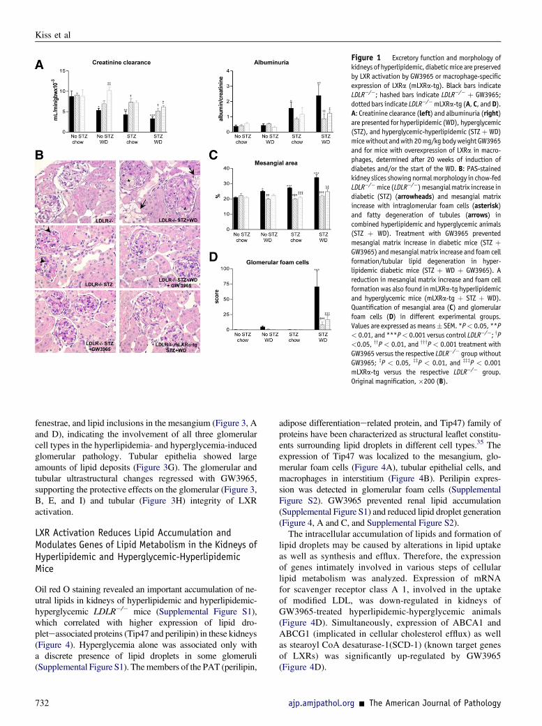
Figure 1 Excretory function and morphology ofkidneys of hyperlipidemic, diabeticmice are preservedby LXR activation by GW3965 or macrophage-specificexpression of LXRa (mLXRa-tg). Black bars indicateLDLR�/�; hashed bars indicate LDLR�/� þ GW3965;dotted bars indicate LDLR�/�mLXRa-tg (A, C, andD).A: Creatinine clearance (left) and albuminuria (right)are presented for hyperlipidemic (WD), hyperglycemic(STZ), and hyperglycemic-hyperlipidemic (STZþ WD)micewithout andwith 20mg/kgbodyweight GW3965and for mice with overexpression of LXRa in macro-phages, determined after 20 weeks of induction ofdiabetes and/or the start of the WD. B: PAS-stainedkidney slices showingnormalmorphology in chow-fedLDLR�/�mice (LDLR�/�) mesangialmatrix increase indiabetic (STZ) (arrowheads) and mesangial matrixincrease with intraglomerular foam cells (asterisk)and fatty degeneration of tubules (arrows) incombined hyperlipidemic and hyperglycemic animals(STZ þ WD). Treatment with GW3965 preventedmesangial matrix increase in diabetic mice (STZ þGW3965) andmesangial matrix increase and foam cellformation/tubular lipid degeneration in hyper-lipidemic diabetic mice (STZ þ WD þ GW3965). Areduction in mesangial matrix increase and foam cellformation was also found inmLXRa-tg hyperlipidemicand hyperglycemic mice (mLXRa-tg þ STZ þ WD).Quantification of mesangial area (C) and glomerularfoam cells (D) in different experimental groups.Values are expressed asmeans� SEM. *P< 0.05, **P< 0.01, and ***P< 0.001 versus control LDLR�/�; yP<0.05, yyP < 0.01, and yyyP < 0.001 treatment withGW3965 versus the respective LDLR�/� group withoutGW3965; zP < 0.05, zzP < 0.01, and zzzP < 0.001mLXRa-tg versus the respective LDLR�/� group.Original magnification, �200 (B).
Kiss et al
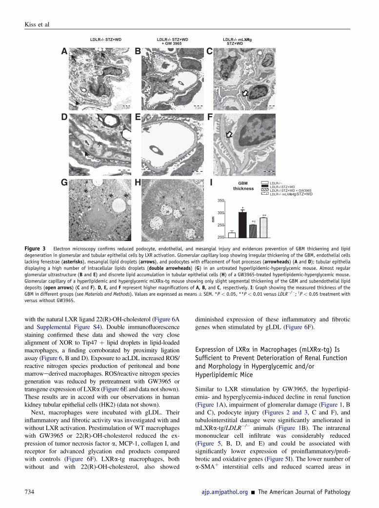
fenestrae, and lipid inclusions in the mesangium (Figure 3, Aand D), indicating the involvement of all three glomerularcell types in the hyperlipidemia- and hyperglycemia-inducedglomerular pathology. Tubular epithelia showed largeamounts of lipid deposits (Figure 3G). The glomerular andtubular ultrastructural changes regressed with GW3965,supporting the protective effects on the glomerular (Figure 3,B, E, and I) and tubular (Figure 3H) integrity of LXRactivation.
LXR Activation Reduces Lipid Accumulation andModulates Genes of Lipid Metabolism in the Kidneys ofHyperlipidemic and Hyperglycemic-HyperlipidemicMice
Oil red O staining revealed an important accumulation of ne-utral lipids in kidneys of hyperlipidemic and hyperlipidemic-hyperglycemic LDLR�/� mice (Supplemental Figure S1),which correlated with higher expression of lipid dro-pleteassociated proteins (Tip47 and perilipin) in these kidneys(Figure 4). Hyperglycemia alone was associated only witha discrete presence of lipid droplets in some glomeruli(Supplemental Figure S1). Themembers of the PAT (perilipin,
732
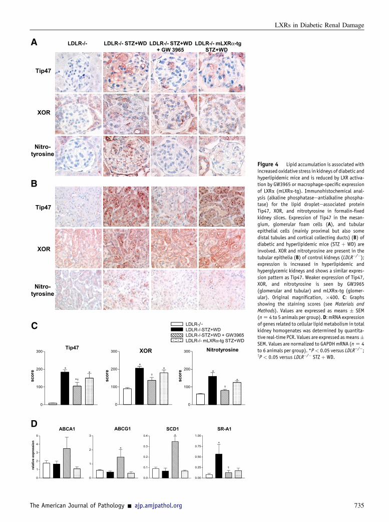
adipose differentiationerelated protein, and Tip47) family ofproteins have been characterized as structural leaflet constitu-ents surrounding lipid droplets in different cell types.35 Theexpression of Tip47 was localized to the mesangium, glo-merular foam cells (Figure 4A), tubular epithelial cells, andmacrophages in interstitium (Figure 4B). Perilipin expres-sion was detected in glomerular foam cells (SupplementalFigure S2). GW3965 prevented renal lipid accumulation(Supplemental Figure S1) and reduced lipid droplet generation(Figure 4, A and C, and Supplemental Figure S2).The intracellular accumulation of lipids and formation of
lipid droplets may be caused by alterations in lipid uptakeas well as synthesis and efflux. Therefore, the expressionof genes intimately involved in various steps of cellularlipid metabolism was analyzed. Expression of mRNAfor scavenger receptor class A 1, involved in the uptakeof modified LDL, was down-regulated in kidneys ofGW3965-treated hyperlipidemic-hyperglycemic animals(Figure 4D). Simultaneously, expression of ABCA1 andABCG1 (implicated in cellular cholesterol efflux) as wellas stearoyl CoA desaturase-1(SCD-1) (known target genesof LXRs) was significantly up-regulated by GW3965(Figure 4D).
ajp.amjpathol.org - The American Journal of Pathology

B
D
A
LDLR-/-
LDLR-/-STZ+WD
+GW3965
LDLR-/-STZ+WD
LDLR-/-mLXR -tg
STZ+WD
C
0
5
10
15
ce
lls
/g
lo
me
ru
lu
s
WT1
**
**
††‡
‡
No STZ No STZ STZ STZchow WD chow WD
0
1
2
3
4
ce
lls
/g
lo
me
ru
lu
s
Desmin
No STZ NoSTZa STZ STZ chow WD chow WD
**
**
***
††
‡‡‡ ‡‡‡ ‡‡‡
†††
LDLR-/-mLXRα-tg
STZ+WD
LDLR-/-STZ+WD
+GW3965
LDLR-/- LDLR-/-STZ+WD
α
Figure 2 Less podocyte loss and injury byLXR activation by GW3965 or macrophage-specificexpression of LXRa (mLXRa-tg) in hyperglycemicand/or hyperlipidemic mice. Black bars indicateLDLR�/�; hashed bars indicate LDLR�/� þ GW3965;dotted bars indicate LDLR�/� mLXRa-tg (B and D).Immunostaining for WT1 (A) and desmin (C) showinga decreased number of podocytes (WT1þ) and anincreased number of injured podocytes (desminþ) inglomeruli of hyperglycemic-hyperlipidemic LDLR�/�
mice (STZþWD) (arrowheads, C); this was preventedby GW3965 or expression of LXRa in macrophages.Original magnification, �400. Quantification ofWT1þ (B) and desminþ (D) cells in different experi-mental groups. Values are expressed asmeans� SEM.*P < 0.05, **P < 0.01, and ***P < 0.001 versuschow-fed LDLR�/�; yyP < 0.01, yyyP < 0.001 treat-ment with GW3965 versus the respective LDLR�/�
group without GW3965; zP < 0.05, zzzP < 0.001mLXRa-tg versus the respective LDLR�/� group.
LXRs in Diabetic Renal Damage
LXR Activation Reduces Oxidative Stress in the Kidneysof Hyperglycemic and/or Hyperlipidemic Mice
The biogenesis of lipid droplets is associated with thecompartmentalization of lipid metabolic enzymes and otherproteins (eg, kinases, small GTPases, and cytokines), indi-cating their potential function as intracellular signalingplatforms.10 We previously identified XOR as an importantenzyme for the production of ROS in hyperlipidemia-associated renal injury.36,37 Herein, we observed increasedexpression and a concomitant close association of theoxidative enzyme XOR and the oxidatively modified proteinproduct nitrotyrosine with Tip47 in glomerular foam cells(Figure 4A), tubular epithelia, and interstitial macrophages(Figure 4B) in the hyperglycemic-hyperlipidemic mousekidneys. The expression of XOR and nitrotyrosine wasreduced in GW3965-treated kidneys (Figure 4, Ae C),suggesting that the inhibition of lipid droplet generation andconsequent reduction of lipid-related oxidative mechanismsmight contribute to the protective effects of LXR activation.
LXR Activation Reduces Inflammation/Fibrosis andModulates Expression of Inflammatory/FibroticCytokines in Kidneys of Hyperglycemic and/orHyperlipidemic Mice
Glomerular and interstitial mononuclear cells (Mac-2þ andF4/80þ macrophages, CD3þ T lymphocytes) were signifi-cantly increased when mice were exposed to hyperglycemiaand hyperlipidemia and were remarkably decreased inkidneys of GW3965-treated mice (Figure 5, AeE).
The American Journal of Pathology - ajp.amjpathol.org
Immmunohistochemical analysis for Ki-67 showed a signifi-cantly increased proliferation rate of glomerular and tubu-lointerstitial cells by hyperlipidemia and/or hyperglycemia ofLDLR�/� mice; however, the overall number of proliferatingcells was low. LXR stimulation with GW3965 had no effecton the proliferation rate of tubular epithelial cells (data notshown) but reduced significantly the proliferation ofglomerular and interstitial cells (Supplemental Figure S3).
Focal fibrotic areas quantified in Masson`s trichromeestained kidney slices anda-SMAþ glomerular and interstitialmyofibroblasts, increased in diabetic and/or hyperlipidemicmice, showed a significant decrease in GW3965-treatedgroups (Figure 5, FeH).
In line with the morphometric analyses, the increasedrenal expression of the proinflammatory/profibrotic genes(receptor for advanced glycation end products, tumornecrosis factor a, MCP-1, transforming growth factor b1) inhyperlipidemic-hyperglycemic LDLR�/� mice was down-regulated by GW3965 (Figure 5I).
Effects of LXR Activation on Lipid Droplets, OxidativeStress, and Inflammatory/Fibrotic Activity ofMacrophages in Vitro
In macrophages, foam cell formation by acLDL was paral-leled by increased expression of Tip47, XOR, and nitro-tyrosine staining (Figure 6A and Supplemental Figure S4).GW3965 led to a reduced number of cells expressing Tip47,XOR, and nitrotyrosine (Figure 6A and SupplementalFigure S4). Macrophages overexpressing LXRa (mLXRa-tg)showed similar reactions without and with prestimulation
733
LDLR-/− LDLR-/-STZ+WD LDLR-/-STZ+WD + GW3965 LDLR-/- mLXRα-tg STZ+WD
GBM
thickness
LDLR-/- STZ+WD LDLR-/- STZ+WD
+ GW 3965
LDLR-/- mLXRα-tg
STZ+WD
*
*
*
200
250
300
350
nm
*
*†
**
Figure 3 Electron microscopy confirms reduced podocyte, endothelial, and mesangial injury and evidences prevention of GBM thickening and lipiddegeneration in glomerular and tubular epithelial cells by LXR activation. Glomerular capillary loop showing irregular thickening of the GBM, endothelial cellslacking fenestrae (asterisks), mesangial lipid droplets (arrows), and podocytes with effacement of foot processes (arrowheads) (A and D); tubular epitheliadisplaying a high number of intracellular lipids droplets (double arrowheads) (G) in an untreated hyperlipidemic-hyperglycemic mouse. Almost regularglomerular ultrastructure (B and E) and discrete lipid accumulation in tubular epithelial cells (H) of a GW3965-treated hyperlipidemic-hyperglycemic mouse.Glomerular capillary of a hyperlipidemic and hyperglycemic mLXRa-tg mouse showing only slight segmental thickening of the GBM and subendothelial lipiddeposits (open arrows) (C and F). D, E, and F represent higher magnifications of A, B, and C, respectively. I: Graph showing the measured thickness of theGBM in different groups (see Materials and Methods). Values are expressed as means � SEM. *P < 0.05, **P < 0.01 versus LDLR�/�; yP < 0.05 treatment withversus without GW3965.
Kiss et al
with the natural LXR ligand 22(R)-OH-cholesterol (Figure 6Aand Supplemental Figure S4). Double immunofluorescencestaining confirmed these data and showed the very closealignment of XOR to Tip47 þ lipid droplets in lipid-loadedmacrophages, a finding corroborated by proximity ligationassay (Figure 6, B and D). Exposure to acLDL increased ROS/reactive nitrogen species production of peritoneal and bonemarrowederived macrophages. ROS/reactive nitrogen speciesgeneration was reduced by pretreatment with GW3965 ortransgene expression of LXRa (Figure 6E and data not shown).These results are in accord with our observations in humankidney tubular epithelial cells (HK2) (data not shown).
Next, macrophages were incubated with gLDL. Theirinflammatory and fibrotic activity was investigated with andwithout LXR activation. Prestimulation of WT macrophageswith GW3965 or 22(R)-OH-cholesterol reduced the ex-pression of tumor necrosis factor a, MCP-1, collagen I, andreceptor for advanced glycation end products comparedwith controls (Figure 6F). LXRa-tg macrophages, bothwithout and with 22(R)-OH-cholesterol, also showed
734
diminished expression of these inflammatory and fibroticgenes when stimulated by gLDL (Figure 6F).
Expression of LXRa in Macrophages (mLXRa-tg) IsSufficient to Prevent Deterioration of Renal Functionand Morphology in Hyperglycemic and/orHyperlipidemic Mice
Similar to LXR stimulation by GW3965, the hyperlipid-emia- and hyperglycemia-induced decline in renal function(Figure 1A), impairment of glomerular damage (Figure 1, Band C), podocyte injury (Figures 2 and 3, C and F), andtubulointerstitial damage were significantly ameliorated inmLXRa-tg/LDLR�/� animals (Figure 1B). The intrarenalmononuclear cell infiltrate was considerably reduced(Figure 5, B, D, and E) and could be associated withsignificantly lower expression of proinflammatory/profi-brotic and oxidative genes (Figure 5I). The lower number ofa-SMAþ interstitial cells and reduced scarred areas in
ajp.amjpathol.org - The American Journal of Pathology
0
100
200
300
sco
re
0
100
200
300
sco
re
Tip47 XOR Nitrotyrosine
LDLR-/- LDLR-/- STZ+WD LDLR-/- STZ+WD
+ GW 3965
LDLR-/- mLXRα-tg
STZ+WD
Tip47
XOR
Tip47
XOR
Nitro-
tyrosine
Nitro-
tyrosine
ABCA1
0
1
2
3
4
5
rela
tiv
e e
xp
ressio
n
ABCG1 SCD1 SR-A1
LDLR-LDLR-/-STZ+WD LDLR-/-STZ+WD + GW3965 LDLR-/- mLXRα-tg STZ+WD
0
100
200
300
sco
re *
†
*
*
†
**
*†
*
0
1
2
3
*
0.0
0.1
0.2
0.3
0.4
0.00
0.25
0.50
0.75
1.00*
*
†
Figure 4 Lipid accumulation is associated withincreased oxidative stress in kidneys of diabetic andhyperlipidemic mice and is reduced by LXR activa-tion by GW3965 or macrophage-specific expressionof LXRa (mLXRa-tg). Immunohistochemical anal-ysis (alkaline phosphataseeantialkaline phospha-tase) for the lipid dropleteassociated proteinTip47, XOR, and nitrotyrosine in formalin-fixedkidney slices. Expression of Tip47 in the mesan-gium, glomerular foam cells (A), and tubularepithelial cells (mainly proximal but also somedistal tubules and cortical collecting ducts) (B) ofdiabetic and hyperlipidemic mice (STZ þ WD) areinvolved. XOR and nitrotyrosine are present in thetubular epithelia (B) of control kidneys (LDLR�/�);expression is increased in hyperlipidemic andhyperglycemic kidneys and shows a similar expres-sion pattern as Tip47. Weaker expression of Tip47,XOR, and nitrotyrosine is seen by GW3965(glomerular and tubular) and mLXRa-tg (glomer-ular). Original magnification, �400. C: Graphsshowing the staining scores (see Materials andMethods). Values are expressed as means � SEM(nZ 4 to 5 animals per group).D: mRNA expressionof genes related to cellular lipid metabolism in totalkidney homogenates was determined by quantita-tive real-time PCR. Values are expressed as means�SEM. Values are normalized to GAPDHmRNA (nZ 4to 6 animals per group). *P< 0.05 versus LDLR�/�;yP < 0.05 versus LDLR�/� STZ þ WD.
LXRs in Diabetic Renal Damage
The American Journal of Pathology - ajp.amjpathol.org 735
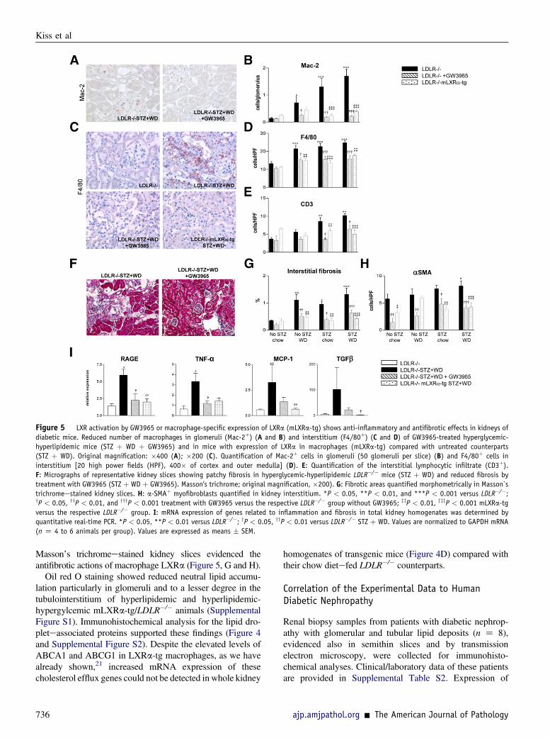
Figure 5 LXR activation by GW3965 or macrophage-specific expression of LXRa (mLXRa-tg) shows anti-inflammatory and antifibrotic effects in kidneys ofdiabetic mice. Reduced number of macrophages in glomeruli (Mac-2þ) (A and B) and interstitium (F4/80þ) (C and D) of GW3965-treated hyperglycemic-hyperlipidemic mice (STZ þ WD þ GW3965) and in mice with expression of LXRa in macrophages (mLXRa-tg) compared with untreated counterparts(STZ þ WD). Original magnification: �400 (A); �200 (C). Quantification of Mac-2þ cells in glomeruli (50 glomeruli per slice) (B) and F4/80þ cells ininterstitium [20 high power fields (HPF), 400� of cortex and outer medulla] (D). E: Quantification of the interstitial lymphocytic infiltrate (CD3þ).F: Micrographs of representative kidney slices showing patchy fibrosis in hyperglycemic-hyperlipidemic LDLR�/� mice (STZ þ WD) and reduced fibrosis bytreatment with GW3965 (STZ þ WD þ GW3965). Masson’s trichrome; original magnification, �200). G: Fibrotic areas quantified morphometrically in Masson`strichromeestained kidney slices. H: a-SMAþ myofibroblasts quantified in kidney interstitium. *P < 0.05, **P < 0.01, and ***P < 0.001 versus LDLR�/�;yP < 0.05, yyP < 0.01, and yyyP < 0.001 treatment with GW3965 versus the respective LDLR�/� group without GW3965; zzP < 0.01, zzzP < 0.001 mLXRa-tgversus the respective LDLR�/� group. I: mRNA expression of genes related to inflammation and fibrosis in total kidney homogenates was determined byquantitative real-time PCR. *P < 0.05, **P < 0.01 versus LDLR�/�; yP < 0.05, yyP < 0.01 versus LDLR�/� STZ þ WD. Values are normalized to GAPDH mRNA(n Z 4 to 6 animals per group). Values are expressed as means � SEM.
Kiss et al
Masson’s trichromeestained kidney slices evidenced theantifibrotic actions of macrophage LXRa (Figure 5, G and H).
Oil red O staining showed reduced neutral lipid accumu-lation particularly in glomeruli and to a lesser degree in thetubulointerstitium of hyperlipidemic and hyperlipidemic-hypergylcemic mLXRa-tg/LDLR�/� animals (SupplementalFigure S1). Immunohistochemical analysis for the lipid dro-pleteassociated proteins supported these findings (Figure 4and Supplemental Figure S2). Despite the elevated levels ofABCA1 and ABCG1 in LXRa-tg macrophages, as we havealready shown,21 increased mRNA expression of thesecholesterol efflux genes could not be detected in whole kidney
736
homogenates of transgenic mice (Figure 4D) compared withtheir chow dietefed LDLR�/� counterparts.
Correlation of the Experimental Data to HumanDiabetic Nephropathy
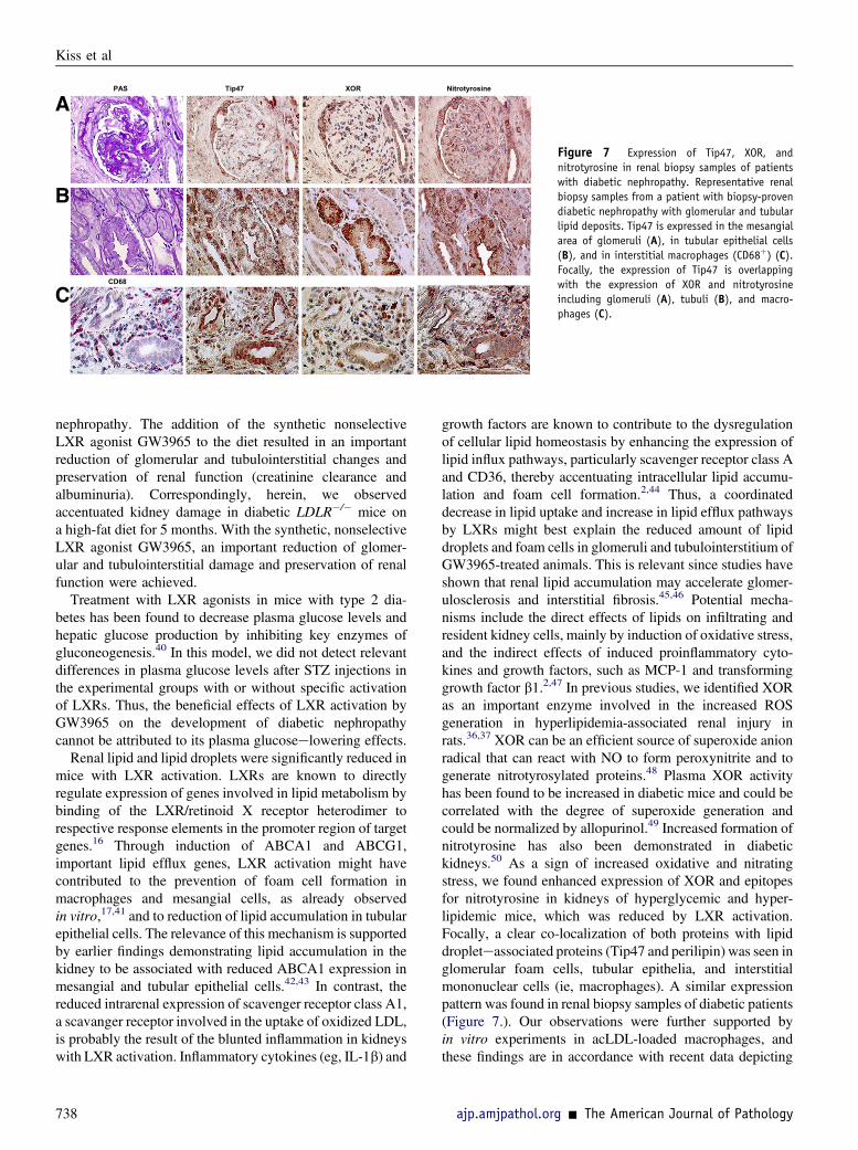
Renal biopsy samples from patients with diabetic nephrop-athy with glomerular and tubular lipid deposits (n Z 8),evidenced also in semithin slices and by transmissionelectron microscopy, were collected for immunohisto-chemical analyses. Clinical/laboratory data of these patientsare provided in Supplemental Table S2. Expression of
ajp.amjpathol.org - The American Journal of Pathology

Figure 6 LXR activation coordinately modulates lipid metabolism, oxidative stress, and inflammatory/fibrotic activity of ac/gLDL-stimulated peritonealmacrophages. A: Expression of Tip47, XOR, and nitrotyrosine in mouse peritoneal macrophages incubated with 50 mg/mL of acLDL for 24 hours. The number ofcells expressing Tip47, XOR, and nitrotyrosine and their staining intensity were reduced when macrophages were prestimulated with GW3965 (3 mmol/L, 16hours) and by LXRa-tg macrophages without and with prestimulation with 10 mmol/L natural ligand 22(R)-OH-cholesterol for 16 hours. The graphs show thestaining scores in different groups (one representative experiment of three). Corresponding micrographs are presented in Supplemental Figure S4. B: Doubleimmunofluorescence staining confirms the increased expression of XOR in lipid-loaded macrophages. A very close alignment of XOR to Tip47-positive lipiddroplets in lipid-loaded macrophages can be observed. C: In addition, proximity ligation assay (PLA), which can visualize and quantify individual proteininteractions in native cells using standard fluorescence microscopy, indicates a very close relation of XOR to lipid droplets in some lipid-loaded macrophages.D: Quantification of PLA spots in different groups (see Materials and Methods). E: ROS was measured in peritoneal macrophages (2 � 104 cells per well) after 10and 60 minutes of stimulation with 50 mg/mL of acLDL. The graph shows one representative experiment of three performed in quadruplicate. F: mRNAexpression of genes related to oxidative stress, inflammation, and fibrosis in peritoneal macrophages stimulated with 30 mg/mL of gLDL for 24 hours wasreduced when the cells were pretreated with GW3965 (3 mmol/L) or 22(R)-OH-cholesterol (HC) (10 mmol/L) for 24 hours. One experiment of three, performedin duplicate. G: Reduced collagen I mRNA expression of gLDL stimulated LXRa-tg macrophages without and with HC (one experiment of three performed induplicate). Values are normalized to S18r mRNA. Results are expressed as means � SEM. *P < 0.05 versus dimethyl sulfoxide (DMSO); yP < 0.05 with versuswithout GW3965.
LXRs in Diabetic Renal Damage
Tip47 was observed as in kidneys of hyperlipidemic-hyperglycemic mice localizing focally and segmentally inmesangial areas, tubular epithelial cells, and interstitialmononuclear cells. As shown in Figure 7, Tip47 expressionwas partly overlapping with the label for XOR and nitro-tyrosine, suggesting a close association of lipid droplets withpotential foci of oxidative processes in diabetic nephropathy.
Discussion
Clinical observations suggest that hyperlipidemia contrib-utes to the progression of diabetic renal disease.2,38 Exper-imentally, it has been demonstrated that hyperlipidemia andhyperglycemia may act synergistically on the initiation and
The American Journal of Pathology - ajp.amjpathol.org
progression of diabetic nephropathy.39 We observed similarchanges in kidneys of LDLR-deficient mice (C57BL6)when injected with STZ and fed the WD for 20 weeks.Under the combined stress of hyperglycemia and hyperlip-idemia, mice developed complex renal damage: glomerularchanges involving all three glomerular cell types, such asmesangial matrix increase and cell proliferation, thickenedGBM, podocyte loss and injury, loss of endothelial fenes-tration and, seemingly, the result of hyperlipidemia, butaccentuated by hyperglycemia, glomerular fat droplets, andfoam cells; the fatty degeneration of the tubular epitheliawas accompanied by a significant increase in interstitialmacrophages and foam cells. These findings underscore thecomplexity and interrelatedness of pathogenic processes,which may play a role in the development of diabetic
737
Tip47 Nitrotyrosine XOR PAS
CD68
Figure 7 Expression of Tip47, XOR, andnitrotyrosine in renal biopsy samples of patientswith diabetic nephropathy. Representative renalbiopsy samples from a patient with biopsy-provendiabetic nephropathy with glomerular and tubularlipid deposits. Tip47 is expressed in the mesangialarea of glomeruli (A), in tubular epithelial cells(B), and in interstitial macrophages (CD68þ) (C).Focally, the expression of Tip47 is overlappingwith the expression of XOR and nitrotyrosineincluding glomeruli (A), tubuli (B), and macro-phages (C).
Kiss et al
nephropathy. The addition of the synthetic nonselectiveLXR agonist GW3965 to the diet resulted in an importantreduction of glomerular and tubulointerstitial changes andpreservation of renal function (creatinine clearance andalbuminuria). Correspondingly, herein, we observedaccentuated kidney damage in diabetic LDLR�/� mice ona high-fat diet for 5 months. With the synthetic, nonselectiveLXR agonist GW3965, an important reduction of glomer-ular and tubulointerstitial damage and preservation of renalfunction were achieved.
Treatment with LXR agonists in mice with type 2 dia-betes has been found to decrease plasma glucose levels andhepatic glucose production by inhibiting key enzymes ofgluconeogenesis.40 In this model, we did not detect relevantdifferences in plasma glucose levels after STZ injections inthe experimental groups with or without specific activationof LXRs. Thus, the beneficial effects of LXR activation byGW3965 on the development of diabetic nephropathycannot be attributed to its plasma glucoseelowering effects.
Renal lipid and lipid droplets were significantly reduced inmice with LXR activation. LXRs are known to directlyregulate expression of genes involved in lipid metabolism bybinding of the LXR/retinoid X receptor heterodimer torespective response elements in the promoter region of targetgenes.16 Through induction of ABCA1 and ABCG1,important lipid efflux genes, LXR activation might havecontributed to the prevention of foam cell formation inmacrophages and mesangial cells, as already observedin vitro,17,41 and to reduction of lipid accumulation in tubularepithelial cells. The relevance of this mechanism is supportedby earlier findings demonstrating lipid accumulation in thekidney to be associated with reduced ABCA1 expression inmesangial and tubular epithelial cells.42,43 In contrast, thereduced intrarenal expression of scavenger receptor class A1,a scavanger receptor involved in the uptake of oxidized LDL,is probably the result of the blunted inflammation in kidneyswith LXR activation. Inflammatory cytokines (eg, IL-1b) and
738
growth factors are known to contribute to the dysregulationof cellular lipid homeostasis by enhancing the expression oflipid influx pathways, particularly scavenger receptor class Aand CD36, thereby accentuating intracellular lipid accumu-lation and foam cell formation.2,44 Thus, a coordinateddecrease in lipid uptake and increase in lipid efflux pathwaysby LXRs might best explain the reduced amount of lipiddroplets and foam cells in glomeruli and tubulointerstitium ofGW3965-treated animals. This is relevant since studies haveshown that renal lipid accumulation may accelerate glomer-ulosclerosis and interstitial fibrosis.45,46 Potential mecha-nisms include the direct effects of lipids on infiltrating andresident kidney cells, mainly by induction of oxidative stress,and the indirect effects of induced proinflammatory cyto-kines and growth factors, such as MCP-1 and transforminggrowth factor b1.2,47 In previous studies, we identified XORas an important enzyme involved in the increased ROSgeneration in hyperlipidemia-associated renal injury inrats.36,37 XOR can be an efficient source of superoxide anionradical that can react with NO to form peroxynitrite and togenerate nitrotyrosylated proteins.48 Plasma XOR activityhas been found to be increased in diabetic mice and could becorrelated with the degree of superoxide generation andcould be normalized by allopurinol.49 Increased formation ofnitrotyrosine has also been demonstrated in diabetickidneys.50 As a sign of increased oxidative and nitratingstress, we found enhanced expression of XOR and epitopesfor nitrotyrosine in kidneys of hyperglycemic and hyper-lipidemic mice, which was reduced by LXR activation.Focally, a clear co-localization of both proteins with lipiddropleteassociated proteins (Tip47 and perilipin) was seen inglomerular foam cells, tubular epithelia, and interstitialmononuclear cells (ie, macrophages). A similar expressionpattern was found in renal biopsy samples of diabetic patients(Figure 7.). Our observations were further supported byin vitro experiments in acLDL-loaded macrophages, andthese findings are in accordance with recent data depicting
ajp.amjpathol.org - The American Journal of Pathology

LXRs in Diabetic Renal Damage
that overexpression of XOR in macrophages can exacerbatefoam cell formation.51 In an earlier study, McManaman andcoworkers52 localized XOR in the membrane of milk fatdroplet membranes. By double immunofluorescence stain-ing, we could provide evidence of a close alignment of XORto Tip47-positive lipid droplets in lipid-loaded macrophages;this finding has been confirmed by a second method, namely,a positive proximity ligation assay result, hinting at a close(30 to 40 nm) association of these two proteins. Documentingthe concomitant presence of an oxidative enzyme and itsproducts with lipid droplets in renal resident and infiltratingcells, the present data point to a potential mechanism howintracellular lipids can be detrimental for the cell; foremostthis can be prevented by LXR activation. In addition, we andothers have shown that XOR can contribute to an increase ofmacrophage infiltration by induction of inflammatory cyto-kine and adhesion molecules.37,53,54
In GW3965-treated hyperlipidemic-hyperglycemic mice,improved renal function and morphology was accompaniedby a significantly diminished intrarenal mononuclear cellinfiltrate and decreased renal expression of proinflammatorymediators (tumor necrosis factor a and MCP-1). Theinflammatory activity (tumor necrosis factor a, MCP-1, andreceptor for advanced glycation end products) of gLDL-stimulated macrophages pretreated with a synthetic ornatural LXR ligand was reduced. The basic mechanismunderlying the anti-inflammatory actions of LXRs has beenshown to occur through the transrepression of NF-kBsignaling16 and consequent reduced transcription of cyto-kines and receptor for advanced glycation end products, oneof the receptors of the advanced glycation end products.55
Of note, LXR activation reduced renal expression oftransforming growth factor b1, the number of interstitialmyofibroblasts, and the extent of fibrotic areas that correspondto the decreased collagen type I expression inGW3965-treatedmacrophages in vitro. These results are in line with earlierobservations in kidney transplantation models in which LXRactivation prevented the development of chronic renal allo-graft damage.20 Recently, Beaven and coworkers56 reportedthe suppression of fibrosis-related genes (eg, COL1A1) inprimary mouse stellate cells by LXR ligands and increasedliver fibrosis after injury in LXRab-deficient mice.
Studies on human renal biopsy samples have shown thatmacrophage accumulation in diabetic kidneys correlates withserum creatinine levels, interstitial myofibroblast accumula-tion, and interstitial fibrosis.8,12,57 Thus, macrophage-mediated injury seems to be an important component in thedevelopment of diabetic nephropathy, which is not sup-pressed effectively by current therapies.6
Since in the kidney, LXR mRNA is expressed in allnephron segments, the protective effects of the nonselectiveLXR agonist GW3965 in hyperlipidemic-hyperglycemickidneys may not be restricted to macrophages; the overalleffect of the nonselective LXR agonist is likely to bea suppression of activity of infiltrating mononuclear cellsand resident renal cells.
The American Journal of Pathology - ajp.amjpathol.org
Nevertheless, transgenic expression of LXRa in macro-phages was sufficient to prevent deterioration of renal functionand morphology related to hyperglycemia and hyperlipidemia.Data concerning isoform-specific effects of LXRs are of prac-tical interest for the generation of isoform-specific pharmaco-logic modulators, without potential adverse metabolic effects.
Activated macrophages can produce endogenous ligandsof LXRs, eg, 24(S)-OH-cholesterol, 22(R)-OH-cholesterol,and 24(S),25-epoxycholesterol, which, in the state of constantexpression of the receptor, apparently can lead to a biologi-cally relevant activation without the need for further ad-ministration of an exogenous ligand.7,58,59 We previouslydemonstrated that macrophage transgenic expression ofLXRa achieved by expression of the mouse LXRa cDNAunder the control of a chicken lysozyme promoter resulted inactivation of LXRa target genes, ABCA1 andABCG1.21 Themacrophage-restricted up-regulation of cholesterol effluxgenes might explain the nonsignificant influence on lipidaccumulation in tubular cells of mLXRa-tg mice comparedwith the GW3965-treated group. These experiments provideevidence for a robust capacity of the macrophage LXRa toinhibit diabetic nephropathy.
In summary, these findings demonstrate that lipid-inducedoxidative mechanisms are operating in tissue injury of dia-betic kidneys. Interrupting the pathophysiologic cascade ofevents induced by excess lipids and inhibiting inflammatorygene expression, LXR activation can prevent the develop-ment of diabetic nephropathy, even if aggravated by hyper-lipidemia. Modulation of macrophage-mediated injury byLXRs seems to be relevant for efficient therapy.
Acknowledgments
We thank Claudia Schmidt, Gabi Schmidt, Sylvia Kaden,and Tjeerd Sijmonsma for expert technical help.
Supplemental Data
Supplemental material for this article can be found athttp://dx.doi.org/10.1016/j.ajpath.2012.11.033.
References
1. Jandeleit-Dahm K, Cao Z, Cox AJ, Kelly DJ, Gilbert RE, Cooper ME:Role of hyperlipidemia in progressive renal disease: focus on diabeticnephropathy. Kidney Int Suppl 1999, 71:S31eS36
2. Abrass CK: Lipid metabolism and renal disease. Contrib Nephrol2006, 151:106e121
3. Kamanna VS, Pai R, Roh DD, Kirschenbaum MA: Oxidative modi-fication of low-density lipoprotein enhances the murine mesangial cellcytokines associated with monocyte migration, differentiation, andproliferation. Lab Invest 1996, 74:1067e1079
4. Gröne HJ, Walli AK, Gröne EF: The role of oxidatively modifiedlipoproteins in lipid nephropathy. Contrib Nephrol 1997, 120:160e175
5. Kamanna VS, Pai R, Ha H, Kirschenbaum MA, Roh DD: Oxidizedlow-density lipoprotein stimulates monocyte adhesion to glomerularendothelial cells. Kidney Int 1999, 55:2192e2202
739

Kiss et al
6. Tesch GH: Macrophages and diabetic nephropathy. Semin Nephrol2010, 30:290e301
7. Shibata N, Glass CK: Macrophages, oxysterols and atherosclerosis.Circ J 2010, 74:2045e2051
8. Schmid H, Boucherot A, Yasuda Y, Henger A, Brunner B,Eichinger F, Nitsche A, Kiss E, Bleich M, Gröne HJ, Nelson PJ,Schlöndorff D, Cohen CD, Kretzler M: Modular activation of nuclearfactor-kB transcriptional programs in human diabetic nephropathy.Diabetes 2006, 55:2993e3003
9. Prieur X, Roszer T, Ricote M: Lipotoxicity in macrophages: evidencefrom diseases associated with the metabolic syndrome. Biochim Bio-phys Acta 2010, 1801:327e337
10. Bozza PT, Bandeira-Melo C: Mechanisms of leukocyte lipid bodyformation and function in inflammation. Mem Inst Oswaldo Cruz2005, 1(Suppl):113e120
11. Zoja C, Garcia PB, Remuzzi G: The role of chemokines in progressiverenal disease. Front Biosci 2009, 14:1815e1822
12. Yonemoto S, Machiguchi T, Nomura K, Minakata T, Nanno M,Yoshida H: Correlations of tissue macrophages and cytoskeletalprotein expression with renal fibrosis in patients with diabetes mellitus.Clin Exp Nephrol 2006, 10:186e192
13. Vallon V, Thomson SC: Renal function in diabetic disease models: thetubular system in the pathophysiology of the diabetic kidney. AnnuRev Physiol 2012, 74:351e375
14. Repa JJ, Mangelsdorf DJ: The liver X receptor gene team: potentialnew players in atherosclerosis. Nat Med 2002, 8:1243e1248
15. Proctor G, Jiang T, Iwahashi M, Wang Z, Li J, Levi M: Regulation ofrenal fatty acid and cholesterol metabolism, inflammation, and fibrosisin akita and OVE26 mice with type 1 diabetes. Diabetes 2006, 55:2502e2509
16. Hong C, Tontonoz P: Coordination of inflammation and metabolismby PPAR and LXR nuclear receptors. Curr Opin Genet Dev 2008, 18:461e467
17. Larrede S, Quinn CM, JessupW, Frisdal E, OlivierM, HsiehV, KimMJ,Van Eck M, Couvert P, Carrie A, Giral P, Chapman MJ, Guerin M, LeGoffW: Stimulation of cholesterol efflux byLXRagonists in cholesterol-loaded human macrophages is ABCA1-dependent but ABCG1-independent. Arterioscler Thromb Vasc Biol 2009, 29:1930e1936
18. Ogawa S, Lozach J, Benner C, Pascual G, Tangirala RK, Westin S,Hoffmann A, Subramaniam S, David M, Rosenfeld MG, Glass CK:Molecular determinants of crosstalk between nuclear receptors andtoll-like receptors. Cell 2005, 122:707e721
19. Terasaka N, Hiroshima A, Koieyama T, Ubukata N, Morikawa Y,Nakai D, Inaba T: T-0901317, a synthetic liver X receptor ligand,inhibits development of atherosclerosis in LDL receptor-deficientmice. FEBS Lett 2003, 536:6e11
20. Kiss E, Popovic Z, Bedke J, Wang S, Bonrouhi M, Gretz N, Stettner P,Teupser D, Thiery J, Porubsky S, Adams J, Gröne HJ: Suppression ofchronic damage in renal allografts by liver X receptor (LXR) activa-tion: relevant contribution of macrophage LXRa. Am J Pathol 2011,179:92e103
21. TeupserD,KretzschmarD,Tennert C,BurkhardtR,WilfertW,FenglerD,NaumannR, Sippel AE, Thiery J: Effect ofmacrophage overexpression ofmurine liver X receptor-a (LXR-a) on atherosclerosis in LDL-receptordeficient mice. Arterioscler Thromb Vasc Biol 2008, 28:2009e2015
22. Collins JL, Fivush AM, Watson MA, Galardi CM, Lewis MC,Moore LB, Parks DJ, Wilson JG, Tippin TK, Binz JG, Plunket KD,Morgan DG, Beaudet EJ, Whitney KD, Kliewer SA, Willson TM:Identification of a nonsteroidal liver X receptor agonist through parallelarray synthesis of tertiary amines. J Med Chem 2002, 45:1963e1966
23. Adams J, Kiss E, Arroyo AB, Bonrouhi M, Sun Q, Li Z, Gretz N,Schnitger A, Zouboulis CC, Wiesel M, Wagner J, Nelson PJ,Gröne HJ: 13-cis retinoic acid inhibits development and progression ofchronic allograft nephropathy. Am J Pathol 2005, 167:285e298
24. Hirose K, Osterby R, Nozawa M, Gundersen HJ: Development ofglomerular lesions in experimental long-term diabetes in the rat.Kidney Int 1982, 21:689e695
740
25. Guo M, Ricardo SD, Deane JA, Shi M, Cullen-McEwen L,Bertram JF: A stereological study of the renal glomerular vasculaturein the db/db mouse model of diabetic nephropathy. J Anat 2005, 207:813e821
26. Weischenfeldt J, Porse B: Bone marrow-derived macrophages (BMM):isolation and applications. CSH Protoc 2008. http://dx.doi.org/10.1101/pdb.prot5080
27. Gersuk GM, Razai LW, Marr KA: Methods of in vitro macrophagematuration confer variable inflammatory responses in association withaltered expression of cell surface dectin-1. J Immunol Methods 2008,329:157e166
28. Söderberg O, Gullberg M, Jarvius M, Ridderstråle K, Leuchowius KJ,Jarvius J, Wester K, Hydbring P, Bahram F, Larsson LG, Landegren U:Direct observation of individual endogenous protein complexes in situby proximity ligation. Nat Methods 2006, 3:995e1000
29. Huo X, Juergens S, Zhang X, Rezaei D, Yu C, Strauch ED, Wang JY,Cheng E, Meyer F, Wang DH, Zhang Q, Spechler SJ, Souza RF:Deoxycholic acid causes DNA damage while inducing apoptoticresistance through NF-kB activation in benign Barrett’s epithelial cells.Am J Physiol Gastrointest Liver Physiol 2011, 301:G278eG286
30. Chomczynski P, Sacchi N: Single-step method of RNA isolation byacid guanidinium thiocyanate-phenol-chloroform extraction. AnalBiochem 1987, 162:156e159
31. Keppler A, Gretz N, Schmidt R, Kloetzer HM, Groene HJ, Lelongt B,Meyer M, Sadick M, Pill J: Plasma creatinine determination in miceand rats: an enzymatic method compares favorably with a high-performance liquid chromatography assay. Kidney Int 2007, 71:74e78
32. Malle E, Ibovnik A, Leis HJ, Kostner GM, Verhallen PF, Sattler W:Lysine modification of LDL or lipoprotein(a) by 4-hydroxynonenal ormalondialdehyde decreases platelet serotonin secretion withoutaffecting platelet aggregability and eicosanoid formation. ArteriosclerThromb Vasc Biol 1995, 15:377e384
33. Lamharzi N, Renard CB, Kramer F, Pennathur S, Heinecke JW,Chait A, Bornfeldt KE: Hyperlipidemia in concert with hyperglycemiastimulates the proliferation of macrophages in atherosclerotic lesions:potential role of glucose-oxidized LDL. Diabetes 2004, 53:3217e3225
34. Fiévet C, Staels B: Liver X receptor modulators: effects on lipidmetabolism and potential use in the treatment of atherosclerosis.Biochem Pharmacol 2009, 77:1316e1327
35. Wolins NE, Brasaemle DL, Bickel PE: A proposed model of fatpackaging by exchangeable lipid droplet proteins. FEBS Lett 2006,580:5484e5491
36. Scheuer H, Gwinner W, Hohbach J, Gröne EF, Brandes RP, Malle E,Olbricht CJ, Walli AK, Gröne HJ: Oxidant stress in hyperlipidemia-induced renal damage. Am J Physiol Renal Physiol 2000, 278:F63eF74
37. Gwinner W, Scheuer H, Haller H, Brandes RP, Groene HJ: Pivotal roleof xanthine oxidase in the initiation of tubulointerstitial renal injury inrats with hyperlipidemia. Kidney Int 2006, 69:481e487
38. Samuelsson O, Mulec H, Knight-Gibson C, Attman PO, Kron B,Larsson R, Weiss L, Wedel H, Alaupovic P: Lipoprotein abnormalitiesare associated with increased rate of progression of human chronicrenal insufficiency. Nephrol Dial Transplant 1997, 12:1908e1915
39. Spencer MW, Mühlfeld AS, Segerer S, Hudkins KL, Kirk E,LeBoeuf RC, Alpers CE: Hyperglycemia and hyperlipidemia actsynergistically to induce renal disease in LDL receptor-deficient BALBmice. Am J Nephrol 2004, 24:20e31
40. Steffensen KR, Gustafsson JA: Putative metabolic effects of the liver Xreceptor (LXR). Diabetes 2004, 53(Suppl 1):S36eS42
41. Wu J, Zhang Y, Wang N, Davis L, Yang G, Wang X, Zhu Y,Breyer MD, Guan Y: Liver X receptor-alpha mediates cholesterolefflux in glomerular mesangial cells. Am J Physiol Renal Physiol 2004,287:F886eF895
42. Johnson AC, Yabu JM, Hanson S, Shah VO, Zager RA: Experimentalglomerulopathy alters renal cortical cholesterol, SR-B1, ABCA1, andHMG CoA reductase expression. Am J Pathol 2003, 162:283e291
43. Takemura T, Yoshioka K, Aya N, Murakami K, Matumoto A,Itakura H, Kodama T, Suzuki H, Maki S: Apolipoproteins and
ajp.amjpathol.org - The American Journal of Pathology
LXRs in Diabetic Renal Damage
lipoprotein receptors in glomeruli in human kidney disease. Kidney Int1993, 43:918e927
44. Ruan XZ, Varghese Z, Moorhead JF: An update on the lipid nephro-toxicity hypothesis. Nat Rev Nephrol 2009, 5:713e721
45. Gröne HJ, Hohbach J, Gröne EF: Modulation of glomerulosclerosisand interstitial fibrosis by native and modified lipoprotein. Kidney IntSuppl 1996, 49:S18eS22
46. Sun L, Halaihel N, Zhang W, Rogers T, Levi M: Role of sterolregulatory element-binding protein 1 in regulation of renal lipidmetabolism and glomerulosclerosis in diabetes mellitus. J Biol Chem2002, 277:18919e18927
47. Chow FY, Nikolic-Paterson DJ, Ozols E, Atkins RC, Rollin BJ,Tesch GH: Monocyte chemoattractant protein-1 promotes the devel-opment of diabetic renal injury in streptozotocin-treated mice. KidneyInt 2006, 69:73e80
48. Godber BL, Doel JJ, Durgan J, Eisenthal R, Harrison R: A new routeto peroxynitrite: a role for xanthine oxidoreductase. FEBS Lett 2000,475:93e96
49. Matsumoto S, Koshiishi I, Inoguchi T, Nawata H, Utsumi H: Confir-mation of superoxide generation via xanthine oxidase in streptozotocin-induced diabetic mice. Free Radic Res 2003, 37:767e772
50. Fujii H, Kono K, Nakai K, Goto S, Komaba H, Hamada Y,Shinohara M, Kitazawa R, Kitazawa S, Fukagawa M: Oxidative andnitrosative stress and progression of diabetic nephropathy in type 2diabetes. Am J Nephrol 2010, 31:342e352
51. Kushiyama A, Okubo H, Sakoda H, Kikuchi T, Fujishiro M, Sato H,Kushiyama S, Iwashita M, Nishimura F, Fukushima T, Nakatsu Y,Kamata H, Kawazu S, Higashi Y, Kurihara H, Asano T: Xanthineoxidoreductase is involved in macrophage foam cell formation andatherosclerosis development. Arterioscler Thromb Vasc Biol 2012, 32:291e298
The American Journal of Pathology - ajp.amjpathol.org
52. McManaman JL, Palmer CA, Wright RM, Neville MC: Functionalregulation of xanthine oxidoreductase expression and localization inthe mouse mammary gland: evidence of a role in lipid secretion. JPhysiol 2002, 545:567e579
53. Sun K, Kiss E, Bedke J, Stojanovic T, Li Y, Gwinner W, Gröne HJ:Role of xanthine oxidoreductase in experimental acute renal-allograftrejection. Transplantation 2004, 77:1683e1692
54. Gibbings S, Elkins ND, Fitzgerald H, Tiao J, Weyman ME, Shibao G,Fini MA, Wright RM: Xanthine oxidoreductase promotes the inflam-matory state of mononuclear phagocytes through effects on chemokineexpression, peroxisome proliferator-activated receptor-{gamma}sumoylation, and HIF-1{alpha}. J Biol Chem 2011, 286:961e975
55. Li J, Schmidt AM: Characterization and functional analysis of thepromoter of RAGE, the receptor for advanced glycation end products.J Biol Chem 1997, 272:16498e16506
56. Beaven SW, Wroblewski K, Wang J, Hong C, Bensinger S,Tsukamoto H, Tontonoz P: Liver X receptor signaling is a determinantof stellate cell activation and susceptibility to fibrotic liver disease.Gastroenterology 2011, 140:1052e1062
57. Nguyen D, Ping F, Mu W, Hill P, Atkins RC, Chadban SJ: Macro-phage accumulation in human progressive diabetic nephropathy.Nephrology (Carlton) 2006, 11:226e231
58. Hultén LM, Lindmark H, Diczfalusy U, Björkhem I, Ottosson M,Liu Y, Bondjers G, Wiklund O: Oxysterols present in atherosclerotictissue decrease the expression of lipoprotein lipase messenger RNA inhuman monocyte-derived macrophages. J Clin Invest 1996, 97:461e468
59. Rowe AH, Argmann CA, Edwards JY, Sawyez CG, Morand OH,Hegele RA, Huff MW: Enhanced synthesis of the oxysterol 24(S),25-epoxycholesterol in macrophages by inhibitors of 2,3-oxidosqualene:lanosterol cyclase. Circ Res 2003, 93:717e725
741