lindee climo clinical pathology in practice
TRANSCRIPT

Clinical Pathology in PracticeNicole Fernandez, DACVP
November 3, 2018
Lindee Climo

My Desk

Clinical Pathology in Practice
▪ Ask the audience
▪ Sample handling
▪ Cytology
▪ Hematology

Ask the Audience
▪ From your smart phone, tablet, or laptop


Sample HandlingResources
Cytology samples
Hematology samples
Laura Boswell

Sample Handling
▪ PDS Protocol Manual is online
▪ Search “pds protocol manual”▪ http://pdsinc.ca/Portals/0/Protocol%20Manual%202017%20with%20bookmarks.pdf

Sample Handling
PDS lab techs recommend:
▪ Separate bag for each patient
▪ Label containers with patient name, date, sample type
• Is this serum or urine?
▪ Wipe off outside of containers
▪ More padding than you think

Sample Handling
Cytology smears
▪ Air dry slides
▪ Adequate padding/proper containers
▪ Pack separately from histopath samples
• Applies to blood smears too

Sample Handling
Fluid samples
▪ Submit in EDTA
▪ Make direct smears
▪ Keep cool

Sample Handling
Hematology samples
▪ CBC- EDTA tube
▪ Make blood smears
▪ Chemistry- red top tube
▪ Separate serum from cells if possible
▪ Keep cool

Cytology
Sample collection
Approach to the slide
A few lumps and bumps
Owen Fink

Sample Collection
▪ FNA and/or FNNA
• Videos in PDS Protocol Manual
▪ Make smears quickly
▪ Air dry
▪ Stain with Diff-Quik


Approach to the Slide1. Gross Examination
• How cellular is the slide?
• Where on the slide is the material distributed?
• Is blood present?
2. 4x objective
• Scan entire smear
• Find areas to evaluate at higher magnification
➢ Intact cells spread in a thin layer
3. 10x-40x objective
• Evaluate several areas identified at 4x
• Identify cell types present
• Classify the lesion (see Figure 1)
4. 100x objective (oil)
• Confirm identification of cells
• Evaluate cellular details
• Identify bacteria

About the images
▪ Cases from WCVM and UCVM
▪ Many taken by Dr Galezowski

Cells needed…

Intact cells needed…

In a thin layer…

Without too much blood

Sample quality
Non-diagnostic (collect new
sample)Diagnostic
Inflammatory ---Cell type?
Septic Non-septic
Non-inflammatory
Epithelial
Hyperplasia, metaplasia, neoplasia
Mesenchymal (spindle or round cell)
Hyperplasia, metaplasia, neoplasia
Submit orevaluate

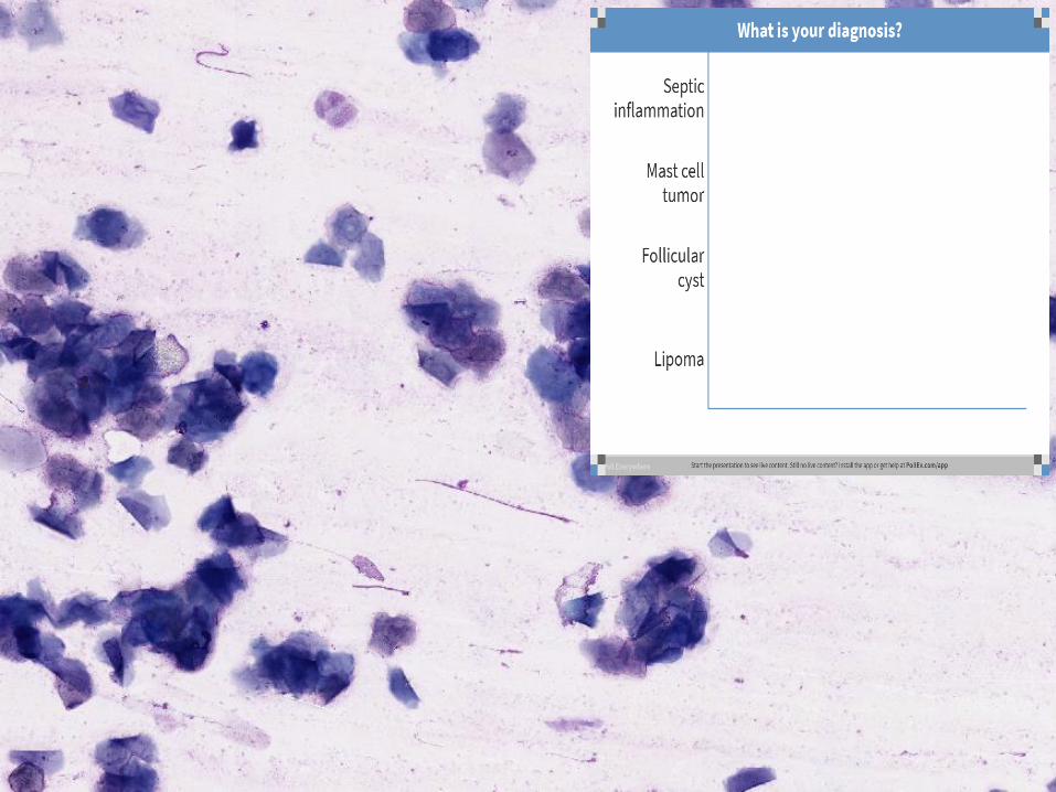
In-house Cytology: The Short List
Lipoma
Mast Cell Tumor
Follicular Cyst
Abscess
Septic Inflammation
Melanoma
Sebaceousadenoma


▪ Lipoma
▪ Large bubble-like cells
▪ Smear appears greasy
▪ May be poorly cellular


▪ Sebaceous adenoma
▪ Clusters of uniform cells
▪ Vacuolated cytoplasm


▪ Follicular cyst
▪ Superficial squamous
▪ Usually anucleate
▪ Keratin debris
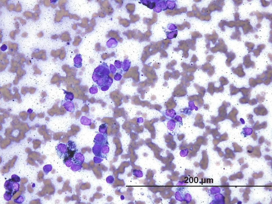

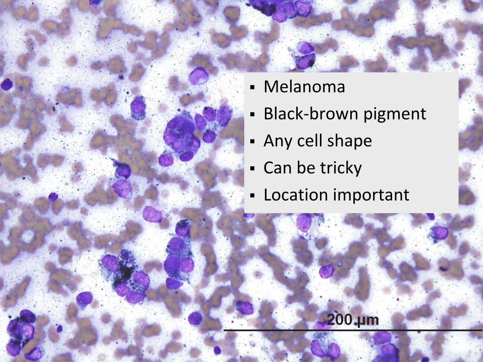
▪ Melanoma
▪ Black-brown pigment
▪ Any cell shape
▪ Can be tricky
▪ Location important


▪ Mast cell tumor
▪ Purple granules
▪ Round cells
▪ Can be tricky
▪ May see eosinophils


▪ Abscess
▪ Numerous neutrophils
▪ May be degenerate
▪ May be septic
▪ Other inflammatory cells may be present
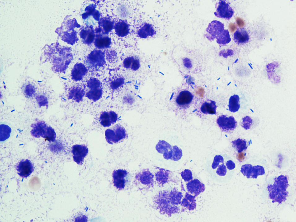


▪ Septic inflammation
▪ Numerous neutrophils
▪ Many degenerate
▪ Intracellular bacteria

Special Considerations

Sub-mandibular mass
Lymph node Salivary gland

Artifacts
US gel Stain precipitate

Questions?Stay tuned for hematology…

Part 2: HematologyRed cells
White cells
Platelets
Anna Lee Larimore

Part 2: Hematology
▪ Ask the audience:


Hematology
▪ Your analyzer can’t tell you everything
▪ RBC morphology
• Regeneration
• Causes of anemia
▪ WBC morphology
• Left shift and toxic change
• Leukemia
▪ Platelet clumping

The Monolayer
Body
Monolayer
Feathered Edge

Hematology
RBC Case 1


Hematology
RBC Case 1


Hematology
RBC Case 1
▪ Regenerative anemia
▪ Polychromasia
▪ Macrocytic
▪ (Hypochromic)

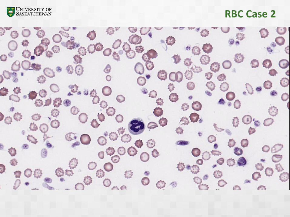
RBC Case 2

RBC Case 2

RBC Case 2
▪ Iron deficiency anemia
▪ Microcytic
▪ Hypochromic
▪ Inadequate regeneration

RBC Case 3


Oxidative Damage

Acanthocytes Echinocytes

Torocytes Hypochromasia

Agglutination Rouleaux

Questions?WBC coming up…

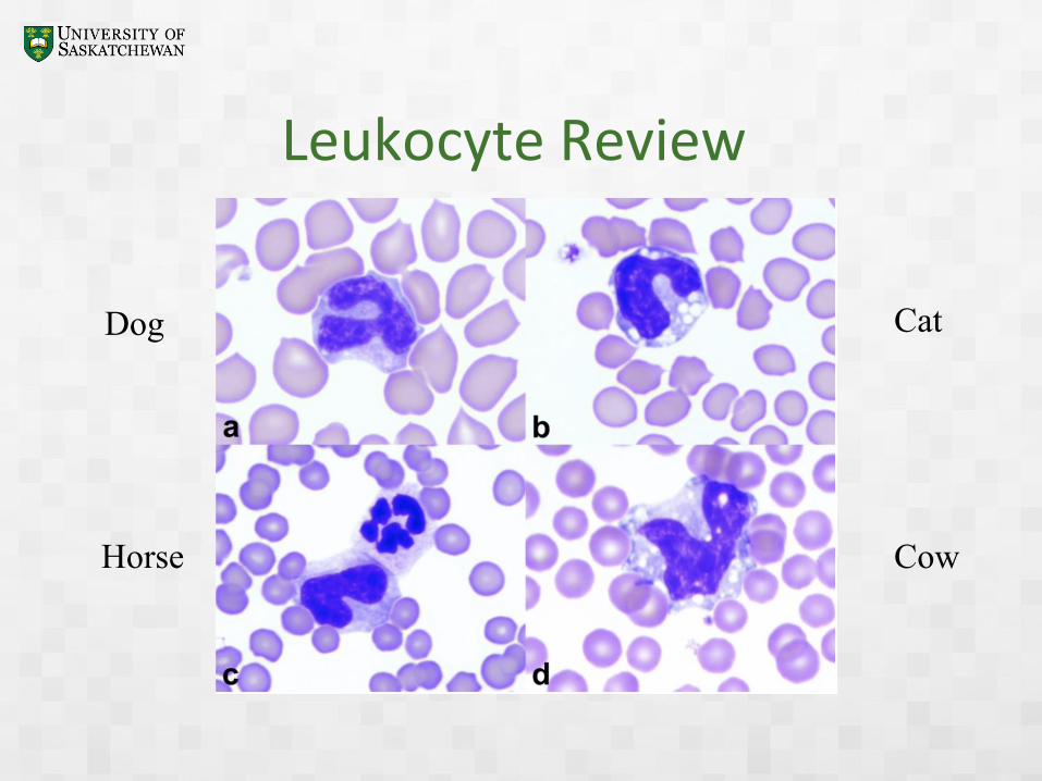
Leukocyte Review
Dog Cat
Horse Cow

Leukocyte Review
Dog Cat
Horse Cow

Leukocyte Review
Dog Cat
Horse Cow
Leukocyte Review
Dog Cat
Horse Cow

Leukocyte Review
Dog Cat
Horse Cow

WBC Case 1


WBC Estimate 10x
▪ Count the leukocytes in at least 3 fields in the monolayer
▪ Divide the total number of leukocytes counted by the number of fields to obtain the average number per field
▪ Divide the average number of leukocytes counted by 4 in order to approximate the number of leukocytes x109/L

WBC Case 1


WBC Case 1Left Shift

Widest part of nucleus Constriction <1/3 widest part of nucleus
Widest part of nucleusConstriction >1/3 widest part of nucleus
Mature seg Band

WBC Case 1Toxic Change

Source of Confusion
Toxic change
▪ In blood (blood smears)
▪ Cytoplasmic changes• Basophilia
• Foaminess
• Dohle bodies
• Toxic granulation
Degenerate neutrophils
▪ In tissues (cytology smears)
▪ Nuclear changes• Swelling
• Pale-staining
• Lysis
• Hunt for bacteria

Source of Confusion
Nucleated RBC Lymphocyte

WBC Case 2


WBC Case 2

Inclusion Confusion
Barr Bodies Bacteria

Inclusion Confusion
Mycoplasma hemofelis Stain precipitate
http://www.eclinpath.com/hematology/sample-collection
-heme/stain-precipitate/

Inclusion Confusion
Anaplasma phagocytophilum Overlapping platelet
Jacob EA. American Journal of Laboratory Medicine
Volume 1, Issue 3, November 2016, Pages: 34-57

Platelets
▪ Feathered edge
▪ 10x

Platelet Estimate
▪ 10x Scan the feathered edge for platelet clumps
▪ 100x Count the number of platelets in at least 5 fields of the monolayer and calculate the average for 1 field. Multiply the average for 1 field by 20 x 109/L.
▪ If platelets are clumped the formula will underestimate the platelet count.
▪ Less than 3 - 4 per 100x field (60 - 80 x 109/L) represents a significant thrombocytopenia

Digital Resources
▪ Veterinary Clinical Pathology ebook
• iBooks
• Google drive link
▪ Cornell e-ClinPath
• http://www.eclinpath.com/


Digital Resources
▪ PDS Protocol Manual
• Search “pds protocol manual” for most recent version
▪ Videos
• FNA & smear making: https://youtu.be/DkXDBom_3JI
• Fine needle nonaspiration: https://youtu.be/jgTnqCWewI4
• Impression smear making: https://youtu.be/prggfKrNlbI
• Cytology fluid handling: https://youtu.be/Dl_Adsa7mpc
• Cytology microscopy: https://youtu.be/vWSD7bjskjs
• Urinalysis: https://www.youtube.com/playlist?list=PLm-jtvx5oGMTmnz0GJvPDzK0O1_AIFz5z

Digital Resources
▪ Hematology Videos
• Manual WBC count: https://youtu.be/WwHI1d5pqSc
• PCV and total protein: https://youtu.be/cau5wWe4Uds
• Making/staining blood smears: https://youtu.be/nbRUiWl2Qrs
• Blood smear evaluation: https://youtu.be/wIZtvTGJL6M


Patty Voje