light scattering by pigment epithelium granules in the human retina
TRANSCRIPT
ACTA OPHTHALMOLOGICA VOL. 4 6 1968
The Department o f Ophthalmology, Rigshospitalet, University o f Copenhagen, Copenhagen, Denmark.
Head: Professor Holger Ehlers, M. D .
LIGHT SCATTERING BY PIGMENT EPITHELIUM GRANULES I N THE HUMAN RETINA
BY
Niels Bulow
Any aspect of the interaction between light and retinal structures has to be evaluated in order to attain a satisfactory interpretation of visual function.
According to the present concept of the physiology of vision, the consensus is, that absorption of light is the only physical effect of the retinal pigment epithelium (see e. g. Davson 1962 or Graham 1965). The pigment epithelium is regarded as a dark, light absorbing tissue, as seen in macroscopic preparations.
I t is the purpose of this paper to point out, that this conception is not ex- haustive, when dealing with the micro-environment of the single photoreceptors, and to emphasize the light scattering properties of the pigment epithelium granules.
METHODS
The present observations were originally made accidentally while studying human retinal pigment epithelial cells in tissue culture. Bulow 1968 a. The observations were favorized by this technique, because it gives the opportunity to examine a monolayer of living cells and a monolayer of pigment granules.
MATERIAL
1 . Human foetus, CR-length 47 mm, F-length 6 mm, weight 7,05 g. Normal eye ex- planted 1 h 30 m after legal abort.
Received January 2th 1968. Dr. Tech. B. Buchmann and Magister F. E. Carlsen, H. C. Orsted Institute, Physical
Laboratorium I1 (Head Professor J. Koch, Dr. Phil.), University of Copenhagen are acknowledged for kindly help.
1048
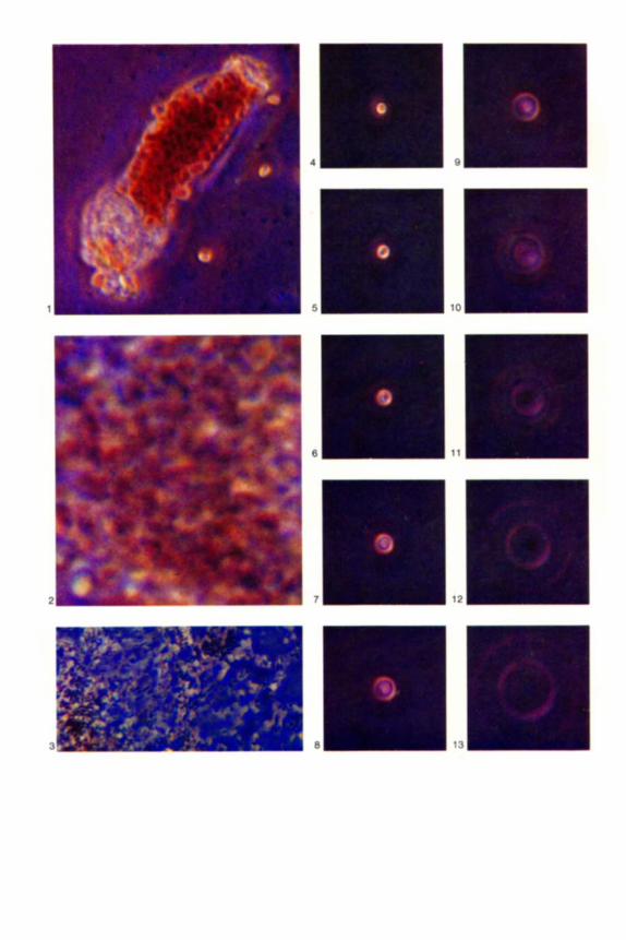
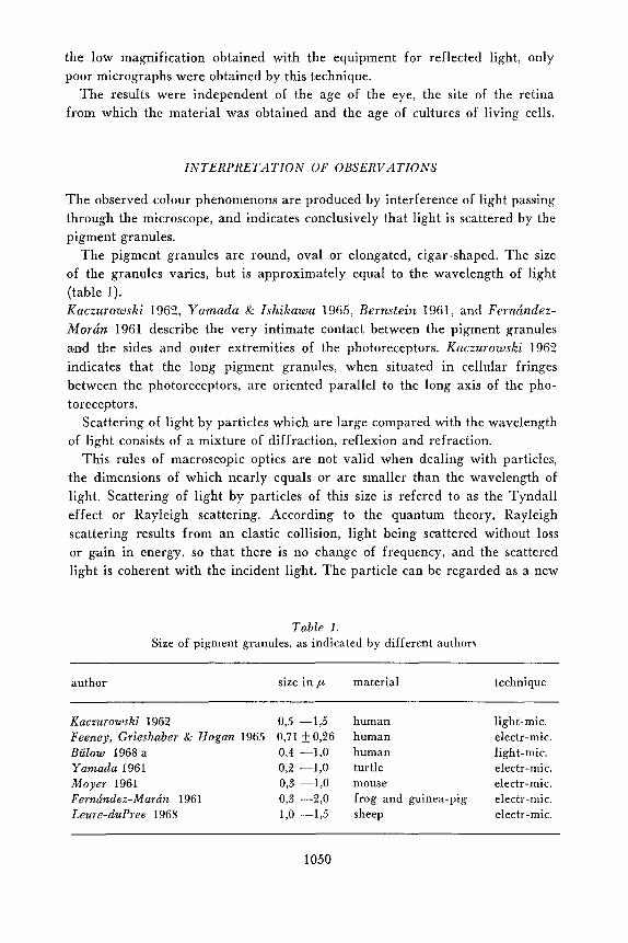
Fig. 1. Pigment epithelial cell in tissue culture, from eye no. 2. Note blue and greenish
granules at the margins of the cell.
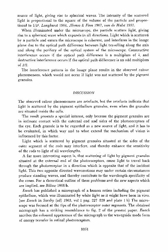
Fig. 2. Part of pigment epithelial cell in tissue culture from eye no. 4. Note the small dots
of different colours.
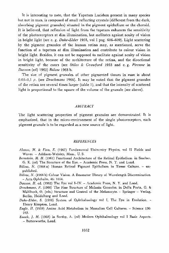
Fig. 3. Pigment granules from eye no. 5 suspended in LBromonaphthalene nu 1,658. Note the
blue-green colour of granules in focus, and compare fig. 4.
Fig. 4. One pigment granule in focus, tissue culture. Pigment granules suspended in Ethanol nD 1,361 showed similar appearence. Note colour of the granule and compare fig. 3.
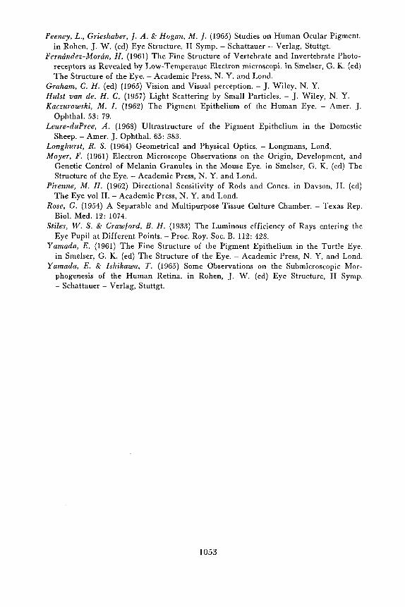
Fig. 5-13. The pigment granule in fig. 4, turned progresevely out of focus. Note interference
pattern described in the text. All micrographs obtained with white light, unstained preparations, phasecontrzst micro- scope X 800 with oil immersion. Figs 1 and 2 photographically enlarged two-fold and
four-fold respectively. Anscochrome GAF T/100 Colour Film.
9 m 10 W
6 W 7
11 m 12 W
2. Human foetus, CR-length 89 mm, F-length 14 mm, Weight 43,25 g. Normal eyc
3. 54 year old man. Normal eye removed 1 h postmortem and used for corneal graft-
4. 59 year old man. Eye removed because of malignant melanoma of the choroid.
5 . 71 year old man. Normal eye removed 2 h postmortem and used for corneal graft-
6. 88 year old woman. Normal eye removed 3 h postmortem and used for corneal
explanted 1 h 30 m after legal abort.
ing. Pigment epithelium explanted 4 h postmortem.
Normal pi,pent epithelium explanted 30 m after operation.
ing. Pigment epithelium examined 24 h postmortem.
grafting. Pigment epithelium examined 48 h postmortem.
TECHNIQUE
A. Eyes 1 to 4 were used f o r tissue culture. Small pieces of retinal pigment epithelium from central and periferal parts of the retina were explanted into modified Rose perfusion chambers (Rose 1954) using modified Eagle’s medium (Eagle 1959) and incubation at 37O C. (Biilow 1968 a).
20 primary cultures were examined immediately after explantation and at various intervals through 30 days, at 250 to 1000 X magnification under phase-contrast and ordinary light microscopes, using white light. B. Eyes 5 and 6. Pigment granules were scraped off from the inner surface of the retinal pigment epithelium, and suspended in a series of mixtures of ethanol nD 1,361 and 1-Bromonaphthalene nD 1,65S.
20 suspensions were examined under microscope, using white light as mentioned above.
OBSERVATIONS
Heavily pigmented cells (fig. 1 ) appeared to be of dark red-brown colour. Some of the pigment granules inside the cell appeared to be green, while other granules appeared to be red, and turning just a little out of focus, the colour of the single granules changed from red to yellow, green or blue.
Scarcely pigmented cells (fig. 2) showed the appearence of innumerable small dots of all colours, and turning a little out of focus, the colour of each dot changed.
Monolayer smears of pigment granules showed just the same appearace. Single pigment granules in focus changed colour, when the suspending me-
dium or the optical system of the microscope were altered (in fig. 4 the granule in focus is yellow, in fig. 3 the granules in focus are blue-green).
When a single pigment granule was turned progresively out of focus (figs. 4 to 13), the colour of the granule changed, and a system of concentric light and dark rings appeared with regular periodicity around the granule. Each light ring was composed of a continuous sequence of rings of all spectral co- lours.
Similar results were obtained with reflected light in the microscope. Due to
1049
the low magnification obtained with the equipment for reflected light, only poor micrographs were obtained by this technique.
The results were independent of the age of the eye, the site of the retina from which the material was obtained and the age of cultures of living cells.
INTERPRETATION OF OBSERVATIONS
The observed colour phenomenons are produced by interference of light passing through the microscope, and indicates conclusively that light is scattered by the pigment granules.
The pigment granules are round, oval or elongated, cigar-shaped. The size of the granules varies, but is approximately equal to the wavelength of light (table 1). Kaczurowski 1962, Yamada & IAhikawa 1965, Bcrnstein 1961, and Ferncindez- Morcin 1961 describe the very intimate contact between the pigment granules and the sides and outer extremities of the photoreceptors. Kaczurowski 1962 indicates that the long pigment granules, when situated in cellular fringes between the photoreceptors, are oriented parallel to the long axis of the pho- toreceptors.
Scattering of light by particles which are large compared with the wavelength of light consists of a mixture of diffraction, reflexion and refraction.
This rules of macroscopic optics are not valid when dealing with particles, the dimensions of which nearly equals or are smaller than the wavelength of light. Scattering of light by particles of this size is refered to as the Tyndall effect or Rayleigh scattering. Accovding to the quantum theory, Rayleigh scattering results from an elastic collision, light being scattered without loss or gain in energy, so that there is no change of frequency, and the scattered light is coherent with the incident light. The particle can be regarded as a new
Table 1. Size o f pigment granules, as indicated by different author\
author size in ,LA material technique
Kaczurowski 1962 0,5 -1,5 human light-mic. Feeney, Grieslzaber & Hogan 1965 0,71 5 0,26 human electr-mic. Bulow 196Xa 0,4 -1,O human light-mic. Yamada 1961 O,2 -1,O turtle electr-mic. Moyer 1961 0,3 -1,O mouse electr-mic. Ferncindez-Marcin 1961 0,3 -2,O frog and guinea-pig electr-mic. Leure-duPree 1968 1,0 -1,5 sheep electr-mic.
1050
source of light, giving rise to spherical waves. The intensity of the scattered light is proportional to the square of the volume of the particle and propor- tional to 1 /14 . Longhurst 1964, Alonso & Finn 1967, van de Hulst 1957.
When illuminated under the microscope, the particle scatters light, giving rise to a spherical wave which expands in all directions. Light which is scattered by a particle and enters the microscope is coherent, and interferes in the image plane due to the optical path difference between light travelling along the axis and along the perifery of the optical system of the microscope. Constructive interference occurs if the optical path difference is a multiplum of 1, and destructive interference occurs if the optical path difference is an odd multiplum of 112.
The interference pattern in the image plane results in the observed colour phenomenons, which would not occur if light was not scattered by the pigment granules.
DISCUSSION
The observed colour phenomenons are artefacts, but the artefacts indicate that light is scattered by the pigment epithelium granules, even when the granules are situated inside the cells.
The result presents a special interest, only because the pigment granules are in intimate contact with the external end and sides of the photoreceptors of the eye. Each granule is to be regarded as a new source of light, and it has to be evaluated, in which way and to what extend the mechanism of vision is influenced by this factor.
Light which is scattered by pigment granules situated at the sides of the outer segment of the rods may interfere, and thereby enhance the sensitivity of the rods to light of all wavelengths.
A far more interesting aspect is, that scattering of light by pigment granules situated at the external end of the photoreceptors, cause light to travel back through the photoreceptor in a direction which is opposite that of the incident light. This two opposite directed wavemotions may under certain circumstances produce standing waves, and thereby contribute to the wavelength specificity of the cones. For a theoretical outline of these problems and the new aspects which are implied, see Biilow 1968 b.
Enoch has published a micrograph of a human retina including the pigment epithelium, which was illuminated by white light as it might have been in vivo. (see Enoch in Sorsby (ed) 1963, vol I pag. 227-228 and plate 1 b) The micro- scope was focused at the tips of the photoreceptor outer segments. The obtained micrograph has a striking resemblance to fig. 2 of the present paper. Enoch ascribes the coloured appearence of the micrograph to the waveguide mode form of energy transfer in retinal photoreceptors.
1051
I t is interesting to note, that the Tapetum Lucidum present in many species but not in man, is composed of small reflecting crystals (different from the dark, absorbing pigment granules) situated in the pigment epithelium or the choroid. I t is believed, that reflexion of light from the tapetum enhances the sensitivity of the photoreceptors a t dim illumination, but melitates against acuity of vision in bright light (see e. g. Duke-Elder 1958, vol I pag. 606-609). Light scattering by the pigment granules of the human retina may, as mentioned, serve the function of a tapetum at dim illumination and contribute to colour vision in bright light. Besides, it can not be supposed to melitate against acuity of vision in bright light, because of the architecture of the retina, and the directional sensitivity of the cones (see Stiles & Grawford 1933 and e. g. Pirenne in Davson (ed) 1962) Riilow 1968 b.
The size of pigment granules of other pigmented tissues in man is about 0.05-0,l ,u. (see Drochmans 1966). I t may be noted that the pigment granules of the retina are several times larger (table I) , and that the intensity of scattered light is proportional to the square of the volume of the granule (see above).
A B S T R A C T
The light scattering properties of pigment granules are demonstrated. I t is emphasized, that in the micro-environment of the single photoreceptors, each pigment granule is to be regarded as a new source of light.
R E F E R E N C E S
Alonso, M . & Finn, E . (1967) Fundamental University Physics, vol I1 Fields and
Bernstein, M . H . (1961) Functional Architecture of the Retinal Epithelium. in Smelser,
Bulow, N . (1968 a) Human Retinal Pigment Epithelium in Tissue Culture. - un-
Biilow, N . (1968 b) Co!our Vision. A Resonator Theory of Wavelength Discremination.
Davson, H . ed. (1962) The Eye vol I-IV. - Academic Press, N. Y. and Lond. Drochmans, P. (1966) The Fine Structure of Melanin Granules. in Della Porta, G. &
Muhlbock, 0. (eds.) Structure and Control of the Melanocyte. - Springer - Verlag, Berlin, Heidelberg and Lond.
Duke-Elder, S. (1958) System of Ophthalmology vol I, The Eye in Evolution. - Henry Kimpton, Lond.
Eagle, H . (1959) Amino Acid Metabolism in Mamalian Cell Cultures. - Science 130: 242.
Enoch, 1. M. (1963) in Sorsby, A. (ed) Modern Ophthalmology vol I Basic Aspects. - Butterworths, Lond.
Waves. - Addison-Welsley, Mass., U. S.
G. K. (ed) The Structure of the Eye. - Academic Press, N. Y. and Lond.
published.
- Acta Ophthalm. 46: 1054.
1052
Feeney, L., Grieshaber, /. A. 6r Hogan, M . /. (1965) Studies on Human Ocular Pigment. in Rohen, J. W. (ed) Eye Structure, I1 Symp. - Schattauer - Verlag, Stuttgt.
Ferrutndez-Morcin, H . (1961) The Fine Structure of Vertebrate and Invertebrate Photo- receptors as Revealed by Low-Temperatue Electron microscopi. in Smelser, G. K. (ed) The Structure of the Eye. - Academic Press, N. Y. and Lond.
Graham, C . H. (ed) (1965) Vision and Visual perception, - J. Wiley, N. Y. Hulst van de, H . C . (1957) Light Scattering by Small Particles. - J. Wiley, N. Y. Kaczurowski, M . I . (1962) The Pigment Epithelium of the Human Eye. - Amer. J.
Leure-duPree, A . (1968) Ultrastructure of the Pigment Epithelium in the Domestic
Longhurst, R. S. (1964) Geometrical and Physical Optics. - Longmans, Lond. Moyer, F. (1961) Electron Microscope Observations on the Origin, Development, and
Genetic Control of Melanin Granules in the Mouse Eye. in Smelser, G. K. (ed) The Structure of the Eye. - Academic Press, N. Y. and Lond.
Pirenne, M. H . (1962) Directional Sensitivity of Rods and Cones. in Davson, H. (ed) The Eye vol 11. - Academic Press, N. Y. and Lond.
Rose, G . (1954) A Separable and Multipurpose Tissue Culture Chamber. - Texas Rep. Biol. Med. 12: 1074.
Stiles, W . S. & Crawford, B. H . (1933) The Luminous efficiency of Rays entering the Eye Pupil at Different Points. - Proc. Roy. SOC. B. 112: 428.
Yamada, E . (1961) The Fine Structure of the Pigment Epithelium in the Turtle Eye. in Smelser, G. K. (ed) The Structure of the Eye. - Academic Press, N. Y. and Lond.
Yamada, E . 6r Zshikawa, T. (1965) Some Observations on the Submicroscopic Mor- phogenesis of the Human Retina. in Rohen, J. W. (ed) Eye Structure, I1 Symp. - Schattauer - Verlag, Stuttgt.
Ophthal. 53: 79.
Sheep. - Amer. J. Ophthal. 65: 383.
1053