leukocyte/ immune system disorders. variations in immune function fetal and neonatal immune...
TRANSCRIPT
LEUKOCYTE/ IMMUNE SYSTEM DISORDERS

VARIATIONS IN IMMUNE FUNCTION
Fetal and Neonatal Immune Function Antibody, phagocytic and complement
functions deficient at birth. Maternal antibodies are protective 5- 6 months
Immune Function in Elderly Diminished T cell and antibody response Increased auto antibodies

Factors Affecting System
Stress Nutrition Drugs Other diseases
• renal failure
• malignancy
• diabetes mellitus
• systemic infections
HYPERSENSITIVITY DISORDERS
Type I Immediate—IgE mediated Type II Sub-Acute—IgG or IgM Type III Sub-Acute—IgG or IgM Type IV—Delayed –T Cell mediated

Type I Hypersensitivity
Allergies, anaphylaxis, anaphylactic shock anaphylactoid reactions
Occurs in seconds to minutes IgE normally binds to surface of basophils
and mast cells Concentrates in lining of respiratory and GI
tracts, skin

Fig 8-1

Fig 8-1 continued

Type I Hypersensitivity
Offending antigen enters body—ingestion, injection, inhalation
• Peanuts, walnuts, latex
• Any insect venom (bee stings)
When appropriate antigen binds to 2 IgE molecules, they cross link
• Histamine, leukotrienes, interleukins, kinins released
Type I Hypersensitivity
Get contraction smooth muscle, increased permeability of vessel walls
Fluid leaks into interstitial space, smooth muscle in respiratory and GI tracts spasms
Angioedema, esp of face, hands, feet Vessels not damaged
If this is local, irritating but not likely to be fatal

Signs of Anaphylaxis begin within seconds to minutes
Body wide reaction to ingested, inhaled, injected ag Acute systemic reaction due to IgE release
Urticaria Itching, laryngeal edema, angioedema Bronchospasm—may die from anoxia Hypotension—may die from hypotensive shock GI and UG cramping (least common)
Administer epinephrine, establish airway, give antihistamines and glucocorticoids

Atopy-chronic, inherited disorder
IgE at mucosal surfaces reacts with normally innocuous compounds
• Pollen, animal dander, certain types of foods
Reaction is localized to surface stimulated Multiple manifestations
Associated with increased incidence of allergic rhinitis and asthma
Atopic eczema (atopic dermatitis)
Associated with allergic rhinitis or asthma T helper 2 cells overproduce IL 31
(believed to be underlying problem) Weeping, eroded, red, scaly patches of skin
• scalp, face, and diaper area most often affected
• exacerbated by weather changes, drying from XS soap and water
• severe itching2º bacterial infection
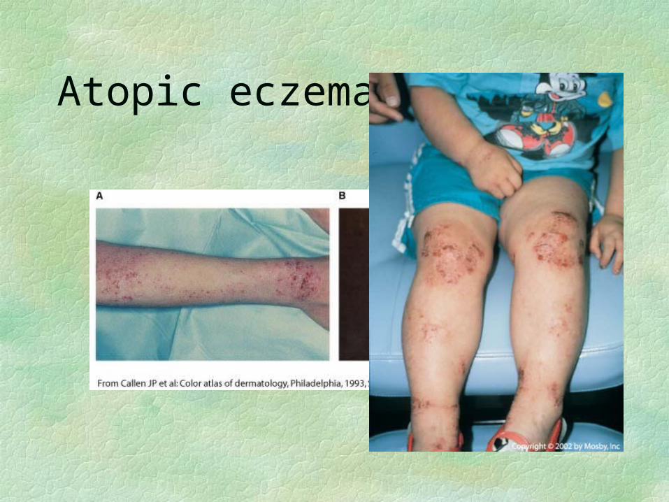
Atopic eczema

Atopic eczema (atopic dermatitis)
May spontaneously disappear as child ages Other allergies commonly appear Areas of knee, elbow, neck, etc. involved if continues Points of flexion/extension
May spread over entire body in adults—remission rare
Treat with dietary restrictions (infants), moisturizers, antihistamines
Antibiotics for skin infections Topical steroids/T cell modulators as needed
Allergic rhinitis—nasal allergy
Seen more in developed nations with high standards of cleanliness
less exposure to pathogens and more exposure to chronic irritants
Less development of the suppressor (regulatory)) T cells
May predispose person to developing asthma
Allergic Rhinitis--S&S
Stuffy, runny nose; red, weepy eyes; itching of eyes and nose
Dark circles under eyes, chronic sinusitis may develop
May report ringing in ears or sensation of pressure in ear
Eosinophils in nasal secretion distinguishes from infection
Subsets of allergies
Seasonal form (hay fever)—pollen, mold Perennial allergies—more likely dust mites,
animal dander
Cytotoxic (Type II) and Immune Complex (Type III) Diseases
Type II involves antibodies binding cells—tissue specific reaction—rapid
Type III involves antibodies binding soluble proteins—serum sickness
• Develops 7-10 days after antigen is introduced• Urticaria, angioedema, fever, generalized
lymphadenopathy• Activation of complement as complexes precipitate in
capillaries• Joint pain, renal dysfunction, GI problems

Fig 8.4
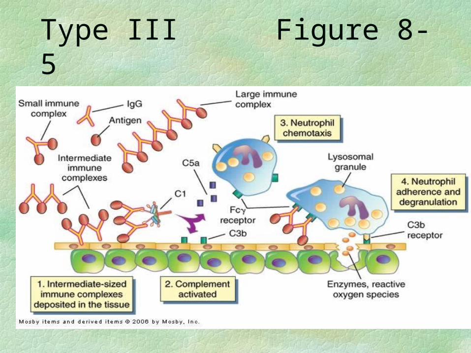
Type III Figure 8-5

Type IV Hypersensitivity—48-72 hours
Allergic Contact Eczema—delayed hypersensitivity T lymphocytes Poison ivy, nickel allergies
Weeping, red, puritic vesicles at site of contact
Localized edema initially, thickening of skin if contact continues avoid offender, steroids as needed
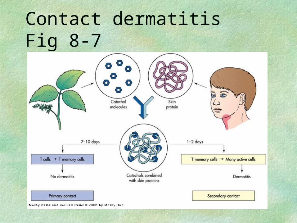
Contact dermatitis Fig 8-7
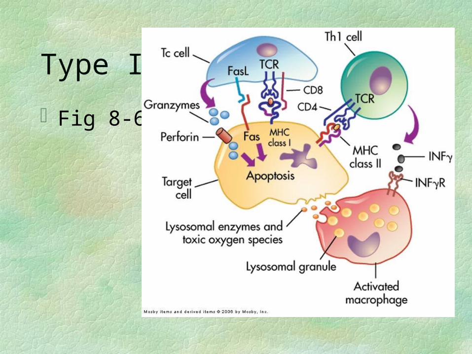
Type IV
Fig 8-6

AUTOIMMUNE DISORDERS
#3 cause of death in the US
AI Disorders -- general
Immune system must tolerate normal antigens by removing all T and B cells that would react with self proteins
AI diseases occur when the immune system starts to attack the cell of the body—break in tolerance
• Occur more frequently in females than males• Precise cause unknown—believe that foreign antigen
resembles a self antigen• Several are associated with HLA antigens--see T 8-5• Presence of one increases risk of others
Relapse and remission common• Seem to get worse when stressed

Immune system removes foreign compound first• Continued surveillance sees normal cell proteins presented by
MHC I molecules as foreign, and attacks those cells
• System eventually turns on to normal proteins
Some suggestion that may start if inflammation accompanies apoptosis reaction
AI diseases be cytotoxic T cell mediated or antigen-antibody reaction or both
Organ specific AI diseases
Damage is limited to one organ/tissue
Rest of the body suffers as a result of damage done to that organ/tissue
Examples
Idiopathic thrombocytopenic purpura Autoimmune hemolytic anemia Type I Diabetes Mellitus Autoimmune thyroiditis Addison’s Disease Celiac Disease

Multiple organ system AI diseases
Most of these involve antibodies that react with the body’s connective
tissue

Examples
Rheumatoid arthritis Systemic Lupus Erythematosis (SLE) Scleroderma

Rheumatoid arthritis
In women 2.5X as often as in men, peak incidence 40-60 years; 1% all adults
Seems to have some association with HLA-DR4 (Minor HC) antigens (varies with ethnic group)
Production of multiple destructive enzymes: collagenase, protease
Attacks synovial membranes Produces granulation tissue that extends into joints-
pannus Destruction of cartilage, ligaments, tendons, bones


Signs & Symptoms
Fever, fatigue, malaise Swollen, painful joints; often bilateral
Hands, knees, feet
Morning stiffness that lasts > 1 hour Deformed joints as cartilage is destroyed

Juvenile rheumatoid arthritis
Systemic onset—boys and girls equally—20% poorest prognosis Multiple organs, any age
Polyarticular onset—girls 2X as often as boys—40% 5 or more joints, and age Little involvement outside joints
Pauciarticular onset—girls 6X as often as boys—40% best prognosis Max of 4 joints, typically < 6 yrs old

Signs and symptoms of JRA
Fatigue, anorexia, weight loss, fever Shifting, symmetrical polyarthritis; any
diarthrotic joint is possible Morning stiffness of joints > 1 hour Erosive arthritis seen on X rays, followed
by deformity
Both age groups
Rheumatoid nodules, usually indicates active or severe disease
Damage to other organs—heart, lungs, eyes, blood vessels
Normocytic, normochromic anemia if bone marrow is involved
Turbid, non-viscous synovial fluid with high WBC count
Increased T cell activity causes increased TNF, which causes increased osteoclast activity

Rheumatoid factor in 85 % of pts
IgM that reacts with altered human IgG Anti-gamma globulin factor Complexes ppt onto joint tissue, stim immune
reaction May show up in other AI connective tissue
diseases Seen in 5-20% of normal people
Increased incidence as get older

Systemic lupus erythematosis
Signs and symptoms are diverse, easily missed in early stages
Butterfly rash across face is classic sign (40% of cases) AB form vs nucleic acids (anti-nuclear
antibodies) RBC, WBC, platelets, coagulation proteins, etc, Mild to rapidly fatal
In US: incidence in Fe 10x M Incidence in Black Fe 8x White Fe
Signs and symptoms of SLE
Symmetric arthritis or joint pain (90%)—not erosive or deforming
Fever, fatigue, weakness (50% have mild anemia), weight loss, possibly severe hair loss
Vasculitis (80%) with butterfly rash that gets worse if exposed to sunlight
Pleurisy chest pain; myocarditis, pericarditis Anemia or other marrow deficiencies

Renal complications
Antinuclear antibodies attach to DNA, deposit in glomerulus
Complement attaches to immune complex, renal inflammation begins
65% of SLE pts develop glomerulonephritis 40% have clinical renal disease 25% have severe renal damage (renal failure)
CNS complications usually fatal Diagnose with + antinuclear antibody test
Scleroderma
Now called Progressive Systemic Sclerosis Usually starts in 20‘s-40’s, least common of
diseases mentioned here Disease is overproduction of collagen, may be
localized or systemic Slow, progressive fibrosis of skin and other organs Proliferation of collagen in walls of blood vessels—lose
elasticity
Localized scleroderma—restricted to skin
May occur as result of repeated exposure to some chemicals
Milder, better prognosis

Generalized scleroderma
Limited cutaneous systemic sclerosis (Raynaud’s phenomenon)
• Vasospasm when extremities are exposed to cold
• Digits become white, then blue, then red in each attack
• Thickening and tightening of skinsausage appearance
Diffuse cutaneous—CT of skin and viscera, rapid progression

Progression of Scleroderma
Gradually spreads up limbs, involves trunk, face Very taut face, wrinkled mouth, smooth forehead Arthritis, joint pain and stiffness common Esophageal dysfunction, GI dysfunction Decreased pulmonary volumes Renal vessels damaged—proteinuria, hematuria,
hypertension Renal failure is leading cause of death for
generalized scleroderma