leukocyte adhesive function in intensive care patients
TRANSCRIPT
Leukocyte adhesive function in Intensive Carepatients with non-infectious and infectioussystemic in�ammationAnqi Li
StickyCell Pty LTdAngela Jacques
StickyCell Pyt LtdBárbara de la Peña Avalos
StickyCell Pyt LtdYih Rue Ong
StickyCell Pty LtdKirstin Elgass
StickyCell Pty LtdEmma Saylor
Princess Alexandra HospitalJason Meyer
Princess Alexandra HospitalJames Walsham
Princess Alexandra HospitalKerina Denny
Royal Brisbane and Women's HospitalJeffery Lipman
The University of QueenslandQiang Cheng ( [email protected] )
StickyCell Pty Ltd, Brisbane https://orcid.org/0000-0001-8730-1620Peter Kruger
Princess Alexandra Hospital
Research
Keywords: Dysregulated host response, sepsis, systemic in�ammation, leukocyte recruitment, leukocyteadhesive function assay (LAFA), sepsis diagnosis
Posted Date: November 1st, 2021

DOI: https://doi.org/10.21203/rs.3.rs-1007613/v1
License: This work is licensed under a Creative Commons Attribution 4.0 International License. Read Full License

1
Leukocyte adhesive function in Intensive Care patients with non-infectious and
infectious systemic inflammation
Anqi Li1,*, Angela Jacques1,*, Bárbara de la Peña Avalos1, Yih Rue Ong1, Kirstin Elgass1,
Emma Saylor2, Jason Meyer2, James Walsham2,4, Kerina Denny3, Jeffery Lipman3,4,5, Qiang
Cheng1 and Peter Kruger2,4
1 StickyCell Pty Ltd, Brisbane, Queensland, Australia
2 Intensive Care Unit, Princess Alexandra Hospital, Woolloongabba, Queensland, Australia
3 Department of Intensive Care Medicine, Royal Brisbane and Women's Hospital, Brisbane,
Queensland, Australia
4 The School of Medicine, University of Queensland, Brisbane, Australia
5 Scientific Consultant, Nimes University Hospital, University of Montpellier, Nimes, France
* These authors contributed equally to this work.
Authors for correspondence:
Peter Kruger: [email protected]
Qiang Cheng: [email protected]
Author contribution: AL, JW, KD, JL, QC and PK designed the study and interpreted the
data. ES, JM, JW and PK recruited patients and collected blood samples. AL, AJ, BP and QC
performed the LAFA blood assays. AL, YRO and KE analysed the assay results. AL, QC,
PK, JL, KD and JW drafted the initial manuscript with all authors editing and approving the
final version.

2
Abstract
The lack of rapid and accurate diagnostic tools to distinguish infectious (known as sepsis) and
non-infectious causes of inflammation in patients with systemic inflammation remains a
significantly unmet clinic need, particularly in the intensive care unit (ICU). As a hallmark of
inflammation, circulating leukocytes must be activated and undergo a cascade of interactions
with blood vessel endothelium before transmigrating into surrounding tissues, a process
called leukocyte recruitment. Given the divergent disease aetiologies, it was hypothesised
that the ability of circulating leukocytes to interact with endothelial cells and cause
inflammation may differ when responding to infectious and non-infectious inflammatory
stimuli, providing potential markers to differentiate these two diseases. In the present study, a
flow-based blood testing platform, named leukocyte adhesive function assay (LAFA), was
used to mimic blood microcirculation in vitro so that the patient leukocyte ability to interact
with multiple endothelial molecules, including P+E selectins, VCAM-1 and IL-8, can be
studied. The leukocyte adhesive functions of multiple leukocyte subsets were quantitatively
assessed using a range of cell kinetic parameters, including cell speed, straightness, dwell
time etc. When analysed on P+E selectin substrate, a significantly lower value of cell
straightness was observed in septic CD4 cells than non-infectious cells (0.78±0.04 vs
0.92±0.01, p < 0.01), suggesting a difference in CD4 cells ability to adhere to selectins in
infectious and non-infectious patients. Additionally, an impaired ability to respond to IL-8
was observed in septic neutrophils compared to non-infectious cells, evidenced by a
significantly reduced cell dwell time (91.7±14.0 vs 150.6±19.0 seconds, p < 0.05). Thus,
our study promisingly showed the ability of LAFA to detect different adhesive function
between leukocytes from ICU patients with infectious and non-infectious systemic
inflammation. LAFA generated a number of novel markers that might distinguish infectious
inflammation from non-infectious causes that warrant further study and novel opportunities
for the rapid and accurate diagnosis of sepsis.

3
Introduction
Dysregulated host response, previously known as Systemic Inflammatory Response
Syndrome (SIRS), is a complex inflammatory systemic response to non-infectious or
infectious foreign insults 1,2. Non-infectious causes include major surgery, trauma, burns and
severe tissue injury, whereas infectious causes (also known as sepsis) include bacterial,
fungal or viral infection 3-5. All causes can result in similar clinical manifestations with the
descriptors recently being redefined with the SEPSIS-3 criteria 1. Due to different aetiology,
the treatments for non-infectious and infectious causes of systemic inflammation differ
dramatically.
Antibiotic therapy and source control are the first line of treatment for sepsis, whilst
antibiotic therapy is not indicated for non-infectious causes. For many patients it is extremely
difficult to rapidly and accurately determine disease pathogenesis and the current practice is
that patients will be given antibiotics when potential infection is suspected, frequently leading
to overuse of antibiotics for patients with non-infectious causes. Antibiotics are continued
until the treating clinician is satisfied that the cause of the systemic inflammation is not
infection. This overuse of antibiotics in patients with non-infectious causes could result in the
emergence of resistant pathogens, as well as other avoidable side effects. It is critical to
develop new technology to aid the early differentiation between non-infectious and infectious
inflammation. This will allow more targeted therapies to be applied earlier, avoiding
unnecessary treatments and may improve survival rates 3.
In order to differentiate sepsis from non-infectious inflammation, most work has focussed on
the determination and characterisation of potential pathogens in patient blood and/or tissues 4.
Currently, blood cultures or other clinically relevant positive microbiology is the gold
standard for the identification of infection in ICU patients with systemic inflammation. A
significant proportion of patients with infection will have negative cultures 5. The turn-around
time for microbiological culture results is usually 12-72 hours. Earlier administration of
antibiotics has been associated with improved survival and the “time critical” nature of both
recognition and treatment of patients with sepsis is increasingly emphasised 1,6,7.
Additionally, even though a number of molecular tools have been developed to characterise
infectious pathogens, the lack of rapidity and sensitivity is a common drawback of these
techniques 8. Biomarkers of host immune response, such as procalcitonin (PCT) and C-
reactive protein (CRP), have been investigated to separate infectious and non-infectious

4
inflammation. Unfortunately, PCT tests lack the negative predictive power to withhold
antibiotics in critically ill patients 9, suggesting the clinical applications of these biomarkers
remain to be better defined 10-12. Thus, there is urgent clinical demand for new sepsis
diagnostic tests, particularly that specifically reflect the patient immune activation during
inflammation.
The recruitment of leukocytes from the circulation to the surrounding tissues is a hallmark of
immune system activation during inflammation 13,14. To be recruited, circulating leukocytes
must undergo a cascade of interactions with blood vessel endothelial cell (EC) surface and
subsequently transmigrate into the tissue. Thus, leukocyte adhesive function is defined as the
leukocyte ability to interact with and transmigrate through the ECs. Initially, the fast
travelling leukocyte will tether and roll on the ECs, which requires the interaction between
leukocytes expressing P-selectin glycoprotein ligand-1 (PSGL-1) and endothelial P or E
selectins 15. The rolling leukocytes will then be activated by chemokines and reduce their
speed. This allows the interaction between leukocyte 41 integrins with their endothelial
ligands, such as vascular cell adhesion molecule-1 (VCAM-1), leading to leukocyte firm
adhesion on endothelial surface 16,17. The adherent leukocytes then crawl along the
endothelial surface until they find an optimal spot to leave the vasculature 13,18. Thus, the
activity of leukocyte PSGL-1 and 41 integrin plays a central role in the regulation of
leukocyte adhesive function, which will then determine the leukocytes potential to
transmigrate and cause inflammation. Additionally, the leukocyte expressing chemokine
receptors may facilitate the leukocyte recruitment process via chemotaxis.
Given the divergent causes of inflammation, it was hypothesised that leukocyte adhesive
functions could be altered differently in non-infectious and infectious patients. In the present
study, a newly developed blood test platform, the leukocyte adhesive function assay (LAFA),
was used to identify such differences. By analysing blood samples from ICU patients with
non-infectious or infectious systemic inflammation using LAFA, we aimed to compare the
ability of specific leukocyte subsets to interact or respond to given endothelial adhesive
substrates, including P+E selectin or VCAM-1 in the presence and absence of IL-8, and
thereby the ability of LAFA to distinguish non-infectious and infectious patients may be
evaluated.

5
Methods and materials
Patient recruitment
This study was approved by Metro South Human Research Ethics Committee, Brisbane
Australia (Reference number: 17/QPATH/571). Patients were recruited for this study by
screening patients who were admitted to the Intensive Care Unit (ICU), at Princess Alexandra
Hospital, Brisbane Australia. Eligible patients were over 18-years of age with a newly
identified systemic inflammatory response who could provide prior consent or had a
surrogate who could provide prior consent. Patients who had pre-existing inflammatory
conditions, such as multiple sclerosis, Crohn's disease, colitis, arthritis, lupus, etc. or were
currently on anti-adhesion therapy, including Natalizumab, Vedolizumab or an anti-adhesion
clinical trial were excluded from the study. All patients enrolled had any two or more of the
following four criteria for systemic inflammation, regardless of the causes of inflammation 19:
1. Body temperature >38°C or <36°C
2. Heart rate >90 per minute
3. Respiratory rate >20 breaths per minute or PaCO2 <32mmHg.
4. White blood cell count >12,000/mm3 or <4,000/mm3 or >10% bands.
The blood samples were collected within 48 hours after the first identification of the systemic
inflammatory response. Whole blood was collected in a Heparin blood collection tubes (5ml)
for LAFA assays and an EDTA tube (4ml) for full blood exam (FBE). A full blood cell exam
was performed using a Mindray BC5000 Haematology Analyser according to manufacturer’s
instructions.
Sepsis Adjudication
Two clinicians (JW / PK) provided a retrospective and independent assessment of all recruited
patients. They were blinded to LAFA results but assessed patients’ medical records and
laboratory results to determine either definite “infection”, definite “no infection” or “possible
infection”. Concordance between the two assessors was required for a patient to be allocated
to the definite infection or no infection group. Where the assessors differed (on 3 occasions)
patients were categorised as having possible infection.
Antibodies, chemical and reagents
Human recombinant VCAM-1, P-selectin, E-selectin and IL-8 were all purchased from R&D
Systems (Minneapolis, MN). Antibodies (Abs) against human leukocyte surface molecules,

6
Anti CD4-Alexa488, CD8-PE, CD14-Alexa488, CD15-APC and CD16-BV510, CD25-APC
were all obtained from either BD Biosciences (San Diego, CA) or BioLegend (San Diego, CA).
Leukocyte adhesive function assay (LAFA)
LAFA utilises microfluidic technology to mimic blood circulation in vitro, allowing a direct
assessment of leukocyte interaction with endothelial adhesive substrates (e.g. P+E selectins,
VCAM-1 and IL-8). Specific leukocyte subsets are fluorescently labelled with different
fluorophores, allowing the detection of multiple cell types concurrently. To perform LAFA, a
Polymethyl methacrylate (PMMA)-bottom microfluidic chip (channel Width×Depth×Length:
1,000×200×18,000µm) from Microfluidic ChipShop (Jena, Germany) was employed.
Recombinant human VCAM-1 protein (10µg/ml) with or without IL-8 (1µg/ml), or a
combination of P-selectin (10µg/ml) and E-selectin (0.5µg/ml) was gently loaded into the
channels and incubated overnight at 4°C 20. To identify specific leukocyte subsets, 100 µl
whole blood was mixed with the two Ab cocktails for 5 minutes (min) at room temperature
(RT) respectively: 1) anti-CD14-Alex488 (1:50), anti-CD15-APC (1:33.3) and Anti-CD16-
BV421 (1:500); 2) anti-CD4-Alex488 (1:50 dilution), anti-CD8-PE (1:66.7) and anti-CD25-
APC (1:500). These two sets of antibody cocktails allow the detection of following leukocyte
subsets: CD14 (most likely monocytes), CD15+CD16+ (mainly neutrophils), CD4 (mainly
CD4 lymphocytes), CD8 (mainly CD8 lymphocytes) and CD4+CD25+ (most likely CD4
regulatory cells) cells. The blood was then pulled into the microfluidic channels by a syringe
pump at a shear stress of 1.5 dyne/cm2.
Fluorescence microscopy time series were recorded on an InCell Analyser 2200 (GE
HealthCare, Seattle, WA) with a 10X objective. All data acquisition was recorded at 1 frame
per two second for 10 minutes, at the centre of the channel (approximately 9mm from the
channel inlet). All experiments were performed in a 37ºC temperature controlled environment.
Cell tracking and data analysis
Cell tracking was accomplished using TrackMate from Fiji image analysis software. Cells were
tracked automatically by detecting quality-filtered fluorescent spots in each frame and then
linked with a maximum distance of 75µm and maximum gap size of 2. All tracks were
subsequently checked manually and corrected for errors. Cell kinetic parameters, including cell
density, speed, diffusion coefficient, straightness, dwell time and track length were determined
for each interacting cell. Each parameter characterises cell migratory behaviours from one
specific aspect. These cell kinetic parameters and their descriptions are list in Table 1.
7
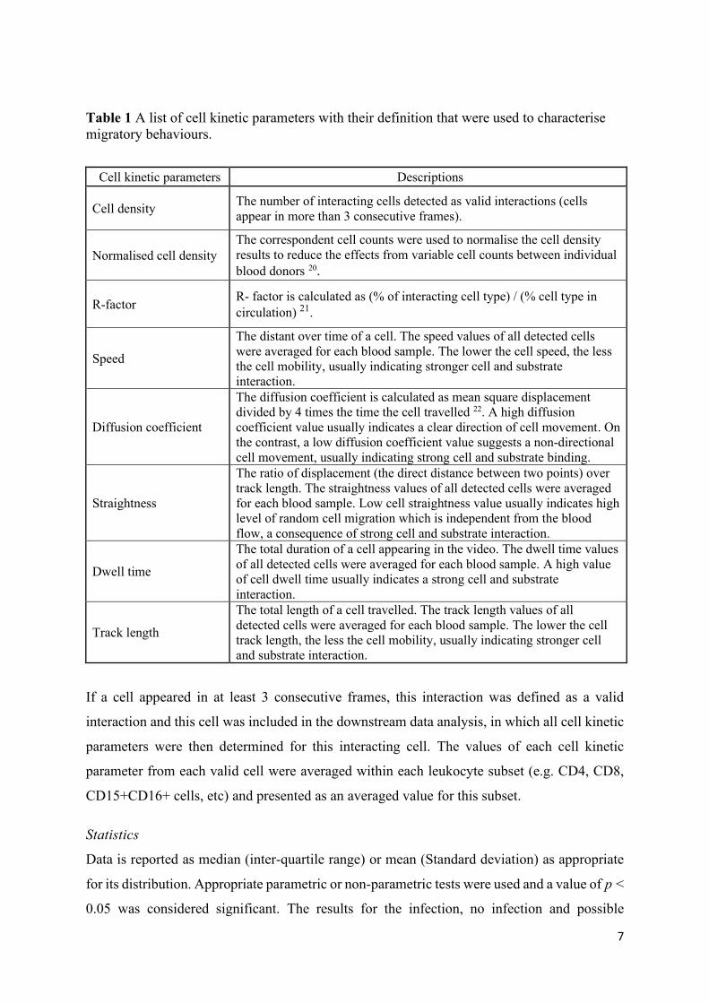
Table 1 A list of cell kinetic parameters with their definition that were used to characterise
migratory behaviours.
Cell kinetic parameters Descriptions
Cell density The number of interacting cells detected as valid interactions (cells
appear in more than 3 consecutive frames).
Normalised cell density
The correspondent cell counts were used to normalise the cell density
results to reduce the effects from variable cell counts between individual
blood donors 20.
R-factor R- factor is calculated as (% of interacting cell type) / (% cell type in
circulation) 21.
Speed
The distant over time of a cell. The speed values of all detected cells
were averaged for each blood sample. The lower the cell speed, the less
the cell mobility, usually indicating stronger cell and substrate
interaction.
Diffusion coefficient
The diffusion coefficient is calculated as mean square displacement
divided by 4 times the time the cell travelled 22. A high diffusion
coefficient value usually indicates a clear direction of cell movement. On
the contrast, a low diffusion coefficient value suggests a non-directional
cell movement, usually indicating strong cell and substrate binding.
Straightness
The ratio of displacement (the direct distance between two points) over
track length. The straightness values of all detected cells were averaged
for each blood sample. Low cell straightness value usually indicates high
level of random cell migration which is independent from the blood
flow, a consequence of strong cell and substrate interaction.
Dwell time
The total duration of a cell appearing in the video. The dwell time values
of all detected cells were averaged for each blood sample. A high value
of cell dwell time usually indicates a strong cell and substrate
interaction.
Track length
The total length of a cell travelled. The track length values of all
detected cells were averaged for each blood sample. The lower the cell
track length, the less the cell mobility, usually indicating stronger cell
and substrate interaction.
If a cell appeared in at least 3 consecutive frames, this interaction was defined as a valid
interaction and this cell was included in the downstream data analysis, in which all cell kinetic
parameters were then determined for this interacting cell. The values of each cell kinetic
parameter from each valid cell were averaged within each leukocyte subset (e.g. CD4, CD8,
CD15+CD16+ cells, etc) and presented as an averaged value for this subset.
Statistics
Data is reported as median (inter-quartile range) or mean (Standard deviation) as appropriate
for its distribution. Appropriate parametric or non-parametric tests were used and a value of p <
0.05 was considered significant. The results for the infection, no infection and possible

8
infection groups are reported, with statistical comparisons made between the infection and no
infection patients.

9
RESULTS
Demographics and stratification of ICU patients
A total of 28 patients were recruited to this study. Clinicians JW and PK reviewed the clinical
records of each patient and independently adjudicated whether the patient had infection, no
infection or were ‘indeterminate/possible infection’. This assessment was done independent of
the LAFA results and the adjudication was not released until all LAFA assays were completed.
As a result, 10 patients were deemed to have infection, 9 no infection and the remaining 9
possible infection. The baseline demographics for the ICU patients are presented in Table 2.
Table 2 Demographic description of Infection, No infection and Possible Infection (or
unknown) ICU patients.
Variables
Infection
(n=10)
No-Infection
(n=9)
Possible
Infection
(n=9)
p
Demographics
Age (years) 66 (58-72) 57 ( 29-57) 50 (45 – 77) 0.10
Gender (male) 4 (40%) 7 (78%) 5 (55%)
Admission characteristics
Medical 4 6 6
Surgical 6 3 3
Severity of Disease
APACHE 3 Score (median(IQR)) 65 ( 44- 89) 53 (41-71) 63 (54-75) 0.66
SOFA Score at inclusion
( median (IQR))
9 (6-12) 4 (3-8) 5 (4-7) 0.08
Organ Support$ and Outcome
Mechanical Ventilation (n) 7 (70%) 5 (56%) 5 (56%)
Vasopressor requirement (n) 9 3 7
Renal Replacement Therapy (n) 0 0 0
ICU Mortality (n) 1 0 1
ICU LOS (days) ( median(IQR)) 3 (2-7.2) 3 (1-7) 4 ( 2.5 – 10) 0.58
$ In the 24 hrs preceding Day 1
The clinical diagnosis and potential causes of systemic inflammation in each ICU patient
are listed in Table 3.
Table 3 The clinical diagnosis and microbiology results for individual study patients.
Study ID ICU Diagnosis Microbiology

10
No
Infection NON-01
Trauma. Fall. Surgery for unstable T8
fracture
Scant Staphylococcus
Aureus in ETA;
considered colonisation
NON-02
Myocardial infarction with out of
hospital cardiac arrest
Nil positive
microbiology
NON-03 Exacerbation of asthma
Nil positive
microbiology
NON-04
Decompressive craniectomy for
malignant middle cerebral artery stroke
Nil positive
microbiology
NON-05 Subarachnoid haemorrhage
Haemophilus and
Klebsiella grown from
ETA considered
colonisation
NON-06
Trauma. Evacuation of subdural
haematoma
Haemophilus in ETA day
3
NON-07
Reduced level of consciousness due to
cerebral metastatic disease
Klebsiella in ETA
NON-08
Pulmonary oedema following coronary
artery bypass grafts
Nil positive
microbiology
NON-09 Lobectomy for carcinoid lung tumour
Nil positive
microbiology
Infection INF-01 Below knee amputation, Gangrenous
foot
Pseudomonas and
mixed enterics from
tissue
INF-02 Urosepsis
E. Coli in blood (and
urine)
INF-03 Perforated viscus with faecal peritonitis
Perforated viscus with
faecal peritonitis
INF-04 Multitrauma. Fall. Spine, thorax,
abdominal and pelvic injuries
Serratia in blood.
Serratia and
Pseudomonas in ETA.
Enterococcus Faecalis
in urine
INF-05 Necrotising fasciitis
Group G Streptococci
and Proteus from tissue
INF-06
Septic arthritis
MRSA in blood and joint
aspirate
INF-07 Fourniers gangrene
Streptococcus
Constellatus and
Proteus from tissue
INF-08
Urosepsis
Providencia in blood
and urine
INF-09
Intraoperative aspiration. Metatarsal
head osteotomies for osteomyelitis
Candida in urine
INF-10 Ischaemic bowel. Perforated colon E. Coli in peritoneum

11
Possible
Infection UN-01
Febrile neutropenia post autologous
stem cell transplant
Nil positive
microbiology
UN-02 Exacerbation of asthma
Initial cultures negative.
Serratia from ETA day 6.
Coagulase negative
Staphylococci from
blood day 5
UN-03 Drug overdose with aspiration
pneumonitis
Coagulase negative
Staphylococci in blood;
considered
contaminant.
Enterobacter Cloacae in
ETA
UN-04 Hanging with aspiration pneumonitis
Coagulase negative
Staphylococci in blood;
considered
contaminant significant
aspiration with mixed
flora in ETA
UN-05
Small bowel resection, colectomy,
nephrectomy
Candida in urine
UN-06
Coronary artery bypass grafts following
myocardial infarction
Nil positive
microbiology
UN-07
Distal pancreatectomy and splenectomy
Nil positive
microbiology
UN-08 Fall, Traumatic brain injury Pseudomonas in ETA
UN-09
Embolisation of bleeding mycotic
hepatic artery pseudoaneurysm
Nil positive
microbiology
ETA = Endotracheal Aspirate, E. coli = Escherichia Coli, MRSA = Methicillin-resistant Staphylococcus
aureus
Migratory behaviours of leukocytes from patients with infectious and non-infectious
inflammation.
Leucocyte behaviour across the study groups is shown in Figure 1A to 1H, using a number of
cell kinetic parameters. When analysed on P+E selectin substrates, the straightness of
interacting CD4 cells in infectious patients was significantly lower than non-infectious cells
(0.78±0.04 vs 0.92±0.01, p < 0.01; Figure 1F), indicating a stronger CD4 cell binding to
selectins in patients with an infectious cause. This notion was further supported by the
observation that the dwell time of infectious CD4 cells was significantly longer than non-
infectious cells (48.3±6.5 vs 29.3±3.6 seconds, p < 0.05; Figure 1G). In addition, significantly
lower numbers of interacting CD4 (55.0±19.1 vs 155.8±45.3, p < 0.05) and CD8 (53.4±13.7
vs 112.0±28.5, p < 0.05) cells were seen in patients with an infectious compared to non-

12
infectious cause of their systemic inflammation (Figure 1A). To reduce the effects from
variable cell counts between individual blood donors (Supplementary Figure 1), the
correspondent cell counts were also used to normalise the cell density results 20. After the
normalisation, no difference in the number of CD4 and CD8 interacting cells were observed
(Figure 1B).
The blood samples were then analysed by LAFA on VCAM-1 substrate, a ligand of
leukocyte α4β1 integrin. VCAM-1 supports firm leukocyte adhesion on the blood vessel wall,
and this LAFA assay allows the assessment of leukocyte α4β1 integrin activity. As shown in
Figure 2H, the track length of interacting CD14 cells from patients with infectious
inflammation was significantly shorter than non-infectious CD14 cells (58.6±9.4µm vs
85.7±9.4µm, p < 0.05), suggesting a reduced CD14 cell mobility in patients with infection.
Additionally, a significantly lower CD15+CD16+ (neutrophils) cell density (29.0±12.4 vs
188.1±81.1, p < 0.05) was detected in patients with infection than non-infectious patients
(Figure 2A), while no such decrease was detected when the cell density was normalised by
neutrophil counts (Figure 2B). The R-factor of CD15+CD16+ cells is significantly lower in
infectious patients than non-infectious patients (0.28±0.07 vs 0.49±0.07, p < 0.05),
suggesting a lower cell propensity in infectious patients to be recruited on VCAM-1 substrate
(Figure 2C). The track length of CD15+CD16+ cells from patients with infections was also
significantly lower than non-infectious cells (43.5±3.1 µm vs 60.0±3.4 µm, p < 0.01),
suggesting a reduced cell mobility in infectious CD15+CD16+ cells on VCAM-1 substrate
(Figure 2H).
Furthermore, the numbers of interacting CD4 (60.0±11.9 vs 146.5±45.6, p < 0.05) and CD8
(30.0±6.9 vs 58.8±12.4, p < 0.05) cells on VCAM-1 substrate were significantly reduced in
patients deemed to have infection compared to non-infectious patients (Figure 2A). The R-
factor of infectious lymphocytes was significantly higher than non-infectious lymphocytes
(17.3±3.1 vs 5.4±1.2, p < 0.01) (Figure 2C).
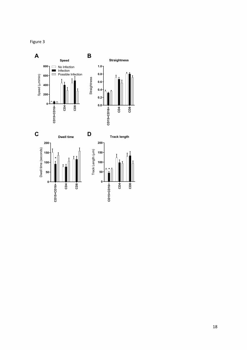
All blood samples were also analysed on VCAM-1+IL-8 substrates. As shown in Figure 3C
and 3D, the dwell time (91.7±14.0 vs 150.6±19.0 seconds, p < 0.05) and track length
(45.1±7.8 vs 62.7±5.8 vs µm, p < 0.05) of CD15+CD16+ neutrophils were significantly
lower in infectious patients than non-infectious patients, indicating a decreased mobility of
infectious CD15+CD16+ cells and an impaired cell ability to respond to IL-8.

13
Discussion
This study explored leukocyte adhesive function assay (LAFA) as a new tool to distinguish
patients with infectious systemic inflammation from those affected by non-infectious causes.
LAFA assesses the functions of several important leukocyte expressing membrane proteins,
including PSGL-1 (selectin receptor), α4β1 integrin (VCAM-1 receptor) and CXCR-1 (IL-8
receptor). Compared with non-infectious patients, an enhanced ability of CD4 cells from
infectious patients to adhere to P+E selectin was detected. Additionally, an impaired response
to IL-8 was observed in neutrophils from infectious patients. These findings may offer a new
understanding on disease pathogenesis and possible new biomarkers for sepsis diagnosis.
Compared with cells from non-infectious patients, an enhanced PSGL-1 activity was seen in
CD4 cells from patients with infection, evidenced by a reduced cell straightness and
increased dwell time (Figure 1F and 1G). The pathological role of PSGL-1 in animal models
of sepsis was demonstrated by previous studies, in which the blockage of PSGL-1 resulted in
less lung damage and better animal survival rate 23,24. However, the role of PSGL-1 in ICU
septic patients remains less well understood. Thus, our results provide direct evidence for a
potential involvement of CD4 PSGL-1 in disease pathogenesis in sepsis patients. Given its
ability to distinguish the likelihood of infection in patients with unknown cause of
inflammation, the divergent activity of PSGL-1 between non-infectious and infectious CD4
cells may also be useful markers for the diagnosis of sepsis.
Our study extends the results of previous work that described an enhanced neutrophil
recruitment by VCAM-1 in patients with infection, but not in patients with non-infectious
systemic inflammation 21. In our study, interacting leukocytes were recorded during the entire
period of flow experiments utilising recently developed LAFA technology to enable a more
complete assessment of leukocyte recruitment and cell migratory behaviours. In contrast to
the previous findings, a slightly decreased number of interacting neutrophils (CD15+CD16+
cells) on VCAM-1 substrate was detected in infectious patients when compared to non-
infectious patients (Figure 2A and 2B). This difference could be due to the divergent
approaches used to detect the interacting cells between the two studies. Our study also noted
that the neutrophil mobility was lower in infectious patients than non-infectious patients
(Figure 2H), suggesting that the reduced neutrophil mobility in neutrophils from infectious
patients on VCAM-1 substrate may serve as a new marker to distinguish infectious patients
from non-infectious patients.

14
IL-8 is a chemokine that is shown to guide the migration of leukocytes (mainly neutrophils) by
forming a concentration gradient, a process known as chemotaxis 25. CXCR1, a receptor for
IL-8, may be expressed on neutrophil cell membranes and plays an important role in the
regulation of leukocyte functions and migratory behaviours. Our study analysed blood samples
by LAFA using VCAM-1 plus IL-8 as substrates, allowing the assessment of leukocyte
CXCR1 activities.
An impaired response to IL-8 induced chemotaxis in neutrophils from patients with infection
has been previously reported 26. Consistently, in the present study, a reduced ability to
respond to IL-8 was detected in infectious neutrophils, compared with non-infectious cells
(Figure 3C and 3D). These results suggest that the neutrophil IL-8 receptors (e.g. CXCR1)
may play different roles in regulation of inflammation in non-infectious and infectious
patients, which may allow it to serve as a useful marker to distinguish these two diseases.
The “Possible Infection (or Unknown)” group provides several unique insights. Firstly, it
is likely this group contains both non-infectious and infectious patients, which may
explain in part why it is indistinguishable from either Non-infectious or Infectious group
in majority of cell parameters when analysed on all adhesive substrate (Figure 1, 2 and 3).
Additionally, patient UN-02 was determined to be “possible infection” by both ICU
specialists even though no evidence of infectious pathogen was found (Table 3). Given a low
CD4 straightness value (0.595) was detected in this patient, and our notion that CD4
straightness may serve as a biomarker for sepsis, this result would support the cause of
inflammation in patient UN-02 is more likely to be infectious than non-infectious.
Despite the small cohort size, this study has provided promising data showing the ability of
LAFA to detect different leukocyte adhesive functions in sepsis and non-infectious systemic
inflammation, providing potential diagnostic biomarkers for sepsis. Larger clinic studies
would be required to further validate these findings and the clinical utility of LAFA
technology. While currently LAFA involves specialised laboratory staff and equipment, it is
possible with further development the assay time can be reduced to less than 20 minutes and
the results turn around may be under 1 hour. Additional automation is also possible which
could mean LAFA may be suitable as a point-of-care test for sepsis diagnosis.

15
Figure legends
Figure 1 Divergent leukocyte migratory behaviours between non-infectious and
infectious ICU patients determined by LAFA using P+E selectins as substrates. Blood
samples were collected from ICU patients and then analysed by LAFA on P+E selectin
substrates. Based on the assessments by two independent ICU specialists, all patients were
then divided into No Infection (n=9), Infection (n=10) and Possible Infection (n=9) groups.
The cell density (A), normalised cell density (B), R-factor (C), cell speed (D), diffusion
coefficient (E), straightness (F), dwell time (G) and track length (H) of CD14,
CD15+CD16+, CD4, CD8 and CD4+CD25+ interacting cells were determined. *, p < 0.05;
**, p < 0.01 compared to No Infection ICU patients.
Figure 2 Divergent leukocyte migratory behaviours between non-infectious and
infectious ICU patients determined by LAFA using VCAM-1 as substrate. Blood
samples were collected from ICU patients and then analysed by LAFA on VCAM-1
substrate. Based on the assessments by two independent ICU specialists, all ICU patients
were then divided into No Infection (n=9), Infection (n=10) and Possible Infection (n=9)
groups. The cell density (A), normalised cell density (B), R-factor (C), cell speed (D),
diffusion coefficient (E), straightness (F), dwell time (G) and track length (H) of CD14,
CD15+CD16+, CD4, CD8 and CD4+CD25+ interacting cells were determined. *, p < 0.05;
**, p < 0.01 compared to No Infection ICU patients.
Figure 3 Divergent leukocyte migratory behaviours between non-infectious and
infectious ICU patients determined by LAFA using VCAM-1+IL-8 as substrates.
Blood samples were collected from ICU patients and then analysed by LAFA on VCAM-
1+IL-8 substrates. Based on the assessments by two independent ICU specialists, all ICU
patients were then divided into No Infection (n=9), Infection (n=10) and Possible Infection
(n=9) groups. The cell speed (A), straightness (B), dwell time (C) and track length (D) were
then determined. *, p < 0.05 compared to No Infection ICU patients.
Supplementary Figure 1 Full blood cell counts. Blood samples were collected from ICU
patients (n=28) and the full blood cell counts were determined by a haematology analyser.
Based on the assessments by two independent ICU specialists, all ICU patients were then
divided into No Infection (n=9), Infection (n=10) and Possible Infection (n=9) groups. *, p <
0.05 compared to no infection ICU patients.

16
Fig
ure
s
Figu
re 1
CD14
CD15+CD16+
CD4
CD8
CD4+CD25+
0
100
200
300
500
10
00
15
00
20
00
Ce
ll de
ns
ity
No. of interacting leukocytes
(Cells/mm2)
No In
fectio
n
Infe
ctio
n
Possib
el In
fectio
n
**
CD14
CD15+CD16+
CD4
CD8
CD4+CD25+
0
200
400
600
800
10
00
No
rma
lise
d c
ell d
en
sity
Normalised interacting leukocytes
(Cells/mm2 / 106 leukocytes/ml)
*
Monocytes
Neutrohils
Lymphocytes
0 1 2 3 4
R-F
ac
tor
Ratio
CD14
CD15+CD16+
CD4
CD8
CD4+CD25+
0
200
400
600
800
Sp
eed
Speed (m/min)
CD14
CD15+CD16+
CD4
CD8
CD4+CD25+
0
50
100
150
200
Diffu
sio
n c
oe
fficie
nt
Diffusion coefficient
(m2/second)
CD14
CD15+CD16+
CD4
CD8
CD4+CD25+
0.0
0.2
0.4
0.6
0.8
1.0
1.2
Stra
igh
tne
ss
Straightness
**
CD14
CD15+CD16+
CD4
CD8
CD4+CD25+
0
50
100
150
200
Dw
ell tim
e
Dwell time
(seconds)
*
CD14
CD15+CD16+
CD4
CD8
CD4+CD25+
0
50
100
150
200
Tra
ck
len
gth
Track Length (m)
AB
CD
EF
GH

17
Figu
re 2
CD14
CD15+CD16+
CD4
CD8
CD4+CD25+
0
100
200
300
Ce
ll de
ns
ity
No. of interacting leukocytes
(Cells/mm2)
No In
fectio
n
Infe
ctio
n
Possib
le In
fectio
n
**
*
CD14
CD15+CD16+
CD4
CD8
CD4+CD25+
0
50
100
150
200
250
No
rma
lise
d c
ell d
en
sity
Normalised interacting leukocytes
(Cells/mm2 / 106 leukocytes/ml)
CD14
CD15+CD16+
CD4
CD8
CD4+CD25+
0
200
400
600
Sp
eed
Speed (m/min)
CD14
CD15+CD16+
CD4
CD8
CD4+CD25+
0
50
100
150
200
Diffu
sio
n c
oe
fficie
nt
Diffusion coefficient
(m2/second)
CD14
CD15+CD16+
CD4
CD8
CD4+CD25+
0.0
0.2
0.4
0.6
0.8
1.0
Stra
igh
tne
ss
Straightness
CD14
CD15+CD16+
CD4
CD8
CD4+CD25+
0
50
100
150
200
Dw
ell tim
e
Dwell time (seconds)
CD14
CD15+CD16+
CD4
CD8
CD4+CD25+
0
50
100
150
Tra
ck
len
gth
Track Length (m)
**
*
AB
CD
EF
GH
Monocytes
Neutrohils
Lymphocytes
0 1 2 3 5
10
15
20
25
R-F
ac
tor
Ratio
*
**
18
Figure 3
CD
15
+C
D1
6+
CD
4
CD
8
0
200
400
600
800
Speed
Sp
ee
d (m
/min
)
No InfectionInfection
Possible Infection
CD
15
+C
D1
6+
CD
4
CD
8
0.0
0.2
0.4
0.6
0.8
1.0
Straightness
Str
aig
htn
ess
CD
15
+C
D1
6+
CD
4
CD
8
0
50
100
150
200
Dwell time
Dw
ell
tim
e (
secon
ds)
*
CD
15
+C
D1
6+
CD
4
CD
8
0
50
100
150
200
Track length
Tra
ck L
ength
(m
)
*
A B
C D

19
Supplementary Figure 1
To
tal L
eu
ko
cy
te
Ne
utr
oh
ils
Ly
mp
ho
cyte
s
Mo
no
cy
tes
0
5
10
15
20
Blood cell counts
Cell
Counts
(10
6 c
ells
/ml) No Infection
Infection
Possible Infection
*

20
Acknowledgement/Declaration
The authors would like to acknowledge the technical assistance of Mr Thomas Andersen
(StickyCell Pty Ltd). This study was financially supported by StickyCell Pty Ltd, Australia.
Dr Qiang Cheng is the Executive Director of StickyCell Pty Ltd.

21
References
1. Singer, M., et al. The Third International Consensus Definitions for Sepsis and Septic Shock
(Sepsis-3). JAMA : the journal of the American Medical Association 315, 801-810 (2016).
2. Bone, R.C., et al. Definitions for sepsis and organ failure and guidelines for the use of
innovative therapies in sepsis. The ACCP/SCCM Consensus Conference Committee. American
College of Chest Physicians/Society of Critical Care Medicine. Chest 101, 1644-1655 (1992).
3. Denny, K.J., De Wale, J., Laupland, K.B., Harris, P.N.A. & Lipman, J. When not to start
antibiotics: avoiding antibiotic overuse in the intensive care unit. Clin Microbiol Infect (2019).
4. Fan, S.L., Miller, N.S., Lee, J. & Remick, D.G. Diagnosing sepsis - The role of laboratory
medicine. Clin Chim Acta 460, 203-210 (2016).
5. de Prost, N., Razazi, K. & Brun-Buisson, C. Unrevealing culture-negative severe sepsis. Crit
Care 17, 1001 (2013).
6. Reinhart, K., Bauer, M., Riedemann, N.C. & Hartog, C.S. New approaches to sepsis: molecular
diagnostics and biomarkers. Clin Microbiol Rev 25, 609-634 (2012).
7. Kumar, A., et al. Duration of hypotension before initiation of effective antimicrobial therapy
is the critical determinant of survival in human septic shock. Crit Care Med 34, 1589-1596
(2006).
8. Sinha, M., et al. Emerging Technologies for Molecular Diagnosis of Sepsis. Clin Microbiol Rev
31(2018).
9. Meisner, M. Update on procalcitonin measurements. Ann Lab Med 34, 263-273 (2014).
10. Sutherland, A., et al. Development and validation of a novel molecular biomarker diagnostic
test for the early detection of sepsis. Crit Care 15, R149 (2011).
11. Aloisio, E., Dolci, A. & Panteghini, M. Procalcitonin: Between evidence and critical issues. Clin
Chim Acta 496, 7-12 (2019).
12. Huang, D.T. & Ramirez, P. Biomarkers in the ICU: less is more? Yes. Intensive Care Med 47,
94-96 (2021).
13. Petri, B., Phillipson, M. & Kubes, P. The physiology of leukocyte recruitment: an in vivo
perspective. Journal of immunology 180, 6439-6446 (2008).
14. Muller, W.A. Leukocyte-endothelial cell interactions in the inflammatory response. Lab Invest
82, 521-533 (2002).
15. Ley, K. The role of selectins in inflammation and disease. Trends Mol Med 9, 263-268 (2003).
16. Norman, M.U., James, W.G. & Hickey, M.J. Differential roles of ICAM-1 and VCAM-1 in
leukocyte-endothelial cell interactions in skin and brain of MRL/faslpr mice. Journal of
leukocyte biology 84, 68-76 (2008).
17. Ruiz-Torres, M.P., Perez-Rivero, G., Rodriguez-Puyol, M., Rodriguez-Puyol, D. & Diez-
Marques, M.L. The leukocyte-endothelial cell interactions are modulated by extracellular
matrix proteins. Cell Physiol Biochem 17, 221-232 (2006).
18. Phillipson, M., et al. Intraluminal crawling of neutrophils to emigration sites: a molecularly
distinct process from adhesion in the recruitment cascade. The Journal of experimental
medicine 203, 2569-2575 (2006).
19. Comstedt, P., Storgaard, M. & Lassen, A.T. The Systemic Inflammatory Response Syndrome
(SIRS) in acutely hospitalised medical patients: a cohort study. Scand J Trauma Resusc Emerg
Med 17, 67 (2009).
20. Cheng, Q., Hoi, A., Hickey, M.J. & Morand, E.F. Lymphocytes from systemic lupus
erythematosus patients display increased spreading on VCAM-1, an effect associated with
active renal involvement. Lupus 21, 632-641 (2012).
21. Ibbotson, G.C., et al. Functional alpha4-integrin: a newly identified pathway of neutrophil
recruitment in critically ill septic patients. Nature medicine 7, 465-470 (2001).
22. Kucik, D.F., Dustin, M.L., Miller, J.M. & Brown, E.J. Adhesion-activating phorbol ester
increases the mobility of leukocyte integrin LFA-1 in cultured lymphocytes. The Journal of
clinical investigation 97, 2139-2144 (1996).

22
23. Wang, X.L., et al. The role of PSGL-1 in pathogenesis of systemic inflammatory response and
coagulopathy in endotoxemic mice. Thromb Res 182, 56-63 (2019).
24. Roller, J., et al. Direct in vivo observations of P-selectin glycoprotein ligand-1-mediated
leukocyte-endothelial cell interactions in the pulmonary microvasculature in abdominal
sepsis in mice. Inflammation research : official journal of the European Histamine Research
Society ... [et al.] 62, 275-282 (2013).
25. Mukaida, N. Interleukin-8: an expanding universe beyond neutrophil chemotaxis and
activation. Int J Hematol 72, 391-398 (2000).
26. Arraes, S.M., et al. Impaired neutrophil chemotaxis in sepsis associates with GRK expression
and inhibition of actin assembly and tyrosine phosphorylation. Blood 108, 2906-2913 (2006).