lesson of the week: playing the odds in clinical … · if you wish to cite this item you are...
TRANSCRIPT

Playing the odds in clinical decision making: Lessons from berry aneurysms undetected by magnetic resonance angiography
Article (Published Version)
http://sro.sussex.ac.uk
Johnson, Michael R, Good, Catriona, Penny, William D, Barnes, Philip R J and Scadding, John W (2001) Playing the odds in clinical decision making: Lessons from berry aneurysms undetected by magnetic resonance angiography. British Medical Journal, 322 (7. pp. 1347-1349. ISSN 0007-1447
This version is available from Sussex Research Online: http://sro.sussex.ac.uk/38070/
This document is made available in accordance with publisher policies and may differ from the published version or from the version of record. If you wish to cite this item you are advised to consult the publisher’s version. Please see the URL above for details on accessing the published version.
Copyright and reuse: Sussex Research Online is a digital repository of the research output of the University.
Copyright and all moral rights to the version of the paper presented here belong to the individual author(s) and/or other copyright owners. To the extent reasonable and practicable, the material made available in SRO has been checked for eligibility before being made available.
Copies of full text items generally can be reproduced, displayed or performed and given to third parties in any format or medium for personal research or study, educational, or not-for-profit purposes without prior permission or charge, provided that the authors, title and full bibliographic details are credited, a hyperlink and/or URL is given for the original metadata page and the content is not changed in any way.

BMJ
Lesson Of The Week: Playing The Odds In Clinical Decision Making: Lessons From BerryAneurysms Undetected By Magnetic Resonance AngiographyAuthor(s): Michael R. Johnson, Catriona D. Good, William D. Penny, Philip R. J. Barnes andJohn W. ScaddingSource: BMJ: British Medical Journal, Vol. 322, No. 7298 (Jun. 2, 2001), pp. 1347-1349Published by: BMJStable URL: http://www.jstor.org/stable/25467070 .
Accessed: 18/06/2014 11:39
Your use of the JSTOR archive indicates your acceptance of the Terms & Conditions of Use, available at .http://www.jstor.org/page/info/about/policies/terms.jsp
.JSTOR is a not-for-profit service that helps scholars, researchers, and students discover, use, and build upon a wide range ofcontent in a trusted digital archive. We use information technology and tools to increase productivity and facilitate new formsof scholarship. For more information about JSTOR, please contact [email protected].
.
Digitization of the British Medical Journal and its forerunners (1840-1996) was completed by the U.S. NationalLibrary of Medicine (NLM) in partnership with The Wellcome Trust and the Joint Information SystemsCommittee (JISC) in the UK. This content is also freely available on PubMed Central.
BMJ is collaborating with JSTOR to digitize, preserve and extend access to BMJ: British Medical Journal.
http://www.jstor.org
This content downloaded from 139.184.30.133 on Wed, 18 Jun 2014 11:39:46 AMAll use subject to JSTOR Terms and Conditions
Clinical review
Lesson of the week
Playing the odds in clinical decision making: lessons from berry aneurysms undetected by magnetic resonance angiography Michael R Johnson, Catriona D Good, William D Penny, Philip RJ Barnes, John W Scadding
The purpose of this report is twofold-to report the
potential for magnetic resonance
angiography to miss
sizeable intracranial aneurysms and to highlight the
value of simple, quantitative clinical reasoning when
interpreting the results of diagnostic tests.
Subarachnoid haemorrhage accounts for a quarter of all cerebrovascular deaths, and over a third of those
who survive have major neurological deficits.12 Intra
cranial aneurysms, the commonest cause of subarach
noid haemorrhage, may present with rupture, mass
effect, or, rarely, with emboli phenomena in large
aneurysms. The typical presentation of rupture is
headache of instantaneous onset that remains continu
ous and is often associated with nausea, vomiting,
meningism, or loss of consciousness. About a third of
patients with aneurysmal subarachnoid haemorrhage will re-bleed, and this is a
major cause of poor outcome.^4 The risk of re-bleeding peaks
on the first
day and then declines.5 Most studies therefore support the need for surgery soon after rupture, and delay in
diagnosis or
misdiagnosis as
migraine or
meningitis can have catastrophic consequences.6
7
Unruptured intracranial aneurysms causing mass
effect may present as pain
or neurological deficit
depending on the site and size of the aneurysm. Such
aneurysms are often large or
giant,8 and, as most intra
cranial aneurysms occur at the junction of the internal
carotid and posterior communicating artery, the
commonest clinical sign is oculomotor palsy. Unrup tured intracranial aneurysms causing
mass effect are at
high risk of subsequent rupture, estimated at 6% a year.4
The optimal method for detecting intracranial
aneurysms is intra-arterial digital subtraction angio
graphy. This procedure carries an associated morbidity of transient or permanent neurological disability (of 1% and 0.5% respectively).10 This associated morbidity
and increasing access to magnetic resonance
imaging has led to interest in the use of magnetic
resonance
angiography for assessing patients at high risk of
symptomatic intracranial aneurysm.
Case reports
Casel
A 21 year old man developed a headache of instantaneous onset The headache improved
over
night, becoming persistent and centred behind the left
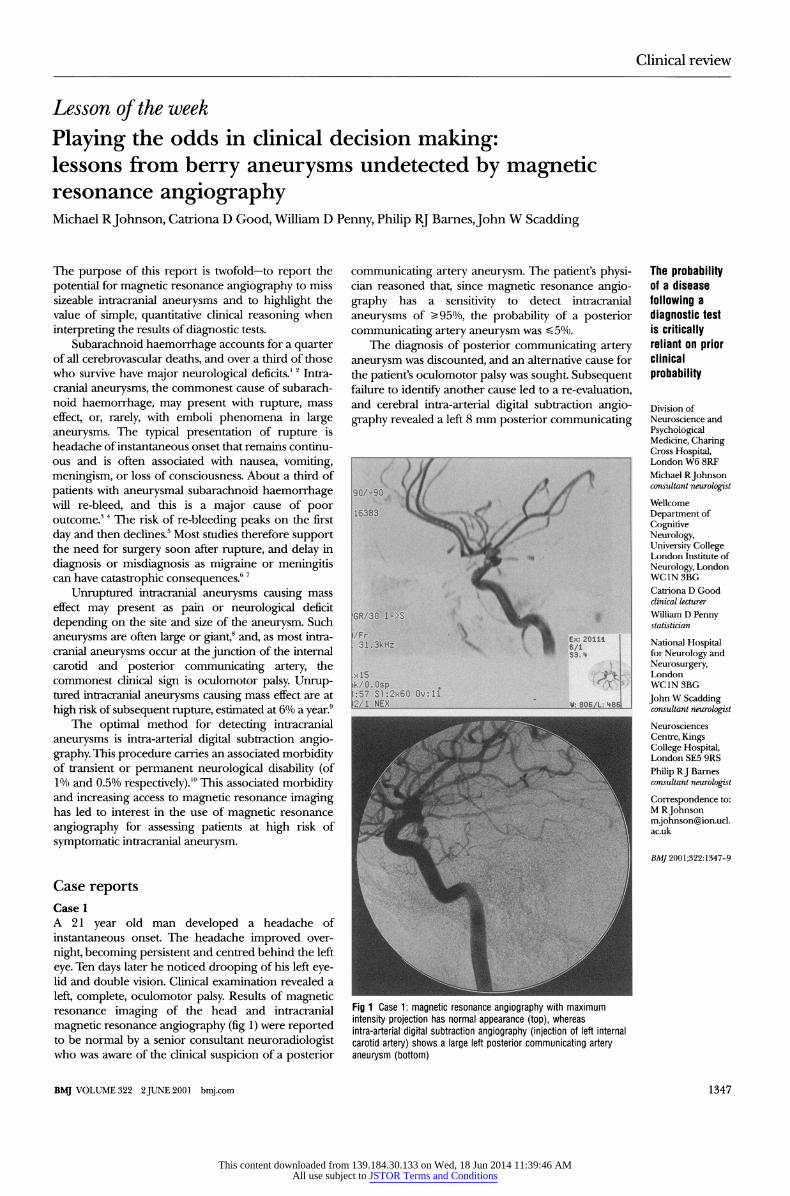
eye. Ten days later he noticed drooping of his left eye lid and double vision. Clinical examination revealed a
left, complete, oculomotor palsy. Results of magnetic resonance
imaging of the head and intracranial
magnetic resonance angiography (fig 1) were reported to be normal by
a senior consultant neuroradiologist who was aware of the clinical suspicion of a
posterior
communicating artery aneurysm The patient's physi cian reasoned that, since magnetic resonance angio
graphy has a sensitivity to detect intracranial
aneurysms of ^95%, the probability of a posterior
communicating artery aneurysm was ^5%
The diagnosis of posterior communicating artery
aneurysm was discounted, and an alternative cause for
the patient's oculomotor palsy was
sought Subsequent failure to identify another cause led to a re-evaluation,
and cerebral intra-arterial digital subtraction angio
graphy revealed a left 8 mm posterior communicating
x15 jrv ik/0.0sp , ir7\
>
:57 Sl:2x60 Ov:ll 12/1 NEX _ *_W 806/1 486
Fig 1 Case 1 magnetic resonance angiography with maximum
intensity projection has normal appearance (top) whereas
intra-arterial digital subtraction angiography (injection of left internal
carotid artery) shows a large left posterior communicating artery
aneurysm (bottom)
The probability of a disease
following a
diagnostic test is critically reliant on prior clinical
probability
Division of Neuroscience and
Psychological Medicine, Channg Cross Hospital, London W6 8RF
Michael R Johnson consultant neurologist
Wellcome
Department of
Cognitive Neurology, University College London Institute of
Neurology, London WC1N3BG
Catriona D Good clinical lecturer
William D Penny statistician
National Hospital for Neurology and
Neurosurgery, London
WC1N3BG
John W Scadding consultant neurologist
Neurosciences
Centre, Kings College Hospital, London SE5 9RS
Philip R J Barnes consultant neurologist
Correspondence to M R Johnson mjohnson@ion ucl acuk
BMJ 2001,322 1347-9
BMJ VOLUME 322 2JUNE2001 bmjcom 1347
This content downloaded from 139.184.30.133 on Wed, 18 Jun 2014 11:39:46 AMAll use subject to JSTOR Terms and Conditions
Clinical review
artery aneurysm (fig 1). The aneurysm was surgically
clipped without complication.
Case 2 A 48 year old woman
developed a headache of instan
taneous onset The headache improved over the course
of a few hours, becoming centred behind the right eye. Two days later she became drowsy, photophobic, and
meningitic. Computed tomography of the head gave normal results. Examination of the cerebrospinal fluid
revealed bloodstained fluid (opening pressure not
recorded), a red blood cell count of 180xlOVl, white blood cell count 210x10Vi (70% lymphocytes, 30%
neutrophils), and glucose concentration 2.2 mmol/1
(serum concentration 8.0 mmol/1). A diagnosis of
"bloody tap" and meningitis was made, and she
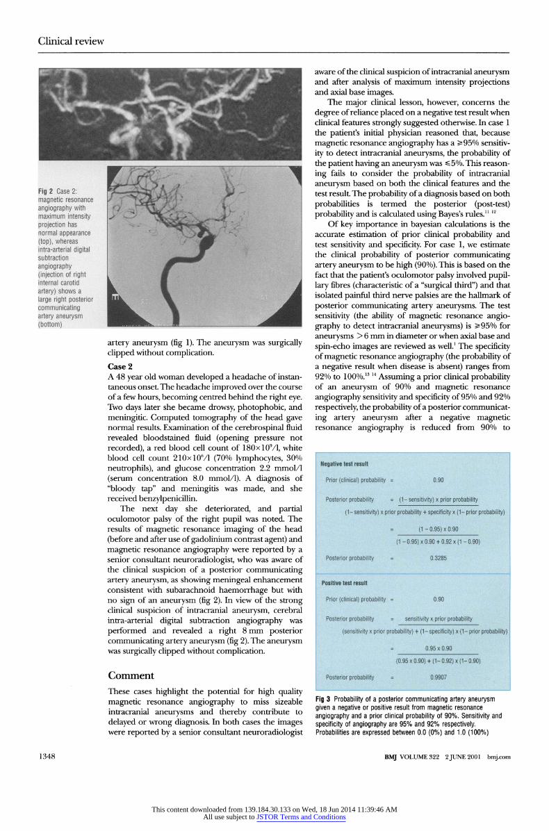
received benzylpenicillin. The next day she deteriorated, and partial
oculomotor palsy of the right pupil was noted. The
results of magnetic resonance
imaging of the head
(before and after use of gadolinium contrast agent) and
magnetic resonance
angiography were
reported by a
senior consultant neuroradiologist, who was aware of
the clinical suspicion of a posterior communicating
artery aneurysm, as showing meningeal enhancement
consistent with subarachnoid haemorrhage but with
no sign of an aneurysm (fig 2). In view of the strong
clinical suspicion of intracranial aneurysm, cerebral
intra-arterial digital subtraction angiography was
performed and revealed a right 8 mm
posterior
communicating artery aneurysm (fig 2). The aneurysm was
surgically clipped without complication.
Comment
These cases highlight the potential for high quality magnetic
resonance angiography
to miss sizeable
intracranial aneurysms and thereby contribute to
delayed or wrong diagnosis. In both cases the images
were reported by
a senior consultant neuroradiologist
aware of the clinical suspicion of intracranial aneurysm and after analysis of maximum intensity projections and axial base images.
The major clinical lesson, however, concerns the
degree of reliance placed on a
negative test result when
clinical features strongly suggested otherwise. In case 1
the patient's initial physician reasoned that, because
magnetic resonance
angiography has a ^95% sensitiv
ity to detect intracranial aneurysms, the probability of
the patient having an aneurysm was ^5%. This reason
ing fails to consider the probability of intracranial
aneurysm based on both the clinical features and the
test result The probability of a diagnosis based on both
probabilities is termed the posterior (post-test) probability and is calculated using Bayes's rules.11
12
Of key importance in bayesian calculations is the
accurate estimation of prior clinical probability and
test sensitivity and specificity. For case 1, we estimate
the clinical probability of posterior communicating
artery aneurysm to be high (90%). This is based on the fact that the patient's oculomotor palsy involved pupil
lary fibres (characteristic of a "surgical third") and that isolated painful third nerve palsies are the hallmark of
posterior communicating artery aneurysms. The test
sensitivity (the ability of magnetic resonance
angio
graphy to detect intracranial aneurysms) is ̂ 95% for
aneurysms > 6 mm in diameter or when axial base and
spin-echo images are reviewed as well.1 The specificity
of magnetic resonance angiography (the probability of a
negative result when disease is absent) ranges from
92% to 100%.1314 Assuming a prior clinical probability of an aneurysm of 90% and magnetic
resonance
angiography sensitivity and specificity of 95% and 92%
respectively, the probability of a posterior communicat
ing artery aneurysm after a negative magnetic
resonance angiography is reduced from 90% to
Negative test result
Prior (clinical) probability = 0 90
Posterior probability = (1- sensitivity) x prior probability
(t- sensitivity) x prior probability + specificity x (1- prior probability)
(1-095)x090
(1-095)x090 + 092x(1-090)
Posterior probability = 0 3285
Positive test result
Prior (clinical) probability = 0 90
Posterior probability = sensitivity x prior probability
(sensitivity x prior probability) + (1- specificity) x (1- prior probability)
095x090
(095x090) + (1-092)x(1-090)
Posterior probability = 0 9907
Fig 3 Probability of a posterior communicating artery aneurysm
given a negative or positive result from magnetic resonance
angiography and a prior clinical probability of 90% Sensitivity and
specificity of angiography are 95% and 92% respectively Probabilities are expressed between 0 0 (0%) and 1 0 (100%)
1348 BMJ VOLUME 322 2 JUNE 2001 bmjcom
This content downloaded from 139.184.30.133 on Wed, 18 Jun 2014 11:39:46 AMAll use subject to JSTOR Terms and Conditions
Clinical review
t 1
_^----'-i
I 09 ^^^~~~ \
o. ^x^Positive test resuit ; s 08
/^ \ 3 0 7 /
0 6/
Ob f 04 / 03 / 0 2 / I
/ ..---"'""* Negative test resuit OL.
0 02 04 06 08 1
Prior probability
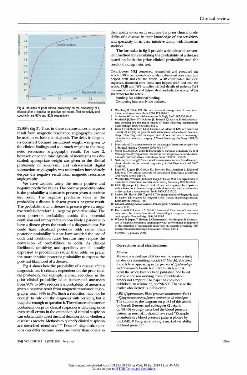
Fig 4 Influence of prior clinical probability on the probability of a
disease after a negative or positive test result Test sensitivity and
specificity are 95% and 92% respectively
32.85% (fig 3). Thus, in these circumstances a negative
result from magnetic resonance
angiography cannot
be used to exclude the diagnosis. The delay in diagno sis occurred because insufficient weight
was given
to
the clinical findings and too much weight to the mag netic resonance
angiography result For case 2,
however, once the misdiagnosis of meningitis was dis
carded, appropriate weight was
given to the clinical
probability of aneurysm, and intra-arterial digital subtraction angiography
was undertaken immediately
despite the negative result from magnetic resonance
angiography. We have avoided using the terms positive and
negative predictive values. The positive predictive value
is the probability a disease is present given
a positive
test result The negative predictive value is the
probability a disease is absent given
a negative result
The probability that a disease is present given a nega
tive result is therefore 1 -
negative predictive value. The
term posterior probability avoids this potential
confusion and simply refers to how likely a patient is to have a disease given the result of a
diagnostic test We
could have calculated posterior odds rather than
posterior probability, but we have avoided the use of
odds and likelihood ratios because they require the conversion of probabilities to odds. As clinical
likelihood, sensitivity, and specificity are all usually expressed
as probabilities rather than odds, we
prefer the more intuitive posterior probability to express the
post-test likelihood of a disease.
Fig 4 shows how the probability of a disease after a
diagnostic test is critically dependent on the prior clini cal probability. For example, a small reduction in the
prior clinical probability of an intracranial aneurysm from 90% to 50% reduces the probability of aneurysm given
a negative result from magnetic resonance
angio
graphy from 33% to 5%. Such a reduction may not be
enough to rule out the diagnosis with certainty, but it
might be enough to question it The reliance of posterior probability on prior clinical suspicion is daunting since even small errors in the estimation of clinical suspicion can
substantially affect the final decision about whether a
disease is present Methods to quantify clinical suspicion
are described elsewhere.1314 Doctors' diagnostic opin ions can differ because some are better than others in
their ability to correcdy estimate the prior clinical prob
ability of a disease, in their knowledge of test sensitivity and specificity, or in their intuitive ability with Bayesian statistics.
The formulas in fig 3 provide a
simple and conven
ient method for calculating the probability of a disease
based on both the prior clinical probability and the result of a
diagnostic test
Contributors: MRJ conceived, researched, and produced the article. CDG contributed data analysis, discussed core ideas, and
helped draft and edit the article. WDP contributed statistical
expertise, discussed core ideas, and helped draft and edit the
article. PRJB andJWS supplied clinical details of patients. JWS discussed core ideas and helped draft and edit the article. JWS is
guarantor for the article.
Funding: No additional funding.
Competing interests: None declared.
1 Wardlaw JM, White PM The detection and management of unruptured intracranial aneurysms Brain 2000,123 205-21
2 Schievmk WI Intracranial aneurysms N EnglJ Med 1997,336 28-40 3 Brodenck JP, Brott TG, Duldner JE, Tomsick T, Leach A Initial and recur
rent bleeding are the major causes of death following subarachnoid
haemorrhage Stroke 1994,25 1342-7 4 Roos YBWEM, Beenen LFM, Groen RJM, Albrecht KW, Vermeulen M
Timing of surgery in patients with aneurysmal subarachnoid haemor
rhage rebleeding is still the major cause of poor outcome m neurosurgi cal units that aim early surgery J Neurol Neurosurg Psychiatry 1997,63 490-3
5 International Co-operative study on the timing of aneurysm surgery Part 2 Surgical results J Neurosurg 1990,73 37-47
6 Mayer PL, Awad IA, Todor R, Harbaugh K, Varnavas G, Lansen TA, et al
Misdiagnosis of symptomatic cerebral aneurysm prevalence and correla tion with outcome at four institutions Stroke 1996,27 1558-63
7 Neil-Dwyer G, Lang D 'Brain attack'-aneurysmal subarachnoid haemor
rhage death due to delayed diagnosis J R Coll Physicians Land 1997, 31 49-52
8 Raps EC, Rogers JD, Galetta SL, Solomon RA, Lennihan L, Klebanoff LM, et al The clinical spectrum of unruptured intracranial aneurysms Arch Neurol 1993,50 265-8
9 Wiebers DO, Whisnant JP, Sundt TM Jr, O'Fallon WM The significance of
unruptured intracranial saccular aneurysms J Neurosurg 1987,66 23-9 10 Cloft HJ, Joseph GJ, Dion JE Risk of cerebral angiography in patients
with subarachnoid haemorrhage, cerebral aneurysm and artenovenous malformation a meta-analysis Stroke 1999,30 317-20
11 Sackett DL, Haynes RB, Tugwell P The interpretation of diagnostic data In Sackett DL, Haynes RB, Tugwell P, eds Clinical epidemiology Boston Litde, Brown, 1985 59-138
12 Gross R Making medical decisions Philadelphia American College of Phy sicians, 1999
13 Honkoshi K, Fukamachi A, Nishi H, Fukasawa I Detection of intracranial
aneurysms by three-dimensional time-of-flight magnetic resonance
angiography Neuroradwlogy 1994,36 203-7 14 W cock DJaspan T, Holland I, Cherryman G, Worthington B Compari
son of magnetic resonance angiography with conventional angiography m the detection of intracranial aneurysms in patients presenting with subarachnoid haemorrhage ClinRadwl 1996,51 330-4
(Accepted 17January 2001)
Corrections and clarifications
Minerva
Minerva was perhaps a bit too keen to report a study on doctors committing suicide (17 March) She cited
the article as appearing in the Journal of Epidemiology and Community Health, but unfortunately at that
point the article had not been published She failed
to realise she was working from prepubhcation
proofs, not a reprint The paper has now been
published-in volume 55, pp 296-300 Thanks to the
reader who alerted us to this error
ABC of hypertension Blood pressure measurement Part 1
-Sphygmomanometry fa ors common to all techniques The caption to the diagram on p 981 of this article
by Gareth Beevers and colleagues (21 April,
pp 981-5) wrongly described the blood pressure
pattern as normal It should have read "Example of ambulatory blood pressure pattern plotted by the DABL Program showing a marked variability
of blood pressure "
BMJ VOLUME 322 2JUNE2001 bmjcom 1349
This content downloaded from 139.184.30.133 on Wed, 18 Jun 2014 11:39:46 AMAll use subject to JSTOR Terms and Conditions