leishmania and the macrophage: a multifaceted interaction
TRANSCRIPT

111ISSN 1746-0913Future Microbiol. (2015) 10(1), 111–129
part of
10.2217/FMB.14.103 © 2015 Future Medicine Ltd
REVIEW
Leishmania and the macrophage: a multifaceted interaction
Maria Podinovskaia*,1 & Albert Descoteaux1
1INRS – Institut Armand-Frappier & Center for Host–Parasite Interactions, 531 boul. des Prairies, Laval, Quebec, H7V 1B7, Canada
*Author for correspondence: Tel.: +1 450 687 5010 ext 4465; Fax: +1 450 686 5501; [email protected]
ABSTRACT Leishmania, the causative agent of leishmaniases, is an intracellular parasite of macrophages, transmitted to humans via the bite of its sand fly vector. This protozoan organism has evolved strategies for efficient uptake into macrophages and is able to regulate phagosome maturation in order to make the phagosome more hospitable for parasite growth and to avoid destruction. As a result, macrophage defenses such as oxidative damage, antigen presentation, immune activation and apoptosis are compromised whereas nutrient availability is improved. Many Leishmania survival factors are involved in shaping the phagosome and reprogramming the macrophage to promote infection. This review details the complexity of the host–parasite interactions and summarizes our latest understanding of key events that make Leishmania such a successful intracellular parasite.
KEYWORDS • GP63 • Leishmania • lipophosphoglycan • macrophage • phagocytosis • phagosome
Leishmaniases encompass a spectrum of human diseases caused by the protozoan parasites of the genus Leishmania. Three major forms of leishmaniases are delineated from the symptoms and clinical manifestations caused by the various Leishmania species. Cutaneous leishmaniasis is char-acterized by the presence of ulcerative lesions of the skin, which, in most cases, are self-healing. Mucocutaneous leishmaniasis, a variant form of cutaneous leishmaniasis, is accompanied by the destruction of the oro-naso-pharyngeal tissues. Visceral leishmaniasis is a chronic infection affect-ing internal organs such as the liver, the spleen and the bone marrow. This disease is fatal if left untreated.
Although Leishmania infection frequently presents itself as devastating disease, many Leishmania species establish asymptomatic long-term parasitism, which may eventually lead to disease follow-ing perturbations in host–parasite interactions and an increase in immune responses leading to tissue damage. The complexity of host–parasite interactions is well demonstrated in L. braziliensis infection of humans, which causes parasite- and immune-driven disease symptoms ranging from mild cutaneous lesions to severe mucosal ulceration of the oro-naso-pharyngeal tissues, sometimes with asymptomatic phases lasting for several years [1]. While parasite persistence correlates with the ability of the parasite to adapt to its environment and to counter host defenses, disease progres-sion is the product of disruption of the dynamic equilibrium between the parasite and the host. Understanding host–parasite interactions is pivotal to addressing disease prevention and improving disease outcomes.
Transmission of the parasite is mediated by female sand flies of either the genus Phlebotomus or the genus Lutzomyia. In the midgut of the sand fly the parasites replicate as promastigotes, which are 10–20 μm in length and 2 μm in width with a long anterior flagellum. Fully infective metacyclic pro-mastigotes are found in the most anterior part of the mid-gut embedded within a parasite-derived gel
For reprint orders, please contact: [email protected]
Future Microbiol. (2015) 10(1)112
REviEW Podinovskaia & Descoteaux
future science group
composed of proteophosphoglycans, which pro-motes regurgitation by the sand fly during feed-ing [2]. Delivery of infectious promastigotes into the vertebrate host occurs when an infected sand fly takes a bloodmeal. The vertebrate hosts of Leishmania parasites parallel those on which the sand fly relies for feeding, and range from wild rodents and canids to humans. Promastigotes are rapidly internalized by phagocytes, where they transform into nonmotile amastigotes, which proliferate within the acidic and hydrolase-rich phagolysosomal compartment.
To persevere within the hostile environment of the macrophage, Leishmania evolved various strategies aimed at countering the microbicidal power of the macrophage and the mounting of an effective immune response. In this review, we will discuss the latest research findings in the mechanisms used by Leishmania to estab-lish infection and survive within macrophages. In the broad overview of various aspects of Leishmania infection of macrophages, the lat-est studies are comprehensively described and highlight the complexity of Leishmania–mac-rophage interactions in different settings for a range of Leishmania species. Consideration is given to the Leishmania–macrophage rela-tionship as a multifaceted interaction. Better knowledge of Leishmania–macrophage interac-tions will further our understanding of the key stages in the parasite life cycle and the disease pathogenesis.
Inttial events in parasite uptake by the macrophage●● Recognition & uptake
During transmission of the parasite from its vector to the vertebrate host, macrophages and neutrophils are rapidly recruited to the site of the sand f ly bite [3]. Proteophosphoglycans secreted by the parasite in the sand fly’s midgut and inoculated into the host during blood meal are powerful stimulators of macrophage recruit-ment, as shown in studies with L. mexicana and L. infantum [4,5]. Initially, the parasites are mainly found in neutrophils and later in mac-rophages. Promastigotes survive in neutrophils but they do not differentiate into amastigotes. Thus, neutrophil infection is transient and fol-lowing neutrophil apoptosis, the parasites may subsequently infect macrophages [6–8]. Thus, the macrophage is an important host cell for the establishment of infection and persistence of the parasite.
Initial interaction of the promastigote with the macrophage occurs via the parasite flagel-lum. This may trigger release of intracellular survival factors by the parasite and subsequent modulation of macrophage phagocytic activity [9]. L. donovani promastigotes killed by glutaral-dehyde fixation or parasites with pharmacologi-cally inhibited motility did not get internalized by murine bone marrow-derived macrophages [10], highlighting the importance of the parasite flagellum in the initial stages of macrophage parasitism.
Different species of Leishmania rely on a range of macrophage receptors, including complement receptors (CRs), mannose receptors, fibronec-tin receptors and Fcγ receptors (FcγRs) [11]. The choice of receptor may impact the course of infection. CR-mediated uptake via CR3 and CR1 inhibits inf lammation and superoxide burst, as well as accumulation of the lysosomal markers LAMP1 and Cathepsin D. This may create more favorable conditions for the parasite within the macrophage phagosome. However, in conjunction with fibronectin receptors, more inflammatory conditions may result in parasite clearance. Mannose receptor signaling may trig-ger inflammatory pathways and more efficient delivery of hydrolytic enzymes into the phago-lysosome. FcγR-mediated phagocytosis leads to enhanced activation of NADPH oxidase on the newly formed phagosome [11].
Whereas phagocytosis of infective meta-cyclic L. donovani promastigotes by murine bone marrow-derived macrophages occurred within 10–20 min after parasite attachment [10], the downstream events in phagosome bio-genesis depended on the macrophage recep-tors used in the recognition of the parasite. In CR3 (CD11b-/-) and FcγR (common chain -/-) deficient macrophages, phagosome fusion with lysosomes occurred significantly faster follow-ing Leishmania phagocytosis than in wild-type macrophages (1 vs 5 h, respectively), as assessed by recruitment of lysosome-associated pro-teins. Interestingly, uptake or viability of the L. donovani or L. major parasites by murine bone marrow-derived macrophages were not affected by the receptor choice [12], pointing to the complexity of Leishmania–macrophage interactions.
Following recognition at host cell surface, promastigotes can be internalized via caveolae that are composed of cholesterol-rich membrane lipid microdomains, as shown for L. chagasi
113
Leishmania & the macrophage REviEW
future science group www.futuremedicine.com
infection of murine bone marrow-derived mac-rophages [13]. Indeed, it has been recently shown that membrane cholesterol is required for L. donovani uptake by J774A.1 macrophage-like cell line [14,15]. Cholesterol depletion and subsequent lipid microdomain disruption by methyl-β-cyclodextrin compromised Leishmania promastigote uptake via nonopsonic pathways. Uptake of opsonized Leishmania, however, was not affected. Interestingly, phagocytosis of amastigotes proceeded independently of lipid microdomains [13]. Likewise, internalization of Escherichia coli was not affected by choles-terol depletion, highlighting the significance of the lipid microdomain-mediated pathway for Leishmania promastigote internalization [16,17].
The significance of lipid microdomains in Leishmania infection is also emphasized in recent studies, which demonstrate that infective Leishmania promastigotes promote lipid micro-domain formation through activation of host acid sphingomyelinase. This enzyme converts sphingomyelin to ceramide, which is a key com-ponent of lipid microdomains [18]. Translocation of acid sphingomyelinase to the cell membrane led to ceramide generation and enhanced para-site uptake. However, at later stages of infection, the parasite also induced de novo ceramide syn-thesis, which in excess might displace choles-terol, disrupt lipid microdomains and impair antigen presentation [18]. Hence, the fine tun-ing of lipid microdomains is likely to be one of the key players in host–parasite interactions in Leishmania infection.
In addition to receptor- and lipid micro-domain-regulated uptake, Leishmania infec-tion also depends on actin-mediated uptake, and the integrity of the actin cytoskeleton of the host macrophage has recently been shown to be essential for L. donovani infectivity [15]. Actin cytoskeleton destabilization induced by cytochalasin D treatment led to a reduction in promastigote attachment to macrophages and the concomitant reduction in intracellular amastigote load. Cellular F-actin levels strongly correlated with the reduction in Leishmania attachment and load in macrophages. In con-trast, uptake of E. coli was unaffected following actin disruption [15].
●● Phagosome maturation & parasite differentiationFollowing phagocytosis, L. donovani promas-tigote-containing phagosomes of murine bone
marrow-derived macrophages failed to acidify and were characterized by a reduced fusogenic-ity towards late endosomes and lysosomes [19,20]. LAMP1 was recruited to the parasitophorous vacuole (PV) with delayed kinetics [19]. The parasite was shown to subsequently reorient itself with the cell body pointing towards the cell nucleus and the flagellum towards the cell periphery. The parasite promoted outward movement of the PV, opposite to the inward cel-lular forces. Such opposing motions resulted in cell injury and prompted lysosome docking and exocytosis. The authors proposed that, at this stage, some lysosomes may fuse with the PV and promote promastigote-to-amastigote differentia-tion, whereas cellular injury may impact plasma membrane integrity and host capacity to combat infection [10].
Promastigote-to-amastigote differentiation is believed to be triggered by the increase in tem-perature and a decrease in pH. Additionally, iron uptake and subsequent generation of hydrogen peroxide by L. amazonensis has been shown to be a major trigger in parasite differentiation [21,22]. Iron mediates generation of reactive oxygen spe-cies (ROS), which are normally deleterious for pathogens, but were proposed to act as a signal-ing molecules regulating parasite differentiation. The Leishmania iron transporter LIT1 mediated iron acquisition by the parasite, which led to par-asite growth arrest and differentiation. In con-trast, LIT1-deficient promastigotes had reduced iron levels, sustained their growth, but eventu-ally died in iron-poor medium. Importantly, LIT1 triggered iron superoxide dismutase to convert ROS to hydrogen peroxide, the presence of which alone was sufficient to trigger differ-entiation in both wild-type and LIT1-deficient promastigotes. In contrast, the ROS-generating drug menadione could only trigger differentia-tion in wild-type but not LIT1-deficient cells, implicating LIT1 in the ability of the parasite to generate hydrogen peroxide and the role of the latter in parasite differentiation [21,22]. Interestingly, iron uptake by the parasite was upregulated in iron-deficient conditions, con-sistent with the low iron levels that the parasite encounters in PVs, where differentiation takes place [23].
Promastigote-to-amastigote differentiation is associated with a reduction in growth rate and the induction of a distinct metabolic state characterized by a decrease in uptake and uti-lization of glucose and amino acids, reduced

Future Microbiol. (2015) 10(1)114
REviEW Podinovskaia & Descoteaux
future science group
organic acid secretion and increased fatty acid beta-oxidation. Catabolism of hexose and fatty acids provide substrates for glutamate synthesis, which is essential for amastigote growth and sur-vival. Notably, in vitro differentiated amastigotes displayed a metabolic profile similar to that of lesion-derived amastigotes, suggesting its cou-pling to differentiation signals rather than nutri-ent availability [24]. Such changes likely facili-tate amastigote survival in the nutrient-poor intracellular niche.
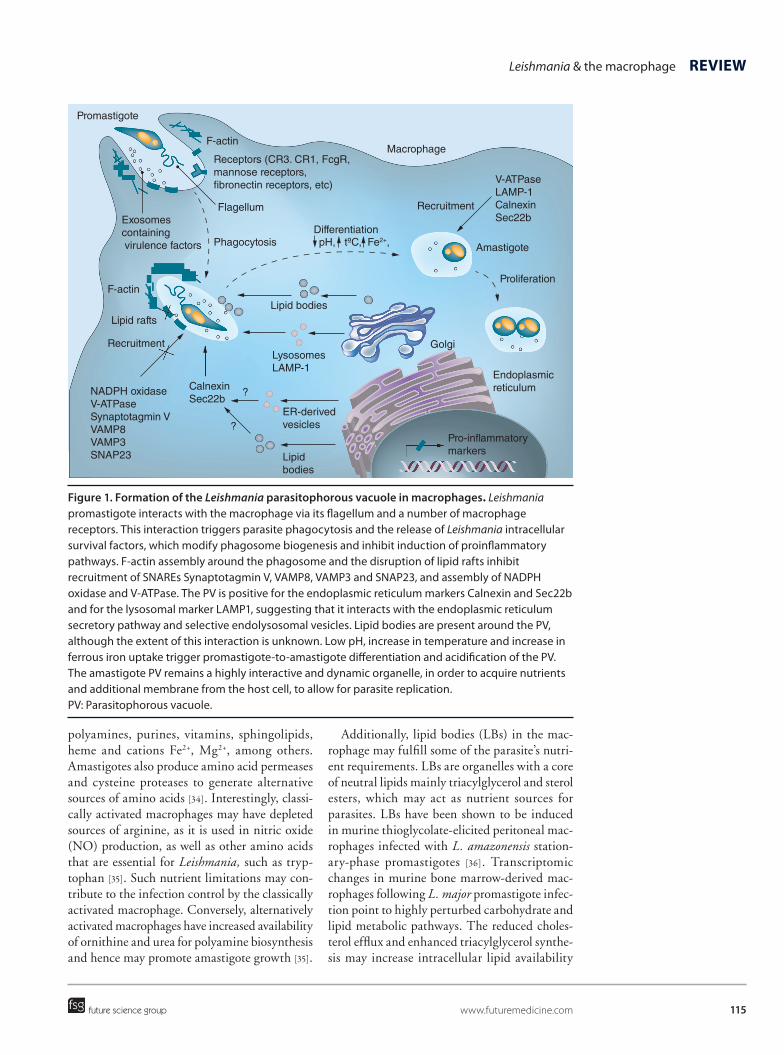
Both promastigotes and amastigotes are able to divert the classical phagosome maturation path-way, which occurs via a set of highly regulated fusion and fission events with vesicles includ-ing endosomes and lysosomes, and form PVs of very specific phenotypes. Fusion is strictly spe-cies- and stage-dependent. This is neatly demon-strated in a recent study by Real and colleagues, in which PVs containing L. major promastigotes, but not L. major amastigotes, could fuse with pre-established L. amazonensis amastigote PVs in murine bone marrow-derived macrophages [8,25]. Furthermore, the L. major promastig-otes within the pre-established L. amazonensis amastigote-harboring PVs failed to differentiate, suggesting that L. amazonensis niche may not provide an appropriate environment for L. major differentiation. L. amazonensis differentiation in dendritic cells correlated with an increase in ERK1/2 phosphorylation, the levels of which remained low in L. major infection [26], suggest-ing important differences in key cellular events during differentiation of the two parasite species. These observations signify the unique nature of host–parasite interactions for different parasite species and their vulnerability to perturbations. Major events in Leishmania PV formation are depicted in Figure 1 and are discussed in more detail below.
The PVs of L. amazonensis and L. major amas-tigotes show distinct dynamics with the former favoring fusion of individual PVs to form spa-cious communal vacuoles and the latter promot-ing fission of the PVs of dividing parasites. These differences are only recently being investigated in greater detail and highlight the complexity in PVs formation and maintenance [27].
Intracellular parasite growth●● Membrane contribution from the
endoplasmic reticulumOnce Leishmania establish infection within macrophages, their subsequent multiplication
requires a source of nutrients and additional membrane for phagosome expansion. For this, the amastigote PV remains a highly dynamic organelle and interacts with the secretory path-way, which contains endoplasmic reticulum (ER) proteins destined for other organelles [28]. RAW 264.7 macrophages infected with L. donovani or L. pifanoi axenic amastigotes for 6 h to estab-lish PVs and incubated with ricin were able to deliver ricin to Leishmania PVs, and this activity was abolished following brefeldin A treatment, which blocks transport from the ER to the Golgi [29]. Importantly, recent studies by Canton et al. demonstrated that disruption of Leishmania PV fusion with ER vesicles resulted in control of L. amazonensis infection of RAW 264.7 mac-rophages [30]. Lysosome recruitment to PVs has been implicated in recent findings [10], and warrants further studies to address membrane contribution from lysosomes to Leishmania PVs.
Further characterization of Leishmania PVs in RAW 264.7 macrophages revealed that phago-somes containing L. donovani and L. pifanoi promastigotes recruit the ER proteins calnexin and Sec22b very early during PV maturation, whereas zymosan-containing phagosomes do not [29]. Loss of the ER SNAREs Sec22b or its cognate partners D12 or Syntaxin 18 or knock-down of Syntaxin 5 had very little effect on the ER or secretion but led to a significant reduction in PV size, as well as a reduction in parasite rep-lication [31]. A similar effect was achieved upon disruption and redistribution of Syntaxin 5 fol-lowing treatment with the small organic mol-ecule Retro-2 [31,32]. These findings highlight the role of the ER SNAREs Sec22b and Syntaxin 5 in the delivery of ER content to Leishmania PVs that supports the infection. Interestingly, Sec22b was also required for cross-presentation and its presence in the phagosome may play a role in parasite control [33].
●● Nutrient acquisitionIntracellular Leishmania parasites have complex nutritional requirements, with amino acids and polyamines being important carbon sources and growth-limiting nutrients. Long-term survival of the parasite within macrophages requires nutri-ent availability within the intracellular niche. The parasite may achieve some of its nutrient requirements through parasite-driven PV fusion with endosomes and ER-derived Sec22b- and calnexin-positive vacuoles [29]. Actively scav-enged metabolites include hexoses, amino acids,
115
Figure 1. Formation of the Leishmania parasitophorous vacuole in macrophages. Leishmania promastigote interacts with the macrophage via its flagellum and a number of macrophage receptors. This interaction triggers parasite phagocytosis and the release of Leishmania intracellular survival factors, which modify phagosome biogenesis and inhibit induction of proinflammatory pathways. F-actin assembly around the phagosome and the disruption of lipid rafts inhibit recruitment of SNAREs Synaptotagmin V, VAMP8, VAMP3 and SNAP23, and assembly of NADPH oxidase and V-ATPase. The PV is positive for the endoplasmic reticulum markers Calnexin and Sec22b and for the lysosomal marker LAMP1, suggesting that it interacts with the endoplasmic reticulum secretory pathway and selective endolysosomal vesicles. Lipid bodies are present around the PV, although the extent of this interaction is unknown. Low pH, increase in temperature and increase in ferrous iron uptake trigger promastigote-to-amastigote differentiation and acidification of the PV. The amastigote PV remains a highly interactive and dynamic organelle, in order to acquire nutrients and additional membrane from the host cell, to allow for parasite replication. PV: Parasitophorous vacuole.
Leishmania & the macrophage REviEW
future science group www.futuremedicine.com
polyamines, purines, vitamins, sphingolipids, heme and cations Fe2+, Mg2+, among others. Amastigotes also produce amino acid permeases and cysteine proteases to generate alternative sources of amino acids [34]. Interestingly, classi-cally activated macrophages may have depleted sources of arginine, as it is used in nitric oxide (NO) production, as well as other amino acids that are essential for Leishmania, such as tryp-tophan [35]. Such nutrient limitations may con-tribute to the infection control by the classically activated macrophage. Conversely, alternatively activated macrophages have increased availability of ornithine and urea for polyamine biosynthesis and hence may promote amastigote growth [35].
Additionally, lipid bodies (LBs) in the mac-rophage may fulfill some of the parasite’s nutri-ent requirements. LBs are organelles with a core of neutral lipids mainly triacylglycerol and sterol esters, which may act as nutrient sources for parasites. LBs have been shown to be induced in murine thioglycolate-elicited peritoneal mac-rophages infected with L. amazonensis station-ary-phase promastigotes [36]. Transcriptomic changes in murine bone marrow-derived mac-rophages following L. major promastigote infec-tion point to highly perturbed carbohydrate and lipid metabolic pathways. The reduced choles-terol efflux and enhanced triacylglycerol synthe-sis may increase intracellular lipid availability
Macrophage
V-ATPaseLAMP-1CalnexinSec22b
Amastigote
Proliferation
Golgi
Endoplasmic reticulum
Pro-in�ammatorymarkersLipid
bodies
ER-derivedvesicles
CalnexinSec22b
?
?
NADPH oxidaseV-ATPaseSynaptotagmin VVAMP8VAMP3SNAP23
Recruitment
Recruitment
Lipid rafts
F-actin
Lipid bodies
DifferentiationpH, tºC, Fe2+,Phagocytosis
Flagellum
Receptors (CR3. CR1, FcgR,mannose receptors, �bronectin receptors, etc)
F-actin
Promastigote
Exosomescontaining virulence factors
LysosomesLAMP-1

Future Microbiol. (2015) 10(1)116
REviEW Podinovskaia & Descoteaux
future science group
and hence facilitate foamy macrophage forma-tion [37]. The LB profile of dendritic leukocytes infected with L. amazonensis amastigotes was transcriptionally distinct from that of lipid overloaded cells following oleate treatment and presented with larger and more numerous LBs [38]. Interestingly, LB accumulation in perito-neal macrophages was enhanced by the saliva of the sand fly Lutzomyia longipalpis, the vec-tor of L. chagasi, further promoting foamy cell generation [39]. The LBs interact, associate and sometimes fuse with phagosomes containing zymosan, silica beads or pathogens [40,41]. As well as containing energy-rich nutrients, LBs also bear Rab GTPases, ER proteins and molecular chaperones, and may provide means for acquisi-tion of phagosomal proteins such as Rab5 and Rab7. Thus, LB interaction with parasite PVs may play a role in phagosome maturation [41] and fusion with other organelles, potentially pro-viding additional nutrient sources. Indeed, LBs induced by L. amazonensis amastigote infection of dendritic leukocytes were observed to be in close apposition to the PV membrane [38]. The extent of interaction of LBs with Leishmania PVs in macrophages has not yet been determined, and the contribution of LBs to Leishmania phagosome biogenesis and to host–parasite interactions remains to be explored.
●● Iron acquisitionIron is an essential element for most organ-isms, including parasites such as Leishmania. This nutrient is required for Leishmania growth and survival. Iron acquisition by Leishmania is facilitated via the parasite ferric iron reduc-tase LFR1, ferrous iron transporter LIT1 and heme transporter LHR1. The three mediators of iron uptake are upregulated in response to low iron. LHR1 is essential for Leishmania via-bility whereas LFR1 and LIT1 are required for intracellular survival [42]. LHR1-null mutants have disrupted heme uptake and are nonviable. Heterozygous mutants (LHR1+/-) were attenu-ated in heme-deficient medium, however differ-entiated normally into amastigotes but did not replicate in macrophages, unless under iron over-load conditions [43]. This suggests that iron avail-ability is essential for parasite growth in the form of heme or high concentrations of labile iron. The host responds by restricting iron availabil-ity to intracellular Leishmania by expressing the NRAMP1 iron efflux pump in maturing phago-somes and lysosomes. The parasite responds
by upregulating LIT1 to counter diminishing phagosomal iron availability in the presence of NRAMP1, as observed in infection of murine bone marrow-derived macrophages by L. ama-zonensis amastigotes [44,45], highlighting the importance of iron availability to Leishmania.
The host has a complex set of iron homeo-static pathways to maximize iron availability to metabolizing cells and at the same time mini-mize the undesired oxidative properties of excess iron. During infection, macrophages play a cen-tral role in withdrawing iron from the circula-tion and limiting iron to infectious agents. The systemic iron regulator, hepcidin, facilitates iron sequestration within macrophages by mediating cell surface degradation of the iron exporter fer-roportin. Such host defense tactics may actually benefit Leishmania as an intracellular parasite of macrophages. Indeed, it has recently been dem-onstrated that L. amazonensis axenic amastigotes causes TLR4-dependent hepcidin upregulation, which triggers ferroportin degradation in murine bone marrow-derived macrophages. Hepcidin deficiency or overexpression of mutant ferropor-tin that is resistant to hepcidin-induced degra-dation inhibited parasite replication. Exogenous hepcidin or expression of dominant-negative fer-roportin enhanced parasite growth and restored growth of parasites defective in iron acquisi-tion [46], highlighting the role of the hepcidin-ferroportin axis in macrophage–Leishmania interactions and the infection outcome.
Alongside promoting iron uptake by the mac-rophage, L. donovani stationary phase promas-tigotes depleted labile iron to activate iron regula-tory proteins IRP1 and IRP2 in primary murine splenic macrophages [47]. These transcription factors promote increased iron uptake in mac-rophages through increased expression of the transferrin receptor TfR1. Holotransferrin (Tf-Fe) supplementation increased and iron chela-tion decreased intracellular Leishmania growth in J774A.1 macrophage-like cells, signifying the importance of transferrin receptor-mediated iron uptake into macrophages for Leishmania survival [47]. Iron acquisition by intracellular Leishmania is summarized in Figure 2.
Macrophage defenses●● Oxidative damage
One of the major tactics used by macrophages to incapacitate pathogens is the generation of ROS and reactive nitrogen intermediates (RNI). Multiple approaches are used by the macrophage

117
Figure 2. Iron acquisition by Leishmania within macrophages. Leishmania expresses the heme transporter LHR and ferrous iron transporter LIT to scavenge iron inside the PV within macrophages. Additionally, the Leishmania ferric reductase LFR converts ferric iron to ferrous iron to facilitate its transport by LIT. Leishmania also produces TXNPx, which inhibits iron export out of the PV by the cation transporter NRAMP-1, thereby augmenting phagosomal iron availability to the parasite and depleting cytosolic iron stores. Depleted intracellular iron triggers upregulation of TfRs and enhances uptake of Tf-bound ferric iron via the endosomal network, with which the PV interacts. Furthermore, elevated expression of the iron regulator hepcidin in infected macrophages causes degradation of the iron exporter ferroportin and leads to iron retention inside the macrophage, increasing iron availability to Leishmania. PV: Parasitophorous vacuole; Tf: Transferrin; TfR: Transferrin receptor; TXNPx: Tryparedoxin peroxidase.
Leishmania & the macrophage REviEW
future science group www.futuremedicine.com
to tightly control production and elimination of these deleterious species, from global mac-rophage activation to responses localized to pathogen-containing phagosomes. As dis-cussed previously, NADPH oxidase assembly at Leishmania-containing phagosome stimulates ROS production and superoxide burst localized to the phagosomal lumen. Additionally, recent findings have demonstrated that NO was pro-duced following Nlrp3 inflammasome assembly in murine bone marrow-derived macrophages infected with L. amazonensis metacyclic pro-mastigotes and helped control the infection [48]. Activated Nlrp3 drove IL-1β production, which through IL-1R and MyD88 induced NOS2 to produce NO [48]. ATP-activated purinergic receptor P2X7 induced inflammasome assembly and participated in the subsequent restriction of L. amazonensis promastigote growth in bone marrow-derived macrophages. Interestingly, P2X7 was also able to induce leukotriene B4
production, which led to a reduction in para-site load [49]. Overall, these findings indicate a concerted effort by multiple macrophage defense mechanisms to induce oxidative damage to the parasite and compromise its ability to survive.
●● Macrophage activationThe macrophage is an extremely plastic cell equipped with homeostatic functions of clearing dead cells and debris in its resting state and micro-bicidal and antigen presentation tasks follow-ing its activation. Classical activation by IFN-γ leads to inflammatory responses and inhibits Leishmania growth, whereas alternative activa-tion by IL-4 inhibits inflammation through IL-10 production and stimulates Leishmania growth [50]. The latest studies addressing macrophage activation following Leishmania infection and its effect on Leishmania growth are discussed below.
Peritoneal resident and inflammatory mac-rophages infected with L. major promastigotes
TfR HepcidinFerroportin
Recyclingendosomes
Macrophages Promastigote
TXNPx
LFR
Fe3+
Fe2+
Fe2+
Fe2+
HemeLIT
LHR
NRAMP-1
Tf
Fe3+
Fe2+

Future Microbiol. (2015) 10(1)118
REviEW Podinovskaia & Descoteaux
future science group
showed increased expression of FasL, TNF, IL-6, and other proinflammatory markers fol-lowing induction of a cellular stress response in macrophages, via the SAPK/JNK activation [51]. Interestingly, the cellular stress response also promoted parasite survival and replication in macrophages [51]. Inflammation-induced IFN-γ led to the activation of members of the PKC family of protein kinases, which were critical for macrophage activation and parasite killing [50,52].
Induction of proinf lammatory functions could be further stimulated by NRAMP-1-mediated cation transport, which led to a revers-ible inhibition of protein tyrosine phosphatases (PTPs), via direct PTP-metal interaction and/or ROS-dependent PTP oxidation. The resulting lower PTP activity led to induction of proin-flammatory pathways and lower survival of L. donovani stationary phase promastigotes in RAW 264.7 macrophages [53]. In the absence of the PTP SHP-1, phagosome acidification was impaired, and pro-Cathepsin D was not pro-cessed to the active enzyme [54]. This is con-sistent with the phagosomal profile of activated macrophages, in which phagosomal degradative capacity is decreased to promote more efficient antigen presentation [55].
Recently, IGF-1, negatively regulated by IFN-γ and macrophage activation, has been implicated in the control of L. major promas-tigote infection in RAW 264.7 macrophages. IGF-1 was expressed in macrophages and colo-calized with parasites. IGF-1 production was inhibited by IFN-γ stimulation, which led to a reduced parasite load. Addition of extrinsic IGF-1 reversed the reduction in parasite load completely [56]. IGF-1-mediated mechanisms of parasite growth control remain to be explored.
●● Foamy cell formationAs discussed above, Leishmania infection of mac-rophages is frequently associated with an increase in LBs in infected macrophages and foamy cell formation. Although from the nutrient perspec-tive, LB induction may potentially be beneficial to the parasites, foamy cells may also form part of host defense against the parasite. LBs are the principal storage organelle for arachidonic acid, which is a paracrine mediator of cell activa-tion. Arachidonic acid can promote phagosome maturation and pathogen killing through ROS production and phagosome–lysosome fusion [41]. Also present in LBs are proinflammatory media-tors such as cyclooxygenases, lipoxygenases,
leukotriene C4 and MAPK (ERK1, ERK2, p85 and p38) [41]. Irgm, the ER protein involved in phagocytic MHC class I presentation is also pre-sent in LBs and may facilitate cross-presentation to other immune cells [57].
Interestingly, the high concentration of pros-taglandin E2 (PGE2) as a product of eicosanoid production within LBs in macrophages acts as a potent inhibitor of NO production, and exog-enous PGE2 increased parasite load in perito-neal inflammatory macrophages infected with stationary phase L. amazonensis promastigotes [36,41]. Hence, the exact contribution of the pres-ence of LBs in macrophages to Leishmania intra-cellular survival may rest on the composition of the LBs, which in turn may be governed by additional factors, such as activation status of the macrophage.
Leishmania evasion of host defenses●● Curbing inflammation
Leishmania employs a number of intervention mechanisms to counter host defenses. Molecular targets and mechanisms for the evasion of mac-rophage defenses by Leishmania are summarized in Table 1. Leishmania targets multiple signaling pathways in the macrophage to reduce infection-induced inflammation. Even as early as during inoculation of the parasites by the sand fly vec-tor, the parasite-produced proteophosphoglycan-rich secretory gel enhances alternative activation and arginase activity of host macrophages to promote L. mexicana survival [4]. Infection of murine peritoneal macrophages with L. amazon-ensis stationary phase promastigotes led to sup-pressed LPS-induced inflammatory responses, such as the production of IL-12, IL-17 and IL-6. Interestingly, Leishmania also augmented LPS-induced proinflammatory cytokines IL-1α, TNF, MIP-1α and MCP-1 and the anti-inflam-matory cytokine IL-10 [58]. Hence, Leishmania may possess selectivity over manipulation of certain cytokines in order to stimulate a unique activation state in the macrophage suitable for the parasite survival.
Recently, L. donovani promastigote infection of murine peritoneal macrophages was shown to induce expression of host PPARγ, which is known to curb inflammation and protect the host from excessive injury. Inhibition of PPARγ facilitated removal of the parasite [65]. Leishmania also induced host PTP activation, including PTP1B, TC-PTP, PTP-PEST and SHP-1. Activation of PTPs leads to a number of events
119
Leishmania & the macrophage REviEW
future science group www.futuremedicine.com
favorable for the parasite, such as the reduction of proinflammatory processes, a reduction in IL-12, NO, TNF, phagolysosomal maturation and MHC class II antigen presentation [50,59]. TRAF3 is yet another recently identified tar-get of L. donovani promastigotes. The parasite inhibited TRAF3 degradation in order to impair TLR4-mediated inflammatory host response in RAW 264.7 cells and in bone marrow-derived macrophages. TRAF3 degradative ubiquitina-tion is required for TLR4 activation. Reduction in TRAF3 by shRNA decreased parasite bur-den [62]. The above studies reveal the multi-tude of host targets that Leishmania exploits in order to evade macrophage activation and the accompanying proinflammatory response.
As Leishmania establishes infection inside the macrophage and proliferates, the macrophage may eventually undergo apoptosis. The para-site delays macrophage apoptosis but ultimately exploits the apoptotic host cell to spread to neigh-boring uninfected macrophages, with minimal exposure to extracellular immune recognition systems. Cell-to-cell transfer of L. amazonensis amastigotes, which were isolated from BALB/c mice and used to infect bone marrow derived
macrophages, was mediated by parasitophorous extrusions, enriched in lysosomal enzymes. The PV components such as LAMP1/2 and Rab7 were shown to be internalized by recipient mac-rophages together with the rescued parasite and stimulate production of the anti-inflammatory cytokine IL-10 by the recipient macrophage [74]. Thus, even at the most vulnerable stages of its life cycle, Leishmania successfully manipu-lates its host to avoid immune recognition and subsequent inflammation.
●● Interfering with host cell signalingMacrophages infected with Leishmania are defective in the expression of activation-associ-ated functions and are unresponsive to IFN-γ [75]. Studies with L. donovani revealed that this parasite targets distinct steps along the JAK-STAT pathway. Upon contact with mac-rophages, L. donovani promastigotes activated the PTP SHP-1, which dephosphorylated JAK2. In addition, proteasome-mediated degradation of STAT1 was rapidly induced, preventing its nuclear translocation. L. donovani promastigotes were also reported to downregulate the IFN-γ receptor and to induce the expression of the
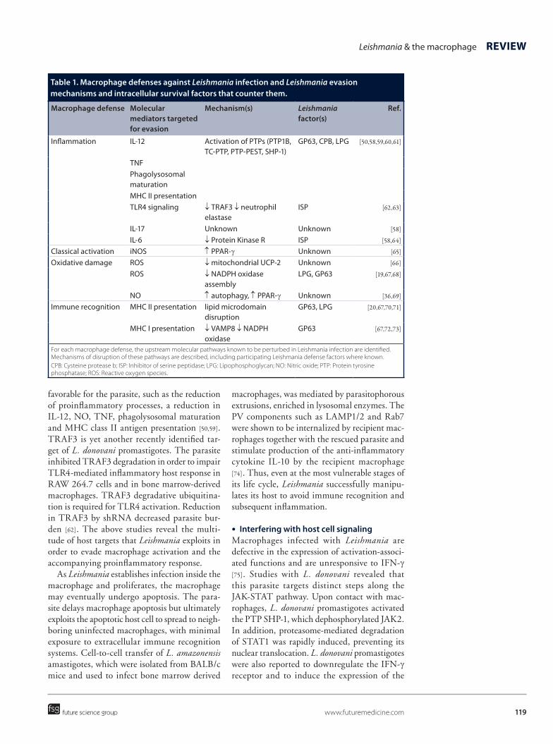
Table 1. Macrophage defenses against Leishmania infection and Leishmania evasion mechanisms and intracellular survival factors that counter them.
Macrophage defense Molecular mediators targeted for evasion
Mechanism(s) Leishmania factor(s)
Ref.
Inflammation IL-12 Activation of PTPs (PTP1B, TC-PTP, PTP-PEST, SHP-1)
GP63, CPB, LPG [50,58,59,60,61]
TNF Phagolysosomal
maturation
MHC II presentation TLR4 signaling ↓ TRAF3 ↓ neutrophil
elastaseISP [62,63]
IL-17 Unknown Unknown [58]
IL-6 ↓ Protein Kinase R ISP [58,64]
Classical activation iNOS ↑ PPAR-γ Unknown [65]
Oxidative damage ROS ↓ mitochondrial UCP-2 Unknown [66]
ROS ↓ NADPH oxidase assembly
LPG, GP63 [19,67,68]
NO ↑ autophagy, ↑ PPAR-γ Unknown [36,69]
Immune recognition MHC II presentation lipid microdomain disruption
GP63, LPG [20,67,70,71]
MHC I presentation ↓ VAMP8 ↓ NADPH oxidase
GP63 [67,72,73]
For each macrophage defense, the upstream molecular pathways known to be perturbed in Leishmania infection are identified. Mechanisms of disruption of these pathways are described, including participating Leishmania defense factors where known.CPB: Cysteine protease b; ISP: Inhibitor of serine peptidase; LPG: Lipophosphoglycan; NO: Nitric oxide; PTP: Protein tyrosine phosphatase; ROS: Reactive oxygen species.
Future Microbiol. (2015) 10(1)120
REviEW Podinovskaia & Descoteaux
future science group
suppressor of cytokine signaling SOCS3 [76]. L. donovani promastigotes can thus efficiently shut off the predominant signaling cascade of one of the most important macrophage activa-tors. Similar to promastigotes, L. donovani amas-tigotes inhibited IFN-γ-induced expression of MHC class II and iNOS. However, infection with L. donovani amastigotes downregulated IFN-γ-induced gene expression without affect-ing STAT1 activation. Rather, amastigotes inhibited IFN-γ-induced STAT1 nuclear trans-location by blocking the interaction of STAT1 with the karyopherin importin-α5 [77]. The underlying mechanisms remain to be elucidated.
●● Avoiding oxidative damageLeishmania responses to oxidative stress vary greatly depending on Leishmania species and host cell type [78]. For example, L. major-infected thioglycolate-elicited peritoneal macrophages infected with L. major stationary phase promas-tigotes produced ROS, whereas in L. amazonen-sis-infected cells ROS production was inhibited [79]. Various approaches are used by different Leishmania species to counter oxidative stress. For example, L. donovani axenic amastigotes were able to impair cellular and mitochondrial ROS via the induction of mitochondrial uncou-pling protein 2 (UCP2). L. donovani degraded the transcription factor USF-1, hence facili-tating recruitment of the transcription factors SREBP2 and Sp1 to the UCP2 promoter, UPC2 upregulation and inhibition of ROS [66].
Leishmania is also able to avoid oxidative dam-age by preventing NADPH oxidase assembly at the phagosomal membrane and generation of ROS within the PV. A recent study by Matheoud and colleagues has demonstrated that L. dono-vani and L. major stationary phase promastig-otes achieve this by cleaving the host SNARE VAMP8, which was necessary for NADPH oxidase recruitment to the phagosome of bone marrow-derived macrophages [67]. Disruption of lipid microdomains by insertion of the sur-face glycolipid lipophosphoglycan (LPG) in the phagosomal membrane by the parasite may also inhibit recruitment of the cytosolic compo-nents of the NADPH oxidase to the PV [19]. In a different approach, L. mexicana pifanoi axenic amastigotes recruited the immature 65-kDa form, but not the mature 91-kDa form, of the gp91phox subunit of the NADPH oxidase complex to the PVs by disrupting gp61phox maturation in RAW 264.7 macrophages. Heme-dependent
maturation of gp91phox was inhibited by the parasite through upregulation of host heme oxygenase 1 and heme degradation [80].
As well as harming the parasite directly, oxi-dative damage by ROS induces apoptosis in macrophages, which destroys the replicative niche of the parasite. Apoptosis was suppressed by L. donovani stationary phase promastigote infection of RAW 264.7 macrophages via the induction of the suppressors of cytokine sign-aling SOCS1 and SOCS3, which enhanced parasite survival [81].
●● Countering antigen presentationAntigen cross-presentation is a critical process for immunity against pathogens. It involves pres-entation of foreign proteins derived from phago-cytosed cargo on MHC class I for detection by cytotoxic CD8+ T cells and for orchestration of a systemic immune response. As a professional antigen presenting cell (APC), the macrophage participates in cross-presentation of Leishmania-derived proteins. Leishmania evades host immu-nity by inhibiting antigen cross-presentation through cleavage of the SNARE VAMP8 in murine bone marrow derived macrophages infected with L. major or L. donovani station-ary phase promastigotes but not L. donovani amastigotes isolated from spleens of infected hamsters. Disruption of VAMP8 prevented NADPH oxidase assembly which led to more efficient phagosomal acidification and proteoly-sis, thereby inhibiting MHC class I presentation and T cell activation [67,72,73]. Both VAMP8 and VAMP3 were excluded from Leishmania PVs. The consequences of VAMP3 exclusion from Leishmania PVs are unknown. Interestingly, MHC class II-dependent antigen presentation was also compromised in Leishmania infection but in VAMP8-independent manner [67,70].
Inhibition of antigen cross-presentation was also achieved via disruption of membrane lipid microdomains by the parasite [71]. Indeed, mem-brane cholesterol levels were found to be reduced in infected cells and the antigen presentation defect could be corrected with liposomal deliv-ery of exogenous cholesterol. Liposomal cho-lesterol was also found to promote ROS and RNI, proinflammatory cytokine expression and intracellular parasite killing, and was implicated in cellular stress and ROS-induced apoptosis of peritoneal exudate cells infected with L. donovani promastigotes [82]. Hence, lower cholesterol lev-els, whether through dysregulated macrophage

121
Leishmania & the macrophage REviEW
future science group www.futuremedicine.com
lipid metabolism or Leishmania-driven cholesterol displacement or depletion, may favor Leishmania survival.
●● Induction of autophagyInduction of autophagy in bone marrow-derived macrophages or peritoneal exudate cells by L. amazonensis amastigotes isolated from mouse footpad and stationary phase pro-mastigotes, respectively, enhanced intracellular parasite survival. Autophagy inhibitors, such as 3-methyladenine (3MA) or wortmannin, reduced parasite load whereas autophagy induc-tors such as rapamycin or starvation did not alter or increased parasite load [36,69]. Induction of autophagy was associated with NO reduction and highlights the role of this pathway in the outcome of infection [36].
Leishmania intracellular survival factorsA number of essential surface molecules pro-tect the parasite from oxidative damage and hydrolytic activity within the phagolysosome. Other survival factors are secreted and directly interact with macrophage proteins within spe-cific signaling pathways to modulate phagosome biogenesis, macrophage defense mechanisms and systemic inflammation. Among others, the most studied Leishmania factors that modulate
host cell physiology include LPG, glycosylino-sitol phospholipids, proteophosphoglycans, cysteine proteases, secreted acid phosphatases and the zinc-dependent metalloprotease GP63 [19,78]. The better studied Leishmania factors are discussed below, and their role in host–parasite interactions summarized in Table 2.
Leishmania expresses multiple intracellular survival factors, which vary according to the species and life cycle stage. For example, amas-tigotes lack LPG but retain a glycocalyx of par-asite-synthesized glycosylinositol phospholipids and host-derived glycosphingolipids, which may protect the parasite from hydrolases and MHC class II presentation [35]. Promastigotes can release microvesicles, or exosomes, into the extracellular milieu to deliver macrophage-modulating molecules into nearby cells before internalization of the parasite. Exposure of mac-rophages to exosomes induced IL-8 secretion but not TNF. This may modify parasite uptake by the macrophage as well as further downstream events during established infection [104–80].
Exosome-based secretion pathway is respon-sible for Leishmania protein export into mac-rophage cytosol. Exosome composition is gov-erned by external cues such as temperature and pH. Exosome release is upregulated at 37°C and low pH, the conditions the parasite encounters
Table 2. Leishmania intracellular survival factors and their role in Leishmania–macrophage interactions.
Name Description Role in host–parasite interactions Ref.
LPG Lipophosphoglycan Activates MAPK, disrupts lipid rafts, ↑ TNF, ↑ IL-1β,↑ IL-6, ↓ TLR9, ↓ recruitment of Syntaptotagmin V, NADPH oxidase and V-ATPase to PV, scavenges ROS, ↑ HO-1
[19–20,60,68,83–88]
GP63 Zinc-dependent metalloprotease Activates PTPs, p130Cas, Cortactin, Caspase 3 ↓ miRNA-122, ↓ TNF, ↓ IL-12, ↓ NO, ↓ mTOR, ↓ AP-1
[50,61,67,89–92]
ISP Inhibitor of serine peptidase ↓ Neutrophil elastase, ↓ trypsin, ↓ chymotrypsin, ↓ TLR4 activation, ↓ Protein kinase R activation
[63–64,93]
Prohibitin Prohibitin ortholog Interacts with host HSP70, ↑ parasite uptake [94]
PKC-like Protein Kinase C ortholog ↑ Parasite phagocytosis [95]
ISCL Inositol phosphosphingolipid phospholipase C-like
↑ Survival and replication [96]
Aldolase Fructose-bisphosphate aldolase Activates SHP-1, ↓ acidification [54,97]
MsrA Methionine sulfoxide reductase A ↑ Resistance to ROS/RNI [98]
ALO Arabino-1,4-lactone oxidase/vitamin C biosynthesis
↑ Resistance to ROS/RNI, ↓ IL-12, ↓ TNF [99]
TXNPx Tryparedoxin peroxidase Detoxifies ROS/RNI, ↓ NRAMP-1, Fe redistribution [100,101]
Thioredoxin ROS scavenging enzyme Stabilizes PTPs, ↓ proinflammatory pathways [81]
CPB Cysteine protease Activates PTPs, ↓ activation, ↓ NO [102]
MIF Macrophage migration inhibitory factor ortholog
Activates MAPK, ↓ apoptosis [103]
Names of Leishmania survival factors are listed alongside their descriptions. Their effects on macrophage defense pathways and Leishmania survival are described.LPG: Lipophosphoglycan; NO: Nitric oxide; PTP: Protein tyrosine phosphatase; PV: Parasitophorous vacuole; RNI: Reactive nitrogen intermediates; ROS: Reactive oxygen species.
Future Microbiol. (2015) 10(1)122
REviEW Podinovskaia & Descoteaux
future science group
following inoculation by the sand fly into a mammalian host, as observed with L. donovani stationary phase promastigotes [105]. Within 4 h of temperature shift from 26°C to 37°C, a rapid increase in protein release was induced in L. mexicana promastigotes via the exosomes budding from the parasite surface. Leishmania-secreted molecules disrupted macrophage intra-cellular signaling pathways, including cleavage of PTPs, altered translocation of NF-κB and AP-1 in macrophages and inhibition of NO production [106,107]. Thus, exosomes provide a means for the parasite to efficiently deliver effec-tor molecules to macrophages and modify their behavior to benefit parasite survival.
●● LipophosphoglycanLPG is the most abundant surface glycolipid of promastigotes and is one of the best stud-ied Leishmania molecules. LPG exhibits wide variation in sugar composition between and within species. For example, LPG from L. bra-ziliensis is devoid of oligosaccharide side chains whereas LPG from L. infantum contains side chains, and they both trigger distinct immune responses in macrophages. L. braziliensis LPG results in higher levels of TNF, IL-1β, IL-6 and NO production and a stronger but more transient MAPK activation than L. infantum LPG, as observed in thioglycolate-elicited peri-toneal macrophages infected with late-log phase promastigotes [60].
LPG may protect the parasite by scavenging ROS and inhibiting NADPH oxidase assembly at the phagosome [68]. LPG accumulated in lipid microdomains during phagocytosis and inter-fered with vesicle attachment and fusion and recruitment of host mediators of phagosome maturation. For example, LPG of L. donovani late stationary phase promastigotes excluded Synaptotagmin V at the phagocytic cup, result-ing in decreased promastigote internalization [19–20,83]. Impaired recruitment of Synaptotagmin V by LPG also excluded V-ATPase from the phago-somes and prevented their acidification [20]. LPG was also the cause of periphagosomal F-actin accumulation, characteristic of the L. donovani PV, believed to play a role in phagosome remod-eling. Disruption of lipid microdomains by cho-lesterol depletion abolished the effects of LPG on phagosome maturation and periphagosomal F-actin accumulation [84].
As well as causing local modifications in macrophage behavior that are restricted to
individual phagosomes, LPG has a more global effect on macrophages by targeting intracellu-lar signaling pathways. Thus, L. mexicana LPG activated ERK and p38 MAPK through their phosphorylation and led to the production of TNF, IL-1β, IL-12p40, IL-12p70 and IL-10 in ERK/ p38 MAPK-dependent manner, as observed in human periferal blood monocyte-derived macrophages infected with L. mexicana stationary phase promastigotes [85]. Production of these cytokines was also TLR2/4-dependent, and LPG has been shown to interact with TLR2 [85]. The changes in cytokine levels affect the activation status of the macrophage as well as the more systemic inflammatory pathways. TLR9 activation has been shown to protect the host, however recent findings reveal that LPG inter-acts with TLR2 to decrease TLR9 to favor sur-vival of L. braziliensis and L. major promastig-otes in bone marrow-derived macrophages and thioglycolate-elicited peritoneal macrophages, respectively [86,87]. L. chagasi LPG upregulated heme oxygenase-1 (HO-1), a key enzyme trig-gered by cellular stress, which was associated with diminished production of TNF and ROS and enhanced parasite survival [88].
Differentiation of promastigotes into amastig-otes is accompanied by the loss of flagellum and a 1000-fold downregulation in LPG levels, under-lying major physical differences between the two life cycle stages and the resulting differences in the two intracellular niches [10].
●● GP63GP63 is a GPI-anchored metalloprotease pre-dominantly expressed by promastigotes and is thought to be released from the parasite via exosomes [78,105]. In infected cells, this intracel-lular survival factor colocalized with ganglio-side GM1-positive lipid microdomains, possibly via its GPI anchor [53,59]. However, disruption of lipid microdomains did not impair GP63-dependent downstream events, such as TC-PTP cleavage, suggesting that additional mechanisms for entry into the host cell cytosol exist. GP63 also interacts with the complement component C3b so it can be taken up directly into cells [59]. Indeed, it has been proposed that parasite phago-cytosis is not required for GP63 uptake and sub-sequent intracellular modifications, suggesting that Leishmania can modulate cell behavior prior to parasite uptake by the macrophage [89].
Once inside macrophages, GP63 cleaves host proteins, including phosphorylated adaptor
123
Leishmania & the macrophage REviEW
future science group www.futuremedicine.com
protein p130Cas, PTP-PEST, cortactin, TC-PTP and caspase-3, as observed in primary embryonic fibroblasts infected with L. major promastigotes and with recombinant GP63 and host proteins [90]. GP63 also participates in p38 MAPK inacti-vation, through cleaving TAB1 [90]. Modulation of PTPs by GP63 led to the inhibition of MAPK activation and downregulation of proinflam-matory cytokine production [50]. Additionally, studies in B10R macrophage cell line infected with L. major promastigotes showed that GP63 cleaves the transcription factor AP-1, which regulates proinflammatory cytokine and NO production [89]. Furthermore, GP63 expressed by L. major stationary phase promastigotes was able to repress induction of type I IFN responses in B10R cells at translational level by targeting mTOR, the negative regulator of translation ini-tiation by the eukaryotic initiation factor 4F [91]. Manipulation of the macrophage by the parasite via GP63 leads to a reduction of TNF, IL-12 and NO production among other changes geared to protect the parasite and promote its survival [61].
GP63 also targets pre-microRNA proces-sor Dicer1 to downregulate microRNA-122, which plays a role in regulation of lipid meta-bolic genes. Restoration of microRNA-122 or Dicer1 increased serum cholesterol and reduced parasite burden in L. donovani-infected mouse liver [92]. GP63 is also responsible for the cleav-age of VAMP8 in murine bone marrow-derived macrophages infected with L. major promastig-otes [67]. As mentioned above, this SNARE is responsible for recruiting NOX2 to phagosomes and its disruption leads to reduced ROS and compromised MHC class I presentation. GP63 also plays a role in MHC class II presentation, although the mechanisms of such effects are yet to be determined.
●● Inhibitors of serine peptidaseLeishmania produces molecules known as inhibi-tors of serine peptidase (ISP), which inhibit a number of host enzymes, including neutrophil elastase, trypsin and chymotrypsin. This was demonstrated in murine peritoneal macrophages infected with L. major stationary-phase meta-cyclic promastigotes enriched by agglutination with peanut lectin [63]. Inhibition of neutrophil elastase prevented TLR4 activation and pro-moted parasite survival. ISP2/3 mutants could differentiate but failed to divide in the absence of serine peptidase inhibition [63]. ISP2/3 defi-ciency in parasites led to unregulated activity of
neutrophil elastase and enhanced parasite uptake and killing rates following increased superox-ide burst [93]. Leishmania ISP is also involved in preventing activation of the host protein kinase, PKR. PKR is a serine/threonine kinase normally activated in response to dsRNA, such as during viral infections, but also to LPS via the TLR2/4 signaling pathway. PKR regulates NF-κB, TNF and IL-6 production. Disruption of PKR activ-ity by L. major purified metacyclic promastigotes prevented activation of bone marrow-derived macrophages and killing of the parasite [64].
●● Other intracellular survival factorsSeveral other Leishmania molecules contribute to parasite fitness and survival inside macrophages. For instance, mammalian ortholog proteins such as prohibitin and the PKC-like enzyme play a role in parasite uptake. Prohibitin interacts with host HSP70 on the macrophage surface and possibly forms part of a recognition com-plex, which is required for parasite binding to macrophages. Overexpression increased infec-tivity, whereas antibody treatment led to lower infectivity of purified metacyclic L. donovani promastigotes in J774A.1 cells [94]. L. mexicana PKC-like enzyme is expressed during the infec-tive stationary phase, exhibits external Ca2+ and phosphatidylserine-dependent PKC activity in murine resident peritoneal macrophages, and plays a role in parasite internalization [95].
L. amazonensis promastigotes produce the mitochondrial enzyme inositol phosphosphin-golipid phospholipase C-like (ISCL), which is responsible for sphingolipid degradation, and mutants in this enzyme were severely attenuated in low pH medium and in bone marrow-derived macrophages [96]. This suggests that ISCL is required for parasite survival in its macrophage replicative niche. L. donovani produces aldolase, which binds and activates the PTP SHP-1 in RAW 264.7 macrophages [97]. This may lead to impaired phagosome acidification [54] and may help the parasite avoid the hostile environment of the macrophage phagolysosome.
Other factors help the parasite counter oxida-tive stress inside the macrophage. L. major pro-duces Methionine Sulfoxide Reductase A, which is required for resistance to oxidative stress. Mutants in this enzyme exhibited increased sensitivity to hydrogen peroxide and a reduced proliferation in RAW 264.7 macrophages. Interestingly, this enzyme was not essential for in vivo lesion formation [98]. L. donovani
Future Microbiol. (2015) 10(1)124
REviEW Podinovskaia & Descoteaux
future science group
produces the ALO enzyme, which is involved in vitamin C biosynthesis. ALO-deficient sta-tionary phase promastigotes induced IFN-γ, IL-12 and TNF production and were suscep-tible to ROS and RNI in J774A.1 cells [99]. L. donovani and L. pifanoi secrete tryparedoxin peroxidase (TXNPx), which is then trafficked out of PVs in vesicles with distinct morpholo-gies, into the cytosol and nucleus, where it acts as an antioxidant that detoxifies peroxides, ROS and RNI [100]. L. donovani peroxidase has peroxidoxin-like peroxidase activity and also downregulates NRAMP1 expression in perito-neal macrophages, possibly to redistribute iron to PVs and dampen immune responses, such as the production of IFN-γ, IL-12 and TNF [101]. Another ROS scavenging enzyme, Leishmania thioredoxin, is induced during infection and is also involved in PTP stabilization [81].
The ability of Leishmania to alter macrophage signaling and counter inf lammation is also mediated by the cysteine protease CPB [102]. L. mexicana promastigotes and amastigotes acti-vate host PTPs, including SHP-1, in B10R cells. Interestingly, PTP-1B is activated by promastig-otes but not amastigotes. Both activate STAT-1α and AP-1. Promastigotes cleave p65 subunit of NF-κB to p35, while amastigotes fully degrade p65. All of these events are mediated by CPB. As a result, IFN-γ-mediated activation is suppressed and NO production is blocked [102]. L. major produces an ortholog of macrophage MIF, which binds MIF receptor, and like its mammalian counterpart it induced ERK1/2 MAPK activa-tion and inhibited activation-induced apoptosis in murine bone marrow-derived macrophages [103]. Overall, such Leishmania tactics create an intracellular niche more conducive to their survival and replication.
ConclusionAs an intracellular parasite of macrophages, Leishmania has to find ways for efficient uptake into the host cell, and subsequently remodel the hostile environment of phagolysosome. Leishmania uses multiple macrophage receptors for recognition and is well adapted to stimulate phagocytosis with the help of the flagellum. Even before the phagosome has fully formed, the macrophage elicits an anti-microbial response through the recruitment of NADPH oxidase and V-ATPase complexes to the phagosomal cup. These measures would normally result in oxidative damage to the pathogen and promote
acidification of the newly formed phagosome, as well as hydrolysis following the delivery of lysosomal enzymes to the phagosome. As well as playing a central role in pathogen destruc-tion, the phagosome also functions in immune recognition by providing substrates for antigen presentation. Leishmania has evolved evasion strategies, such as the expression of LPG, to selectively modify the recruitment of various phagosome maturation factors and the fusion with lysosomes. The resulting PV protects the parasite from oxidative damage and immune rec-ognition by antigen presentation. Furthermore, Leishmania releases intracellular survival factors, such as GP63, to target host signaling and cause global cellular modifications, including sup-pressed immune activation, retention of iron, foamy cell formation and enhanced autophagy. Such changes enhance nutrient availability to the parasite and prevent stimulation of proinflam-matory responses. Multiple other defense factors have been shown to contribute to the intra-mac-rophage survival of different Leishmania strains and life stages, helping the parasite modify and adapt to its niche and making it a highly successful parasite.
Future perspectiveThe recent studies of Leishmania-infected mac-rophages highlight the vast complexity of host–parasite interactions, with many of the described pathways overlapping or interacting with each other. We have learned that the LPG-mediated disruption of lipid microdomains prevents microbicidal events, such as the NADPH oxi-dase recruitment to the phagosomal cup, but at the same time parasite uptake by the macrophage is compromised. Lower NADPH oxidase activ-ity in the phagosome protects the parasite from ROS damage and also results in suppressed anti-gen presentation. LBs may simultaneously act as nutrient sources for Leishmania and as provid-ers of arachidonic acid and other proinflamma-tory factors for the macrophage. LPG-mediated TLR2/4 activation may improve iron availability to the parasite, through iron retention by the macrophage, but it may also lead to macrophage activation. Many proinflammatory pathways are in turn selectively countered by GP63, ISP, CPB and other Leishmania defense molecules.
Although a lot of progress has been made in Leishmania research in the past few years, much remains to be explored in the area of Leishmania–macrophage interactions. What is
125
Leishmania & the macrophage REviEW
future science group www.futuremedicine.com
the trigger for parasite differentiation? What is causing foamy cell formation? What other mac-rophage functions are compromised? How do these findings translate to in vivo research and to humans? As we learn more about the changes exerted on the macrophage by Leishmania infec-tion, we will gain greater appreciation for key
macrophage functions and their role in immune response. As more Leishmania defense molecules are discovered, their importance in intracellu-lar parasite survival is determined, and mecha-nisms for their actions are described, we will have a more comprehensive understanding of strategies employed by the parasite to survive in
EXECUTivE SUMMARYInitial events in parasite uptake by the macrophage
● Leishmania is phagocytosed by the macrophage via a range of receptors. Receptor choice affects phagosome biogenesis. Macrophage interaction with the parasite flagellum may trigger the release of parasite intracellular survival factors that modulate macrophage phagocytic activity.
● Leishmania phagosome maturation is modified by the mechanical action of the flagellum and a restricted fusion with vesicles of the endosomal pathway. Phagosomal pH and iron availability trigger promastigote-to-amastigote differentiation.
Intracellular parasite growth
● Leishmania growth depends on its interaction with ER-derived vesicles, presumably as a source of nutrients and additional membrane for the parasitophorous vacuole (PV).
● Leishmania acquires nutrients from vesicles of the endolysosomal and ER pathways, and possibly lipid bodies, following their fusion with the PV.
● Leishmania acquires iron through heme and ferrous iron transporters. The parasite upregulates iron uptake and retention by the macrophage to enhance iron availability.
Macrophage defenses
● The macrophage promotes assembly of NADPH oxidase and Nlrp3 inflammasome complexes, which produce reactive oxygen species (ROS) and reactive nitrogen intermediates to impair the parasite.
● The infected macrophage induces inflammation and IFN-γ-mediated activation, leading to enhanced ROS production, phagosome maturation and antigen presentation.
● The macrophage modulates lipid metabolism to generate lipid bodies, which produce arachidonic acid and other proinflammatory mediators.
Leishmania evasion of host defenses
● Leishmania upregulates host PPAγ and protein tyrosine phosphatases, which leads to suppressed inflammation.
● Leishmania interferes with the JAK/STAT pathway, preventing macrophage activation.
● Leishmania inhibits ROS generation by preventing NADPH oxidase assembly.
● Leishmania inhibits MHC class II expression and modulates the phagosome proteolytic function to suppress substrate production for efficient antigen presentation.
● Leishmania induces autophagy in macrophages, which is associated with nitric oxide reduction.
Leishmania intracellular survival factors
● Lipophosphoglycan scavenges ROS and disrupts lipid microdomains to suppress phagosome maturation. Lipophosphoglycan also interacts with TLR2/4 to interfere with host cell signaling.
● GP63 is released via exosomes and cleaves multiple host proteins, including protein tyrosine phosphatases, leading to downregulation of proinflammatory responses and antigen presentation.
● Other defense factors promote phagocytosis, modulate PV biogenesis and counter macrophage defenses including oxidative stress, activation, apoptosis and inflammation.
Future Microbiol. (2015) 10(1)126
REviEW Podinovskaia & Descoteaux
future science group
macrophages. As the macrophage responds to infection, tissue damage often occurs, contribut-ing to clinical manifestations of Leishmaniasis disease. Advancing our understanding host-driven immune responses to the infection will support our efforts in minimizing symptoms of this devastating disease.
Expanding our knowledge of macrophage functions and Leishmania survival strategies will help us make more informed decisions in vac-cine and drug development efforts. Currently, there are twelve million people infected with Leishmania, with two million new cases a year, however there is no available vaccine and drug resistance is emerging. Leishmaniasis is preva-lent in 98 countries in Asia, Africa, South and Central America and southern Europe, with at least 17 different species of Leishmania caus-ing the disease. The inter-species variation in drug sensitivity often means limitations in drug choice. Additionally, most available drugs have severe side effects, further complicating treat-ment. Manipulation of the immune response by the parasite makes it difficult to design an effective vaccine. Enhanced understanding of
essential interaction pathways in Leishmania infection of macrophages will help design new drugs to disrupt interactions that favor the parasite, boost macrophage microbicidal and immune functions that support parasite elimination and inhibit Leishmania molecules that are essential for intracellular Leishmania survival.
AcknowledgementsThe authors thank Christine Matte for critical reading of the manuscript.
Financial & competing interests disclosureA Descoteaux is supported by grants from the Canadian Institutes for Health Research and the Natural Sciences and Engineering Research Council of Canada. M Podinovskaia is supported by a postdoctoral fellowship from the Fondation Armand-Frappier. The authors have no other relevant affiliations or financial involvement with any organization or entity with a financial interest in or financial conflict with the subject matter or materials discussed in the manu-script apart from those disclosed.
No writing assistance was utilized in the production of this manuscript.
ReferencesPapers of special note have been highlighted as:•• of considerable interest
1 De Oliveira CI, Brodskyn CI. The immunobiology of Leishmania braziliensis infection. Front. Immunol. 3, 145 (2012).
2 Bates PA. Transmission of Leishmania metacyclic promastigotes by phlebotomine sand flies. Int. J. Parasitol. 37(10), 1097–1106 (2007).
3 Peters NC, Egen JG, Secundino N et al. In vivo imaging reveals an essential role for neutrophils in leishmaniasis transmitted by sand flies. Science 321(5891), 970–974 (2008).
4 Rogers M, Kropf P, Choi BS et al. Proteophosophoglycans regurgitated by Leishmania-infected sand flies target the L-arginine metabolism of host macrophages to promote parasite survival. PLoS Pathog. 5(8), e1000555 (2009).
5 Rogers ME, Corware K, Muller I, Bates PA. Leishmania infantum proteophosphoglycans regurgitated by the bite of its natural sand fly vector, Lutzomyia longipalpis, promote parasite establishment in mouse skin and skin-distant tissues. Microbes Infect. 12(11), 875–879 (2010).
6 Peters NC, Sacks DL. The impact of vector-mediated neutrophil recruitment on
cutaneous leishmaniasis. Cell. Microbiol. 11(9), 1290–1296 (2009).
7 Mollinedo F, Janssen H, De La Iglesia-Vicente J, Villa-Pulgarin JA, Calafat J. Selective fusion of azurophilic granules with Leishmania-containing phagosomes in human neutrophils. J. Biol. Chem. 285(45), 34528–34536 (2010).
8 Beattie L, Kaye PM. Leishmania-host interactions: what has imaging taught us? Cell. Microbiol. 13(11), 1659–1667 (2011).
9 Rotureau B, Morales MA, Bastin P, Spath GF. The flagellum-mitogen-activated protein kinase connection in trypanosomatids: a key sensory role in parasite signalling and development? Cell. Microbiol. 11(5), 710–718 (2009).
10 Forestier CL, Machu C, Loussert C, Pescher P, Spath GF. Imaging host cell-Leishmania interaction dynamics implicates parasite motility, lysosome recruitment, and host cell wounding in the infection process. Cell Host Microbe 9(4), 319–330 (2011).
•• ImagingthedynamicsofearlyeventsinLeishmaniainfectionofmacrophagesimplicatespromastigotepolarityandmotilityinparasiteuptakeandphagosomematuration.
11 Ueno N, Wilson ME. Receptor-mediated phagocytosis of Leishmania: implications for
intracellular survival. Trends Parasitol. 28(8), 335–344 (2012).
•• Comprehensivereviewofhowmacrophagereceptorsusedduringphagocytosisimpacttheintracellularfateoftheparasite.
12 Polando R, Dixit UG, Carter CR et al. The roles of complement receptor 3 and Fcgamma receptors during Leishmania phagosome maturation. J. Leukoc. Biol. 93(6), 921–932 (2013).
13 Rodriguez NE, Gaur Dixit U, Allen LA, Wilson ME. Stage-specific pathways of Leishmania infantum chagasi entry and phagosome maturation in macrophages. PLoS ONE 6(4), e19000 (2011).
14 Chattopadhyay A, Jafurulla M. Role of membrane cholesterol in leishmanial infection. Adv. Exp. Med. Biol. 749, 201–213 (2012).
15 Roy S, Kumar GA, Jafurulla M, Mandal C, Chattopadhyay A. Integrity of the Actin Cytoskeleton of Host Macrophages is Essential for Leishmania donovani Infection. Biochim. Biophys. Acta 1838(8), 2011–2018 (2014).
16 Pucadyil TJ, Tewary P, Madhubala R, Chattopadhyay A. Cholesterol is required for Leishmania donovani infection: implications in leishmaniasis. Mol. Biochem. Parasitol. 133(2), 145–152 (2004).
127
Leishmania & the macrophage REviEW
future science group www.futuremedicine.com
17 Pucadyil TJ, Chattopadhyay A. Cholesterol: a potential therapeutic target in Leishmania infection? Trends Parasitol. 23(2), 49–53 (2007).
18 Majumder S, Dey R, Bhattacharjee S et al. Leishmania-induced biphasic ceramide generation in macrophages is crucial for uptake and survival of the parasite. J. Infect. Dis. 205(10), 1607–1616 (2012).
19 Moradin N, Descoteaux A. Leishmania promastigotes: building a safe niche within macrophages. Front. Cell Infect. Microbiol. 2, 121 (2012).
20 Vinet AF, Fukuda M, Turco SJ, Descoteaux A. The Leishmania donovani lipophosphoglycan excludes the vesicular proton-ATPase from phagosomes by impairing the recruitment of synaptotagmin V. PLoS Pathog. 5(10), e1000628 (2009).
21 Mittra B, Andrews NW. IRONy OF FATE: role of iron-mediated ROS in Leishmania differentiation. Trends Parasitol. 29(10), 489–496 (2013).
22 Mittra B, Cortez M, Haydock A, Ramasamy G, Myler PJ, Andrews NW. Iron uptake controls the generation of Leishmania infective forms through regulation of ROS levels. J. Exp. Med. 210(2), 401–416 (2013).
23 Flannery AR, Huynh C, Mittra B, Mortara RA, Andrews NW. LFR1 ferric iron reductase of Leishmania amazonensis is essential for the generation of infective parasite forms. J. Biol. Chem. 286(26), 23266–23279 (2011).
24 Saunders EC, Ng WW, Kloehn J, Chambers JM, Ng M, Mcconville MJ. Induction of a stringent metabolic response in intracellular stages of Leishmania mexicana leads to increased dependence on mitochondrial metabolism. PLoS Pathog. 10(1), e1003888 (2014).
25 Real F, Mortara RA, Rabinovitch M. Fusion between Leishmania amazonensis and Leishmania major parasitophorous vacuoles: live imaging of coinfected macrophages. PLoS Negl. Trop. Dis. 4(12), e905 (2010).
•• CoinfectionofmacrophageswithL. amazonensisandL. majorrevealsspecificityofinteractionsbetweenparasitophorousvacuolesandspecies-dependenttriggersofdifferentiation.
26 Boggiatto PM, Jie F, Ghosh M et al. Altered dendritic cell phenotype in response to Leishmania amazonensis amastigote infection is mediated by MAP kinase, ERK. Am. J. Pathol 174(5), 1818–1826 (2009).
27 Real F, Mortara RA. The diverse and dynamic nature of Leishmania parasitophorous vacuoles studied by
multidimensional imaging. PLoS Negl. Trop. Dis. 6(2), e1518 (2012).
28 Canton J, Kima PE. Interactions of pathogen-containing compartments with the secretory pathway. Cell. Microbiol. 14(11), 1676–1686 (2012).
29 Ndjamen B, Kang BH, Hatsuzawa K, Kima PE. Leishmania parasitophorous vacuoles interact continuously with the host cell’s endoplasmic reticulum; parasitophorous vacuoles are hybrid compartments. Cell. Microbiol. 12(10), 1480–1494 (2010).
30 Canton J, Ndjamen B, Hatsuzawa K, Kima PE. Disruption of the fusion of Leishmania parasitophorous vacuoles with ER vesicles results in the control of the infection. Cell. Microbiol. 14(6), 937–948 (2012).
•• Disruptionofendoplasmicreticulum/GolgiSNAREsresultsinthereductioninparasitophorousvacuolesizeandparasitereplication.
31 Canton J, Kima PE. Targeting host syntaxin-5 preferentially blocks Leishmania parasitophorous vacuole development in infected cells and limits experimental Leishmania infections. Am. J. Pathol. 181(4), 1348–1355 (2012).
32 Stechmann B, Bai SK, Gobbo E et al. Inhibition of retrograde transport protects mice from lethal ricin challenge. Cell 141(2), 231–242 (2010).
33 Cebrian I, Visentin G, Blanchard N et al. Sec22b regulates phagosomal maturation and antigen crosspresentation by dendritic cells. Cell 147(6), 1355–1368 (2011).
34 Besteiro S, Williams RA, Coombs GH, Mottram JC. Protein turnover and differentiation in Leishmania. Int. J. Parasitol. 37(10), 1063–1075 (2007).
35 Naderer T, Mcconville MJ. The Leishmania-macrophage interaction: a metabolic perspective. Cell. Microbiol. 10(2), 301–308 (2008).
•• Adetailedreviewonnutritionalrequirementsofamastigotesandmechanismsemployedtoobtainthenutrientsfromtheintraphagosomalniche.
36 Pinheiro RO, Nunes MP, Pinheiro CS et al. Induction of autophagy correlates with increased parasite load of Leishmania amazonensis in BALB/c but not C57BL/6 macrophages. Microbes Infect 11(2), 181–190 (2009).
37 Rabhi I, Rabhi S, Ben-Othman R et al. Transcriptomic signature of Leishmania infected mice macrophages: a metabolic point of view. PLoS Negl. Trop. Dis. 6(8), e1763 (2012).
38 Lecoeur H, Giraud E, Prevost MC, Milon G, Lang T. Reprogramming neutral lipid metabolism in mouse dendritic leucocytes hosting live Leishmania amazonensis amastigotes. PLoS Negl. Trop. Dis. 7(6), e2276 (2013).
39 Araujo-Santos T, Prates DB, Andrade BB et al. Lutzomyia longipalpis saliva triggers lipid body formation and prostaglandin E(2) production in murine macrophages. PLoS Negl. Trop. Dis. 4(11), e873 (2010).
40 Podinovskaia M, Lee W, Caldwell S, Russell DG. Infection of macrophages with Mycobacterium tuberculosis induces global modifications to phagosomal function. Cell. Microbiol. 15(6), 843–859 (2013).
41 Melo RC, Dvorak AM. Lipid body-phagosome interaction in macrophages during infectious diseases: host defense or pathogen survival strategy? PLoS Pathog. 8(7), e1002729 (2012).
42 Flannery AR, Renberg RL, Andrews NW. Pathways of iron acquisition and utilization in Leishmania. Curr. Opin. Microbiol. 16(6), 716–721 (2013).
43 Miguel DC, Flannery AR, Mittra B, Andrews NW. Heme uptake mediated by LHR1 is essential for Leishmania amazonensis virulence. Infect. Immun. 81(10), 3620–3626 (2013).
44 Marquis JF, Gros P. Intracellular Leishmania: Your iron or mine? Trends Microbiol. 15(3), 93–95 (2007).
45 Huynh C, Sacks DL, Andrews NW. A Leishmania amazonensis ZIP family iron transporter is essential for parasite replication within macrophage phagolysosomes. J. Exp. Med. 203(10), 2363–2375 (2006).
46 Ben-Othman R, Flannery AR, Miguel DC, Ward DM, Kaplan J, Andrews NW. Leishmania-mediated inhibition of iron export promotes parasite replication in macrophages. PLoS Pathog. 10(1), e1003901 (2014).
•• LeishmaniainfectiontriggersironretentioninmacrophagesthroughTLR4-dependentironexportregulationandpromotesparasitegrowth.
47 Das NK, Biswas S, Solanki S, Mukhopadhyay CK. Leishmania donovani depletes labile iron pool to exploit iron uptake capacity of macrophage for its intracellular growth. Cell. Microbiol. 11(1), 83–94 (2009).
48 Lima-Junior DS, Costa DL, Carregaro V et al. Inflammasome-derived IL-1beta production induces nitric oxide-mediated resistance to Leishmania. Nat. Med. 19(7), 909–915 (2013).
49 Chaves MM, Marques-Da-Silva C, Monteiro AP, Canetti C, Coutinho-Silva R.
Future Microbiol. (2015) 10(1)128
REviEW Podinovskaia & Descoteaux
future science group
Leukotriene B4 modulates P2X7 receptor-mediated Leishmania amazonensis elimination in murine macrophages. J. Immunol. 192(10), 4765–4773 (2014).
50 Liu D, Uzonna JE. The early interaction of Leishmania with macrophages and dendritic cells and its influence on the host immune response. Front. Cell. Infect. Microbiol. 2, 83 (2012).
51 Filardy AA, Costa-Da-Silva AC, Koeller CM et al. Infection with Leishmania major induces a cellular stress response in macrophages. PLoS ONE 9(1), e85715 (2014).
52 Hardy PO, Diallo TO, Matte C, Descoteaux A. Roles of phosphatidylinositol 3-kinase and p38 mitogen-activated protein kinase in the regulation of protein kinase C-alpha activation in interferon-gamma-stimulated macrophages. Immunology 128(Suppl. 1), e652–e660 (2009).
53 Gomez MA, Li S, Tremblay ML, Olivier M. NRAMP-1 expression modulates protein-tyrosine phosphatase activity in macrophages: impact on host cell signaling and functions. J. Biol. Chem. 282(50), 36190–36198 (2007).
54 Gomez CP, Tiemi Shio M, Duplay P, Olivier M, Descoteaux A. The protein tyrosine phosphatase SHP-1 regulates phagolysosome biogenesis. J. Immunol. 189(5), 2203–2210 (2012).
55 Yates RM, Hermetter A, Taylor GA, Russell DG. Macrophage activation downregulates the degradative capacity of the phagosome. Traffic 8(3), 241–250 (2007).
56 Reis LC, Ramos-Sanchez EM, Goto H. The interactions and essential effects of intrinsic insulin-like growth factor-I on Leishmania (Leishmania) major growth within macrophages. Parasite Immunol. 35(7–8), 239–244 (2013).
57 Bougneres L, Helft J, Tiwari S et al. A role for lipid bodies in the cross-presentation of phagocytosed antigens by MHC class I in dendritic cells. Immunity 31(2), 232–244 (2009).
58 Lapara NJ, 3rd, Kelly BL. Suppression of LPS-induced inflammatory responses in macrophages infected with Leishmania. J. Inflamm. 7(1), 8 (2013).
59 Gomez MA, Olivier M. Proteases and phosphatases during Leishmania-macrophage interaction: paving the road for pathogenesis. Virulence 1(4), 314–318 (2010).
60 Ibraim IC, De Assis RR, Pessoa NL et al. Two biochemically distinct lipophosphoglycans from Leishmania braziliensis and Leishmania infantum trigger different innate immune
responses in murine macrophages. Parasit. Vectors 6, 54 (2013).
61 Isnard A, Shio MT, Olivier M. Impact of Leishmania metalloprotease GP63 on macrophage signaling. Front. Cell. Infect. Microbiol. 2, 72 (2012).
62 Gupta P, Giri J, Srivastav S et al. Leishmania donovani targets tumor necrosis factor receptor-associated factor (TRAF) 3 for impairing TLR4-mediated host response. FASEB J. 28(4), 1756–1768 (2014).
63 Eschenlauer SC, Faria MS, Morrison LS et al. Influence of parasite encoded inhibitors of serine peptidases in early infection of macrophages with Leishmania major. Cell. Microbiol. 11(1), 106–120 (2009).
64 Faria MS, Calegari-Silva TC, De Carvalho Vivarini A, Mottram JC, Lopes UG, Lima AP. Role of protein kinase R in the killing of Leishmania major by macrophages in response to neutrophil elastase and TLR4 via TNFalpha and IFNbeta. FASEB J. 28(7), 3050–3063 (2014).
65 Chan MM, Adapala N, Chen C. Peroxisome Proliferator-Activated Receptor-gamma-Mediated Polarization of Macrophages in Leishmania Infection. PPAR Res. 2012, 796235 (2012).
66 Ball WB, Mukherjee M, Srivastav S, Das PK. Leishmania donovani activates uncoupling protein 2 transcription to suppress mitochondrial oxidative burst through differential modulation of SREBP2, Sp1 and USF1 transcription factors. Int. J. Biochem. Cell Biol. 48, 66–76 (2014).
67 Matheoud D, Moradin N, Bellemare-Pelletier A et al. Leishmania evades host immunity by inhibiting antigen cross-presentation through direct cleavage of the SNARE VAMP8. Cell Host Microbe 14(1), 15–25 (2013).
•• TheroleofVAMP8inphagosomebiogenesisandantigenpresentationanditsdisruptionbyLeishmaniainGP63-dependentmanner.
68 Lodge R, Diallo TO, Descoteaux A. Leishmania donovani lipophosphoglycan blocks NADPH oxidase assembly at the phagosome membrane. Cell. Microbiol. 8(12), 1922–1931 (2006).
69 Cyrino LT, Araujo AP, Joazeiro PP, Vicente CP, Giorgio S. In vivo and in vitro Leishmania amazonensis infection induces autophagy in macrophages. Tissue Cell 44(6), 401–408 (2012).
70 Meier CL, Svensson M, Kaye PM. Leishmania-induced inhibition of macrophage antigen presentation analyzed at the single-cell level. J. Immunol. 171(12), 6706–6713 (2003).
71 Chakraborty D, Banerjee S, Sen A, Banerjee KK, Das P, Roy S. Leishmania donovani affects antigen presentation of macrophage by disrupting lipid rafts. J. Immunol. 175(5), 3214–3224 (2005).
72 Rybicka JM, Balce DR, Chaudhuri S, Allan ER, Yates RM. Phagosomal proteolysis in dendritic cells is modulated by NADPH oxidase in a pH-independent manner. EMBO J. 31(4), 932–944 (2012).
73 Savina A, Jancic C, Hugues S et al. NOX2 controls phagosomal pH to regulate antigen processing during crosspresentation by dendritic cells. Cell 126(1), 205–218 (2006).
74 Real F, Florentino PT, Reis LC et al. Cell-to-cell transfer of Leishmania amazonensis amastigotes is mediated by immunomodulatory LAMP-rich parasitophorous extrusions. Cell. Microbiol. 16(10), 1549–1564 (2014).
75 Shio MT, Hassani K, Isnard A et al. Host cell signalling and leishmania mechanisms of evasion. J. Trop. Med. 2012, 819512 (2012).
76 Bhattacharya P, Gupta G, Majumder S et al. Arabinosylated lipoarabinomannan skews Th2 phenotype towards Th1 during Leishmania infection by chromatin modification: Involvement of MAPK signaling. PLoS ONE 6(9), e24141 (2011).
77 Matte C, Descoteaux A. Leishmania donovani amastigotes impair gamma interferon-induced STAT1alpha nuclear translocation by blocking the interaction between STAT1alpha and importin-alpha5. Infect. Immun. 78(9), 3736–3743 (2010).
78 Olivier M, Atayde VD, Isnard A, Hassani K, Shio MT. Leishmania virulence factors: focus on the metalloprotease GP63. Microbes Infect. 14(15), 1377–1389 (2012).
•• AcomprehensivereviewoftheLeishmaniavirulencefactorsincludingGP63.
79 Almeida TF, Palma LC, Mendez LC, Noronha-Dutra AA, Veras PS. Leishmania amazonensis fails to induce the release of reactive oxygen intermediates by CBA macrophages. Parasite Immunol. 34(10), 492–498 (2012).
80 Pham NK, Mouriz J, Kima PE. Leishmania pifanoi amastigotes avoid macrophage production of superoxide by inducing heme degradation. Infect. Immun. 73(12), 8322–8333 (2005).
81 Srivastav S, Basu Ball W, Gupta P, Giri J, Ukil A, Das PK. Leishmania donovani prevents oxidative burst-mediated apoptosis of host macrophages through selective induction of suppressors of cytokine signaling (SOCS)
129future science group www.futuremedicine.com
Leishmania & the macrophage REviEW
proteins. J. Biol. Chem. 289(2), 1092–1105 (2014).
82 Ghosh J, Guha R, Das S, Roy S. Liposomal cholesterol delivery activates the macrophage innate immune arm to facilitate intracellular Leishmania donovani killing. Infect. Immun. 82(2), 607–617 (2013).
83 Vinet AF, Jananji S, Turco SJ, Fukuda M, Descoteaux A. Exclusion of synaptotagmin V at the phagocytic cup by Leishmania donovani lipophosphoglycan results in decreased promastigote internalization. Microbiology 157(Pt 9), 2619–2628 (2011).
84 Winberg ME, Holm A, Sarndahl E et al. Leishmania donovani lipophosphoglycan inhibits phagosomal maturation via action on membrane rafts. Microbes Infect. 11(2), 215–222 (2009).
85 Rojas-Bernabe A, Garcia-Hernandez O, Maldonado-Bernal C et al. Leishmania mexicana lipophosphoglycan activates ERK and p38 MAP kinase and induces production of proinflammatory cytokines in human macrophages through TLR2 and TLR4. Parasitology 141(6), 788–800 (2014).
86 Weinkopff T, Mariotto A, Simon G et al. Role of toll-like receptor 9 signaling in experim-ental Leishmania braziliensis infection. Infect. Immun. 81(5), 1575–1584 (2013).
87 Srivastava S, Pandey SP, Jha MK, Chandel HS, Saha B. Leishmania expressed lipophosphoglycan interacts with Toll-like receptor (TLR)-2 to decrease TLR-9 expression and reduce anti-leishmanial responses. Clin. Exp. Immunol. 172(3), 403–409 (2013).
88 Luz NF, Andrade BB, Feijo DF et al. Heme oxygenase-1 promotes the persistence of Leishmania chagasi infection. J. Immunol. 188(9), 4460–4467 (2012).
89 Contreras I, Gomez MA, Nguyen O, Shio MT, Mcmaster RW, Olivier M. Leishmania-induced inactivation of the macrophage transcription factor AP-1 is mediated by the parasite metalloprotease GP63. PLoS Pathog. 6(10), e1001148 (2010).
90 Halle M, Gomez MA, Stuible M et al. The Leishmania surface protease GP63 cleaves multiple intracellular proteins and actively
participates in p38 mitogen-activated protein kinase inactivation. J. Biol. Chem. 284(11), 6893–6908 (2009).
91 Jaramillo M, Gomez MA, Larsson O et al. Leishmania repression of host translation through mTOR cleavage is required for parasite survival and infection. Cell Host Microbe 9(4), 331–341 (2011).
92 Ghosh J, Bose M, Roy S, Bhattacharyya SN. Leishmania donovani targets Dicer1 to downregulate miR-122, lower serum cholesterol, and facilitate murine liver infection. Cell Host Microbe 13(3), 277–288 (2013).
93 Faria MS, Reis FC, Azevedo-Pereira RL, Morrison LS, Mottram JC, Lima AP. Leishmania inhibitor of serine peptidase 2 prevents TLR4 activation by neutrophil elastase promoting parasite survival in murine macrophages. J. Immunol. 186(1), 411–422 (2011).
94 Jain R, Ghoshal A, Mandal C, Shaha C. Leishmania cell surface prohibitin: role in host-parasite interaction. Cell. Microbiol. 12(4), 432–452 (2010).
95 Alvarez-Rueda N, Biron M, Le Pape P. Infectivity of Leishmania mexicana is associated with differential expression of protein kinase C-like triggered during a cell-cell contact. PLoS ONE 4(10), e7581 (2009).
96 Pillai AB, Xu W, Zhang O, Zhang K. Sphingolipid degradation in Leishmania (Leishmania) amazonensis. PLoS Negl. Trop. Dis. 6(12), e1944 (2012).
97 Nandan D, Tran T, Trinh E, Silverman JM, Lopez M. Identification of Leishmania fructose-1,6-bisphosphate aldolase as a novel activator of host macrophage Src homology 2 domain containing protein tyrosine phosphatase SHP-1. Biochem. Biophys. Res. Commun. 364(3), 601–607 (2007).
98 Sansom FM, Tang L, Ralton JE, Saunders EC, Naderer T, Mcconville MJ. Leishmania major methionine sulfoxide reductase A is required for resistance to oxidative stress and efficient replication in macrophages. PLoS ONE 8(2), e56064 (2013).
99 Manhas R, Anand S, Tripathi P, Madhubala R. Deletion of Vitamin C biosynthesis enzyme, Arabino-1, 4-lactone oxidase in Leishmania donovani results in increased pro-inflammatory responses from host immune cells. Mol. Microbiol. 91(6), 1227–1239 (2014).
100 Kima PE, Bonilla JA, Cho E et al. Identification of Leishmania proteins preferentially released in infected cells using change mediated antigen technology (CMAT). PLoS Negl. Trop. Dis. 4(10), (2010).
101 Singh N, Bajpai S, Kumar V, Gour JK, Singh RK. Identification and functional characterization of Leishmania donovani secretory peroxidase: delineating its role in NRAMP1 regulation. PLoS ONE 8(1), e53442 (2013).
102 Abu-Dayyeh I, Hassani K, Westra ER, Mottram JC, Olivier M. Comparative study of the ability of Leishmania mexicana promastigotes and amastigotes to alter macrophage signaling and functions. Infect. Immun. 78(6), 2438–2445 (2010).
103 Kamir D, Zierow S, Leng L et al. A Leishmania ortholog of macrophage migration inhibitory factor modulates host macrophage responses. J. Immunol. 180(12), 8250–8261 (2008).
104 Silverman JM, Reiner NE. Exosomes and other microvesicles in infection biology: organelles with unanticipated phenotypes. Cell. Microbiol. 13(1), 1–9 (2011).
105 Silverman JM, Clos J, De’oliveira CC et al. An exosom.e-based secretion pathway is responsible for protein export from Leishmania and communication with macrophages. J. Cell Sci. 123(Pt 6), 842–852 (2010).
106 Lambertz U, Silverman JM, Nandan D et al. Secreted virulence factors and immune evasion in visceral leishmaniasis. J. Leukoc. Biol. 91(6), 887–899 (2012).
107 Hassani K, Antoniak E, Jardim A, Olivier M. Temperature-induced protein secretion by Leishmania mexicana modulates macrophage signalling and function. PLoS ONE 6(5), e18724 (2011).