leftward flow determines laterality in conjoined twins · supplemental figures and legends figure...
TRANSCRIPT
Current Biology, Volume 27
Supplemental Information
Leftward Flow Determines
Laterality in Conjoined Twins
Matthias Tisler, Thomas Thumberger, Isabelle Schneider, Axel Schweickert, and MartinBlum
SUPPLEMENTAL FIGURES AND LEGENDS
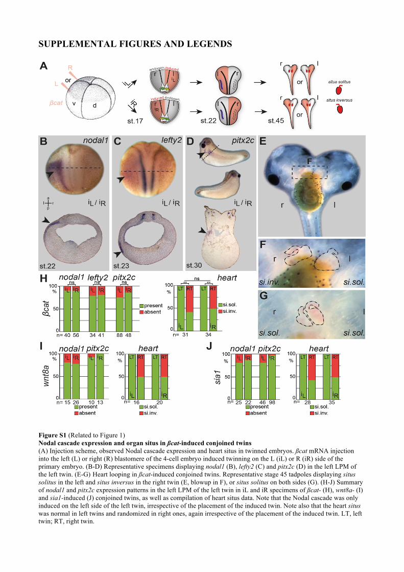
Figure S1 (Related to Figure 1) Nodal cascade expression and organ situs in ßcat-induced conjoined twins (A) Injection scheme, observed Nodal cascade expression and heart situs in twinned embryos. ßcat mRNA injection into the left (L) or right (R) blastomere of the 4-cell embryo induced twinning on the L (iL) or R (iR) side of the primary embryo. (B-D) Representative specimens displaying nodal1 (B), lefty2 (C) and pitx2c (D) in the left LPM of the left twin. (E-G) Heart looping in ßcat-induced conjoined twins. Representative stage 45 tadpoles displaying situs solitus in the left and situs inversus in the right twin (E, blowup in F), or situs solitus on both sides (G). (H-J) Summary of nodal1 and pitx2c expression patterns in the left LPM of the left twin in iL and iR specimens of ßcat- (H), wnt8a- (I) and sia1-induced (J) conjoined twins, as well as compilation of heart situs data. Note that the Nodal cascade was only induced on the left side of the left twin, irrespective of the placement of the induced twin. Note also that the heart situs was normal in left twins and randomized in right ones, again irrespective of the placement of the induced twin. LT, left twin; RT, right twin.
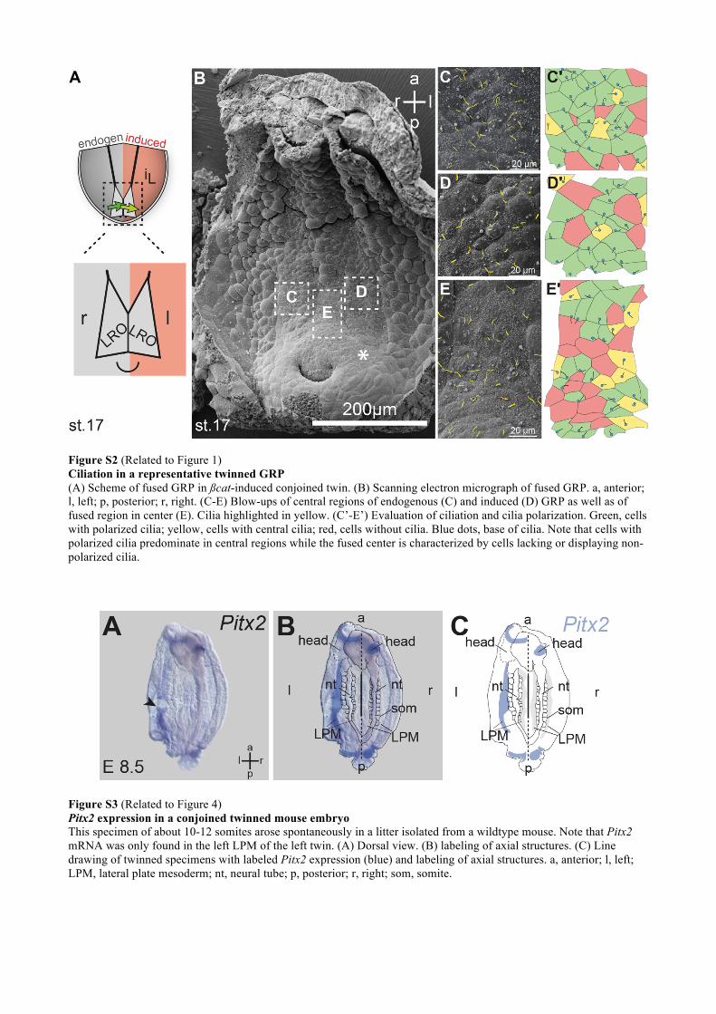
Figure S2 (Related to Figure 1) Ciliation in a representative twinned GRP (A) Scheme of fused GRP in ßcat-induced conjoined twin. (B) Scanning electron micrograph of fused GRP. a, anterior; l, left; p, posterior; r, right. (C-E) Blow-ups of central regions of endogenous (C) and induced (D) GRP as well as of fused region in center (E). Cilia highlighted in yellow. (C’-E’) Evaluation of ciliation and cilia polarization. Green, cells with polarized cilia; yellow, cells with central cilia; red, cells without cilia. Blue dots, base of cilia. Note that cells with polarized cilia predominate in central regions while the fused center is characterized by cells lacking or displaying non-polarized cilia.
Figure S3 (Related to Figure 4) Pitx2 expression in a conjoined twinned mouse embryo This specimen of about 10-12 somites arose spontaneously in a litter isolated from a wildtype mouse. Note that Pitx2 mRNA was only found in the left LPM of the left twin. (A) Dorsal view. (B) labeling of axial structures. (C) Line drawing of twinned specimens with labeled Pitx2 expression (blue) and labeling of axial structures. a, anterior; l, left; LPM, lateral plate mesoderm; nt, neural tube; p, posterior; r, right; som, somite.

SUPPLEMENTAL EXPERIMENTAL PROCEDURES
Manipulations of Xenopus embryos
Xenopus laevis eggs were obtained and fertilized in vitro as described [S1]. Embryos were injected
in 2% Ficoll in 1×MBSH buffer and cultured in 0.1× MBSH (pH7.4). Developing embryos were
reared at 14-18°C and staged according to [S2]. Embryo culture and microinjection followed
standard procedures [S1]. mRNAs were prepared using the Ambion message machine kit. For
microinjections, drop size was calibrated to about 4 nl/injection. mRNAs were transcribed from a
CS2+-expression vector and injected at 60 ng/µl (ßcat), 20 ng/µl (wnt8a) and 0.4 ng/µl (sia1). For
gene knock-down experiments, previously published MOs were used at the following
concentrations: dnah9MO, 1 pmol/embryo [S3]; dand5MO, 0.5 pmol/embryo [S4]; control MO, 0.5
- 1 pmol/embryo (Gene Tools LLC). To block cilia-driven leftward flow using methylcellulose
(MC), stage 14 embryos were injected into the archenteron with control buffer (1xMBSH) or 1.5%
MC dissolved in buffer and cultured until the stages indicated, as described [S5]. Statistical
calculations were performed using Pearson’s chi-square test comparing the number of affected
embryos against the number of wildtype embryos (Statpages.com).
RNA in situ hybridization and histological analysis
Embryos were fixed in 4% PFA for 2 hrs and processed following standard protocols [S6].
Digoxigenin-labelled (Roche) RNA probes were prepared from linearized plasmids using SP6 or T7
RNA polymerase (Promega). For histological analyses embryos were embedded in gelatine-
albumin and sectioned (35µm) on a vibratome (Leica).
Scanning Electron Microscopy (SEM)
SEM analysis was performed following published protocols [S7]. In brief, embryos were dissected
and immediately fixed in 2.5% glutaraldehyde in Soerensen’s buffer (0.1 M sodium phosphate
buffer; pH 7.4). Specimens were postfixed in 1% OsO4, critical point dried, sputter coated, and
examined using a Zeiss DSM 940A SEM.
Animal experimentation
Handling, care and experimental manipulations of Xenopus laevis frogs were approved by the
Regional Government Stuttgart, Germany (A379/12 ZO Molekulare Embryologie) and performed
according to German laws and regulations (§6, article 1, sentence 2, nr. 4 of the animal protection
act).
SUPPLEMENTAL REFERENCES
[S1] Sive, H.L, Grainger, R.M, Harland, R.M. Sive, H. (2000). Early development of Xenopus
laevis: a laboratory manual (New York: Cold Spring Harbor Laboratory Press). [S2] Nieuwkoop, P.D, Faber, J. (1994). Normal table of Xenopus laevis (Daudin). A systematical
and chronological survey of the development from the fertilized egg till the end of metamorphosis, (New York: Garland Pub.).
[S3] Vick, P., Schweickert, A., Weber, T., Eberhardt, M., Mencl, S., Shcherbakov, D., Beyer, T. and Blum, M. (2009). Flow on the right side of the gastrocoel roof plate is dispensable for symmetry breakage in the frog Xenopus laevis. Dev. Biol. 33, 281-91.
[S4] Vonica, A., and Brivanlou, A.H. (2007). The left-right axis is regulated by the interplay of Coco, Xnr1 and derrière in Xenopus embryos. Dev. Biol. 303, 281-94.
[S5] Schweickert, A., Weber. T., Beyer, T., Vick, P., Bogusch, S., Feistel, K., and Blum, M. (2007). Cilia-Driven Leftward Flow Determines Laterality in Xenopus. Curr. Biol. 17, 60-6.
[S6] Belo, J.A., Bouwmeester, T., Leyns, L., Kertesz, N., Gallo, M., Follettie, M., and De Robertis, E.M. (1997). Cerberus-like is a secreted factor with neutralizing activity expressed in the anterior primitive endoderm of the mouse gastrula. Mech. Dev. 68, 45-57.
[S7] Sulik, K., Dehart, D.B., Iangaki, T., Carson, J.L., Vrablic, T., Gesteland, K., and Schoenwolf, G.C. (1994). Morphogenesis of the murine node and notochordal plate. Dev. Dyn. 201,260-78.