lêda maria braga t (universidade federal de santa catarina ... · 1/11/2008 · (universidade...
TRANSCRIPT

MATTE: EXISTA FALA NEUTRA PARA A POESIA? 175
COMPREHENDING THE TOPIC OF A PARAGRAPH:
A FUNCTIONAL IMAGING STUDY OF A COMPLEX LANGUAGE PROCESS
(Compreendendo o tópico de um parágrafo: Um estudo de
neuroimagem funcional sobre um processo lingüístico complexo)
Lêda Maria Braga TOMITCH
(Universidade Federal de Santa Catarina)
Sharlene D. NEWMAN
(Indiana University-EUA)
Patricia A. CARPENTER and Marcel Adam JUST(Carnegie Mellon University-EUA)
ABSTRACT: This study uses fMRI (functional magnetic resonance imaging) to investigate
the brain activity in a set of cortical areas in the task of main idea identification, when
the topic sentence was presented in first versus in last position in a three-sentence paragraph.
The participants were eight right-handed undergraduate students from Carnegie Mellon
University, six male and 2 female, all native speakers of English. Each participant
read twelve paragraphs, six in which the topic sentence was paragraph initial and six
in which it was paragraph final, and each paragraph was presented word by word in
the center of a screen, inside the scanner. The major finding of the current study is the
differential response observed in the left and right hemispheres as to the location of the
topic sentence within the paragraph. The left temporal region showed greater activation
when the topic sentence was in final position than in initial position. The right temporal
region, on the other hand, was affected only by sentence type, showing a greater response
to topic sentences than support sentences, regardless of their location within the paragraph.
KEY-WORDS: FMRI; Main idea identification; Discourse processing.
RESUMO: Este estudo utiliza a ressonância magnética funcional para investigar a
atividade cerebral em áreas corticais durante a execução da tarefa de extração de idéias
principais, quando a idéia principal é apresentada no início ou ao final de um parágrafo
com três orações. Os participantes da pesquisa foram oito alunos de graduação da
Universidade de Carnegie Mellon, todos destros, falantes nativos do inglês. Cada
participante leu doze parágrafos, seis com a idéia principal no início e seis com a idéia
principal no final do parágrafo, e cada parágrafo foi apresentado palavra por palavra,
D.E.L.T.A., 24:2, 2008 (175-197)
C_delta_24-2.p65 1/11/2008, 18:55175

176 D.E.L.T.A., 24:2
no centro de uma tela, dentro do aparelho de ressonância magnética. O maior achado do
presente estudo refere-se à resposta diferenciada observada nos hemisférios direito e esquerdo.
A região temporal esquerda mostrou maior atividade cerebral quando a idéia principal
era apresentada em posição final do que em posição inicial. A região temporal direita,
por sua vez, só foi afetada pelo tipo de oração, mostrando uma maior atividade durante
o processamento da idéia principal do que das idéias secundárias, independente da sua
posição no parágrafo, inicial ou final.
PALAVRAS-CHAVE: Ressonância magnética funcional; Identificação de idéias principais;
Processamento textual.
1. Introduction
Main idea identification is at the very heart of human thinking, beinga skill required in everyday situations such as reading a message, interpretingan interlocutor’s utterance, listening to the news and attending a lecture.It is part of human nature to try to integrate incoming information orbuild a macrostructure containing the main points of the input, so thatthis information can be more easily stored in memory, related to otherknowledge, and retrieved when needed (Kintsch & van Dijk 1978; vanDijk & Kintsch 1983). Despite its importance in human interaction, theprocess of main idea identification is little understood. Here, a textcomprehension task is used to explore the process of identifying the mainidea, or topic of a text.
Cognitive brain imaging has provided researchers new possibilities fortrying to understand what happens in the human brain during theperformance of various complex tasks. While there is an extensiveneuroimaging literature investigating comprehension processes at the wordlevel (e.g. Petersen et al 1989,1990; Pugh et al 1996, 2000; Binder 1997;Fiez & Petersen 1998; Brunswick et al 1999; Hagoort et al 1999; Shaywitzet al 2000; Waldie & Mosley 2000), and sentence level (Mazoyer et al1993; Bottini et al 1994, Just et al 1996; Stromswold et al 1996; Bavelieret al 1997; Schlosser et al 1998; Caplan et al 1999; Keller et al 2001; andMichael et al 2001), there are few neuroimaging studies that have gonebeyond the sentence level and examined discourse comprehension (Mazoyer
C_delta_24-2.p65 1/11/2008, 18:55176

TOMITCH; NEWMAN & JUST: COMPREENDING THE TOPIC OF A PARAGRAPH 177
et al 1993; Nichelli et al 1995; Dehaene et al 1997; St George et al. 1999;Robertson et al 2000; Mason & Just 2004; Tomitch, Newman & Just2004). Lesion studies, however, have provided some insight into theunderlying neural architecture that supports discourse comprehension. Ithas been found that patients with right hemisphere damage often experiencedifficulties at the discourse level even though their ability to comprehendat the word and sentence level are intact. For example, right hemispherepatients often have problems maintaining the theme of discourse (Brownell& Martino 1998), suggesting that the right hemisphere is responsible formain idea identification and maintenance.
The current study examines main idea identification by examiningthe effect of the position of the topic sentence in three-sentence paragraphs.There were two conditions that differed in the position of the main idea/topic sentence in the paragraph: in first position – topic first and in lastposition– topic last. The topic sentence contained a superordinate themethat unified the concepts in the other sentences. The placement of thetopic sentence dramatically affects how the coherence of the discourse isestablished and therefore, the ease at which the text is comprehended. Theexperimental design was intended to contrast the processes of integratingtext details into a previously established theme, versus first storing detailsor dynamically integrating them prior to reading the statement of thetopic, and then relating them to the topic.
Computational models have been proven to be very important inunderstanding cognition. As such, the Structure Building Framework (SBF)of Gernsbacher (1990, 1995, 1997) is used here to frame the current studyand to guide its interpretation. SBF can account for several phenomena(e.g., the advantage of the first mention and the clause recency effect)related to the comprehension processes that establish text coherence.According to the SBF, discourse comprehension builds cohesive mentalrepresentations using three general processes: laying the foundation of atext representation, mapping incoming information from the text toprevious information, and initiating a new substructure if the incominginformation is not adequately coherent with previous information. SBFstates that the first step in building a mental representation of the text isto lay a foundation to which subsequent information presented in the textcan be attached. Laying a foundation presumably consists of selecting or
C_delta_24-2.p65 1/11/2008, 18:55177
178 D.E.L.T.A., 24:2
constructing an organizing structure or schema for the text. Once thefoundation has been built, the structure is then elaborated by mappingincoming information to the schema and shifting to develop newsubstructures when needed. SBF can be used to make several predictionsregarding the results of the current study. SBF states that attempts to laythe foundation begin with the first phrase of the first sentence, regardlessof the content of that phrase. Therefore, SBF would predict that theinitiation of this content-free foundation formation process has no preferenceas to which sentence type comes first, either topic or support sentence.Also, according to SBF, in the topic last condition the topic of the text isavailable after the initiation of foundation formation. Therefore, a secondprediction is that more substructures may be expected to be generatedduring the topic last condition compared to the topic first condition whichindicates more “shifting” processing. This suggests that the increase inshifting is expected to result in a higher level of activation for the topicsentence when it is in the final position compared to when it is located inthe initial position. This also suggests that the text representation generatedwhen the topic is last is more complex and may be more memory intensive.
SBF is a psychological model of discourse processing and therefore,does not address the neural basis of text comprehension. One neural-basedhypothesis concerning comprehension that does relate to discourseprocessing is the coarse coding hypothesis proposed by Beeman (1993;1998; Beeman et al 1994). The coarse coding hypothesis proposes thatthe two hemispheres differ in the comprehensiveness of the representationsthey activate. According to this hypothesis, the left hemisphere uses fine(precise) semantic coding to selectively activate a small number of relevantmeanings or features when processing language. The right hemisphere, onthe other hand, is proposed to use a coarse semantic coding scheme inwhich it weakly activates a broad spectrum of related meanings and features,inducing diffusely activated semantic fields distributed over manyrepresentations. This type of coding scheme is thought to support inferencegeneration, which is often necessary to connect events within a passage.
The goal of the current study is to attempt to characterize the neuralarchitecture that supports discourse processing, particularly topicidentification. This was done by using fMRI to examine paragraphs thatvaried the location of the topic sentence.
C_delta_24-2.p65 1/11/2008, 18:55178
TOMITCH; NEWMAN & JUST: COMPREENDING THE TOPIC OF A PARAGRAPH 179
2. Method
Participants. The participants were eight right-handed undergraduatestudents from Carnegie Mellon University, six male and 2 female, all nativespeakers of English. All participants gave written, informed consentaccording to guidelines approved by the University of Pittsburgh and theCarnegie Mellon Institutional Review Boards. Participants were paid fortheir participation.
Materials. The experimental stimuli consisted of paragraphs adaptedfrom naturally occurring expository texts. Each paragraph was rewrittenin a way that it represented a complete text on its own, “a coherent pieceof writing exhibiting both structure and texture…” (Davies 1995: 94).All sentences in each paragraph were equated for approximately the samenumber of words, ranging from 19 to 21, and also equated for number ofcharacters, ranging from 110 to 130.
The main idea of each paragraph was identified on the basis ofCunningham and Moore’s (1986) definition of a main idea pertaining tothe thesis sentence: “The single sentence in a paragraph or passage whichtells most completely what the paragraph or passage as a whole states or isabout” (p.7). The two supporting sentences in each paragraph presenteddetails and information which illustrated, exemplified or gave support tothe main point.
In the topic first condition, the first sentence introduced the theme ofthe paragraph and the following two sentences presented supportingarguments and details related to the main idea, being easily integratedinto one uniform whole as shown in Table 1. In the topic last condition,the supporting arguments and details were presented in the first twosentences making it difficult to integrate them before reading the lastsentence, which made the main idea of the paragraph explicit. Two versionsof the experimental task were developed. In each version the paragraphscontained the same sentences. The two versions were different in that thelocation of the topic sentences changed from the first position to the finalposition and visa versa. In this way each paragraph occurred under each ofthe two conditions: Topic first and Topic last. Each participant saw onlyone of the two versions of each paragraph. After reading each paragraph,participants answered true/false probe questions about the thematicinformation presented (see Table 1 below).
C_delta_24-2.p65 1/11/2008, 18:55179
180 D.E.L.T.A., 24:2
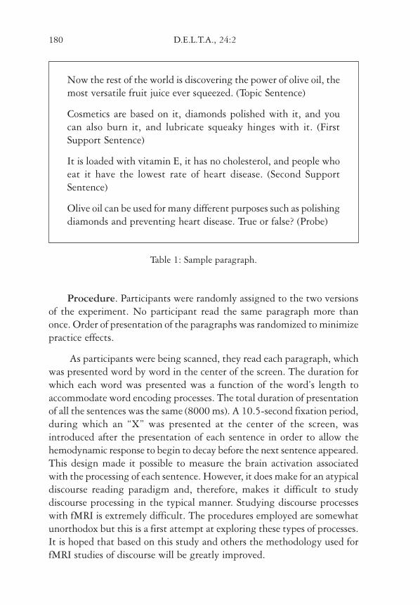
Now the rest of the world is discovering the power of olive oil, themost versatile fruit juice ever squeezed. (Topic Sentence)
Cosmetics are based on it, diamonds polished with it, and youcan also burn it, and lubricate squeaky hinges with it. (FirstSupport Sentence)
It is loaded with vitamin E, it has no cholesterol, and people whoeat it have the lowest rate of heart disease. (Second SupportSentence)
Olive oil can be used for many different purposes such as polishingdiamonds and preventing heart disease. True or false? (Probe)
Table 1: Sample paragraph.
Procedure. Participants were randomly assigned to the two versionsof the experiment. No participant read the same paragraph more thanonce. Order of presentation of the paragraphs was randomized to minimizepractice effects.
As participants were being scanned, they read each paragraph, whichwas presented word by word in the center of the screen. The duration forwhich each word was presented was a function of the word’s length toaccommodate word encoding processes. The total duration of presentationof all the sentences was the same (8000 ms). A 10.5-second fixation period,during which an “X” was presented at the center of the screen, wasintroduced after the presentation of each sentence in order to allow thehemodynamic response to begin to decay before the next sentence appeared.This design made it possible to measure the brain activation associatedwith the processing of each sentence. However, it does make for an atypicaldiscourse reading paradigm and, therefore, makes it difficult to studydiscourse processing in the typical manner. Studying discourse processeswith fMRI is extremely difficult. The procedures employed are somewhatunorthodox but this is a first attempt at exploring these types of processes.It is hoped that based on this study and others the methodology used forfMRI studies of discourse will be greatly improved.
C_delta_24-2.p65 1/11/2008, 18:55180

TOMITCH; NEWMAN & JUST: COMPREENDING THE TOPIC OF A PARAGRAPH 181
After reading each paragraph, participants answered true or false towhether a probe statement represented the main point in the paragraph.The probe questions contained details from each of the three sentencesof the paragraph in order to encourage participants to generate acomplete text representation. A probe deadline procedure was used and,on the basis of the results in a behavioral pilot study, participants had amaximum of 7 seconds to respond to the probe, after which it disappearedfrom the screen.
Each participant read twelve paragraphs, six in which the topic sentencewas paragraph initial and six in which it was paragraph final. Althoughthe number of trials per condition was not large, it was found to yieldsignificant activation within the regions of interest. However, the numberof trials may have caused “real” activation to not be detected due to lowsignal to noise. Because the goal of the study was to determine if there areprocessing differences related to the location of the topic sentence withintemporal cortex, the current design was thought to be sufficient as a baselineexperiment with future studies being planned based upon the currentfindings.
Four 24 second fixation periods (again, an “X” centered on the screen)were placed at the beginning, end, and at the two trisection points of thestudy, to obtain a baseline measure of activation.
Data Acquisition. Functional data were acquired in seven contiguousoblique axial slices situated in order to maximize coverage of the languageprocessing regions (primarily Broca’s and Wernicke’s areas). The scanningwas conducted on a 1.5T General Electric Signa scanner, with a TR=1500ms, TE=50 ms, flip angle=90o, FOV=40x20 cm, matrix size=128x64voxels, and 1 mm gap, resulting in a voxel size of 3.125 x 3.125 x 5 mm.The slice prescription was set so that the posterior, superior temporal cortexand the inferior frontal gyrus (primarily the superior portion) wereadequately covered. Scanning was synchronized with stimulus presentation,the acquisition of the first slice occurring at the onset of each sentencepresentation.
For anatomical localization, 3D SPGR structural images were acquiredwith the following parameters: 124 slices (1.5 mm thick), TR=25 ms,
C_delta_24-2.p65 1/11/2008, 18:55181
182 D.E.L.T.A., 24:2
TE=4 ms, flip angle=40o, and FOV=24 cm. Functional maps were co-registered with the corresponding structural images.
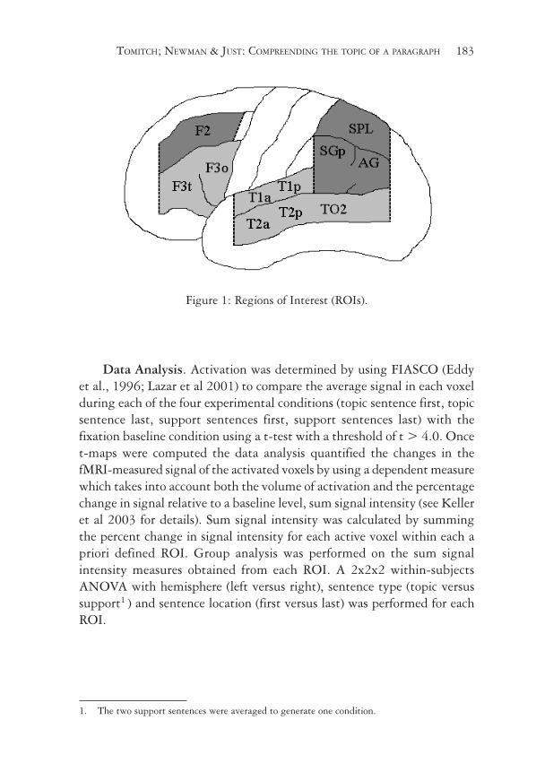
Anatomical Regions of Interest (ROI). To compare the amount ofactivation in a given area between the two experimental conditions,anatomically-defined ROIs were drawn for each participant using theparcellation scheme described by Rademacher, Galaburda, Kennedy,Filipek, and Caviness (1992) and further refined by Caviness, Meyer, Makris,and Kennedy (1996). The schematic drawing in Figure 1 below displaysmost of the ROIs that were examined. This method uses limiting sulci andanatomically landmarked coronal planes to segment cortical regions andhas been shown to provide more accurate anatomical localization thandoes morphing all participants’ brains into a common space (Nieto-Castanon et al 2003).
A staff research assistant defined the ROIs after extensive training onthe Rademacher/Caviness parcellation scheme. The anatomical informationin the structural images was displayed in the three orthogonal planessimultaneously and the ROIs were manually drawn on each functionalslice. The inter-rater reliability of this ROI-defining procedure betweentwo trained staff members was evaluated for four ROIs in two participantsin another study. The reliability measure was obtained by dividing the sizeof the set of voxels that overlapped between the two raters by the mean oftheir two set sizes. The resulting eight reliability measures were in the 78-91% range, with a mean of 84%, as high as the reliability reported by thedevelopers of the parcellation scheme (Just et al 2001).
The primary regions of interest were the inferior frontal gyrus (Broca’sarea) and the superior temporal gyrus, which includes Wernicke’s area.However, data from all cortical areas covered by the seven slices obtainedare reported for completeness (see Figure 1 below). The inferior frontalROI (LIFG) included areas F3t and F3o, referring to the Caviness et al.(1996) nomenclature, or approximately BA 44 and a portion of 45. Thetemporal ROI (LT) included the superior (T1a and T1p, BA 22) and middletemporal gyri (T2a, T2p, and TO2; BA 21, and 37). The superior andportions of the middle temporal gyri were combined into one ROI becauseprevious studies of language processing have often found activation centeredin the superior temporal sulcus between them (Just et al 2001). The ROIswere drawn separately for the left and right hemispheres.
C_delta_24-2.p65 1/11/2008, 18:55182
TOMITCH; NEWMAN & JUST: COMPREENDING THE TOPIC OF A PARAGRAPH 183
Figure 1: Regions of Interest (ROIs).
Data Analysis. Activation was determined by using FIASCO (Eddyet al., 1996; Lazar et al 2001) to compare the average signal in each voxelduring each of the four experimental conditions (topic sentence first, topicsentence last, support sentences first, support sentences last) with thefixation baseline condition using a t-test with a threshold of t > 4.0. Oncet-maps were computed the data analysis quantified the changes in thefMRI-measured signal of the activated voxels by using a dependent measurewhich takes into account both the volume of activation and the percentagechange in signal relative to a baseline level, sum signal intensity (see Kelleret al 2003 for details). Sum signal intensity was calculated by summingthe percent change in signal intensity for each active voxel within each apriori defined ROI. Group analysis was performed on the sum signalintensity measures obtained from each ROI. A 2x2x2 within-subjectsANOVA with hemisphere (left versus right), sentence type (topic versussupport1 ) and sentence location (first versus last) was performed for eachROI.
1. The two support sentences were averaged to generate one condition.
C_delta_24-2.p65 1/11/2008, 18:56183

184 D.E.L.T.A., 24:2
3. Results
Behavioral Results. The reaction time and error rate related to the
comprehension probe did not differ between conditions, as shown in Figure
2 below.
Figure 2: Reaction time to the comprehension probeand error rate as a function of topic sentence location.
fMRI Results. There were two main findings. First, the serial position
of the topic sentence had a much larger impact on the left hemisphere
language regions than on their right homologues, with the left temporal
area showing the most significant effect (see Figures 3 and 4). Second, the
right temporal region showed more activation to topic sentences than
supporting sentences, regardless of their location within the paragraph
(see also, Tomitch, Just & Newman 2004). These findings are reported in
more detail below, focusing on the higher-level language processing regions,
namely the inferior frontal gyrus (LIFG or Broca’s area) and temporal cortex
(Wernicke’s area). Table 2 below shows the activation levels and centroids
of the activation for each ROI.
PR2_delta_24-2.p65 29/8/2009, 18:02184

TOMITCH; NEWMAN & JUST: COMPREENDING THE TOPIC OF A PARAGRAPH 185
Table 2: Activation levels (Sum Percent Signal Intensity).
First Last
ROI Support Topic Support Topic x y z
L. Temporal Cortex 43.51 44.71 38.75 55.88 52.5 34.12 1.4
R. Temporal Cortex 14.36 21.78 15.87 22.92 -50.44 37.9 6.37
L. Inferior Frontal Gyrus 20.04 13.15 16.09 25.02 46.16 -12.91 21.88
R. Inferior Frontal Gyrus 3.1 2.64 2.24 2.7 -43.46 -19.05 23.98
L. Parietal Cortex 6.12 6.9 7.69 8.74 38.26 59.42 18.39
R. Parietal Cortex 1.92 3.18 1.2 1.61 -29.95 66.3 26.97
L. Dorsolateral Prefrontal
Cortex 1.83 1.14 1.63 2.31 39.47 -34.12 23.63
R. Dorsolateral Prefrontal
Cortex 0.78 0.71 0.9 0.67 -30.35 -45.08 18.67
L. Frontal Eye Fields 3.83 4.3 3.23 4.41 38.79 -5.11 38.21
R. Frontal Eye Fields 1.05 2 2.4 1.74 -44.68 -8.35 35.74
L. Extrastriate 20.89 24.45 20.16 23.53 30.94 76.22 -8.65
R. Extrastriate 7.93 11.1 9.84 12.72 -39.52 68.92 -6.06
Figure 3: Talairach averaged activation maps (A) Activationelicited by the topic sentence when presented first; B) Activation
elicited by the topic sentence when presented last.).
C_delta_24-2.p65 1/11/2008, 18:56185
186 D.E.L.T.A., 24:2
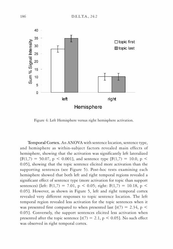
Figure 4: Left Hemisphere versus right hemisphere activation.
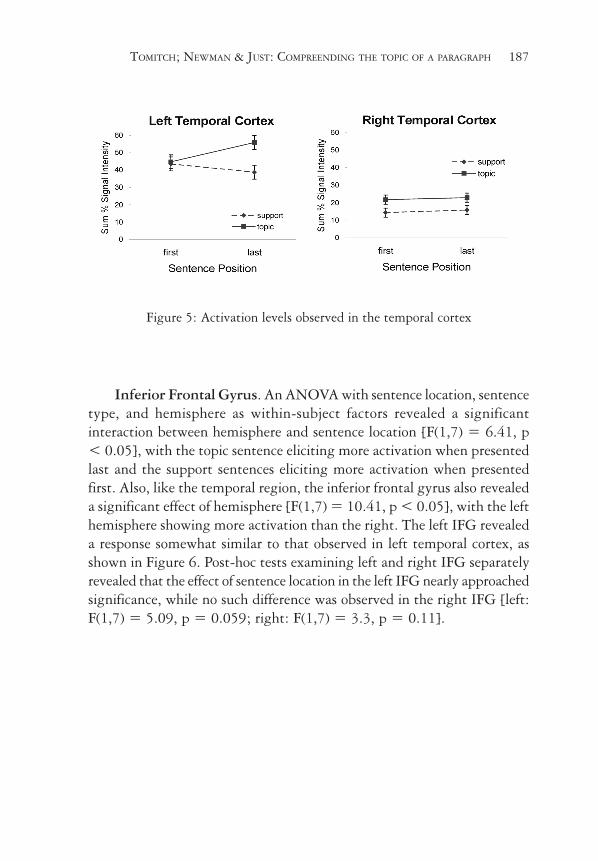
Temporal Cortex. An ANOVA with sentence location, sentence type,and hemisphere as within-subject factors revealed main effects ofhemisphere, showing that the activation was significantly left lateralized[F(1,7) = 50.07, p < 0.001], and sentence type [F(1,7) = 10.0, p <0.05], showing that the topic sentence elicited more activation than thesupporting sentences (see Figure 5). Post-hoc tests examining eachhemisphere showed that both left and right temporal regions revealed asignificant effect of sentence type (more activation for topic than supportsentences) [left: F(1,7) = 7.01, p < 0.05; right: F(1,7) = 10.18, p <0.05]. However, as shown in Figure 5, left and right temporal cortexrevealed very different responses to topic sentence location. The lefttemporal region revealed less activation for the topic sentences when itwas presented first compared to when presented last [t(7) = 2.34, p <0.05]. Conversely, the support sentences elicited less activation whenpresented after the topic sentence [t(7) = 2.1, p < 0.05]. No such effectwas observed in right temporal cortex.
C_delta_24-2.p65 1/11/2008, 18:56186
TOMITCH; NEWMAN & JUST: COMPREENDING THE TOPIC OF A PARAGRAPH 187
Figure 5: Activation levels observed in the temporal cortex
Inferior Frontal Gyrus. An ANOVA with sentence location, sentencetype, and hemisphere as within-subject factors revealed a significantinteraction between hemisphere and sentence location [F(1,7) = 6.41, p< 0.05], with the topic sentence eliciting more activation when presentedlast and the support sentences eliciting more activation when presentedfirst. Also, like the temporal region, the inferior frontal gyrus also revealeda significant effect of hemisphere [F(1,7) = 10.41, p < 0.05], with the lefthemisphere showing more activation than the right. The left IFG revealeda response somewhat similar to that observed in left temporal cortex, asshown in Figure 6. Post-hoc tests examining left and right IFG separatelyrevealed that the effect of sentence location in the left IFG nearly approachedsignificance, while no such difference was observed in the right IFG [left:F(1,7) = 5.09, p = 0.059; right: F(1,7) = 3.3, p = 0.11].
C_delta_24-2.p65 1/11/2008, 18:56187

188 D.E.L.T.A., 24:2
Figure 6: Activation levels observed in the inferior frontal gyrus
4. Discussion
The current study is a first attempt at exploring the underlyingprocesses associated with topic identification. The major finding of thecurrent study is the differential response observed in the left and righthemispheres to the location of the topic sentence within a paragraph. Thetwo primary left hemisphere brain regions examined here (temporal cortexand inferior frontal gyrus) responded in similar ways, although to differentdegrees, to the topic location in this study: greater activation occurredwhen the topic sentence was in the final position than in the initial position.The right temporal region, on the other hand, was affected only by thesentence type, showing a greater response to topic sentences than supportsentences, regardless of their location within the paragraph.
The Structure Building Framework– SBF (Gernsbacher 1990, 1995,1997) allows for the framing of the current study by making a number ofpredictions. One such prediction is related to the differential involvementof the “shifting” process in paragraphs whose topic sentence is in the finalposition. This is because when the topic is in the final position the lesscoherent is the text, meaning the sentences seem less related which thenleads to a greater number of substructures generated within the textrepresentation (i.e., greater “shifting”). Therefore, this increase in the
C_delta_24-2.p65 1/11/2008, 18:56188

TOMITCH; NEWMAN & JUST: COMPREENDING THE TOPIC OF A PARAGRAPH 189
amount of “shifting” taking place during the topic last condition would beexpected to result in greater activation for the topic last condition comparedto the topic first condition. This predicted response was observed in theleft temporal region, not the right, as shown in Figure 5. The left temporalregion revealed a differential response to the topic location, with the topicsentence eliciting more activation when it was in the final position thanwhen it was in the initial position. In addition, the support sentences showedthe opposite effect: greater activation when they were located in the initialposition compared to when they were in the final position. Thus, while theleft hemisphere is involved in comprehension processes at all times, theintroduction of coherence gaps increases its processing load. This increasedprocessing load may be a function of an increased need to shift or reorganizethe text representation in memory.
The increased involvement of the left hemisphere during the topiclast condition appears to be at odds with previous reports in the literature.For example, St George et al (1999), in an fMRI study of semanticintegration in reading in which participants read vague and ambiguousparagraphs in the Bransford and Johnson (1972) unlabeled text style, founda different pattern of activation for titled and untitled paragraphs, wheretitled paragraphs were similar to the topic first condition and untitledparagraphs were similar to the topic last condition here. While the currentresults revealed greater left hemisphere involvement for the topic lastcompared to topic first condition, St George and colleagues found a similareffect in the right hemisphere; untitled paragraphs produced higher levelsof activation in the right hemisphere than titled paragraphs. Although thetwo studies appear to be at odds, there are a number of methodologicaldifferences that may account for the disparate results. First, the currentstudy requires participants to respond to a comprehension question afterreading the paragraph; there is no such requirement in the previous study.Secondly, the topic (title) is always provided in the current study therefore,there is always opportunity to generate a coherent text representation. Inthe previous study the untitled condition never provides a unifying themeto bring all of the information together making it impossible to generate acoherent representation. Finally, the anatomical regions scanned in thetwo studies are quite different. The slices acquired in the St. George studycovered the middle and inferior temporal cortex and the inferior portion ofBroca’s area only. The current study covered portions of the middle temporalcortex, the superior temporal-parietal cortex (Wernike’s area) and the
C_delta_24-2.p65 1/11/2008, 18:56189

190 D.E.L.T.A., 24:2
superior portion of Broca’s area, neither of which were scanned in theprevious study. This difference in the anatomical regions examined is asignificant one because these regions have been shown to be involved indifferent processes, particularly in the left hemisphere. For example, theanterior, inferior portion of Broca’s area (BA 45) has been found to beinvolved in semantic processing (Newman et al 2003; Fiez 1997; Wagneret al 2000) while the posterior, superior portion (BA 44) is more involvedin syntactic processing (Dapretto et al 1999; Embick et al 2000; Newmanet al 2003). Although there are fewer reports investigating the processingin the right hemisphere, similar differences may be expected. As a result ofthese methodological differences it is difficult to directly compare the studiesand further research is needed to clarify this issue.
The current study seems to suggest a role for the left temporal andinferior frontal region in the shifting process. One possible explanation forthe left temporal region’s involvement in shifting may be found in theregion’s involvement in memory processing more generally. Consider thepossibility that when reading a text various concepts activate a certainfeature space within memory and as incoming information comes in, thereis a check to see how much the new concept’s feature space overlaps withprevious information. If the overlap is above some threshold, then it ispossible that it is just added to the current representation. If not, then a“shift” happens and a new, or a more elaborate memory substructure iscreated. It may be that the left hemisphere is responsible for holding thisrepresentation on-line. There are many studies that associate left temporaland inferior frontal cortex to memory functions including semantic memory.For example, the superior portion of the inferior frontal gyrus is thoughtto become more involved during sentence comprehension when memory-demanding dependencies between elements in a sentence must beestablished (Fiebach, et al 2004; Cooke et al 2001; Fiebach et al 2001). Inthe current study, when the topic is in the final position similar memory-demanding dependencies must be established in order to relate the sentenceswithin the paragraph.
In an fMRI study of the cognitive process of mapping (‘…identify…recurring concepts and have a means for interrelating them’, p.3), Robertsonet al (2000) gave participants two sets of narrative paragraphs. In one set,the sentences contained the definite article ‘the’ modifying nouns. Anotherset of sentences contained the indefinite article ‘a’. Robertson et al (2000)
C_delta_24-2.p65 1/11/2008, 18:56190

TOMITCH; NEWMAN & JUST: COMPREENDING THE TOPIC OF A PARAGRAPH 191
and Gernsbacher and Robertson (1992) suggested that the definite article‘the’ serves as a cue to map a representation of the current input and foundthe involvement of the right hemisphere. One of the consequences of havingthe topic sentence in the final position is that the preceding supportingsentences have unresolved anaphors. For example, the support sentence“It is loaded with vitamin E, it has no cholesterol, and people who eat ithave the lowest rate of heart disease” does not state what “it” refers to. Incontrast, when the topic sentence is in the initial position, such as “Nowthe rest of the world is discovering the power of olive oil, the most versatilefruit juice ever squeezed”, the topic of the paragraph is explicitly stated(olive oil in this case), facilitating the resolution of anaphors in thesubsequent support sentences (it-olive oil). Therefore, in the currentexperiment, the mapping process may be expected to be cued more oftenwhen the topic sentence is located in the final position because only thencan the recurring concepts be identified and interrelated.
The results presented here do suggest significant processing differencesbetween the left and right hemisphere during text processing. Accordingto the coarse coding hypothesis the left hemisphere activates a small numberof representations to a high degree making it ideal for working/short-termmemory language functions. Also, previous studies at the word and sentencelevel have shown that the left hemisphere is involved in memory intensivelanguage processing such as semantic and syntactic processing. Theseprocesses occur at both the sentence and discourse level. However, becausethe left hemisphere is so involved in memory it may be that the textrepresentation is stored/processed in the left hemisphere. If this is the casethen its increased involvement when the topic is located at the end of aparagraph would be due to the reorganization or even the generation ofthis memory structure.
The right hemisphere, on the other hand, is thought to activate abroad range of concepts at a weak level making it ideal for inferencegeneration that connects events and details within a text. The fact that itis equally involved during the processing of the topic sentence regardlessof its location may be a result of different processes. For example, it maybe that when the topic is first the right hemisphere is involved in layingthe foundation of the text representation and beginning to weakly activatea number of concepts related to the topic. Conversely, when the topic is inthe final position the right hemisphere may be involved in the mapping
C_delta_24-2.p65 1/11/2008, 18:56191

192 D.E.L.T.A., 24:2
process of SBF in order to perform anaphor resolution and to begin torelate the information that was presented previous to the topic. This wouldcoincide with the shifting processes taking place in the left hemispherethat are reorganizing/generating a memory structure. Knowledge of howto relate the new information with the old is needed before the textrepresentation can be edited and this knowledge may be generated by theright hemisphere.
5. Conclusions
Discourse comprehension is a collaborative effort involving both theleft and right hemisphere language areas, as these fMRI results show. Thesystematic activation observed in both hemispheres in our study suggeststhat the two hemispheres work in an integrated manner, each beingdifferentially responsible for various aspects of language processing (Mazoyeret al 1993), but working together to achieve the more global role ofdiscourse comprehension. The integrated manner in which the biologyappears to execute discourse processing also suggests that the underlyingcognitive processes are non-modular. For example, the data presented heresuggests that the foundation is not formed irrespective of the content ofthe initial sentence and that the lexical/semantic processing of eachindividual sentence in a text dynamically influences the text representation.
Recebido em novembro de 2005Aprovado em outubro de 2007
E-mail: [email protected]
REFERENCES
BAVELIER, D.; CORINA, D.; JEZZARD, P.; PADMANABHAN, S.; CLARK, V. P.;KARNI, A.; PRINSTER, A.; BRAUN, A.; LALWANI, A.; RAUSCHECKER, J. P.;TURNER, R. & NEVILLE, H. 1997. Sentence reading: A functional MRIstudy at 4 Tesla. Journal of Cognitive Neuroscience 9: 664-686.
BEEMAN, M. 1993. Semantic processing in the right hemisphere maycontribute to drawing inferences during comprehension. Brain andLanguage, 44: 80-120.
C_delta_24-2.p65 1/11/2008, 18:56192
TOMITCH; NEWMAN & JUST: COMPREENDING THE TOPIC OF A PARAGRAPH 193
_____. 1998. Coarse semantic coding and discourse comprehension. In:M. Beeman & C. Chiarello, (Eds.), Right hemisphere languagecomprehension: Perspectives from cognitive neuroscience, pp. 255-284.Mahwah, NJ: Lawrence Erlbaum Associates.
BEEMAN, M.; FRIEDMAN, R. B.; GRAFMAN, J.; PEREZ, E.; DIAMOND, S. &LINDSAY, M. B. 1994. Summation priming and coarse coding in theright hemisphere. Journal of Cognitive Neuroscience, 6: 26-45.
BINDER, J. R. 1997. Neuroanatomy of language processing studied withfunctional MRI. Clinical Neuroscience, 4: 87-94.
BOTTINI, G; CORCORAN, R.; STERZI, R.; PAULESU, E.; SCHENONE, P.; SCARPA,P., et al. 1994. The role of the right hemisphere in the interpretationof figurative aspects of language. A positron emission t omographyactivation study. Brain, 117: 1241-53.
BRANSFORD, J. D. & JOHNSON, M. K. 1972. Contextual prerequisites forunderstanding: some investigations of comprehension and recall.Journal of Verbal Learning and Verbal Behavior, 11: 717-726.
BROWNELL, H. & MARTINO, G. 1998. Deficits in inference and socialcognition: The effects of right hemisphere brain damage on discourse.In: M. Beeman & C. Chiarello (Eds.), Right hemisphere languagecomprehension: Perspectives from cognitive neuroscience (pp. 309-328).Mahwah, NJ: Erlbaum.
BRUNSWICK, N.; MCCRORY, E.; PRICE, C. J. & FRITH, C. D. 1999. Explicitand implicit processing of words and pseudowords by adultdevelopmental dyslexics. A search for Wernicke’s Wortschatz? Brain,122: 1901-1917.
CAPLAN, D.; ALPERT, N. & WATERS, G. 1999. PET studies of syntacticprocessing with auditory sentence presentation. NeuroImage, 9: 343-351.
CAVINESS, V. S. Jr.; MEYER, J.; MAKRIS, N. & KENNEDY, D. N. 1996. MRI-based topographic parcellation of human neocortex: An anatomicallyspecified method with estimate of reliability. Journal of CognitiveNeuroscience, 8: 566-87.
COOKE, A.; ZURIF, E. B.; DEVITA, C.; ALSOP, D.; KOENIG, P.; DETRE, J.; GEE,J.; PINANGO, M.; BALOGH, J. & GROSSMAN, M. 2001. Neural basis forsentence comprehension: Grammatical and short-term memorycomponents. Human Brain Mapping, 15: 80-94.
CUNNINGHAM, J.W. & MOORE, D.W. 1986. The confused world of mainidea. In: J.F. Baumann (Ed.), Teaching main idea comprehension. Newark,DEL: International Reading Association.
C_delta_24-2.p65 1/11/2008, 18:56193
194 D.E.L.T.A., 24:2
DAPRETTO, M. & BOOKHEIMER, S. 1999. Form and content: Dissociatingsyntax and semantics in sentence comprehension. Neuron, 24:427-432.
DAVIES, Florence. 1995. Introducing reading. UK: Penguin Books.DEHAENE, S.; DUPOUX, E.; MEHLER, J.; COHEN, L.; PAULESU, E.; PERANI, D.;
VAN DE MOORTELE, P-F.; LEHERICY, S. & LEBIHAN, D. 1997. Anatomicalvariablity in the cortical representation of first and second language.NeuroReport, 8: 3809-3815.
EDDY, W.; FITZGERALD, M.; GENOVESE, C.; MOCKUS, A. & NOLL, D. 1996.Functional imaging analysis software – computational olio. In:Proceedings in Computational Statistics. Physica-Verlag, Heidelberg,pp. 39-49.
EMBICK, D.; MARANTZ, A.; MIYASHITA, Y. et al. 2000. A syntacticspecialization for Broca’s area. Proceedings of the National Academy ofScience. USA, 97,6150-6154.
FIEBACH, C. J.; SCHLESEWSKY, M. & FRIEDERICI, A. D. 2001. Syntactic workingmemory and the establishment of filler-gap dependencies: Insightsfrom ERPs and fMRI. Journal of Psycholinguistic Research, 30: 321–338.
FIEBACH, C.J.; VOS, S.H. & FRIEDERICI, A.D. 2004. Neural correlates ofsyntactic ambiguity in sentence comprehension for low and high spanreaders. Journal of Cognitive Neuroscience, 16: 1562-1575.
FIEZ, J. & PETERSEN, S. E. 1998. Neuroimaging studies of word reading.Proceedings of the National Academy of Science, USA, 95, 914-921.
GERNSBACHER, M. A. 1990. Language comprehension as structure building.Hillsdale, NJ: Lawrence Erlbaum Associates.
GERNSBACHER, M. A. 1995. The Structure Building Framework: What itis, what it might also be, and why. In: B. K. Britton, & A. C. Graesser,(Eds.), Models of text understanding (pp. 289-311). Hillsdale, NJ:Erlbaum.
GERNSBACHER, M. A. 1997. Two decades of structure building. DiscourseProcesses, 23: 265-304.
GERNSBACHER, M. A. & ROBERTSON, R. R. W. 1992. Knowledge activationversus sentence mapping when representing fictional characters’emotional states. Language and Cognitive Processes, 7: 353-371.
HAGOORT, P.; INDEGREY, P.; BROWN, C.; HERZOG, H.; STEINMETZ, H. & SEITZ,R. 1999. The neural circuitry involved in the reading of German wordsand pseudowords: a PET study. Journal of Cognitive Neuroscience, 11:383-398.
C_delta_24-2.p65 1/11/2008, 18:56194
TOMITCH; NEWMAN & JUST: COMPREENDING THE TOPIC OF A PARAGRAPH 195
FIEZ, J. A. 1997. Phonology, semantics, and the role of the left inferiorprefrontal cortex. Human Brain Mapping, 5: 79-83.
JUST, M. A.; CARPENTER, P. A.; KELLER, T. A.; EDDY, W. F. & THULBORN, K.R. 1996. Brain activation modulated by sentence comprehension.Science, 274: 114-116.
JUST, M. A.; CARPENTER, P. A.; MAGUIRE, M., DIWADKAR, V. & MCMAINS, S.2001. Mental rotation of objects retrieved from memory: an fMRIstudy of spatial processing. Journal of Experimental Psychology: General,130: 493-504.
KELLER, T. A.; CARPENTER, P. A. & JUST, M. A. 2001. The neural bases ofsentence comprehension: an fMRI examination of syntactic and lexicalprocessing. Cerebral Cortex, 11: 223-237
KELLER, T. A.; CARPENTER, P. A. & JUST, M. A. 2003. Brain imaging oftongue-twister sentence comprehension: Twisting the tongue and thebrain. Brain and Language, 84: 189-203.
KINTSCH, W. & VAN DIJK, T. A. 1978. Toward a model of textcomprehension and production. Psychological Review, 85: 363-394.
LAZAR, N. A.; EDDY, W. F.; GENOVESE, C. R. & WELLING, J. S. 2001. StatisticalIssues in fMRI for Brain Imaging. International Statistical Review, 69:105-27.
MASON, R. A. & JUST, M. A. 2004. How the brain processes causal inferencesin text: A multiple process theory of the function of the languagenetwork in both hemispheres. Psychological Science, 15: 1-7.
MAZOYER, B.M.; TZOURIO, N.; FRAK, V.; SYROTA, A.; MURAYAMA, N.; LEVRIER,O.; SALAMON, G.; DEHAENE, S.; COHEN, L. & MEHLER, J. 1993. Thecortical representation of speech. Journal of Cognitive Neuroscience, 5(4):
467-479.MICHAEL, E. B.; KELLER, T. A.; CARPENTER, P. A. & JUST, M. A. 2001. An
fMRI investigation of sentence comprehension by eye and by ear:Modality fingerprints on cognitive processes. Human Brain Mapping,13: 239-252.
NEWMAN, S. D.; JUST, M. A.; KELLER, T. A.; ROTH, J. & CARPENTER, P. A.2003. Differential effects of syntactic and semantic processing on thesubregions of Broca’s area. Cognitive Brain Research, 4: 297-307.
NICHELLI, P.; GRAFMAN, J.; PIETRINI, P.; CLARK, K.; LEE, K. Y. & MILETICH,R. 1995. Where the brain appreciates themoral of a story. Neuroreport,6: 2309-2313.
C_delta_24-2.p65 1/11/2008, 18:56195

196 D.E.L.T.A., 24:2
NIETO-CASTANON, A.; GHOSH, S.S.; TOURVILLE, J.A. & GUENTHER, F. H.2003. Region of interest based analysis of functional imaging data,NeuroImage, 19: 1303-1316.
PETERSEN, S. E.; FOX, P. T.; POSNER, M. I.; MINTUN, M. & RAICHLE, M. E.1989. Positron emission tomographic studies of the processing of singlewords. Journal of Cognitive Neuroscience, 1: 153-170.
PETERSEN, S. E.; FOX, P. T.; SNYDER, A. Z. & RAICHLE, M. E. 1990. Activationof extrastriate and frontal cortical areas by visual words and word-likestimuli. Science, 249: 1041-1044.
PUGH, K.; MENCL, E. W.; SHAYWITZ, B. A.; SHAYWITZ, S. E.; FULBRIGHT, R.K.; SKUDLARSKI, P.; CONSTABLE, R. T.; MARCHIONE, K.; JENNER A.R.;SHANKWEILER, D. P.; KATZ, L.; FLETCHER, J.; LACADIE, C. & GORE, J. C.2000. The angular gyrus in developmental dyslexia: Task-specificdifferences in functional connectivity in posterior cortex. PsychologicalScience, 11: 51-56.
PUGH, K.; SHAYWITZ, B.; CONSTABLE, T.; SHAYWITZ, S.; SKUDLARSKI, P.;FULLBRIGHT, R.; BRONEN, R.; SHANKWEILER, D.; KATZ, L.; FLETCHER, J.& GORE, J. 1996. Cerebral organization of component processes inreading. Brain, 119: 1221-1238.
RADEMACHER, J.; GALABURDA, A. M.; KENNEDY, D. N.; FLILIPEK, P. A. &CAVINESS, V. S. 1992. Human cerebral cortex: localization, parcellation,and morphometry with magnetic resonance imaging. Journal of CognitiveNeuroscience, 4: 352-74.
ROBERTSON, D. A.; GERNSBACHER, M. A.; GUIDOTTI, S. J.; ROBERTSON, R. R.W.; IRWIN, W.; MOCK, B. J. & CAMPANA, M. E. 2000. Functionalneuroanatomy of the cognitive process of mapping during discoursecomprehension. Psychological Science, 11: 255-260.
SCHLOSSER, M. J.; AOYAGI, N.; FULBRIGHT, R. K.; GORE, J. C. & MCCARTHY,G. 1998. Functional MRI of auditory comprehension. Human BrainMapping, 6: 1-13.
SHAYWITZ, B. A.; PUGH, K. R.; JENNER, A. R.; FULLBRIGHT, R. K.; FLETCHER,J. M.; GORE, J. C. & SHAYWITZ, S. E. 2000. The neurobiology of readingand reading disability (Dyslexia). In: M.L. Kamil, P.B. Mosenthal,P.D. Pearson & R. Barr (Eds.), Handbook of Reading Research, Vol. III.Mahwah, N.J.: Lawrence Erlbaum Associates.
ST GEORGE, M.; KUTAS, M.; MARTINEZ, A. & SERENO, M. I. 1999 Semanticintegration in reading: engagement of the right hemisphere duringdiscourse processing. Brain, 122: 1317-25.
C_delta_24-2.p65 1/11/2008, 18:56196

TOMITCH; NEWMAN & JUST: COMPREENDING THE TOPIC OF A PARAGRAPH 197
STROMSWOLD, K.; CAPLAN, D.; ALPERT, N. & RAUCH, S. 1996. Localizationof syntactic comprehension by positron emission tomography. Brainand Language, 52: 452-473.
TOMITCH, L. M. B.; JUST, M. A. & NEWMAN, S. 2004. A neuroimagemfuncional na investigação do processo de leitura. In: Rodrigues, C. &Tomitch, L.M.B (orgs.). Linguagem e cérebro humano: contribuiçõesmultidisciplinares. Porto Alegre, RS: ARTMED Editora.
VAN DIJK, T. A. & KINTSCH, W. 1983. Strategies of discourse comprehension.New York: Academic Press.
WAGNER, A; KOUSTAAL, W.; MARIL, A. et al. 2000. Semantic repetitionpriming for verbal and pictorial knowledge. Journal of CognitiveNeuroscience, 9: 714-26.
WALDIE, K. & MOSLEY, J. 2000. Hemispheric specialization for readingsource. Brain and Language, 75: 108-122.
C_delta_24-2.p65 1/11/2008, 18:56197