lecture 6 - bone - north seattle collegefacweb.northseattle.edu/jdahms/fall07/lectures/lecture 6 -...
TRANSCRIPT
1
Lecture 6: Osseous Tissue and Bone Structure
Topics:
Skeletal cartilageStructure and function of bone tissuesTypes of bone cellsStructures of the two main bone tissuesBone membranesBone formation Minerals, recycling, and remodelingHormones and nutritionFracture repairThe effects of aging
The Skeletal System
Skeletal system includes:bones of the skeletoncartilages, ligaments, and connective tissues
Skeletal Cartilage
Contains no blood vessels or nervesSurrounded by the perichondrium (dense irregular connective tissue) that resists outward expansionThree types – hyaline, elastic, and fibrocartilage
Hyaline Cartilage
Provides support, flexibility, and resilienceIs the most abundant skeletal cartilageIs present in these cartilages:
Articular – covers the ends of long bonesCostal – connects the ribs to the sternumRespiratory – makes up larynx, reinforces air passagesNasal – supports the nose
Elastic Cartilage
Similar to hyaline cartilage, but contains elastic fibersFound in the external ear and the epiglottis
2
Fibrocartilage
Highly compressed with great tensile strengthContains collagen fibersFound in menisci of the knee and in intervertebral discs
Growth of Cartilage
Appositional – cells in the perichondrium secrete matrix against the external face of existing cartilageInterstitial – lacunae-bound chondrocytes inside the cartilage divide and secrete new matrix, expanding the cartilage from withinCalcification of cartilage occurs
During normal bone growthDuring old age
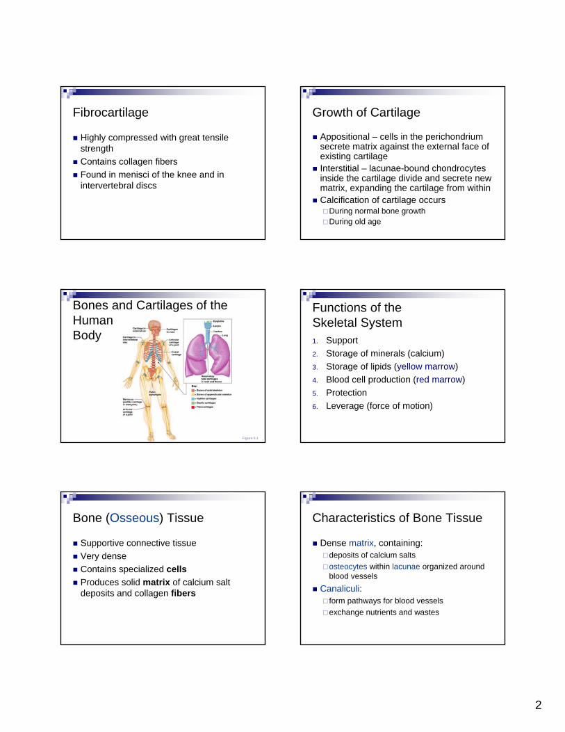
Bones and Cartilages of the Human Body
Figure 6.1
Functions of the Skeletal System1. Support2. Storage of minerals (calcium)3. Storage of lipids (yellow marrow) 4. Blood cell production (red marrow)5. Protection6. Leverage (force of motion)
Bone (Osseous) Tissue
Supportive connective tissue Very denseContains specialized cellsProduces solid matrix of calcium salt deposits and collagen fibers
Characteristics of Bone Tissue
Dense matrix, containing:deposits of calcium saltsosteocytes within lacunae organized around blood vessels
Canaliculi: form pathways for blood vesselsexchange nutrients and wastes
3
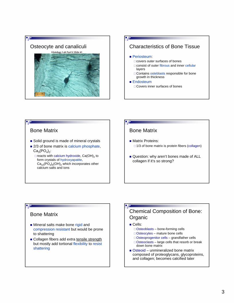
Osteocyte and canaliculi Characteristics of Bone Tissue
Periosteum: covers outer surfaces of bones consist of outer fibrous and inner cellularlayersContains osteblasts responsible for bone growth in thickness
EndosteumCovers inner surfaces of bones
Bone Matrix
Solid ground is made of mineral crystals2/3 of bone matrix is calcium phosphate, Ca3(PO4)2:
reacts with calcium hydroxide, Ca(OH)2 to form crystals of hydroxyapatite, Ca10(PO4)6(OH)2 which incorporates other calcium salts and ions
Bone Matrix
Matrix Proteins:1/3 of bone matrix is protein fibers (collagen)
Question: why aren’t bones made of ALL collagen if it’s so strong?
Bone Matrix
Mineral salts make bone rigid and compression resistant but would be prone to shatteringCollagen fibers add extra tensile strengthbut mostly add tortional flexibility to resist shattering
Chemical Composition of Bone: Organic
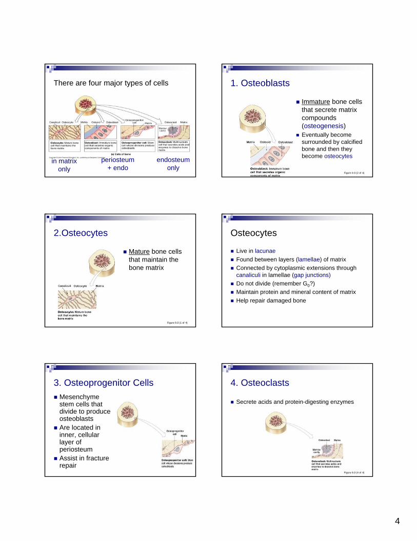
Cells:Osteoblasts – bone-forming cellsOsteocytes – mature bone cellsOsteoprogenitor cells – grandfather cellsOsteoclasts – large cells that resorb or break down bone matrix
Osteoid – unmineralized bone matrix composed of proteoglycans, glycoproteins, and collagen; becomes calcified later
4
There are four major types of cells
in matrix only
endosteumonly
periosteum+ endo
1. Osteoblasts
Immature bone cells that secrete matrix compounds (osteogenesis)Eventually become surrounded by calcified bone and then they become osteocytes
Figure 6–3 (2 of 4)
2.Osteocytes
Mature bone cells that maintain the bone matrix
Figure 6–3 (1 of 4)
Osteocytes
Live in lacunaeFound between layers (lamellae) of matrixConnected by cytoplasmic extensions through canaliculi in lamellae (gap junctions)Do not divide (remember G0?)Maintain protein and mineral content of matrixHelp repair damaged bone
3. Osteoprogenitor CellsMesenchymestem cells that divide to produce osteoblastsAre located in inner, cellular layer of periosteumAssist in fracture repair
4. Osteoclasts
Secrete acids and protein-digesting enzymes
Figure 6–3 (4 of 4)
5
Osteoclasts
Giant, mutlinucleate cellsDissolve bone matrix and release stored minerals (osteolysis)Often found lining in endosteum lining the marrow cavity Are derived from stem cells that produce macrophages
Homeostasis
Bone building (by osteocytes and -blasts) and bone recycling (by osteoclasts) must balance:
more breakdown than building, bones become weakexercise causes osteocytes to build bone
Bone cell lineage summary
Osteoprogenitor cells
osteoblasts
osteocytes
Osteoclasts are related to macrophages (blood cell derived)
Gross Anatomy of Bones: Bone Textures
Compact bone – dense outer layerSpongy bone – honeycomb of trabeculae filled with yellow bone marrow
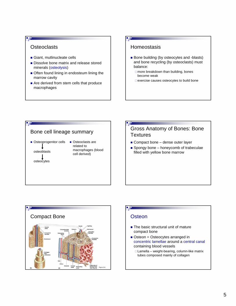
Compact Bone
Figure 6–5
Osteon
The basic structural unit of mature compact boneOsteon = Osteocytes arranged in concentric lamellae around a central canalcontaining blood vessels
Lamella – weight-bearing, column-like matrix tubes composed mainly of collagen
6
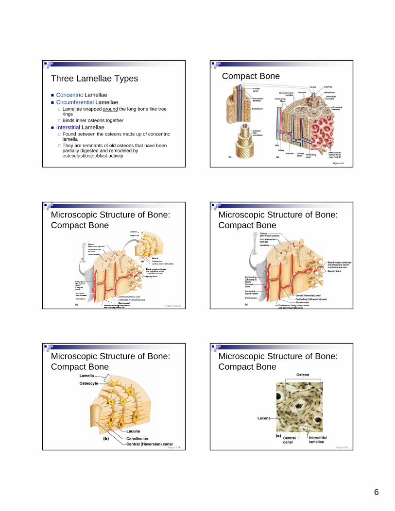
Three Lamellae Types
Concentric LamellaeCircumferential Lamellae
Lamellae wrapped around the long bone line tree ringsBinds inner osteons together
Interstitial LamellaeFound between the osteons made up of concentric lamellaThey are remnants of old osteons that have been partially digested and remodeled by osteoclast/osteoblast activity
Compact Bone
Figure 6–5
Microscopic Structure of Bone: Compact Bone
Figure 6.6a, b
Microscopic Structure of Bone: Compact Bone
Figure 6.6a
Microscopic Structure of Bone: Compact Bone
Figure 6.6b
Microscopic Structure of Bone: Compact Bone
Figure 6.6c
7
Spongy Bone
Figure 6–6
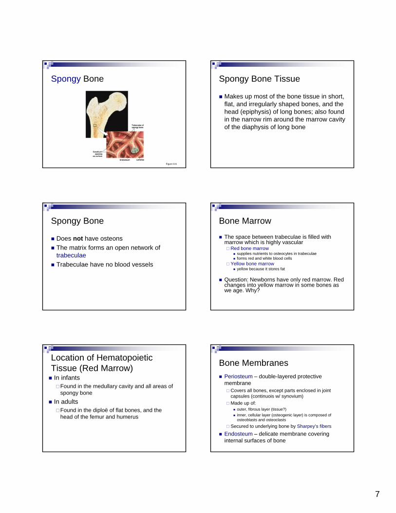
Spongy Bone Tissue
Makes up most of the bone tissue in short, flat, and irregularly shaped bones, and the head (epiphysis) of long bones; also found in the narrow rim around the marrow cavity of the diaphysis of long bone
Spongy Bone
Does not have osteonsThe matrix forms an open network of trabeculaeTrabeculae have no blood vessels
Bone Marrow
The space between trabeculae is filled with marrow which is highly vascular
Red bone marrowsupplies nutrients to osteocytes in trabeculaeforms red and white blood cells
Yellow bone marrowyellow because it stores fat
Question: Newborns have only red marrow. Red changes into yellow marrow in some bones as we age. Why?
Location of HematopoieticTissue (Red Marrow)In infants
Found in the medullary cavity and all areas of spongy bone
In adultsFound in the diploë of flat bones, and the head of the femur and humerus
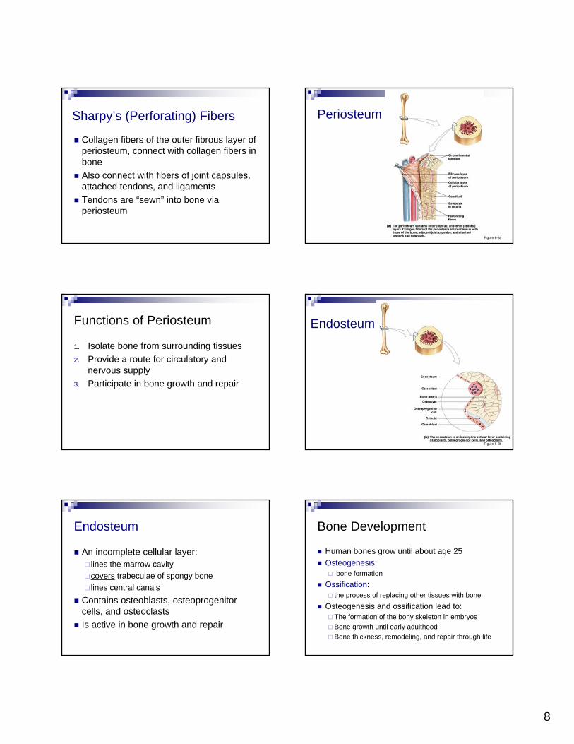
Bone MembranesPeriosteum – double-layered protective membrane
Covers all bones, except parts enclosed in joint capsules (continuois w/ synovium)Made up of:
outer, fibrous layer (tissue?)inner, cellular layer (osteogenic layer) is composed of osteoblasts and osteoclasts
Secured to underlying bone by Sharpey’s fibersEndosteum – delicate membrane covering internal surfaces of bone
8
Sharpy’s (Perforating) Fibers
Collagen fibers of the outer fibrous layer of periosteum, connect with collagen fibers in boneAlso connect with fibers of joint capsules, attached tendons, and ligamentsTendons are “sewn” into bone via periosteum
Periosteum
Figure 6–8a
Functions of Periosteum
1. Isolate bone from surrounding tissues2. Provide a route for circulatory and
nervous supply3. Participate in bone growth and repair
Endosteum
Figure 6–8b
Endosteum
An incomplete cellular layer:lines the marrow cavitycovers trabeculae of spongy bonelines central canals
Contains osteoblasts, osteoprogenitorcells, and osteoclastsIs active in bone growth and repair
Bone Development
Human bones grow until about age 25Osteogenesis:
bone formationOssification:
the process of replacing other tissues with boneOsteogenesis and ossification lead to:
The formation of the bony skeleton in embryosBone growth until early adulthoodBone thickness, remodeling, and repair through life

9
Calcification
The process of depositing calcium salts Occurs during bone ossification and in other tissues
Formation of the Bony Skeleton
Begins at week 8 of embryo developmentOssification
Intramembranous ossification – bone develops from a fibrous membraneEndochondral ossification – bone forms by replacing hyaline cartilage
Intramembranous OssificationNote: you don’t have to know the steps of this process in detail
Also called dermal ossification (because it occurs in the dermis)
produces dermal bones such as mandible and clavicle
Formation of most of the flat bones of the skull and the claviclesFibrous connective tissue membranes are formed by mesenchymal cells
The Birth of Bone
When new bone is born, either during development or regeneration, it often starts out as spongy bone (even if it will later be remodeled into compact bone)
Endochondral OssificationNote: you DO have to know this one
Begins in the second month of developmentUses hyaline cartilage “bones” as models for bone construction then ossifies cartilage into bone Common, as most bones originate as hyaline cartilageThis is like a “trick” the body uses to allow long bones to grow in length when bones can only grow by appositional growth
Bone formation in a chick embryo
Stained to represent hardened bone (red) and cartilage (blue)
: This image is the cover illustration from The Atlas of Chick Development by Ruth Bellairs and Mark Osmond, published by Academic Press (New York) in 1998
10
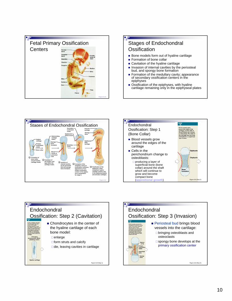
Fetal Primary Ossification Centers
Figure 6.15
Stages of Endochondral Ossification
Bone models form out of hyaline cartilageFormation of bone collarCavitation of the hyaline cartilageInvasion of internal cavities by the periosteal bud, and spongy bone formationFormation of the medullary cavity; appearance of secondary ossification centers in the epiphysesOssification of the epiphyses, with hyaline cartilage remaining only in the epiphyseal plates
Stages of Endochondral Ossification
Figure 6.8
Formation ofbone collararound hyalinecartilage model.
Hyalinecartilage
Cavitation ofthe hyaline carti-lage within thecartilage model.
Invasion ofinternal cavitiesby the periostealbud and spongybone formation.
Formation of themedullary cavity asossification continues;appearance of sec-ondary ossificationcenters in the epiphy-ses in preparationfor stage 5.
Ossification of theepiphyses; whencompleted, hyalinecartilage remains onlyin the epiphyseal platesand articular cartilages.
Deterioratingcartilagematrix
Epiphysealblood vessel
Spongyboneformation
Epiphysealplatecartilage
Secondaryossificatoncenter
Bloodvessel ofperiostealbud
Medullarycavity
Articularcartilage
Spongybone
Primaryossificationcenter
Bone collar
1
2
34
5
EndochondralOssification: Step 1 (Bone Collar)
Blood vessels grow around the edges of the cartilage Cells in the perichondrium change to osteoblasts:
producing a layer of superficial bone (bone collar) around the shaft which will continue to grow and become compact bone (appositional growth) Figure 6–9 (Step 2)
EndochondralOssification: Step 2 (Cavitation)
Chondrocytes in the center of the hyaline cartilage of each bone model:
enlargeform struts and calcifydie, leaving cavities in cartilage
Figure 6–9 (Step 1)
EndochondralOssification: Step 3 (Invasion)
Periosteal bud brings blood vessels into the cartilage:
bringing osteoblasts and osteoclastsspongy bone develops at the primary ossification center
Figure 6–9 (Step 3)
11
EndochondralOssification: Step 4a (Remodelling)
Figure 6–9 (Step 4)
Remodeling creates a marrow (medullary) cavity:
bone replaces cartilage at the metaphysesDiaphysis elongates
EndochondralOssification: Step 4b (2° Ossification)
Capillaries and osteoblastsenter the epiphyses:
creating secondary ossification centers (perinatal)
Figure 6–9 (Step 5)
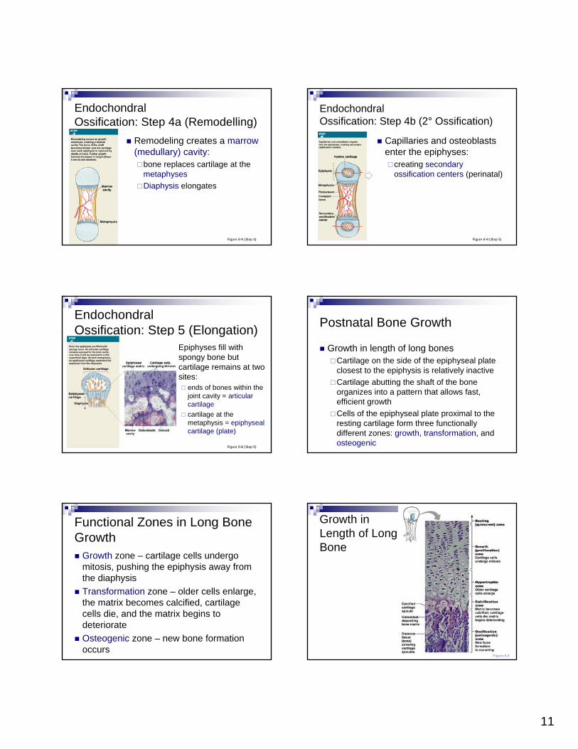
EndochondralOssification: Step 5 (Elongation)
Epiphyses fill with spongy bone but cartilage remains at two sites:
ends of bones within the joint cavity = articularcartilagecartilage at the metaphysis = epiphysealcartilage (plate)
Figure 6–9 (Step 6)
Postnatal Bone Growth
Growth in length of long bonesCartilage on the side of the epiphyseal plate closest to the epiphysis is relatively inactiveCartilage abutting the shaft of the bone organizes into a pattern that allows fast, efficient growth Cells of the epiphyseal plate proximal to the resting cartilage form three functionally different zones: growth, transformation, and osteogenic
Functional Zones in Long Bone Growth
Growth zone – cartilage cells undergo mitosis, pushing the epiphysis away from the diaphysisTransformation zone – older cells enlarge, the matrix becomes calcified, cartilage cells die, and the matrix begins to deteriorateOsteogenic zone – new bone formation occurs
Growth in Length of Long Bone
Figure 6.9
12
Postnatal bone growth
Remember that bone growth can only occur from the outside (appositional growth). So this type of endochondralgrowth is a way for bones to grow from the inside and lengthen because it is the cartilage that is growing, not the bone
Key Concept
As epiphyseal cartilage grows through the division of chondrocytes it pushes the ends of the bone outward in length. At the “inner” (shaft) side of the epiphysealplate, recently born cartilage gets turned into bone, but as long as the cartilage divides and extends as fast or faster than it gets turned into bone, the bone will grow longer
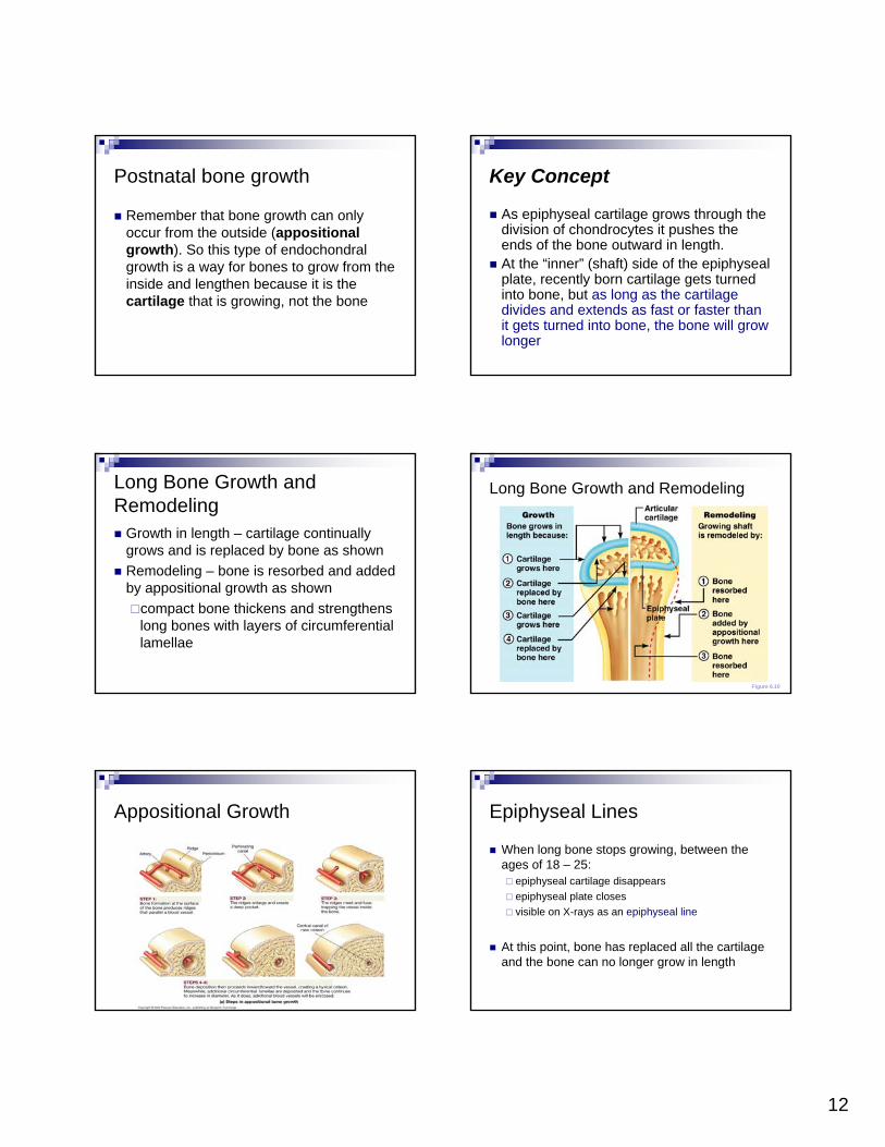
Long Bone Growth and Remodeling
Growth in length – cartilage continually grows and is replaced by bone as shown Remodeling – bone is resorbed and added by appositional growth as shown
compact bone thickens and strengthens long bones with layers of circumferential lamellae
Long Bone Growth and Remodeling
Figure 6.10
Appositional Growth Epiphyseal Lines
When long bone stops growing, between the ages of 18 – 25:
epiphyseal cartilage disappears epiphyseal plate closesvisible on X-rays as an epiphyseal line
At this point, bone has replaced all the cartilage and the bone can no longer grow in length
13
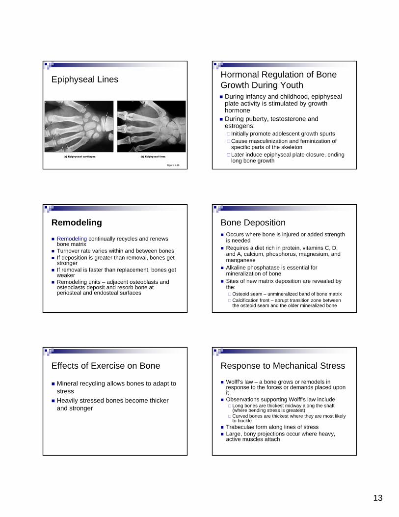
Epiphyseal Lines
Figure 6–10
During infancy and childhood, epiphyseal plate activity is stimulated by growth hormoneDuring puberty, testosterone and estrogens:
Initially promote adolescent growth spurtsCause masculinization and feminization of specific parts of the skeletonLater induce epiphyseal plate closure, ending long bone growth
Hormonal Regulation of Bone Growth During Youth
RemodelingRemodeling continually recycles and renews bone matrixTurnover rate varies within and between bonesIf deposition is greater than removal, bones get strongerIf removal is faster than replacement, bones get weakerRemodeling units – adjacent osteoblasts and osteoclasts deposit and resorb bone at periosteal and endosteal surfaces
Bone DepositionOccurs where bone is injured or added strength is neededRequires a diet rich in protein, vitamins C, D, and A, calcium, phosphorus, magnesium, and manganeseAlkaline phosphatase is essential for mineralization of boneSites of new matrix deposition are revealed by the:
Osteoid seam – unmineralized band of bone matrixCalcification front – abrupt transition zone between the osteoid seam and the older mineralized bone
Effects of Exercise on Bone
Mineral recycling allows bones to adapt to stressHeavily stressed bones become thicker and stronger
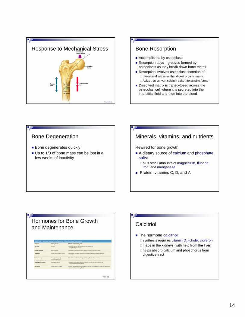
Response to Mechanical Stress
Wolff’s law – a bone grows or remodels in response to the forces or demands placed upon itObservations supporting Wolff’s law include
Long bones are thickest midway along the shaft (where bending stress is greatest)Curved bones are thickest where they are most likely to buckle
Trabeculae form along lines of stressLarge, bony projections occur where heavy, active muscles attach
14
Response to Mechanical Stress
Figure 6.12
Bone ResorptionAccomplished by osteoclastsResorption bays – grooves formed by osteoclasts as they break down bone matrixResorption involves osteoclast secretion of:
Lysosomal enzymes that digest organic matrixAcids that convert calcium salts into soluble forms
Dissolved matrix is transcytosed across the osteoclast cell where it is secreted into the interstitial fluid and then into the blood
Bone Degeneration
Bone degenerates quickly Up to 1/3 of bone mass can be lost in a few weeks of inactivity
Minerals, vitamins, and nutrients
Rewired for bone growthA dietary source of calcium and phosphatesalts:
plus small amounts of magnesium, fluoride, iron, and manganese
Protein, vitamins C, D, and A
Hormones for Bone Growth and Maintenance
Table 6–2
Calcitriol
The hormone calcitriol:synthesis requires vitamin D3 (cholecalciferol)made in the kidneys (with help from the liver)helps absorb calcium and phosphorus from digestive tract
15
The Skeleton as Calcium Reserve
Bones store calcium and other mineralsCalcium is the most abundant mineral in the bodyCalcium ions in body fluids must be closely regulated because:Calcium ions are vital to:
membranesneuronsmuscle cells, especially heart cellsblood clotting
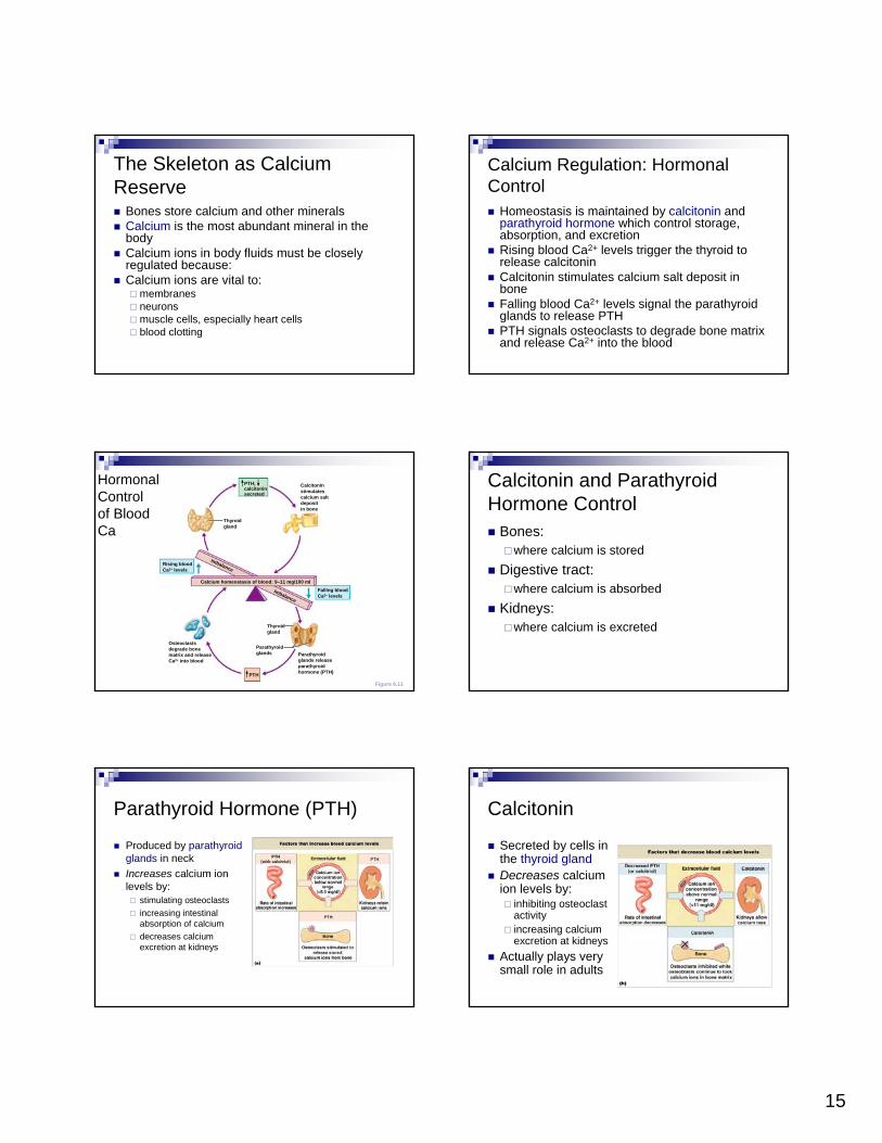
Calcium Regulation: Hormonal Control
Homeostasis is maintained by calcitonin and parathyroid hormone which control storage, absorption, and excretionRising blood Ca2+ levels trigger the thyroid to release calcitoninCalcitonin stimulates calcium salt deposit in boneFalling blood Ca2+ levels signal the parathyroid glands to release PTHPTH signals osteoclasts to degrade bone matrix and release Ca2+ into the blood
Hormonal Control of Blood Ca
Figure 6.11
PTH;calcitoninsecreted
Calcitoninstimulatescalcium saltdepositin bone
Parathyroidglands releaseparathyroidhormone (PTH)
Thyroidgland
Thyroidgland
Parathyroidglands
Osteoclastsdegrade bonematrix and releaseCa2+ into blood
Falling bloodCa2+ levels
Rising bloodCa2+ levels
Calcium homeostasis of blood: 9–11 mg/100 ml
PTH
Imbalance
Imbalance
Calcitonin and Parathyroid Hormone Control
Bones:where calcium is stored
Digestive tract:where calcium is absorbed
Kidneys:where calcium is excreted
Parathyroid Hormone (PTH)
Produced by parathyroid glands in neckIncreases calcium ion levels by:
stimulating osteoclastsincreasing intestinal absorption of calcium decreases calcium excretion at kidneys
Secreted by cells in the thyroid glandDecreases calcium ion levels by:
inhibiting osteoclastactivityincreasing calcium excretion at kidneys
Actually plays very small role in adults
Calcitonin
16
Fractures
Fractures:cracks or breaks in bonescaused by physical stress
Fractures are repaired in 4 steps
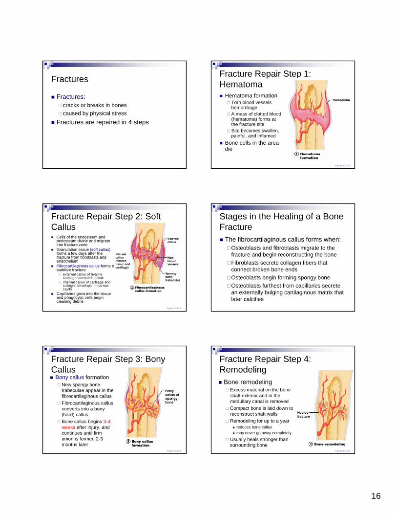
Fracture Repair Step 1: Hematoma
Hematoma formationTorn blood vessels hemorrhageA mass of clotted blood (hematoma) forms at the fracture siteSite becomes swollen, painful, and inflamed
Bone cells in the area die
Figure 6.13.1
Fracture Repair Step 2: Soft Callus
Cells of the endosteum and periosteum divide and migrate into fracture zoneGranulation tissue (soft callus) forms a few days after the fracture from fibroblasts and endotheliumFibrocartilaginous callus forms to stabilize fracture
external callus of hyaline cartilage surrounds breakinternal callus of cartilage and collagen develops in marrow cavity
Capillaries grow into the tissue and phagocytic cells begin cleaning debris
Figure 6.13.2
Stages in the Healing of a Bone Fracture
The fibrocartilaginous callus forms when:Osteoblasts and fibroblasts migrate to the fracture and begin reconstructing the boneFibroblasts secrete collagen fibers that connect broken bone endsOsteoblasts begin forming spongy boneOsteoblasts furthest from capillaries secrete an externally bulging cartilaginous matrix that later calcifies
Fracture Repair Step 3: Bony Callus
Bony callus formationNew spongy bone trabeculae appear in the fibrocartilaginous callusFibrocartilaginous callus converts into a bony (hard) callusBone callus begins 3-4 weeks after injury, and continues until firm union is formed 2-3 months later
Figure 6.13.3
Fracture Repair Step 4: Remodeling
Bone remodelingExcess material on the bone shaft exterior and in the medullary canal is removedCompact bone is laid down to reconstruct shaft wallsRemodeling for up to a year
reduces bone callusmay never go away completely
Usually heals stronger than surrounding bone
Figure 6.13.4

17
Clinical advances in bone repairElectrical stimulation of fracture site.
results in increased rapidity and completeness of bone healingelectrical field may prevent parathyroid hormone from activating osteoclasts at the fracture site thereby increasing formation of bone and minimizing breakdown of bone,
Ultrasound.Daily treatment results in decreased healing time of fracture by about 25% to 35% in broken arms and shinbones. Stimulates cartilage cells to make bony callus.
Free vascular fibular graft technique. Uses pieces of fibula to replace bone or splint two broken ends of a bone. Fibula is a non-essential bone, meaning it does not play a role in bearing weight; however, it does help stabilize the ankle.
Bone substitutes.synthetic material or crushed bones from cadavers serve as
bone fillers (Can also use sea coral).
Aging and Bones
Bones become thinner and weaker with ageOsteopenia begins between ages 30 and 40 Women lose 8% of bone mass per decade, men 3%
Osteoporosis
Severe bone loss which affects normal functionGroup of diseases in which bone reabsorption outpaces bone depositThe epiphyses, vertebrae, and jaws are most affected, resulting in fragile limbs, reduction in height, tooth lossOccurs most often in postmenopausal womenBones become so fragile that sneezing or stepping off a curb can cause fracturesOver age 45, occurs in:
29% of women18% of men
Notice what happens in osteoporosis
Osteoporosis: Treatment
Calcium and vitamin D supplementsIncreased weight-bearing exerciseHormone (estrogen) replacement therapy (HRT) slows bone lossNatural progesterone cream prompts new bone growthStatins increase bone mineral densityPPIs may decrease density
Hormones and Bone Loss
Estrogens and androgens help maintain bone massBone loss in women accelerates after menopause
18
Cancer and Bone Loss
Cancerous tissues release osteoclast-activating factor:
stimulates osteoclastsproduces severe osteoporosis
Paget’s Disease
Characterized by excessive bone formation and breakdownAn excessively high ratio of spongy to compact bone is formedReduced mineralization causes spotty weakening of boneOsteoclast activity wanes, but osteoblastactivity continues to work
Developmental Aspects of Bones
Mesoderm gives rise to embryonic mesenchymal cells, which produce membranes and cartilages that form the embryonic skeletonThe embryonic skeleton ossifies in a predictable timetable that allows fetal age to be easily determined from sonogramsAt birth, most long bones are well ossified (except for their epiphyses)
Developmental Aspects of Bones
By age 25, nearly all bones are completely ossifiedIn old age, bone resorption predominatesA single gene that codes for vitamin D docking determines both the tendency to accumulate bone mass early in life, and the risk for osteoporosis later in life
SUMMARY
Skeletal cartilageStructure and function of bone tissuesTypes of bone cellsStructures of compact bone and spongy boneBone membranes, peri- and endosteumOssification: intramembranous and endochondralBone minerals, recycling, and remodelingHormones and nutritionFracture repairThe effects of aging
Figure 6–16 (1 of 9)
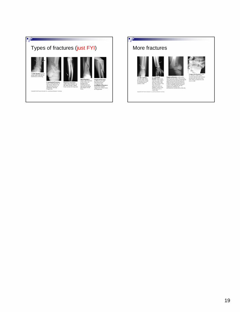
The Major Types of Fractures
Simple (closed): bone end does not break the skinCompound (open): bone end breaks through the skinNondisplaced – bone ends retain their normal positionDisplaced – bone ends are out of normal alignmentComplete – bone is broken all the way throughIncomplete – bone is not broken all the way throughLinear – the fracture is parallel to the long axis of the boneTransverse – the fracture is perpendicular to the long axis of the boneComminuted – bone fragments into three or more pieces; common in the elderly
19
Types of fractures (just FYI) More fractures