lecture 6 — defects in crystals....lecture 6 — defects in crystals. 1 introduction in the first...
TRANSCRIPT

Lecture 6 — Defects in Crystals.
1 Introduction
In the first five lectures, we have focussed our attentions on the methodologies used to describecrystal symmetry and on the scattering techniques employed to study the arrangement of atomsand spins in crystals (electronic, nuclear and spin densities). Throughout this discussion, wehave explicitly assumed that the crystalline order is “perfect” — in other words, that translationalsymmetry is strictly valid. The only allowance we made for deviations from one unit cell to thenext is through the introduction of the Debye-Waller factors, the effect of which, as we have seen,is to “spread out” the scattering densities in a fashion similar to the atomic scattering factors. Inthis lecture, we will explicitly consider crystal ”imperfections” of different kinds, starting fromthe simplest form of translational symmetry breaking (the effects arising from the finite sizeof the crystals) and considering in turn other, more complex types of defects: “point” defects,correlated defects and “line” or “plane” defects (dislocations or stacking faults). As we shallsee, all these defects give rise to particular kinds of scattering, most often away from the Braggpeaks. We will also introduce a new experimental technique — transmission electron microscopy— and describe its relevance in the study of crystal defects.
1.1 Space and time scales: coherence
When dealing with crystal imperfections, one of the first questions one should ask is whetherthey will contribute coherently (i.e., amplitudes are summed) or incoherently (i.e., intensities aresummed) to the diffraction patterns.
Let us make this clear with a simple example: consider an inhomogeneous alloy, in which thecomposition and the lattice constants varies on the scale of millimetres. Clearly, the differentregions of the crystal will scatter independently, each region being in essence a “perfect crystal”.The result, therefore, with be the incoherent superposition of different patterns. In this example,each hkl will produce a set of independent Bragg peaks (corresponding to the different latticeconstants), which will typically appear as a broadening of the scattered reflections. Let us nowimagine to “shrink” the lengthscale of the composition and lattice parameters fluctuations downto nanometre sizes. Here, the different compositions will scatter coherently, and the appropriatepicture is that of an average lattice. In the different regions, the atoms will be displaced awayfrom the average positions, and this, as we know by now, will produce a reduction of the Braggintensity at high q, in complete analogy to the Debye-Waller factors. Furthermore, the displace-ments will be correlated (i.e., nearby atoms will tend to be displaced in the same direction). Later
1

in this lecture, we will learn that this produces diffuse scattering away from the Bragg peaks. Atdifferent lengthscales, he diffraction patterns will be therefore qualitatively different. At whatpoint, between millimetres and nanometres, does this qualitative transition occur? We can intuitthat the lengthscale where the transition occurs might be set by the probe, i.e., that there mat beintrinsic coherence lengths for X-rays, neutrons, electrons etc. A very similar argument maybe construed for timescales. If the positions of the atoms in the crystal “fluctuate”, e.g., dueto phonons, do the different configurations occurring at different times contribute coherently orincoherently to the diffraction pattern? If the timescale of the fluctuations if of the order of sec-onds, the latter will be true, but, as the timescale of the fluctuations is reduced, this will no longerbe the case. What is the typical coherence time of the different probes?
A complete treatment of the correlation lengths and times is to complex to be presented here, butthe following points should provide a good idea of the issues involved.
• One distinguishes between transverse and longitudinal coherence lengths, perpendicular andparallel, respectively, to the direction of the beam.
• For a quasi-parallel beam geometry (as it is typical of a diffraction instrument), the trans-verse coherence length ξt is proportional to the wavelength and inversely proportional tothe beam divergence α (ξt = λ/α). Here, the beam divergence is defined as the anglesubtended by the source as seen from the sample. The derivation is analogous to that ofthe double-slit experiment, and corresponds to the distance between slits where the inter-ference pattern is lost. For for typical diffraction instruments, the beam divergence variesbetween a few mrad (lab diffractometers) down to a tenth of mrad (synchrotrons), so forλ = 1A, ξt varies between a few tens of nm up to about 1 µm. Highly coherent X-raybeams (several tens of µm) are employed for special studies (lensless imaging, “speckle”patterns), revealing the shape and internal structure of large crystal domains. Neutronbeams have comparatively relaxed divergences, and typical coherence lengths are 1000-2000 A.
• For geometries employing focussing, (e.g., electron diffraction) it is possible to manipulate thecoherence domain by varying the focal plane where the diffraction pattern is formed (seebelow). If the diffraction pattern is in focus, the coherence length is essentially limited bythe aberration of the lenses, and can be as large as for X-rays, in spite of the fact that thewavelength employed are 1-2 orders of magnitude smaller. If the sample is in focus, thetransverse coherence length is much smaller (down to atomic sizes), so that each detectorpixel receives a coherent contribution of a column of atoms in the direction of the beam.In this case, the coherence domain coincides with the resolving power of the instrument(see below).
2

• The longitudinal coherence length is inversely proportional to the relative wavelength spreadof the beam: ξl = 1
2λ/(∆λ/λ). High-resolution monochromators at synchrotron sources
typically yield ∆λ/λ ∼ 0.5− 1× 10⁻⁴, so at 1 A ξl is about 1 µm. The wavelength spreadof neutron and electron beams is typically 10 times and 100 times larger, respectively.
• The coherence time can be calculated using the uncertainty principle as τ = ~/∆E or fromthe relation τ = ξl/v, yielding the same result apart for a factor of the order 1. Adoptingthe first approach, we obtain
τ =1
π2
mλ2
~
(∆λ
λ
)−1
for particles with mass
τ =1
2π
λ
c
(∆λ
λ
)−1
for photons (1)
The prefactor is 1.6× 10⁻¹⁴ [secA−2] for neutrons, 0.9× 10⁻¹⁷ [secA−2] for electrons and5×10−20 [secA−1] for photons. Using the wavelengths and monochromaticity values men-tioned above, we find that coherence times for neutrons, photons and 100 Kev electronsare in the picosecond, femtosecond and tens of attosecond ranges, respectively.
2 Finite size effects
Up to this point, we have consider the crystal lattice to be of infinite extent. As we have seen, thecross section becomes then a series of delta functions, centered on the RL nodes. We can relaxour approximation by starting form eq. 6 in Lecture 5 (the cross section) and solve explicitly forthe finite summations. in fact, we need not make any particular approximation other than the factthat the crystal should be composed of N1,N2 and N3 unit cell in the a1, a2 and a3 directions,respectively. Remembering that
N∑n=−N
e−inx =sin
((2N + 1)x
2
)
sin x2
(2)
after some straightforward math (in each direction, x = q · ai ) we obtain
dσ
dΩ=
[∏i
sin2(Ni
12q · ai
)
sin2(12q · ai)
]|F (q)|2 [ε · ε′]2 (3)
3

The diffraction pattern from a small single crystal taken with coherent radiation, i.e., radia-tion with coherence length (=wavepacket length/width) larger than the crystal, will display 3-dimensional fringes, as typical of the function sin2(Nx)/ sin2 x. However, for typical experi-ments with incoherent radiation, the number of unit cells is set by the coherence length of eachphoton rather than by the crystal size, and the oscillations will be smeared out. In this case, wecan replace the oscillatory functions with a Gaussian function with the same maximum and samearea:
sin2 Nx
sin2 x→ N2e−(Nx)2/π (4)
Observing that∏
i Ni = Nc, the total number of unit cell in the crystal, we obtain
dσ
dΩ= N2
c
[∏i
e−(Ni12q· ai)
2/π
]|F (q)|2 [ε · ε′]2 (5)
where the Gaussian functions have variance and FWHM
σ2i =
2π
N2i a2
i
FWHM =4√
π ln 2
Niai
(6)
We can therefore conclude that:
• The cross section at a given q is proportional to N2c .
• The width in q is inversely proportional to the number of unit cells along that direction.
• The integrated cross section in three dimensions (remember the Gaussian integral√
2πσ2)is therefore proportional to Nc, which reproduces the result we obtained for the infinitecrystal (eq. 8, lecture 5).
3 Beyond the perfect-crystal approximation: diffuse scatter-ing
Finite-size effects only generate scattering in the vicinity of the ”ideal” Bragg positions, leav-ing the integrated intensity of the reflection unaltered. In this section, we will demonstrate that
4

additional scattering is generated whenever either the atomic scattering factors or the positionsof individual atoms deviate from the average values and/or the “ideal” lattice sites. In general,this additional scattering is present throughout reciprocal space, and is therefore known as dif-fuse scattering. In the remainder of this section, we will consider the deviations from perfectperiodicity as static. In other words, we will imagine that the scattering process occurs coher-ently from a frozen snapshot of the crystal. To estimate the full scattered intensity, at the end,we will undertake a time/thermal averaging process on the scattered intensities. This is a verygood approximation in the case of X-rays (and electrons), since the coherence times are muchshorter than phonon frequencies. For neutrons, where this is clearly not the case, a more complextreatment is required, but qualitatively similar considerations (with some caveats) can be applied.
3.1 Classification of disorder in crystals
We will here consider two types of disorder:
• Substitutional disorder is typical of alloys and, more generally, of solid solutions. It occurswhen more that one atonic species can reside on the same crystallographic site, and givesrise to a fluctuation of the atomic scattering factor around an “average” value, the latter re-flecting the average composition of the alloy. For example, in a 67%Cu-33%Au “random”alloy, Cu and Au occupy the same crystallographic sites of the FCC structure. To calcu-late the Bragg scattering from such an alloy, one should use the average atomic scatteringfactor (fave(0) = 45.7). However, locally, the scattering factor varies between that of Cu(fCu(0) = 29) and Au (fAu(0) = 79). This local fluctuation, as we shall see, gives rise todiffuse scattering. Substitutional disorder is almost always static, since atoms in alloys donot usually move from one site to the next, but counter-examples do exist (e.g., in the caseof fast ion conductors).
• Dynamic displacive disorder is present in all materials even at zero temperatures, due tophonons and zero-point motion. In addition to scattering contrast fluctuations, substitu-tional disorder can itself produce static displacive disorder, since different species have ingeneral different atomic radii.
3.2 Simple diffuse scattering calculations
We shall now calculate the full scattering cross section for an (infinite) crystal in the presenceof both substitutional and displacive disorder. To simplify the formalism, we will perform thiscalculation in the case of a crystal with one atom per unit cell, but the extension to more complex
5

structures is completely straightforward. Let us therefore re-write the cross section in eq. 6,Lecture 5 in this case.
dσ
dΩ= r2
0P(γ)
(∑j
∑i
e−iq·(ri−rj)
)fi(q)f ∗j (q) (7)
In eq. 7, ri represent the actual positions of the atoms, while we indicate with Ri the position ofthe lattice nodes. Moreover, we will write
ri = Ri + ui (8)
where the ui are the displacements away from the lattice nodes and are “small” (see below). Withthe additional notation Rij = Ri − Rj and uij = ui − uj we can write:
dσ
dΩ= r2
0P(γ)∑Rij
e−iq·Rij
∑
Pairs with Rij
[fi(q)f ∗j (q)e−iq·uij
](9)
In eq. 9, we have rearranged the double summation, so that the rightmost sum runs over allpairs of unit cells joined by a given vector Rij , whereas the leftmost sum runs over these vectors.Using, for example, periodic boundary conditions, we find that for each Rij , there are exactly N
pairs of sites joined by it (N being the total number of atoms in the crystal. We can thereforere-write eq. 9 as:
dσ
dΩ= Nr2
0P(γ)∑Rij
e−iq·Rij⟨fi(q)f ∗j (q)e−iq·uij
⟩Rij
(10)
We can subtract from eq. 10 the Bragg scattering cross section to obtain the expression for thenon-Bragg or diffuse scattering (we stress, once again, that the average atomic scattering factoris used to calculate the Bragg scattering):
(dσ
dΩ
)
diff
= Nr20P(γ)
∑Rij
e−iq·Rij⟨fi(q)f ∗j (q)e−iq·uij − |fave(q)|2e−2W
⟩Rij
(11)
where the sign 〈〉Rijindicates the average of the quantity within it over all the pairs in the coher-
ence domain joint by the vector Rij .
6

3.2.1 Diffuse scattering from uncorrelated substitutional disorder
Here, we will make the slightly unrealistic assumption that the atoms are not displaced at allfrom their ideal lattice sites, and consider the diffuse scattering arising from purely substitutionaldisorder (uij = 0). We can write
(dσ
dΩ
)
diff
= Nr20P(γ)
∑Rij
e−iq·Rij⟨fi(q)f ∗j (q)− |fave(q)|2⟩Rij
(12)
If the sites have random occupancy, for i 6= j the average is identically zero for all Rij , so onlythe autocorrelation term i = j survives, yielding
(dσ
dΩ
)
diff
= Nr20P(γ)
[(|f(q)|2)ave − |fave(q)|2] (13)
For example, for a binary alloy with species A and B and concentrations CA and 1 − CA, weobtain
(dσ
dΩ
)
diff
= Nr20P(γ)CA(1− CA)|fA(q)− fB(q)|2 (14)
As we can see, these cross sections are slowly varying in q, since they are only modulated by theatomic scattering factors, and have their maximum near q = 0.
3.2.2 Diffuse scattering from uncorrelated displacements
Let us now assume that we have a single atomic species (fi = fj = fave) and that the displace-ments of atoms i and j are not correlated with each other. If i 6= j,
⟨e−iq·uij
⟩=
⟨e−iq·ui
⟩ ⟨e−iq·uj
⟩= e−2W (15)
so Rij vectors joining different atoms do not give any contributions to the diffuse scattering. Theonly surviving term is the autocorrelation term, for which uij = 0. Therefore,
(dσ
dΩ
)
diff
= Nr20P(γ)|f(q)|2 (
1− e−2W)
(16)
7

As we can see from eq. 16, the diffuse scattering from uncorrelated displacements is a slowlyvarying function of q; it is zero near the origin of the reciprocal space and increases with q (it isin the first approximation proportional to q2), only to decay again with the decay of the atomicscattering factors.
3.3 General treatment of displacive disorder and thermal diffuse scatter-ing
In the two simple examples we proposed above, we have treated the deviations from the averagestructure as completely uncorrelated from site to site. The result, in both cases, is a form ofdiffuse scattering that has very little structure in reciprocal space. In the extreme case of neutronstudies of substitutional disorder, the atomic form factor is a constant, and the resulting diffusescattering is completely flat.
In realistic cases, correlations are almost always present: even in disordered alloys, a givenspecies may have a “preference” to be surrounded by atoms of the same kind or, quite frequently,of a different kind, so near-neighbour probabilities are generally different from the average.These correlations are lost after a few unit cells, and they are referred to as short-range or-der. In this case, beside the autocorrelation term, additional terms in the summation (eq. 12) willbe non-zero, giving rise to a more structured diffuse scattering pattern. Note that each Rij pairthat corresponds to a definite correlation will contribute with a Fourier component exp(R · Rij)
(or cos R · Rij in the centrosymmetric case) to the diffuse scattering.
The correlated displacements due to thermal vibrations are of even longer range: an ideal plane-wave phonon involves all sites of the crystal, and therefore involves all terms in the summation(eq. 11). The correlation terms uij will typically involve sums and differences of sines andcosines, and since they appear in the exponential, the summation over an infinite number of pairscannot be performed analytically. The typical approach is to expand the exponential in Taylorseries. This is done as follows: for simplicity, let us rewrite eq. 11 in the case of pure displacivedisorder (no substitutional disorder):
(dσ
dΩ
)
diff
= Nr20P(γ)
∑Rij
e−iq·Rij |f(q)|2[⟨
e−iq·uij⟩
Rij− e−2W
](17)
Next, we will use again eq. 13, Lecture 5, which is valid for small displacements or harmonicdisplacements:
⟨e−iq·uij
⟩= e−
12〈[q·uij ]
2〉 (18)
8

Finally, we expand the exponential in eq. 18 in Taylor series:
(dσ
dΩ
)
diff
= Nr20P(γ)
∑Rij
e−iq·Rij |f(q)|2[−1
2
⟨[q · uij]
2⟩ + 2W
]
+
[1
8
⟨[q · uij]
2⟩2 − 2W 2
]+ . . .
= I1 + I2 + . . . (19)
The different terms in eq. 19 are known as first-order, second-order etc., diffuse scattering.
3.3.1 First-order thermal diffuse scattering
In well-ordered crystals, the most important contribution to the diffuse scattering is that fromthe displacement patterns due to phonons. This type of diffuse scattering is known as ThermalDiffuse Scattering or TDS. The first-order term in eq. 19 is often the dominant contribution,particularly at moderate temperatures. The calculation of diffuse scattering for monoatomic solidis not inaccessible at the present level, but it is rather lengthy. Here, we summarise the main stepsand present the final result, so that its structure can be analysed.
• Only phonons with the same wavevector k and the same branch index contribute coherentlyto first-order TDS. The scattering from one such phonon is not difficult to calculate: itgenerate satellite peaks at ±k around any given Bragg peaks.
• One needs to sum the scattered intensity of phonons with different wavevectors, taking intoaccount the fact that their amplitude is related to the thermal population.
• The final result is:
(dσ
dΩ
)
1stTDS
= Nr20P(γ)
~2M
∑j
1
ωj(q)coth
(~ωj(q)
2kBT
)|Fj(q)|2 (20)
where the sum is over the different phonon branches and
Fj(q) = f(q)e−W [q · ej(q)] (21)
is the so-called one-phonon structure factor and ej(q) is the polarisation vector of phononbranch j at the wavevector q. In order to evaluate the quantities ej(q) and ωj(q), one needsto reduce q to the first Brillouin zone in the usual way.
9

• The function 1ωj(q)
coth(~ωj(q)
2kBT
)decreases rapidly with increasing frequency. Therefore low-
energy, acoustic phonons provide the largest contribution to first-order TDS. For thesame reason, the TDS diverges at the Bragg peak positions and is minimum between theBragg peaks.
• When the same reduced wavevector is considered in different Brillouin zones, one finds thatthe cross section is proportional to q2, similar to the diffuse scattering due to uncorrelateddisplacements (eq. 21).
4 Scattering from non-crystalline solids and liquids
Our discussion of the scattering cross sections for ”defective” crystal would not be completewithout a mention of the most extreme form of defects — the complete loss of crystalline order,as in the case of non-crystalline solids (glasses) or liquids. In this case, it is no longer possibleto define a crystal lattice; nevertheless, the formalism to calculate the scattering cross section isvery similar to the one we have already encountered (eq. 9). The main difference is that, forobvious reasons, decomposing position vectors in Rij and uij will no longer make sense, and wewill therefore employ the original notation rij
dσ
dΩ= r2
0P(γ)N∑
m=1
∑rij
fi(q)f ∗j (q)e−iq·rij (22)
where the first sum runs over all the atoms in the sample. For simplicity, we will proceed fromhere assuming that all the atoms in the sample are the same, as appropriate, for example, for amonoatomic liquid like mercury. The extension to polyatomic liquids or glasses is rather straight-forward but requires a heavier notation. With our assumption, we can extract N |f(q)|2 from allthe summations and replace the sums with averages; we will also isolate the autocorrelation term(rij = 0) which yields 1:
dσ
dΩ= Nr2
0P(γ)|f(q)|21 +
∑
rij 6=0
〈e−iq·rij〉 (23)
We now want to take advantage of the fact that, in a disordered sample, if a vector rij connectingtwo atoms is present, then all other vectors with the same modulus rij will also be present withequal probability somewhere in the sample. This will enable us average over the angular variablesand replace the summation in eq. 23 with a sum over rij . In doing so, we have to account for the
10
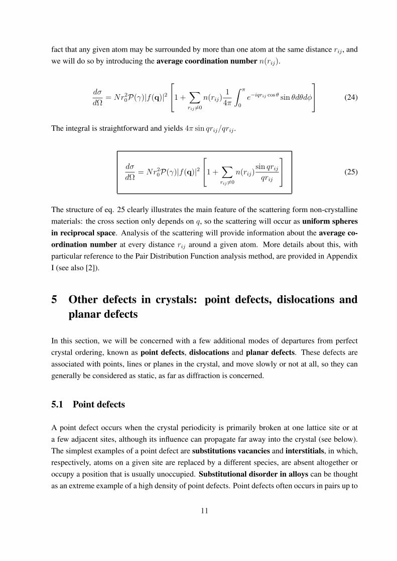
fact that any given atom may be surrounded by more than one atom at the same distance rij , andwe will do so by introducing the average coordination number n(rij).
dσ
dΩ= Nr2
0P(γ)|f(q)|21 +
∑
rij 6=0
n(rij)1
4π
∫ π
0
e−iqrij cos θ sin θdθdφ
(24)
The integral is straightforward and yields 4π sin qrij/qrij .
dσ
dΩ= Nr2
0P(γ)|f(q)|21 +
∑
rij 6=0
n(rij)sin qrij
qrij
(25)
The structure of eq. 25 clearly illustrates the main feature of the scattering form non-crystallinematerials: the cross section only depends on q, so the scattering will occur as uniform spheresin reciprocal space. Analysis of the scattering will provide information about the average co-ordination number at every distance rij around a given atom. More details about this, withparticular reference to the Pair Distribution Function analysis method, are provided in AppendixI (see also [2]).
5 Other defects in crystals: point defects, dislocations andplanar defects
In this section, we will be concerned with a few additional modes of departures from perfectcrystal ordering, known as point defects, dislocations and planar defects. These defects areassociated with points, lines or planes in the crystal, and move slowly or not at all, so they cangenerally be considered as static, as far as diffraction is concerned.
5.1 Point defects
A point defect occurs when the crystal periodicity is primarily broken at one lattice site or ata few adjacent sites, although its influence can propagate far away into the crystal (see below).The simplest examples of a point defect are substitutions vacancies and interstitials, in which,respectively, atoms on a given site are replaced by a different species, are absent altogether oroccupy a position that is usually unoccupied. Substitutional disorder in alloys can be thoughtas an extreme example of a high density of point defects. Point defects often occurs in pairs up to
11

maintain stoichiometry or charge neutrality, as in the case of Frenkel defects (a vacancy and aninterstitial) or Schottky defects (vacancy of ions with opposite charge). In the case of extendeddefects, the coordination environment around the defect site is modified to approximate the pre-ferred environment of the new species. For example, if a cation with a preference for tetrahedralcoordination is substituted on an octahedral site, the six surrounding anions can be distorted andone or two can be missing altogether, forming an extended defect.
If present in sufficient concentration, point defects can give rise to diffuse scattering, in com-plete analogy to substitutional disorder. Another form of diffuse scattering associated with pointdefects is Huang scattering: this arises from the static displacement field due to the elastic de-formation of the lattice around the defect, and its treatment is analogous to that of first-orderTDS.
5.2 Dislocations
Dislocations are linear defects that commonly form in metals as a result of the application ofstresses, e.g, due to repeated bending. They play an important role in both the strength and thefailure of materials: work hardening (such as beating a red-hot piece of metal on an anvil)has been used for centuries by blacksmiths to introduce dislocations into materials, increasingtheir yield strengths. On the other hand, the infamous metal fatigue fracture (responsible, forexample, for the De Havilland Comet disasters) are initiated by dislocations, which eventuallyform persistent slip bands that nucleate short cracks
Two main types of dislocations are identified: edge dislocations and screw dislocation, althoughmost real dislocation are intermediate between these types. They are characterised by two direc-tion vectors: the dislocation direction — the direction of the linear structure in question, and theBurgers vector — the principal direction of the strain (displacement) field near the dislocation.
5.2.1 Burgers vectors for edge and screw dislocations
The classic definition of the Burgers vector is the following (see fig. 1 and 2): Let us considera part of the crystal that is free from any defect, and let us define a closed loop of atoms. Letus now insert the dislocation so that the dislocation line is inside and perpendicular to the loop.The strain (displacement) field generated by the defect will displace the existing atoms, so thatthe rectangle will be deformed, whilst, by definition, remaining a closed loop. If the rectangle isdeformed into a trapezium, the Burgers vector will be perpendicular to the dislocation line (edgedislocation, fig. 1). If the rectangle is deformed into a 3-dimensional shape, the Burgers vectorwill be parallel to the dislocation line (screw dislocation, fig. 2).
12

Figure 1: An edge dislocation and its associated Burgers vector. An edge dislocation arises whena plane of atoms is abruptly terminated within the crystal. The displacement field around theedge dislocation is defined by its Burgers vector, and, is perpendicular to the dislocation line.
Figure 2: A screw dislocation and its associated Burgers vector. We can imagine that a screwdislocation is generated by “slicing” a plane through the crystal up to a certain line, and by dis-placing the atoms on one side of the plane by one unit cell along the direction of the line. Thedisplacement field around the edge dislocation is defined by its Burgers vector, and is perpen-dicular to the dislocation line.
5.3 Stacking faults
The last family of crystal defects we will consider in this lecture are the planar defects. Asthe word implies, the defect originated at a planar locus in the crystal, although the strain fieldcan propagate some way away from the plane. Although other types of planar defects exist (forexample, the twin planes), here we will only consider the stacking faults.
Staking faults are defects in the “stacking” of atomic layers — most commonly a fault in thestacking sequence of the layers, a missing layer or an added layer, either of the same type or ofa different type with respect to the bulk layers. Unsurprisingly, stacking faults are common
13

in layered structures, but are also encountered in isotropic structures: in fact, perhaps the bestknown type of stacking fault occurs in FCC metals, and is a fault of the ...ABCABCABC...stacking sequence of the (111) layers, as, for example, in ...ABCABABCABC... Stacking faultsof this type can be described as a displacement (slip) of an entire plane of atoms perpendicularto the fault plane, so that the fault is described by two vectors: the stacking vector (perpendicularto the fault plane) and the slip vector, describing the displacement direction of the plane of atoms.The slip vector plays a similar role to the Burgers vectors in the imaging of stacking faults byTEM (see below). In this case of the (111) faults in FCC metals, the slip vector is along the(211) direction or its equivalents (fig. 3).
Figure 3: A: a portion of the unfaulted FCC structure, with stacking sequence ...ABCAB-CABC....B A stacking fault, in which the “A” layer is inserted instead of a “C”, and the cor-responding possible slip vectors.
6 Experimental techniques: Electron Microscopy
6.1 Rationale and brief history of electron microscopy
In the absence of aberrations, the resolving power of a microscope is given by the well-knownformula R = κλ/n sin α, where n0 sin α is the numerical aperture, n0 is the refractive index inobject space and α is the semi-angle subtended by the object at the lens (or stop). The constant κ
depends on the coherence length of the light, but the typical value of 0.61 is generally adopted.
14

For visible light (e.g. 550 nm), the largest achievable numerical apertures is about 1.6, yielding a“ diffraction limited” resolution of about 200 nm. X-ray beams have much shorter wavelengths,but X-ray lenses are complex, high-precision objects and are limited to very small numericalapertures. With wavelengths of a few hundredths of A (Table 1) and the possibility of achievingnumerical apertures comparable to those of optical microscopy, a microscope based on an elec-tron beam should be able to resolve feature well below the inter-atomic spacing. In practice, theresolution of an electron microscope is limited by optical aberrations rather than diffraction, butresolutions below 1 A have been achieved in recent years.
After the verification of the De Broglie hypothesis, the next important step towards a practicalelectron microscope has been the the development of the magnetic lens (H. Busch, 1926, fig.4), followed by the demonstration of focussing by electric fields. These discoveries led to rapidprogress, so much so that the first electron microscope was constructed by M. Knoll and E.Ruska in 1931. This first instrument worked in transmission, like the modern-day TransmissionElectron Microscope or TEM, and had a resolution of several nm. The TEM was later developedas a commercial device, and now reaches routinely resolutions of the order of 1-2 A, with sub-Aresolutions having been demonstrated in specialised instruments.
Figure 4: Schematic representation of a TEM magnetic lens. The trajectory of the electrons inthis lens is quite complex, so that the image is rotated as well as magnified.
A different type of electron microscope, known as Scanning Electron Microscope or SEM, wasbuilt in 1938 by M. von Ardenne, following a suggestion by Knoll. In the SEM, the electron beamis electrically “scanned” across the surface of the sample producing a small spot, and a response(backscattered electrons, Auger electrons, X-rays etc.) is measured. An image of the sampleis then formed on the screen using the measured variable. The importance of both TEM andSEM for materials science cannot be overstated. However the TEM has been far more importantto solve problems in structural condensed matter physics, and we will therefore limit our brief
15

discussion to the Transmission Electron Microscope.
Table 1: Electron wavelengths and energies and the corresponding TEM techniques. For non-relativistic electrons, λ = h(2meV )−1 = 12.26V −1 A. HEED wavelengths are corrected forrelativistic effects (∼ 5% at 105 V.)
V(volts) λ (A) v/c name
10 3.9 0.006 LEED102 1.2 0.02 LEED103 0.39 0.06 MEED104 0.12 0.19 MEED105 0.037 0.54 HEED106 0.009 0.94 HEED
6.2 The TEM: working principle and operating parameters
Very schematically, a TEM can be described as follows (fig. 5):
• A quasi-monochromatic beam of electron is produced and accelerated to a working voltageby an electron gun. The electrons are emitted by either a thermoionic filament or, inmodern instrument, by a field-emission source, which is more monochromatic and hasmuch higher brightness. The relation between accelerating voltage and wavelength is:
λ =h√
2meV=
12.26√V
A (26)
Typical accelerating voltages and corresponding wavelengths for various applications areshown in tab. 1.
• A series of magnetic lenses, collectively known as the condenser, create a spot size on thesample that can be varied with the excitation current of the magnetic lenses, but can be assmall as 1 µm. This spot defines the field of view of the instrument, and it is ultimatelyimaged on the screen, yielding magnifications in excess of 300,000. The beam divergenceon the sample is of the order of 10−3 radians, so it can be often considered as quasi-parallel.
• The sample stage consists of a sample holder (the sample — either a very thin section or aseries of grains from a powder sample, is usually supported by a fine carbon mesh) and atitling stage, which enables the sample to be oriented so that different “zones” are broughtinto Bragg condition (see below). Since the sample itself is very thin (usually < 1000A),a significant fraction of the electrons are either transmitted or undergo Bragg scattering —in both cases emerging as quasi-parallel rays. Because of the very short wavelength andthe decaying atomic scattering factor, electron scattering is strongly peaked in the forward
16

direction and the whole diffraction pattern is concentrated within 1-2 deg for 100 KeVelectrons.
• Immediately below the sample the objective lens brings parallel rays emerging from the sam-ple into focus on the so-called back focal plane. In other words, both the direct beam andeach of the Bragg reflections will be brought into a distinct spot on the back focal plane,forming a diffraction pattern. Here, a series of diaphragms (diffraction apertures) canbe inserted to select the direct beam and/or one or one or more of the Bragg spots. Themicroscope is said to operate in (bright-field mode) if the transmitted beam is allowed topropagate beyond the diffraction apertures.
• Below the back focal plane, a series of lenses and apertures known collectively as the pro-jector magnify and focus the electrons the so that the image of the spot fills the viewingscreen.
• The image is formed on an electron detector, which was a simple phosphor screen in the earlymodels, and has now evolved in a much more sophisticated and sensitive device such as aCCD or a solid-state detector.
Figure 5: Schematic representation of a TEM in diffraction mode (left) and imaging mode (right).
17

6.2.1 Diffraction and imaging modes of operation
Broadly speaking, the TEM can be operated in two modes (see again fig. 5):
Diffraction mode: the projector brings into focus onto the detector a magnified image of theback focal plane, so that the diffraction pattern from the sample appears on the screen.
Imaging mode: here, the projector brings into focus onto the detector a magnified image of thesample spot, so that an “image” of the sample is created. Unlike conventional microscopy,where the contrast is primarily due to differential absorption of light, for crystalline sam-ple, contrast in TEM is primarily due to a modulation of the strength of diffractionfrom different parts of the sample. The role of the diffraction apertures should becomeclear at this point: by propagating electrons that have undergone different Bragg reflec-tions, one may obtain different types of contrasts. Thiks is particularly important in thestudy of dislocations and stacking faults (see below).
6.3 Electron diffraction: basic principles
The kinematic of electron diffraction is governed by the fact that, in a typical TEM, electronwavelengths are much shorted than the accessible d-spacings — in other words, |ki| =
|kf | À |q|. In addition, the thickness of the sample is much smaller than the spot size, sothat (by finite-size effects) the Bragg spots will be elongated in the direction of ki. The resultingEwald construction is shown in fig. 7. From this, it is clear that a sizeable portion of a RL
plane can be brought into (approximate) Bragg scattering condition by setting ki to be per-pendicular to this plane. An estimation of the scattered intensity for each Bragg spot, using thekinematic approximation, is provided in Appendix II. The plane in question always contains theorigin of the RL, and is known as zeroth-order Laue zone [xyz], where [xyz] is the real-spacedirection orthogonal to it. .
When operating in diffraction mode, a TEM generates a fairly undistorted picture of aslice of reciprocal space corresponding to a zeroth-order zone (see fig. 6). If the electronenergy is not very high, peaks from the first-order zone may become visible near the edgeof the diffraction pattern.Example: in a monoclinic crystal, the electron beam is directed along the [110] directionin real space. The corresponding zone will contain the origin of the RL and the two RLvectors (110) and (001).
Because of the large cross section for electron scattering, dynamical diffraction is alwaysrequired for quantitative calculation of electron scattering intensities. This means that struc-tural solution from electron diffraction is a highly specialised field of electron microscopy.
18

Figure 6: Sets of electron diffraction patterns of the multiferroic material YMn0.75Ti0.25O3showing the (a) [001], (b) [110], (c) [110], and (d) [540] zone-axis. From T. Asaka, et. alPhysical Review B 71, 014114 (2005)
6.4 TEM imaging: basic principles
When operating in imaging mode, the projector lenses of the microscope create onto the detectoran image of the sample. If a wide open diffraction aperture is employed, the transmitted beam
19

Figure 7: Ewald construction for electron diffraction.
and all the diffracted beam coming from one specific point in the sample are made to interfereat one specific pixel on the detector, and the contrast on that pixels results from the interferenceof these rays. By inserting appropriate diffraction apertures, one can select the transmitted beamalone and/or one or more diffracted beams. The simplest situation is that of dark-field imag-ing with a single reflection, since only one diffracted beam needs to be considered. Even thisapparently simple scenario is rather complex to assess quantitatively:
• The “circle of confusion” of the imaging process is far from the diffraction limit, and is alwaysdominated by the aberration of the optics. For a typical TEM, this is of the order of 1-2 A.
• In the direction of the electron beam, the depth of field of the optics is generally sufficient tokeep the whole thickness of the sample close to focussing conditions, although fine-tuningof the focus can have a dramatic effect on the contrast.
• To assess the contrast in the first approximation, therefore, one has to consider interferencebetween the scattered beams from a column of atoms, contained in a cylinder defined bythe circle of confusion of the optics and the thickness of the sample.
In the case of a perfect lattice, the imaging contrast has the periodicity of the lattice, andthis can be easily distinguished at sufficient resolution.
Fig. 8 shows a typical example — a high-resolution lattice image of Si3N4. A clearly distin-guishable grain boundary runs irregularly from the bottom left to the middle right of the image.The two grains have different orientations, and show different periodicity. In some images, onewould be tempted to identify the repeated pattern with the content of the unit cell, as if one waslooking at individual atoms, but this is never the case: a complex calculation involving dynamicaldiffraction end the exact focus condition is always require for realistic lattice image simulations.
20

Figure 8: High-resolution image of a grain boundary in Si3N4.
6.4.1 TEM imaging of dislocations and stacking faults
One of the most successful applications of the TEM has been the imaging and identification oflattice defects — particularly dislocations and stacking faults. As we have seen, a dislocation orstacking fault is characterised by the fact that a portion of the crystal, either around the dislocationline or across the stacking fault, is displaced with respect to the rest of the crystal by a vector b,which is known as the Burgers vector in the case of dislocations and the slip vector in the caseof stacking faults. The structure factor Ff of the “faulted” region will be therefore related to thatof the unfaulted region by:
Ff (q) = F (q)e−iq·b (27)
We remind that here the scattering vector q is selected using the diffraction apertures. Theintensity scattered from a column (corresponding to the contrast of a pixel) can then be writtenas
I/I0 ∝ |F |2∣∣∣∣∣
N1∑N=0
e−iNq·az +
N2∑N=1
e−iNq·(az+b) +Nz∑
N=2
e−iNq·az
∣∣∣∣∣
2
(28)
were we have assumed that the faulted region is delimited by N1 and N2 along the column.We could elaborate further on eq. 28, but here it will suffice to say that the fault will havemaximum contrast if the vector q along the Burgers or slip vector b, and will be invisibleif q ⊥ b. By taking a series of dark-field lattice images using different Bragg reflections, it istherefore possible to identify both the direction of the dislocation line or fault plane (which issimply the direction of the contrast in the lattice image) and the Burgers or slip vector, which
21

are perpendicular to the Bragg reflections for which the defects become invisible (see fig. 9).
Figure 9: Reproduced from [1], fig 4.16. Images of dislocations in stainless steel. On eachpicture, the (hkl) of the reflection employed to create the image is marked with an arrow. Left:Image taken with a (111) reflection: many dislocations are in contrast. Right: Image taken witha (113): most dislocations are out of contrast. This analysis indicates that most dislocations have(111) Burgers vector.
7 Bibliography
P.J. Grundy & G.A. Jones [1] A handy booklet on electron microscopy, a bit old but still use-ful.
S.J.L. Billinge & M.F. Thorpe [2] A good collection of articles on diffuse scattering and scat-tering from disordered materials.
References
[1] P.J. Grundy and G.A. Jones T. Hahn, ed., Electron Microscopy in the Study of Materials,(Edward Arnold Ltd. London: UK, 1976).
[2] S.J.L. Billinge and M.F. Thorpe eds., “Local structure from diffraction”, Kluwer AcademicPublishers New York, Boston, Dordrecht, London, Moscow, 2002.
22

8 Appendix I: the pair distribution function analysis
Let us continue on from eq. 25 and introduce a continuous function ρ(rij) (of dimensions [m−3])representing the “local number densities” of atoms at a distance rij around a given atom (exclud-ing itself). The function ρ(rij), known as the pair distribution function will be zero at shortdistances and will have a sharp peak corresponding to the first coordination shell, with furtheroscillations corresponding to more distant correlations At long distances, ρ(rij) will convergeto the average density ρa. For a typical glass, this occurs at distance of the order of 10-20 A,whereas for a crystal the oscillations persist to very long distances. Observing that(we drop thesubscripts ij at this point)
n(r) = 4πr2ρ(r)dr (29)
we replace the sum with an integral in eq. 25 and write:
dσ
dΩ= Nr2
0P(γ)|f(q)|2[1 +
∫ ∞
0
dr4πr2ρ(r)sin qr
qr
](30)
If we now write ρ(r) = ρa + ∆ρ(r), where ∆ρ(r) represents the deviation from the averagedensity, we can show that the term containing ρa fluctuates rapidly (except at very low q, wheredata are usually not measured since they merge with the transmitted beam), and averages outbetween different domains of scattering. We thus obtain the final form:
dσ
dΩ= Nr2
0P(γ)|f(q)|2[1 +
∫ ∞
0
dr4πr2∆ρ(r)sin qr
qr
](31)
Let us now introduce the function
S(q) =(Nr2
0P(γ)|f(q)|2)−1 dσ
dΩ(32)
which can be directly extracted from a scattering experiment, after appropriate normalisation andcorrection of the data. By using the properties of the Fourier transform we find:
ρ(r) = ρa +1
2π2r
∫ ∞
0
dq [S(q)− 1] q sin qr (33)
Clearly, it is impossible to measure S(q) up to q = ∞, so the Fourier transform procedure asapplied to “real” data will introduce truncation errors, which can be mitigated by applying
23

various procedures.
The Pair Distribution Function (or PDF) analysis method is widely used not only for liquidand amorphous substances, but to disordered (and sometimes rather well ordered) crystallinematerials, where it claims to provided an unbiassed determination of the “true” bond lengths.When PDF analysis is applied to crystals, both Bragg and diffuse scattering are included yieldinga picture of the local structure (as opposed to the average structure from Bragg scattering).
9 Appendix II: electron scattering from a thin section of sam-ple
In this section, we give a quantitative calculation for the scattered intensity in the kinematicapproximation. To be realistic, this calculation can only be applied to a very thin section of thesample, since, as we already mentioned, the elastic scattering cross section of electrons is so largethat dynamic effects are dominant. In this scenario, the electron beam illuminates a region of thesample of, say, 1-2 µm in size, and are partly transmitted, partly scattered through a section a afew tens of A. We also recall that electron scattering is strongly peaked in the forward directionand is concentrated within 1-2 deg for 100 KeV electrons. The Ewald construction appropriatefor this problem is shown in fig. 7. When the incident beam is along a real lattice vector, severalBragg peaks of the “zeroeth-order zone” (i.e., the RL plane perpendicular to that vector andcontaining the origin) will be close to scattering conditions, so that a portion of the diffractionpattern for that plane will appear on the screen. Bragg peaks that are progressively more distantfrom the origin will be cut by the Ewald sphere “above” the ideal Bragg position. Here, we wantto calculate the integrated intensity, i.e., the number of electrons collected by the film or CCD,of each of these reflections in the kinematic approximation.
Let us define the z axis in the direction of the beam, x and y being the lateral coordinates. Inanalogy to what we have done before, we will consider the lateral extent of the sample to beinfinite, so that the cross section will contain a 2-dimensional delta function. By contrast, wewill consider explicitly the finite size of the sample along z. We therefore write
dσ
dΩ=
(2π)2NxNy
axay
δ(qx)δ(qy)sin2(1
2Nzqzaz)
sin2(12qzaz)
|F |2
' (4π)2NxNy
v0az
δ(qx)δ(qy)sin2(1
2Nzqzaz)
q2z
|F |2 (34)
Where F is the structure factor and has the dimension of a length. In the geometry we areconsidering (fig. 7)
24

dΩ ' dqxdqy
k2i
=λ2
4π2dqxdqy (35)
We can therefore integrate in dΩ
σ =4NxNyλ
2
v0az
sin2(12Nzqzaz)
q2z
|F |2 (36)
Here, v0 = axayaz is the unit cell volume. Now, let multiply by the incident flux, which we willwrite as Φ0 = I0/(NxNyaxay), where I0 is the number of incident electrons per second. Thisis appropriate, since here the lateral dimension of the sample (i.e., NxNy) is defined by the spotsize. We thus obtain the scattered intensity (again, in electrons per second) in a given reflection.
Ihkl
I0
=
∣∣∣∣2λFhkl
v0
∣∣∣∣2 sin2(1
2qzt)
q2z
(37)
Where we have introduced t = Nzaz — the thickness of the sample. The quantity
ξg =v0
2λFhkl
(38)
has the dimensions of a length, and is known as the extinction distance. As we can see, the in-tensity has a central maximum and a series of subsidiary maxima. The height and width (distancebetween the two nearest minima) of the central maximum are:
H =
(t
ξg
)2
W =4π
t(39)
One can see that when the thickness t is equal to ξg, H = 1 and the scattered intensity is equal tothe incident intensity (total reflection). This is a clear signal that the kinematic approximationis no longer applicable. Typical extinction distances are of the order of 10 nm. It is a matterof simple geometry to show that the principal and subsidiary maxima can be intersected by theEwald sphere, giving rise to scattering corresponding to a given Bragg peak in the diffractionpattern.
25