lecture 12 –domains –protein structure. some common structural motifs of folded proteins d) the ...
TRANSCRIPT
Lecture 12
– Domains– Protein structure

Some common structural motifs of folded proteins
d) The motif
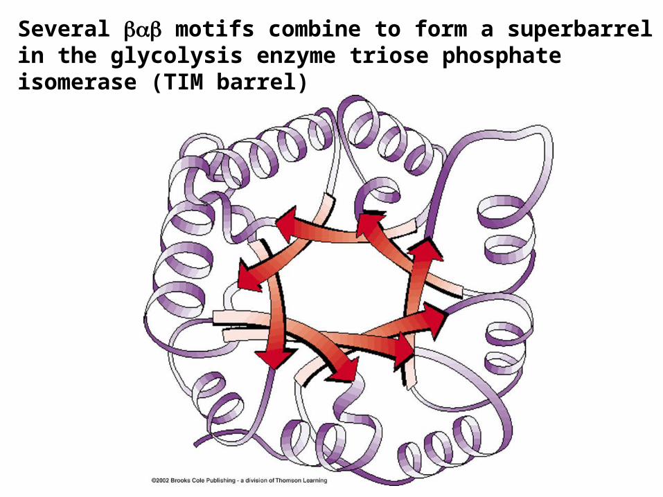
Several motifs combine to form a superbarrel in the glycolysis enzyme triose phosphate isomerase (TIM barrel)

Quaternary structure• Spatial arrangement of protein subunits.• Polypeptide subunits associate in a geometrically specific manner.• Why subunits?• Easier to repair self-assembling single subunit vs. a large
polypeptide.• Increasing a protein’s size through subunits is more efficient for
specifying the active site.• Provides a structural basis for regulating activity.

Domains in proteins.
• Common sequence regions in native proteins can fold up to form compact structures called “domains”.
• Domains can range in size from 50-400 amino acids, have upper limit in forming compact hydrophobic core.
• Domains are a type of folding motif, typically have separate hydrophobic core.
• Larger proteins are composed of multiple domains, often connected by flexible linker peptide regions.
• Classic example: antibodies

Structural elements of IgGs:
Naturally occurring immunoglobulins (IgG molecules) have identical heavy chains and light chains giving rise to multiple binding sites with identical specificities for antigen.
Antibody Immunoglobulin Domains
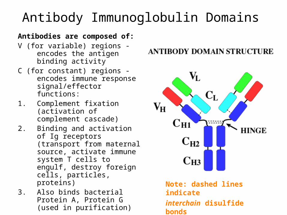
Antibodies are composed of: V (for variable) regions - encodes the
antigen binding activityC (for constant) regions - encodes
immune response signal/effector functions:
1. Complement fixation (activation of complement cascade)
2. Binding and activation of Ig receptors (transport from maternal source, activate immune system T cells to engulf, destroy foreign cells, particles, proteins)
3. Also binds bacterial Protein A, Protein G (used in purification)
Note: dashed lines indicate
interchain disulfide bonds
Antibody Immunoglobulin Domains

• There is a conserved glycosylation site in the CH2 domain of IgG (purple region).
• A carbohydrate is covalently attached here by postranslational modification.
Antibody Immunoglobulin Domains
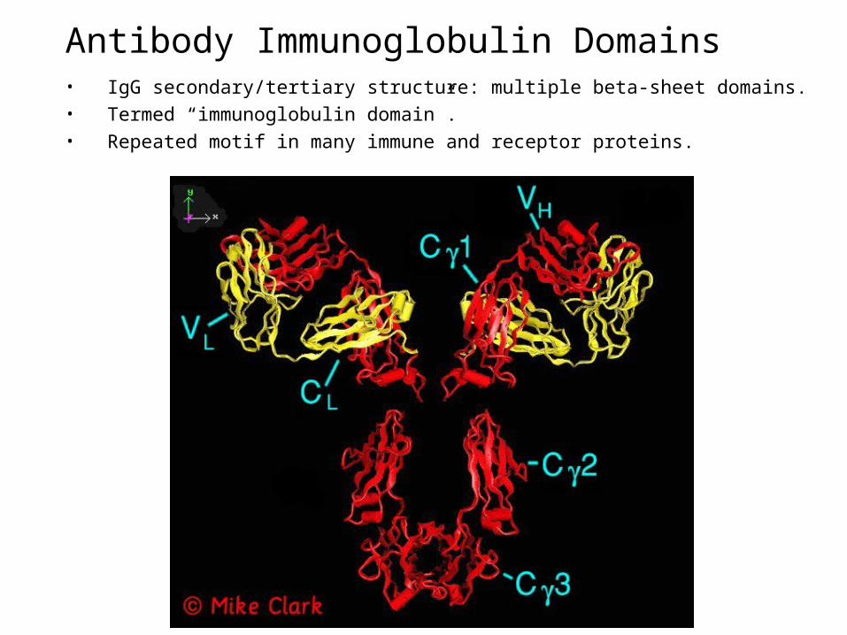
• IgG secondary/tertiary structure: multiple beta-sheet domains.• Termed “immunoglobulin domain”.• Repeated motif in many immune and receptor proteins.
Antibody Immunoglobulin Domains

Modes of Flexibility of IgG structure
Antibody Immunoglobulin Domains

Subunit interactions• Identical subunits in a protein are called protomers• Proteins with identical subunits are oligomers.• Hemoglobin is a dimer (oligomer of two protomers) of
protomers.
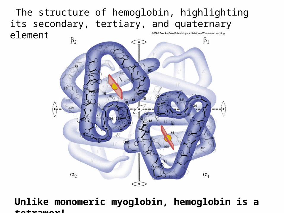
The structure of hemoglobin, highlighting its secondary, tertiary, and quaternary elements.
Unlike monomeric myoglobin, hemoglobin is a tetramer!
Alternate VMD generated structure of hemoglobin, highlighting its secondary, tertiary, and quaternary elements.
Unlike monomeric myoglobin, hemoglobin is a tetramer!
QuickTime™ and aTIFF (LZW) decompressor
are needed to see this picture.
Symmetry in proteins• Most oligomeric proteins, protomers are symmetrically arranged.• Occupy geometrically equivalent positions in the oligomer.• Have point symmetry around one point-no mirror symmetry but
have rotational symmetry.– Cyclic symmetry– Dihedral symmetry– Other rotational symmetries
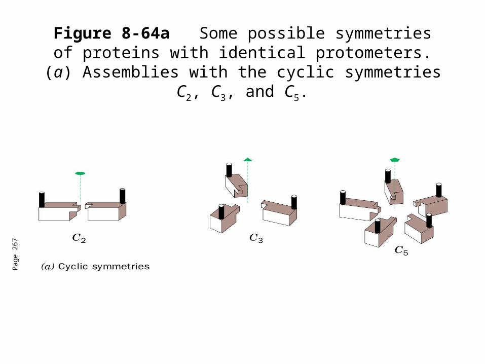
Figure 8-64aSome possible symmetries of proteins with identical protometers. (a) Assemblies with the
cyclic symmetries C2, C3, and C5.
Pag
e 26
7
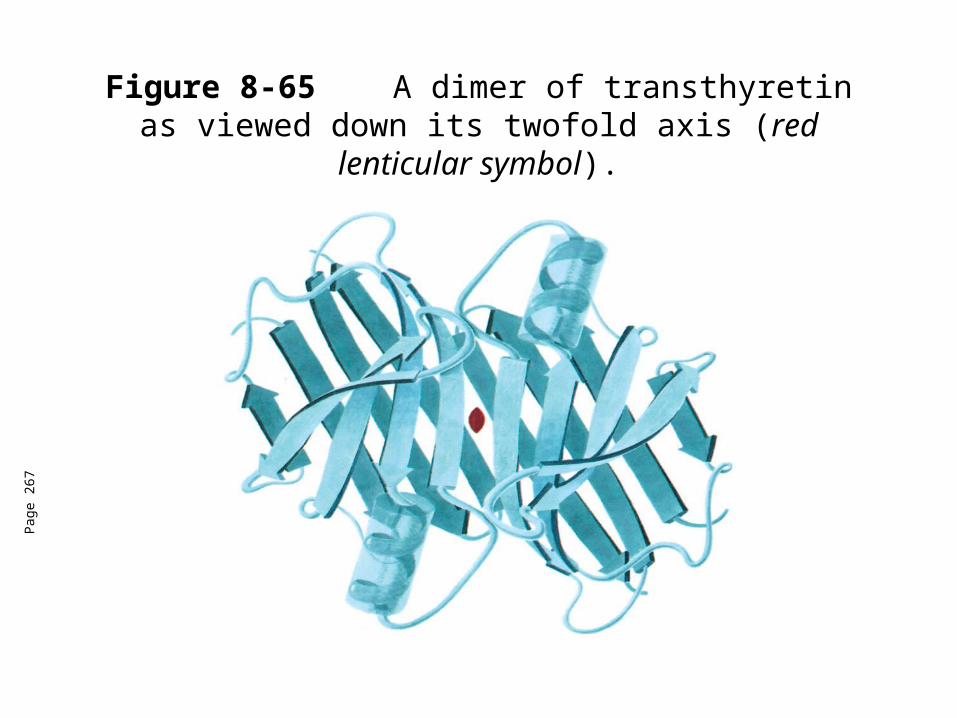
Figure 8-65 A dimer of transthyretin as viewed down its twofold axis (red lenticular symbol).
Pag
e 26
7
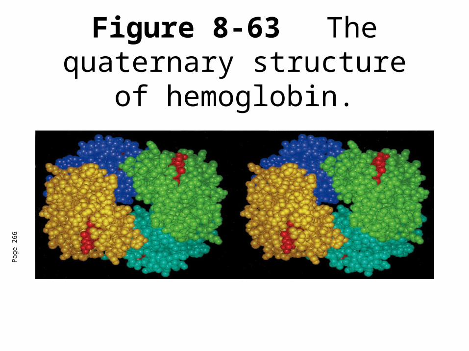
Figure 8-63 The quaternary structure of hemoglobin.
Pag
e 26
6
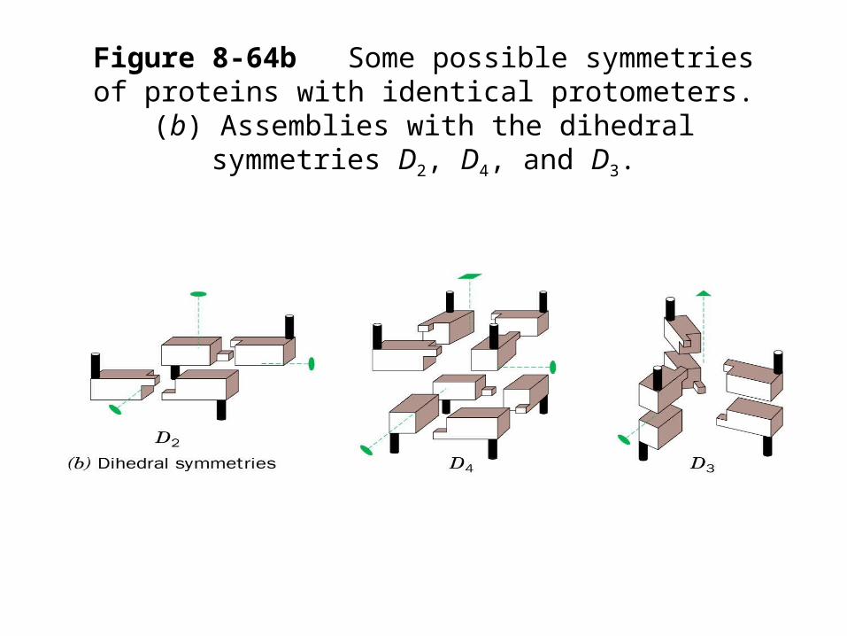
Figure 8-64bSome possible symmetries of proteins with identical protometers. (b) Assemblies with the
dihedral symmetries D2, D4, and D3.

Figure 8-66a X-Ray structure of glutamine synthease from Salmonella
typhimurium.
Pag
e 26
8
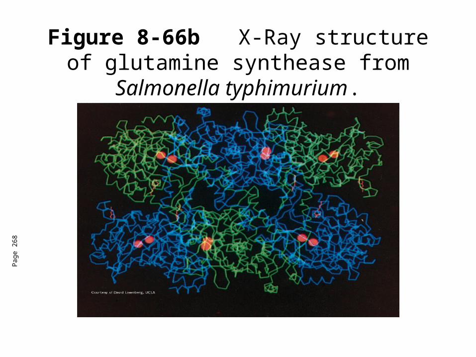
Figure 8-66b X-Ray structure of glutamine synthease from Salmonella
typhimurium.
Pag
e 26
8
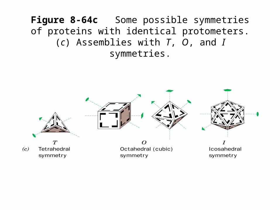
Figure 8-64cSome possible symmetries of proteins with identical protometers. (c) Assemblies with T, O,
and I symmetries.

Structure of protoporphyrin IX.
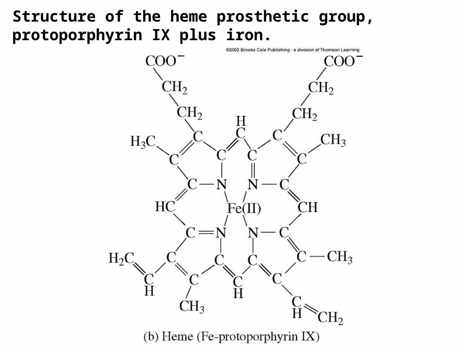
Structure of the heme prosthetic group, protoporphyrin IX plus iron.

The oxygen binding curves of myoglobin and hemoglobin obtained by measuring the percent of heme sites filled with O2 at varying O2 concentrations
Myoglobin has greater affinity for O2 than hemoglobin at all partial pressures (conc.) of oxygen.
The sigmoidal curve for hemoglobin indicates a cooperative binding of O2.
-Allosteric effect!

The heme binding site for oxygen in hemoglobin
•Note the Fe(II) in the protoporphyrin structure is linked to a histidine in helix F by a coordinate covalent bond. •When O2 is not
bound, the 6th position is protected from water oxidation by a 2nd His from helix E.
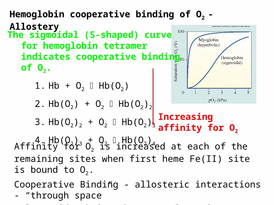
Hemoglobin cooperative binding of O2 - Allostery
The sigmoidal (S-shaped) curve for hemoglobin tetramer indicates cooperative binding of O2.
1. Hb + O2 Hb(O2)
2. Hb(O2) + O2 Hb(O2)2
3. Hb(O2)2 + O2 Hb(O2)3
4. Hb(O2)3 + O2 Hb(O2)4
Affinity for O2 is increased at each of the remaining sites when first heme Fe(II) site is bound to O2.
Cooperative Binding - allosteric interactions - “through space”
Polypeptide chains change conformation upon heme binding O2
Increasing affinity for O2

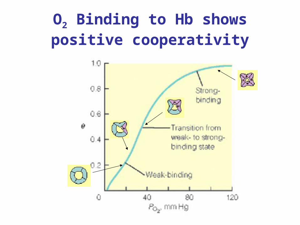
O2 Binding to Hb shows positive cooperativity
• Hb binds four O2 molecules
• O2 affinity increases as each O2 molecule binds
• Increased affinity due to conformation change• Deoxygenated form = T (tense) form = low affinity• Oxygenated form = R (relaxed) form = high affinity
O2 Binding to Hb shows positive cooperativity
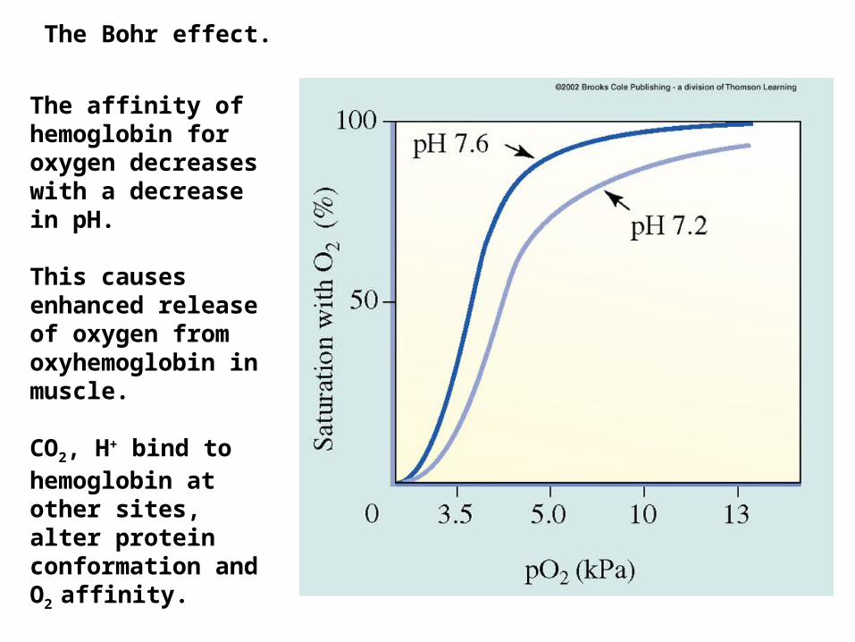
The affinity of hemoglobin for oxygen decreases with a decrease in pH.
This causes enhanced release of oxygen from oxyhemoglobin in muscle.
CO2, H+ bind to hemoglobin at other sites, alter protein conformation and O2 affinity.
The Bohr effect.
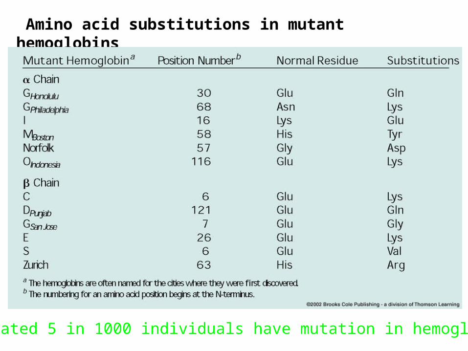
Amino acid substitutions in mutant hemoglobins
Estimated 5 in 1000 individuals have mutation in hemoglobin
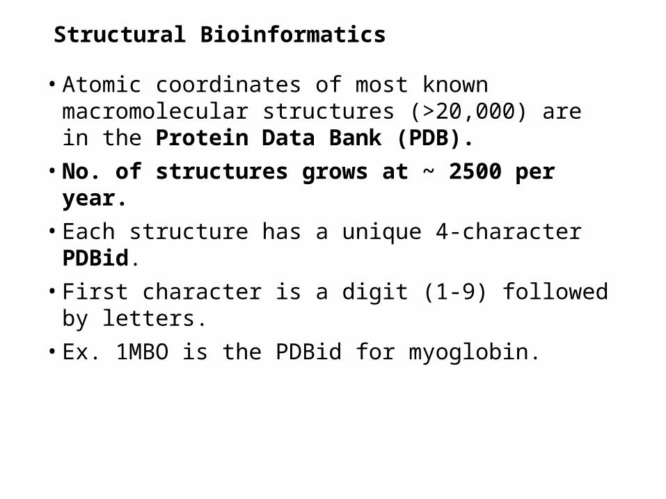
Structural Bioinformatics
• Atomic coordinates of most known macromolecular structures (>20,000) are in the Protein Data Bank (PDB).
• No. of structures grows at ~ 2500 per year.
• Each structure has a unique 4-character PDBid.
• First character is a digit (1-9) followed by letters.
• Ex. 1MBO is the PDBid for myoglobin.

Structural Bioinformatics
• Files contain info that describes the macromolecule
• Date the coordinate file was deposited
• Organism
• Authors
• Key journal references
• PDB file consists of a series of ATOM (for standard residues) and HETATM (for heterogens; not among the std. amino acids).

Table 8-4 (top) Structural Bioinformatics Websites
(URLs).
Pag
e 25
6
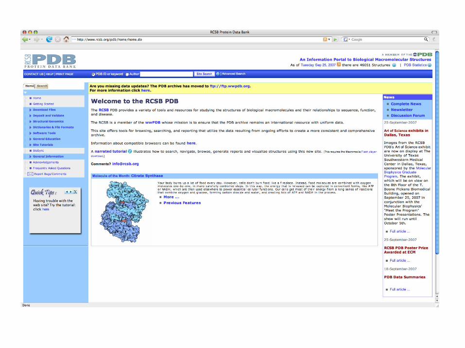

Table 8-4 (middle) Structural Bioinformatics Websites (URLs).
Pag
e 25
6

Structural classification and comparison
• CATH (Class, Architecture, Topology, Homologous superfamily)
• Class-highest level-places the selected protein into 1 of 4 categories of secondary structure (mostly , mostly , and having few secondary structures.
• Architecture-description of the gross arrangement of secondary structure, independent of topology.
• Topology-indicative of overall shape and connectivity of protein’s secondary structures.
• Homologous superfamily-proteins of known structure that are homologous (share a common ancestor) to a selected protein.
What is the CATH classification for 1MBO?
Structural classification and comparison
• CE (Combinatorial Extension of the optimal path)
• Finds all proteins in the PDB that can be structurally aligned with the query structure (structural alignment).
• Can be displayed with RasMol
• FSSP (Fold classification based on Structure-Structure alignment of Proteins)
• Lists protein structures in PDB which in part, structurally resemble the query protein.
• Can be displayed using Chime.
Structural classification and comparison
• SCOP (Structural Classification Of Proteins)
• Classifies protein structures based mainly on manually generated toplogical considerations (6-levels)
1. Class-all-, all-, (having helices and strands that are largely interspersed), (having helices and strands that are largely segregated), and multi-domian (having domains of different classes)
2. Fold-groups that have similar arrangements of 2ndary structural elements.
3. Superfamily-distant evolutionary relationships basd on structural criteria and functional features
4. Family-near evolutionary relationships based on sequence and structure
5. Protein
6. Species

Table 8-4 (bottom) Structural Bioinformatics Websites (URLs)
Pag
e 25
8

Exam• Study the HW assignments-similar problems will make up the bulk
of the exam. • All notes and chapters in the book are fair game for the exam.