lecithin-based wet chemical precipitation of ... · lecithin-based wet chemical precipitation of...
TRANSCRIPT

SHORT COMMUNICATION
Lecithin-based wet chemical precipitationof hydroxyapatite nanoparticles
Wojasiński Michał & Duszyńska Ewa & Ciach Tomasz
Received: 5 February 2015 /Revised: 25 February 2015 /Accepted: 2 March 2015 /Published online: 15 March 2015# The Author(s) 2015. This article is published with open access at Springerlink.com
Abstract Hydroxyapatite Ca10(PO4)6(OH)2 nanoparticleshave been successfully synthesized by the wet chemical pre-cipitation method at 60 °C in the presence of biocompatiblenatural surfactant—lecithin. The composition and morpholo-gy of nanoparticles of hydroxyapatite synthesized with leci-thin (nHAp-PC) was studied by X-ray diffraction (XRD),Fourier transform infrared spectroscopy (FTIR), and scanningelectron microscopy (SEM). Size distribution for nanoparti-cles was measured by nanoparticle tracking analysis inNanoSight system. We discuss in details influence of lecithinconcentration in reaction system on nHAp-PCmorphology, aswell as on size distributions and suspendability of nanoparti-cles. Product exhibits crystalline structure and chemical com-position of hydroxyapatite, with visible traces of lecithin. Dif-ference in surfactant amounts results in changes in particlesmorphology and their average size.
Keywords Hydroxyapatite . Nanoparticles . Synthesis .
Lecithin .Wet chemical precipitation
Introduction
In current research, the calcium phosphate materials, includ-ing hydroxyapatite (HAp), gather a lot of attention. Biocom-patibility, osteoinductivity, osteoconductivity, and non-toxicproperties of HAp create an opportunity to apply this materialin wide range of industries [1]. Beginning with biomedical
applications, HAp in different physical forms finds applica-tion in tissue engineering [2], drug delivery [3], implants [4],and bone cements [5]. High stability of HAp in a wide rangeof physical conditions makes it a desirable material for otherindustries. Apart from biomedical application, one can utilizeHAp in catalysis [6], in adsorption processes [7], or in chro-matography [8]. For the most cases of its applications, hy-droxyapatite has a form of nanoparticles (nHAp), sometimeswith different sizes and morphologies: from plates, throughrod-shape nanoparticles, to spherical nanoparticles.
In order to apply any kind of material for practical usage,there is a great need to produce it quickly, without high-costmanufacturing equipment and in stable, controllable condi-tions. In the case of nHAp synthesis, the methods might bedivided into three categories: (1) simple chemical processes,(2) advanced processes, and (3) processes with application ofnovel technologies. Such a division depends on the complica-tion of the synthesis system or difficulties on maintaining thesynthesis conditions. Looking at the simplest approaches fornHAp synthesis—mostly one-step processes—one can extractas the most common wet chemical precipitation technique[9–11], hydrothermal method [12, 13], sol-gel method [14],and microemulsion method [15]. In all of these methods, ad-dition of templates, surfactants, or crystallization mediationagents is possible for improvement of product physical prop-erties control. However, for applications in field of biomedicalengineering, nHAp need to be synthesized without addition ofany toxic additives. Biomimicking process [16], aerosol py-rolysis [17], and combustion preparation [18] are the exam-ples of the advanced processes. Effort made for those ad-vances aimed on higher nHAp quality in terms of morpholo-gy, connected with its application. Nevertheless, the cost ofnHAp synthesis in this manner has risen. The last group ofnHAp synthesis processes contains methods using novel en-ergy sources for nanoparticle formation: ultrasounds [19], mi-crowaves [20], and laser ablation [21]. Those methods yield
W. Michał :D. Ewa :C. Tomasz (*)BioMedical Engineering Laboratory, Faculty of Chemical andProcess Engineering, Warsaw University of Technology,Waryńskiego 1, 00-645 Warszawa, Polande-mail: [email protected]
Colloid Polym Sci (2015) 293:1561–1568DOI 10.1007/s00396-015-3557-0

with the highest quality of the nHAp (narrow size distribution,uniform particles morphology, etc.), on the other hand, rise theproduction cost.
Product properties, chemical as well as physical, play a keyrole in its application. Synthesis process of the product shouldgive a possibility to control product properties in a reproduc-ible and dependable manner, without cost rising. Among sim-ple chemical processes, wet chemical precipitation method forsynthesis of hydroxyapatite nanoparticles has raised up muchinterest owing to its good repeatability, low reaction tempera-ture, and reaction system simplicity [9]. In wet chemical pre-cipitation of nHAp, chemical and morphological properties ofthe product can be tailored by variation of synthesis condi-tions: temperature; pressure; and pH [22], as well as by addi-tives, polymers, surfactants, and small molecules. Researchgroups tested various polymers in order to control morpholo-gy of the synthesized hydroxyapatite nanoparticles or to pro-vide a template for synthesis, including the following: polyvi-nyl alcohol (PVA) [16], polyethylene glycol (PEG) [9],polystyrene-acetoacetoxyethyl methacrylate (PS-AAEM)[1], etc. Addition of surfactants into synthesis system is an-other way of product properties control and includes sub-stances like the following: cetyltrimethylammonium bromide(CTAB) [9], sodium dodecyl sulfate (SDS) [10], and ethanol-amine [22]. In some cases of wet chemical precipitation pro-cess and hydrothermal method, one can add small moleculesubstances like sodium citrate to affect physical characteristicsof nHAp [13].
In light of currently used technologies describedabove, we propose a novel approach to the wet chemi-cal precipitation method for synthesis of hydroxyapatitenanoparticles. The goal of this paper is to describe thewet chemical precipitation process based on the applica-tion of solution of lecithin—zwitterionic surfactant, mix-ture of phosphatidylcholines (PC) ubiquitous in cellularmembranes—as a controlling agent of product proper-ties. Proposed process results with hydroxyapatite nano-particles chemically modified with lecithin—a productsuitable for various applications. Furthermore, phospha-tidylcholines bonded to the nanoparticle surface increasebiocompatibility and suspendability of the synthesisproduct. Additionally, the paper presents results of in-vestigation of the product chemical and crystalline struc-ture (by Fourier transform infrared spectroscopy and X-ray diffraction) and morphology (by scanning electronmicroscopy) as a function of the amount of lecithinadded to the precipitation system. We present size dis-tributions and results of the estimation of suspendabilityproperty of the resulting nanoparticle powders in water(estimated by NanoSight measurements). Method forproduction of nanoparticles of hydroxyapatite bondedwith lecithin (nHAp-PC) presented in this paper yieldswith product for many kinds of applications: from
biomedical products to chemicals from and for variousindustries.
Materials and methods
Materials
Diammonium phosphate (NH4)2HPO4, calcium nitratetetrahydrate Ca(NO3)2·4H2O, ammonium water NH3 H2O(Chempur, Poland), lecithin (Serva, Germany), and commer-cial hydroxyapatite nanopowder (particle size <200 nm,Sigma-Aldrich, Germany) were used as received withoutany further purification.
Synthesis of HAp-PC nanoparticles
nHAp-PC nanoparticles were synthesized by the wet chemicalprecipitation method, under atmospheric pressure, with calci-um nitrate (Ca(NO3)2·4H2O) as calcium source anddiammonium phosphate ((NH4)2HPO4) as phosphoroussource and lecithin as an additive to control product proper-ties. (NH4)2HPO4, Ca(NO3)2·4H2O were dissolved indemineralized water, while lecithin was suspended. In syn-thesis process, the Ca/P molar ratio was maintained at 1.67,corresponding to molar ratio in natural-sourced hydroxy-apatite, and pH value of reaction mixture was adjusted to10.0 with aqueous ammonia NH3 H2O. Level of pH inreaction system promotes nHAp nucleation through thefollowing mechanism [16]:
10Ca2þ þ 6PO3−4 þ 2OH−→Ca10 PO4ð Þ6 OHð Þ2
Reaction procedure was as follows: 0.0075 moles ofCa(NO3)2·4H2O was dissolved in 25 ml of demineralized wa-ter. Suspensions of lecithin in 37.5 ml of demineralized waterwere prepared, with concentrations 0.30, 0.75, 1.50, 3.00, and9.00 % w/w. Ca(NO3)2·4H2O solution was added into reactor,and lecithin suspension was slowly and gently added to thereaction system. The initial value of pH of the resulting solu-tion was adjusted to 10.0 with ammonia water. The tempera-ture was controlled and set at 60 °C and mixture was stirred tomaintain stable conditions. Then, 0.0046 moles of(NH4)2HPO4 was dissolved in 15 ml of demineralized waterand the initial pH of the solution was adjusted to 10.0.Diammonium phosphate solution was added drop-wise intothe mixture of calcium nitrate and lecithin at a feeding rate of2 ml/min during the stirring. Then, mixture was stirred con-tinually for 1.5 h in 60 °C, and then naturally cooled to roomtemperature and left under stirring overnight for aging theproduct. The resulting suspensionwas centrifuged (CentrifugeMPW-251, MPW Med.Instruments, Poland), decantated, and
1562 Colloid Polym Sci (2015) 293:1561–1568

washed with hexane (washing process was repeated fivetimes) to remove unbounded residues of lecithin. The finalproduct (yellowish powder) was dried at 50 °C for 24 h.
Measurements
The values of initial pH were measured with SevenMulti(Mettler Toledo, Switzerland) with measuring range of pH0.000–14.000, resolution 0.001, and accuracy ±0.002. Thedegree of crystallinity was characterized by X-ray diffraction(XRD, D8 Advanced, Bruker-ASX) using Cu Kα radiation(λ=0.15406 nm) at the X-ray tube voltage 40 kV and tubecurrent 40 mA. The XRD data were collected at room temper-ature over the 2θ range of 3–60° at a step size of 0.02°/s. Thecharacteristic groups of HA and lecithin were analyzed byFourier transform infrared spectroscopy (FTIR, NicoletTM6700 spectroscope, Thermo Scientific, USA). Morphologyof final dry product was studied by scanning electron micro-scope (SEM, Zeiss Ultra Plus, Germany). Average size, sizedistribution, and assessment of suspendability of synthesizednHAp-PC powders were carried out based on nanoparticletracking analysis (NTA). For the analysis, NTA 2.3 softwareon NanoSight system (LM10, Malvern Instruments Ltd., UK)was applied. The system measures diffusion coefficient forparticles and then calculates, from Stokes-Einstein equation,their sphere-equivalent hydrodynamic diameter. In this paper,calculated hydrodynamic diameter is considered as a size ofthe analyzed nanoparticles. Powder samples were suspended,by sonication for 10 min, in ultra pure water (Direct-Q
System, Merck Millipore, Germany) to obtain 10 ml of highlydiluted suspension prior NTA measurements.
Results and discussion
The XRD diffraction pattern of nHAp-PC synthesized in thepresence of lecithin and diffraction pattern of commerciallyavailable nHAp are shown in Fig. 1. Comparison with XRDpattern of nHAp and data in standard JCPDS file no. 09-0432obtained for pure crystalline hydroxyapatite proved consisten-cy between two samples. Some differences in peak intensityand positions between analyzed nHAp-PC sample and com-mercial nHAp, as well as literature reference, might be causedby: (1) amorphous structure, (2) preferred orientation of smallcrystallites in studied powder; (3) crystallites shape anisotro-py—different size in different crystalline directions; (4) crys-tallites microstress anisotropy—atomic systematic displace-ment in lattice node; and (5) presence of lecithin bonded tosurface of the nHAp-PC. Despite the fact of those slight dif-ferences in the crystallinity of nHAp-PC comparing to com-mercial nHAp, and HAp form data file, the structure of nHAp-PC corresponds with previously reported hydroxyapatitenanoparticles obtained in different manners [9, 12, 20].
FTIR analysis of nHAp-PC shows peaks position consis-tent with commercially available hydroxyapatite (Fig. 2). Thestrongest, characteristic peak for nHAp derived from PO4
3−
appears at position of 1,030 cm−1 for commercially availablenHAp and nHAp-PC produced in lecithin-based wet chemical
Fig. 1 XRD patterns of commercially available hydroxyapatite powder (nHAp) and hydroxyapatite nanoparticles (nHAp-PC) synthesized with3.00 % w/w lecithin
Colloid Polym Sci (2015) 293:1561–1568 1563

Fig. 2 FTIR spectra of lecithin-based (3.00 % w/w) wet chemical precipitation synthesized hydroxyapatite nanoparticles (nHAp-PC), commerciallysourced hydroxyapatite nanoparticles (nHAp), and pure soybean lecithin
Fig. 3 SEM images of lecithin-basedwet chemical precipitation synthesized hydroxyapatite nanoparticles: a 0.30%w/w lecithin, b 0.75%w/w lecithin,c 1.50 % w/w lecithin, d 3.00 % w/w lecithin, e 9.00 % w/w lecithin, and f enlargement of 9.00 % w/w lecithin; note the difference in scale
1564 Colloid Polym Sci (2015) 293:1561–1568

precipitation. Peaks derived from P-O stretching bonds appearat 570 and 609 cm−1 for nHAp and nHAp-PC, which indicatecorrespondence and chemical similarity of commerciallyavailable hydroxyapatite and hydroxyapatite synthesized inthe presence of lecithin. Spectra of lecithin shows peak fromC = O stretching bonds in ester groups at 1,737 cm−1, peaksfrom C-O stretching bond at 1,376 and 1,227 cm−1, and peaksfrom C-H bond of fatty-acid’s long chains at 2,852 and 2,
921 cm−1 (Fig. 2). The weak bands corresponding to lecithinappear in nHAp-PC spectra, which suggest presence of leci-thin bonded to the surface of the final product. The effect ofsurfactant bonding to the resulting nanoparticles is establishedin literature. Liu et al. [9] reported presence of residues ofcetyltrimethylammonium bromide (CTAB)—toxic surfac-tant—in hydroxyapatite nanoparticles synthesized in wetchemical precipitation. Apart from lecithin presence, the
Fig. 4 NanoSight size distribution, dominant particle morphology, and average particle size of lecithin-based wet chemical precipitation synthesizedhydroxyapatite nanoparticles: a 0.30 % w/w lecithin, b 0.75 % w/w lecithin, c 1.50 % w/w lecithin, d 3.00 % w/w lecithin, e 9.00 % w/w lecithin
Colloid Polym Sci (2015) 293:1561–1568 1565
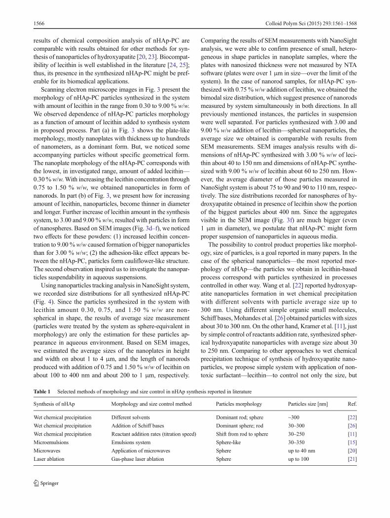
results of chemical composition analysis of nHAp-PC arecomparable with results obtained for other methods for syn-thesis of nanoparticles of hydroxyapatite [20, 23]. Biocompat-ibility of lecithin is well established in the literature [24, 25];thus, its presence in the synthesized nHAp-PC might be pref-erable for its biomedical applications.
Scanning electron microscope images in Fig. 3 present themorphology of nHAp-PC particles synthesized in the systemwith amount of lecithin in the range from 0.30 to 9.00 % w/w.We observed dependence of nHAp-PC particles morphologyas a function of amount of lecithin added to synthesis systemin proposed process. Part (a) in Fig. 3 shows the plate-likemorphology, mostly nanoplates with thickness up to hundredsof nanometers, as a dominant form. But, we noticed someaccompanying particles without specific geometrical form.The nanoplate morphology of the nHAp-PC corresponds withthe lowest, in investigated range, amount of added lecithin—0.30%w/w.With increasing the lecithin concentration through0.75 to 1.50 % w/w, we obtained nanoparticles in form ofnanorods. In part (b) of Fig. 3, we present how for increasingamount of lecithin, nanoparticles, become thinner in diameterand longer. Further increase of lecithin amount in the synthesissystem, to 3.00 and 9.00 %w/w, resulted with particles in formof nanospheres. Based on SEM images (Fig. 3d–f), we noticedtwo effects for these powders: (1) increased lecithin concen-tration to 9.00%w/w caused formation of bigger nanoparticlesthan for 3.00 % w/w; (2) the adhesion-like effect appears be-tween the nHAp-PC, particles form cauliflower-like structure.The second observation inspired us to investigate the nanopar-ticles suspendability in aqueous suspensions.
Using nanoparticles tracking analysis in NanoSight system,we recorded size distributions for all synthesized nHAp-PC(Fig. 4). Since the particles synthesized in the system withlecithin amount 0.30, 0.75, and 1.50 % w/w are non-spherical in shape, the results of average size measurement(particles were treated by the system as sphere-equivalent inmorphology) are only the estimation for these particles ap-pearance in aqueous environment. Based on SEM images,we estimated the average sizes of the nanoplates in heightand width on about 1 to 4 μm, and the length of nanorodsproduced with addition of 0.75 and 1.50 % w/w of lecithin onabout 100 to 400 nm and about 200 to 1 μm, respectively.
Comparing the results of SEMmeasurements with NanoSightanalysis, we were able to confirm presence of small, hetero-geneous in shape particles in nanoplate samples, where theplates with nanosized thickness were not measured by NTAsoftware (plates were over 1 μm in size—over the limit of thesystem). In the case of nanorod samples, for nHAp-PC syn-thesized with 0.75 % w/w addition of lecithin, we obtained thebimodal size distribution, which suggest presence of nanorodsmeasured by system simultaneously in both directions. In allpreviously mentioned instances, the particles in suspensionwere well separated. For particles synthesized with 3.00 and9.00 % w/w addition of lecithin—spherical nanoparticles, theaverage size we obtained is comparable with results fromSEM measurements. SEM images analysis results with di-mensions of nHAp-PC synthesized with 3.00 % w/w of leci-thin about 40 to 150 nm and dimensions of nHAp-PC synthe-sized with 9.00 % w/w of lecithin about 60 to 250 nm. How-ever, the average diameter of those particles measured inNanoSight system is about 75 to 90 and 90 to 110 nm, respec-tively. The size distributions recorded for nanospheres of hy-droxyapatite obtained in presence of lecithin show the portionof the biggest particles about 400 nm. Since the aggregatesvisible in the SEM image (Fig. 3f) are much bigger (even1 μm in diameter), we postulate that nHAp-PC might formproper suspension of nanoparticles in aqueous media.
The possibility to control product properties like morphol-ogy, size of particles, is a goal reported in many papers. In thecase of the spherical nanoparticles—the most reported mor-phology of nHAp—the particles we obtain in lecithin-basedprocess correspond with particles synthesized in processescontrolled in other way. Wang et al. [22] reported hydroxyap-atite nanoparticles formation in wet chemical precipitationwith different solvents with particle average size up to300 nm. Using different simple organic small molecules,Schiff bases,Mohandes et al. [26] obtained particles with sizesabout 30 to 300 nm. On the other hand, Kramer et al. [11], justby simple control of reactants addition rate, synthesized spher-ical hydroxyapatite nanoparticles with average size about 30to 250 nm. Comparing to other approaches to wet chemicalprecipitation technique of synthesis of hydroxyapatite nano-particles, we propose simple system with application of non-toxic surfactant—lecithin—to control not only the size, but
Table 1 Selected methods of morphology and size control in nHAp synthesis reported in literature
Synthesis of nHAp Morphology and size control method Particles morphology Particles size [nm] Ref.
Wet chemical precipitation Different solvents Dominant rod; sphere ~300 [22]
Wet chemical precipitation Addition of Schiff bases Dominant sphere; rod 30–300 [26]
Wet chemical precipitation Reactant addition rates (titration speed) Shift from rod to sphere 30–250 [11]
Microemulsions Emulsions system Sphere-like 30–350 [15]
Microwaves Application of microwaves Sphere up to 40 nm [20]
Laser ablation Gas-phase laser ablation Sphere up to 100 [21]
1566 Colloid Polym Sci (2015) 293:1561–1568

morphology too. For more detailed comparison of discussedtechniques, please see Table 1. Cytotoxicity of surfactants likeCTAB or sodium dodecyl sulfate (SDS) results from theirinteractions with cellular membranes leading to cell wall rup-ture. This effect might be observed even when surfactants arebonded or adsorbed on the biomaterial surface. Lecithin—mixture of phosphatidylcholines, components of cellularmembranes [24]—when present on the surface of any bioma-terial, attracts cells to interact with the material without caus-ing any damage to the cell membrane [25]. Aforementionedreported approaches to wet chemical precipitation allow toaffect the particles morphology, but only in range from nano-rods to nanopheres. It is important to notice that morphologycontrol—in specific way—in the nHAp formation processesrequires more complicated solutions, like previously men-tioned, e.g., microemulsions, microwaves or laser ablation[15, 20, 21].
Conclusions
In this paper, we proposed a novel approach to wet chemicalprecipitation of hydroxyapatite nanoparticles in the presenceof lecithin in the reaction system. We showed resulting nano-particles crystallinity and chemistry analysis results, provingthat lecithin-based approach allows formation of good qualityhydroxyapatite nanoparticles, slightly modified by traces oflecithin (nHAp-PC). Based on SEM image investigation andadditional particle tracking analysis experiments in NanoSightsystem, we described possibilities to control the product mor-phology and the average particle size by varying lecithin con-centration in the reaction system only. We postulate the in-creased biocompatibility of the resulting nHAp-PC materialby modification of nanoparticles by bonding with lecithin.This modification also increases suspendability property ofnanoparticles in various media.
The patent of the presented method is pending.
Acknowledgments The authors are thankful to IgaWasiak, MSc. Eng.,from Faculty of Chemical and Process Engineering, Warsaw Universityof Technology for the NanoSight measurements, and Andrzej Ostrowski,PhD Eng., from Faculty of Chemistry, Warsaw University of Technologyfor the XRD measurements.
Open Access This article is distributed under the terms of the CreativeCommons Attribution License which permits any use, distribution, andreproduction in any medium, provided the original author(s) and thesource are credited.
References
1. Schachschal S, Pich A, Adler H-J (2007) Growth of hydroxyapatitenanocrystals on polymer particle surface. Colloid Polym Sci 285:1175–1180. doi:10.1007/s00396-007-1685-x
2. Kusmanto F, Walker G, Gan Q et al (2008) Development of compos-ite tissue scaffolds containing naturally sourced mircoporous hy-droxyapatite. Chem Eng J 139:398–407. doi:10.1016/j.cej.2007.11.041
3. Malmsten M (2013) Inorganic nanomaterials as delivery systems forproteins, peptides, DNA, and siRNA. Curr Opin Colloid In 18:468–480. doi:10.1016/j.cocis.2013.06.002
4. Vallet-Regi M, Gonzalez-Calbet JM (2004) Calcium phosphates assubstitution of bone tissues. Prog Solid State Ch 32:1–31. doi:10.1016/j.progsolidstchem.2004.07.001
5. Rauschmann MA, Wichelhaus TA, Stirnal V et al (2005)Nanocrystalline hydroxyapatite and calcium sulphate as biodegrad-able composite carrier material for local delivery of antibiotics inbone infections. Biomaterials 26:2677–2684. doi:10.1016/j.biomaterials.2004.06.045
6. Zahouily M, BahlaouanW, Bahlaouan B, RayadhA (2005) Catalysisby hydroxyapatite alone and modified by sodium nitrate: a simpleand efficient procedure for the construction of carbon-nitrogen bondsin heterogeneous catalysis. Arkivoc xiii:150–161
7. Kawai T, Ohtsuki C, Kamitakahara M et al (2006) Removal of form-aldehyde by hydroxyapatite layer biomimetically deposited on poly-amide film. Environ Sci Technol 40:4281–4285. doi:10.1021/es050098n
8. Jungbauer A, Hahn R, Deinhofer K (2004) Performance and charac-terization of a nanophased porous hydroxyapatite for protein chro-matography. Biotechnol Bioeng 87:364–375. doi:10.1002/bit.20121
9. Liu Y, Hou D, Wang G (2004) A simple wet chemical synthesis andcharacterization of hydroxyapatite nanorods. Mater Chem Phys 86:69–73. doi:10.1016/j.matchemphys.2004.02.009
10. Mateus AYP, Barrias CC, Ribeiro C et al (2008) Comparative studyof nanohydroxyapatite microspheres for medical applications. JBiomed Mater Res A 86A:483–493. doi:10.1002/jbm.a.31634
11. Kramer E, Podurgiel J, Wei M (2014) Control of hydroxyapatitenanoparticle morphology using wet synthesis techniques. Reactantaddition rate effects. Mater Lett 131:145–147. doi:10.1016/j.matlet.2014.05.105
12. Wang Y, Zhang S, Wei K et al (2006) Hydrothermal synthesis ofhydroxyapatite nanopowders using cationic surfactant as a template.Mater Lett 60:1484–1487. doi:10.1016/j.matlet.2005.11.053
13. Jin X, Zhuang J, Zhang Z et al (2015) Hydrothermal synthesis ofhydroxyapatite nanorods in the presence of sodium citrate and itsaqueous colloidal stability evaluation in neutral pH. J Colloid InterfSci 443:125–130. doi:10.1016/j.jcis.2014.12.010
14. Kuriakose TA, Kalkura SN, PalanichamyM et al (2004) Synthesis ofstoichiometric nano crystalline hydroxyapatite by ethanol-based sol–gel technique at low temperature. J Cryst Growth 263:517–523. doi:10.1016/j.jcrysgro.2003.11.057
15. García C, García C, Paucar C (2012) Controlling morphology ofhydroxyapatite nanoparticles through hydrothermal microemulsionchemical synthesis. Inorg Chem Commun 20:90–92. doi:10.1016/j.inoche.2012.02.024
16. Mollazadeh S, Javadpour J, Khavandi A (2007) In situ synthesis andcharacterization of nano-size hydroxyapatite in poly(vinyl alcohol)matrix. Ceram Int 33:1579–1583. doi:10.1016/j.ceramint.2006.06.006
17. Varma HK, Suresh Babu S (2005) Synthesis of calcium phosphatebioceramics by citrate gel pyrolysis method. Ceram Int 31:109–114.doi:10.1016/j.ceramint.2004.03.041
18. Cüneyt Tas A (2000) Combustion synthesis of calcium phosphatebioceramic powders. J Eur Ceram Soc 20:2389–2394. doi:10.1016/S0955-2219(00)00129-1
19. Gopi D, Govindaraju KM, Victor CAP et al (2008) Spectroscopicinvestigations of nanohydroxyapatite powders synthesized by con-ventional and ultrasonic coupled sol–gel routes. Spectrochim Acta A70:1243–1245. doi:10.1016/j.saa.2008.02.015
Colloid Polym Sci (2015) 293:1561–1568 1567

20. Smoleń D, Chudoba T, Malka I et al (2013) Highly biocom-patible, nanocrystalline hydroxyapatite synthesized in asolvothermal process driven by high energy density micro-wave radiation. Int J Nanomed 8:653–668. doi:10.2147/IJN.S39299
21. Bapat PV, Kraft R, Camata RP (2012) Gas-phase laser syn-thesis of aggregation-free, size-controlled hydroxyapatite nano-particles. J Nanopart Res 14:1–8. doi:10.1007/s11051-012-1163-3
22. Wang P, Li C, Gong H et al (2010) Effects of synthesis conditions onthe morphology of hydroxyapatite nanoparticles produced by wetchemical process. Powder Technol 203:315–321. doi:10.1016/j.powtec.2010.05.023
23. Rajeswari A, Kumar VG, Karthick Vet al (2013) Hydrothermal syn-thesis of hydroxyapatite plates prepared using low molecular weightheparin (LMWH). Colloid Surface B 111:764–768. doi:10.1016/j.colsurfb.2013.06.040
24. Chapman D (1998) New biomaterials based upon biomembranemimicry. Prog Colloid Polym Sci 108:17–20
25. Butruk-Raszeja B, Trzaskowski M, Ciach T (2014) Cell membrane-mimicking coating for blood-contacting polyurethanes. J BiomaterAppl 29:801–812. doi:10.1177/0885328214549611
26. Mohandes F, Salavati-Niasari M (2014) Simple morphology-controlled fabrication of hydroxyapatite nanostructures with the aidof new organic modifiers. Chem Eng J 252:173–184. doi:10.1016/j.cej.2014.05.026
1568 Colloid Polym Sci (2015) 293:1561–1568