learning under distributed weak supervision
TRANSCRIPT

1
Learning under Distributed Weak SupervisionMartin Rajchl, Matthew C. H. Lee, Franklin Schrans, Alice Davidson, Jonathan Passerat-Palmbach,
Giacomo Tarroni, Amir Alansary, Ozan Oktay, Bernhard Kainz, and Daniel Rueckert
Abstract—The availability of training data for supervision isa frequently encountered bottleneck of medical image analysismethods. While typically established by a clinical expert rater, theincrease in acquired imaging data renders traditional pixel-wisesegmentations less feasible. In this paper, we examine the use of acrowdsourcing platform for the distribution of super-pixel weakannotation tasks and collect such annotations from a crowd ofnon-expert raters. The crowd annotations are subsequently usedfor training a fully convolutional neural network to address theproblem of fetal brain segmentation in T2-weighted MR images.Using this approach we report encouraging results compared tohighly targeted, fully supervised methods and potentially addressa frequent problem impeding image analysis research.
Index Terms—Weak Supervision, Image Segmentation, Ma-chine Learning, Convolutional Neural Networks
I. INTRODUCTION
MODERN learning-based methods for medical imageanalysis rely on large amounts of labelled data to
properly cover different sources of variability in the data (e.g.due to the pose of the subject, the presence of pathology, etc.).This situation is particularly exacerbated when analysis ondata is required for which no open labelled atlas databasesexist that could be adopted for supervision. However, theoption of an expert rater to pixel-wise label a training set,is often not feasible. To address this problem, methods em-ploying weak forms of annotations (e.g. image-level tags[1], bounding boxes [2], [3], [4], drawn scribbles [5], [6],etc.) aim to reduce the annotation effort and increasinglygain attention. For instance, recent studies have shown thatemploying bounding box annotations is approximately 15times faster than using pixel-wise manual segmentations [7],[2]. In conjunction with using simple forms of annotations,web-based collaborative platforms for crowdsourcing havebeen investigated in their ability to obtain large amounts ofannotations for labelling image databases [8], [9], [10]. Whilesuch interfaces often have limited capacity to interact withthe image data, using weak annotations immediately suggestsitself, because of its simplicity. However, in contrast to taskssuch as the annotation of natural images [7], [11] and theidentification of surgical instruments [9] in surgical videosequence, the correct interpretation of medical images requiresspecialised training and experience, and therefore might posea challenge for non-expert crowds. Nevertheless, in contrastto the diagnostic interpretation of medical images, medicalimage analysis pipelines often require the identification ofanatomical structures, requiring less expertise (i.e. it requires
All authors are with the Dept. of Computing, Imperial College London,SW7 2AZ, London, UK
(*)Corresponding author: Martin Rajchl e-mail: [email protected]
less expertise to identify an organ in an MR image, than apotential pathology).
A. Contributions:In this paper, we entertain the notion that non-experts can be
used for some annotation tasks on medical images. These taskscan be simplified by employing super-pixel weak annotationsand the total annotation effort can be distributed to many raters(also commonly referred to as crowd) using a web browseras an interface. We investigate this concept in the contextof the fetal brain segmentation problem in T2-weighted MRimages. Using a fully convolutional neural network (FCN)we achieve state-of-the-art accuracy performance under fullexpert supervision and report comparably high values forlearning from expert weakly supervised data (i.e. super-pixelannotations). Further, we distribute the super-pixel annotationtasks to 12 non-expert raters and achieve similar performanceto that of experts.
II. METHODS
In the following sections, we describe means of distributingannotation tasks and facilitating learning from acquired weakannotations using a state-of-the-art fully convolutional neuralnetwork [12].
A. Distributed Weak Annotations:For a flexible solicitation of annotation tasks, we propose a
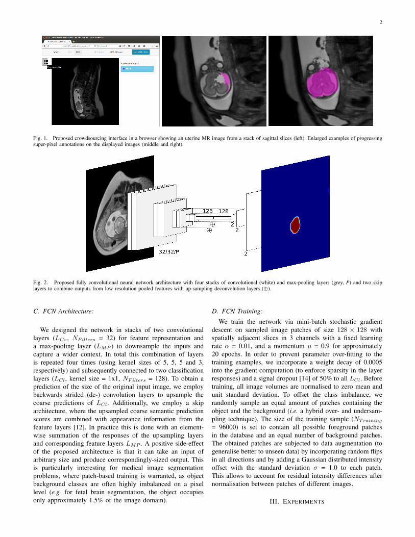
crowdsourcing platform where users can interact and annotateimage data. To accelerate the annotation process, we provide aSLIC super-pixel segmentation [13] and let users select thosebelonging to the object we are interested in. We implement theSLIC computation using Javascript to outsource the computa-tional load to the client machine and concentrate on backendtasks on the server side (e.g. data conversion, collection, etc.).The web-based user interface is based on the well-knownLabelMe framework [11] and was modified to interact withvolumetric medical image data and to compute and collectsuper-pixel annotations. Fig. 1 depicts the interface reducedto accommodate the particular annotation task at hand and anexample of a user labelling super-pixel belonging to a fetalbrain on a T2w MR image slice.
B. Learning with Fully Convolutional Neural Networks:We propose a fully convolutional neural network (FCN)
architecture to address the segmentation problem in a generaland extendible manner [12]. Such an approach has recentlybeen introduced for semantic object segmentation problemson natural images, exceeding the state-of-the-art in accuracyperformance while exhibiting remarkable training and infer-ence speed thanks to its fully convolutional nature [12].
arX
iv:1
606.
0110
0v1
[cs
.CV
] 3
Jun
201
6
2
Fig. 1. Proposed crowdsourcing interface in a browser showing an uterine MR image from a stack of sagittal slices (left). Enlarged examples of progressingsuper-pixel annotations on the displayed images (middle and right).
Fig. 2. Proposed fully convolutional neural network architecture with four stacks of convolutional (white) and max-pooling layers (grey, P) and two skiplayers to combine outputs from low resolution pooled features with up-sampling deconvolution layers (⊕).
C. FCN Architecture:
We designed the network in stacks of two convolutionallayers (LCv , NFilters = 32) for feature representation anda max-pooling layer (LMP ) to downsample the inputs andcapture a wider context. In total this combination of layersis repeated four times (using kernel sizes of 5, 5, 5 and 3,respectively) and subsequently connected to two classificationlayers (LCl, kernel size = 1x1, NFilters = 128). To obtain aprediction of the size of the original input image, we employbackwards strided (de-) convolution layers to upsample thecoarse predictions of LCl. Additionally, we employ a skiparchitecture, where the upsampled coarse semantic predictionscores are combined with appearance information from thefeature layers [12]. In practice this is done with an element-wise summation of the responses of the upsampling layersand corresponding feature layers LMP . A positive side-effectof the proposed architecture is that it can take an input ofarbitrary size and produce correspondingly-sized output. Thisis particularly interesting for medical image segmentationproblems, where patch-based training is warranted, as objectbackground classes are often highly imbalanced on a pixellevel (e.g. for fetal brain segmentation, the object occupiesonly approximately 1.5% of the image domain).
D. FCN Training:
We train the network via mini-batch stochastic gradientdescent on sampled image patches of size 128 × 128 withspatially adjacent slices in 3 channels with a fixed learningrate α = 0.01, and a momentum µ = 0.9 for approximately20 epochs. In order to prevent parameter over-fitting to thetraining examples, we incorporate a weight decay of 0.0005into the gradient computation (to enforce sparsity in the layerresponses) and a signal dropout [14] of 50% to all LCl. Beforetraining, all image volumes are normalised to zero mean andunit standard deviation. To offset the class imbalance, werandomly sample an equal amount of patches containing theobject and the background (i.e. a hybrid over- and undersam-pling technique). The size of the training sample (NTraining
= 96000) is set to contain all possible foreground patchesin the database and an equal number of background patches.The obtained patches are subjected to data augmentation (togeneralise better to unseen data) by incorporating random flipsin all directions and by adding a Gaussian distributed intensityoffset with the standard deviation σ = 1.0 to each patch.This allows to account for residual intensity differences afternormalisation between patches of different images.
III. EXPERIMENTS

3
A. Image Data:
Images from 37 fetal subjects were acquired on a PhilipsAchieva 1.5T with the mother lying 20◦ tilt on the left sideto avoid pressure on the inferior vena cava or on her backdepending on her comfort. Single-shot fast spin echo (ssFSE)T2-weighted sequences are used to acquire stacks of imagesthat are aligned to either the main axes of the fetus or of themother. Usually three to six stacks are acquired for the wholeuterus with a voxel size of 1.25 × 1.25 × 2.50 mm. fetal MRIdata can be corrupted by motion because of unpredictable fetalmovements and maternal respiratory motion. The stacks withthe least motion artefacts were selected for our experiments.A clinical expert rater manually annotated the fetal brain toestablish a reference standard segmentation.
B. Evaluation:
We recruited 12 users with technical degrees and exposureto medical imaging research as a non-expert crowd and askedthem to annotate consecutive slices of T2w fetal MR volumesusing the proposed web interface (see Sec. II-A) to labelsuper-pixels of size 12 × 12 px belonging to a fetal brain.Prior to access to the data, the users were asked to completea short tutorial showing expert segmentations of the fetalbrain in different slice directions. Furthermore, to evaluate thedetrimental impact of the SLIC weak annotations, a secondexperiment was performed using super-pixels extracted fromthe reference segmentations from the expert rater (based ona threshold of 50% of area coverage between each super-pixel and the reference). These serve as training data forlearning under expert weak supervision. Finally, we comparetraining on full expert supervision data (i.e. directly from thereference standard) using the proposed FCN architecture inSec. II-B. The trained network models are then used to inferthe fetal brain on unseen volumes. To reduce the variationin the experimental setup and suppress possible factors im-pacting on accuracy, we sampled the same patch locations forall annotation types and computed the same augmentations.Additionally, prior to training all networks were initialisedwith the same random weights. For validation, we used a three-fold cross-validation setup and computed the Dice SimilarityCoefficient (DSC = 2|P∩M |
|P |+|M | ) between predicted regionsP and expert manual segmentations M . We used the Caffelibrary [15] for the creation and training of the proposed FCNarchitecture (see Sec. II-B) and performed all experiments on aUbuntu 14.04 machine with 256 GB memory and an NVIDIATesla K80 (12 GB memory).
IV. RESULTS
Table I shows the accuracy as DSC for all compared ap-proaches, respectively. While learning under full expert super-vision exhibits the most accurate performance, using weaklysupervised simulated expert annotations and weak annotationsfrom non-expert raters yield comparably high results. Figure 3shows selected examples of segmentation results and segmen-tation errors for all compared methods.
The reliability of the web-based super-pixel annotationscollected from non-expert users and those extracted from the
TABLE IMEAN ACCURACY RESULTS FOR FETAL BRAIN SEGMENTATION COMPARED
TO THE EXPERT REFERENCE STANDARD AS DSC [%].
Expert Non-expert
Supervision type Full Weak WeakDSC [%] 92.7 ± 2.3 90.3 ± 2.8 90.6 ± 2.3
expert is depicted in Figure 4. All slices that either containedthe object in the reference standard or were annotated by auser were evaluated in their accuracy against the referencestandard. Note, that the users did not perform an equal numberof annotation tasks.
A. Runtime:
The average time spent annotating an image slice withthe proposed web-interface was 7.2 ± 3.4 seconds, includingloading, annotation and task submission. FCN training timewas approximately six hours and inference can be done underone minute for the largest acquired MR stack (512 × 512× 200 vx). Generation of the manual ground truth tookapproximately three full working days.
V. DISCUSSION
All compared methods perform well in qualitative com-parison with other studies employing highly targeted, fullysupervised approaches. The works in [16], [17] and [18]reported mean DSC scores of 93.0%, 90.7% and 80.4%,respectively. Note, that the FCN prediction could additionallybe post-processed with a graphical method, which has beenshown to improve results in other segmentation problems[2], [3]. As expected, a higher accuracy could be achievedwhen learning under full supervision, however differencesappear marginal compared to those reported using boundingbox annotations [2], [3]. Surprisingly, both weakly supervisednetworks present with very similar accuracy (see Tab. I),when random factors such as sampling and augmentation areaccounted for. Particularly interesting is the presentation oflearned segmentation errors. We expect the exclusion of thecerebro-spinal fluid when using non-expert annotations (c.f.Fig. 3, magenta) is due to differences in image interpretationof the crowd on where the brain boundary is on axial slices.Similarly, the oversegmentation of the skull (c.f. Fig. 3, cyan)might be due to systematic oversegmentations from computedexpert super-pixels. Systematic annotation errors could beaddressed by integration of quality assurance measures and/orannotation regularisation post collection. Considering the baseaccuracy of the collected fetal brain annotations from non-experts, we observe similar performance to that of an expert(c.f. Fig. 4), indicating that some anatomical annotation taskscan be performed by crowds with less expertise.
The observed efficiency of distributed weak annotation taskswith the proposed crowdsourcing interface is remarkable. Con-sidering the measured average annotation time of 7.2 seconds,with a collective of 12 users, the annotation of the entiredatabase took less than one hour to annotate (total of 10.7hours) the entire database (more than 5000 slices) an expert

4
Fig. 3. Top and middle row: Example results of expert manual (white), expert fully supervised (yellow), expert weakly supervised (cyan) and non-expertweakly supervised segmentations. Prediction errors are marked with arrows (green). Bottom row: Non-expert weakly supervised segmentation results on axialand sagittal slices, including twins (middle).
annotator took three work days to establish the same witha multi-planar interface. These observations might indicatea paradigm shift on how we enable learning based methodsfor medical image analysis to address the ever-growing datacollected for imaging studies.
At this juncture, we note that contrary to relying on com-mercial crowdsourcing platforms such as Amazon MTurk, weaim to focus on a more flexible platform that can better takeadvantage of contributions from image scientists and thoseinterested in supporting medical research, thereby fostering en-gagement with a wider general public. The proposed approachhas the ability to enable this while simultaneously enablingthe development of machine learning based methods at muchlarger scales.
A. Conclusions:
We have investigated the web-based distribution of weakannotation tasks to a crowd of non-expert users to establish
training data for a learning-based segmentation method. Theproposed approach largely reduces the annotation load onexpert users and was successfully employed for segmentationof fetal brains from motion-corrupted T2w MR image stacks.The encouraging results and the consistent annotation perfor-mance of the crowd suggest that this approach could be readilyported to other challenges and potentially address a frequentlyencountered bottleneck in medical image analysis studies.
ACKNOWLEDGEMENTS
We gratefully acknowledge the support of NVIDIA Corpo-ration with the donation of a Tesla K40 GPU used for thisresearch. This research was also supported by the NationalInstitute for Health Research (NIHR) Biomedical ResearchCentre based at Guy’s and St Thomas’ NHS Foundation Trustand King’s College London. The views expressed are thoseof the author(s) and not necessarily those of the NHS, theNIHR or the Department of Health. Furthermore, this work

5
Fig. 4. Accuracy of SLIC weak annotations of the simulated expert user (EXP) and non-expert users (U) in DSC [0,1].
was supported by Wellcome Trust and EPSRC IEH award[102431] for the iFIND project and the Developing HumanConnectome Project, which is funded through a Synergy Grantby the European Research Council (ERC) under the EuropeanUnion’s Seventh Framework Programme (FP/2007-2013) /ERC Grant Agreement number 319456.
REFERENCES
[1] T. Schlegl, S. M. Waldstein, W.-D. Vogl, U. Schmidt-Erfurth, andG. Langs, “Predicting semantic descriptions from medical images withconvolutional neural networks,” in Information Processing in MedicalImaging. Springer, 2015, pp. 437–448.
[2] G. Papandreou, L.-C. Chen, K. Murphy, and A. L. Yuille, “Weakly-andsemi-supervised learning of a DCNN for semantic image segmentation,”arXiv preprint arXiv:1502.02734, 2015.
[3] J. Dai, K. He, and J. Sun, “Boxsup: Exploiting bounding boxes to super-vise convolutional networks for semantic segmentation,” in Proceedingsof the IEEE International Conference on Computer Vision, 2015, pp.1635–1643.
[4] M. Rajchl, M. C. Lee, O. Oktay, K. Kamnitsas, J. Passerat-Palmbach,W. Bai, B. Kainz, and D. Rueckert, “Deepcut: Object segmentation frombounding box annotations using convolutional neural networks,” arXivpreprint arXiv:1605.07866, 2016.
[5] L. M. Koch, M. Rajchl, W. Bai, C. F. Baumgartner, T. Tong, J. Passerat-Palmbach, P. Aljabar, and D. Rueckert, “Multi-atlas segmentation usingpartially annotated data: Methods and annotation strategies,” arXivpreprint arXiv:1605.00029, 2016.
[6] M. Rajchl, J. Yuan, J. White, E. Ukwatta, J. Stirrat, C. Nambakhsh, F. Li,and T. Peters, “Interactive hierarchical max-flow segmentation of scartissue from late-enhancement cardiac mr images,” IEEE Transactions onMedical Imaging, vol. 33, no. 1, pp. 159–172, 2014.
[7] T.-Y. Lin, M. Maire, S. Belongie, J. Hays, P. Perona, D. Ramanan,P. Dollar, and C. L. Zitnick, “Microsoft coco: Common objects incontext,” in Computer Vision–ECCV 2014. Springer, 2014, pp. 740–755.
[8] M. T. McKenna, S. Wang, T. B. Nguyen, J. E. Burns, N. Petrick, andR. M. Summers, “Strategies for improved interpretation of computer-aided detections for CT colonography utilizing distributed human intel-ligence,” Medical image analysis, vol. 16, no. 6, pp. 1280–1292, 2012.
[9] L. Maier-Hein, S. Mersmann, D. Kondermann, S. Bodenstedt,A. Sanchez, C. Stock, H. G. Kenngott, M. Eisenmann, and S. Speidel,“Can Masses of Non-Experts Train Highly Accurate Image Classifiers?”in Medical Image Computing and Computer-Assisted Intervention–MICCAI 2014, 2014, pp. 438–445.
[10] D. Haehn, J. Beyer, H. Pfister, S. Knowles-Barley, N. Kasthuri, J. Licht-man, and M. Roberts, “Design and evaluation of interactive proofreadingtools for connectomics,” Computer Graphics, IEEE Transactions on,2014.
[11] B. C. Russell, A. Torralba, K. P. Murphy, and W. T. Freeman, “Labelme:a database and web-based tool for image annotation,” Internationaljournal of computer vision, vol. 77, no. 1-3, pp. 157–173, 2008.
[12] J. Long, E. Shelhamer, and T. Darrell, “Fully convolutional networksfor semantic segmentation,” in Proceedings of the IEEE Conference onComputer Vision and Pattern Recognition, 2015, pp. 3431–3440.
[13] R. Achanta, A. Shaji, K. Smith, A. Lucchi, P. Fua, and S. Susstrunk,“SLIC superpixels compared to state-of-the-art superpixel methods,”Pattern Analysis and Machine Intelligence, IEEE Transactions on,vol. 34, no. 11, pp. 2274–2282, 2012.
[14] N. Srivastava, G. Hinton, A. Krizhevsky, I. Sutskever, and R. Salakhut-dinov, “Dropout: A simple way to prevent neural networks from over-fitting,” The Journal of Machine Learning Research, vol. 15, no. 1, pp.1929–1958, 2014.
[15] Y. Jia, E. Shelhamer, J. Donahue, S. Karayev, J. Long, R. Girshick,S. Guadarrama, and T. Darrell, “Caffe: Convolutional architecture forfast feature embedding,” in Proceedings of the ACM InternationalConference on Multimedia. ACM, 2014, pp. 675–678.
[16] K. Keraudren, M. Kuklisova-Murgasova, V. Kyriakopoulou, C. Mala-mateniou, M. Rutherford, B. Kainz, J. Hajnal, and D. Rueckert, “Au-tomated fetal brain segmentation from 2D MRI slices for motioncorrection,” NeuroImage, vol. 101, pp. 633–643, 2014.
[17] B. Kainz, K. Keraudren, V. Kyriakopoulou, M. Rutherford, J. V. Hajnal,and D. Rueckert, “Fast Fully Automatic Brain Detection in Foetal MRIUsing Dense Rotation Invariant Image Descriptors,” in IEEE ISBI’14,2014, pp. 1230 –1233.
[18] Y. Taleb, M. Schweitzer, C. Studholme, M. Koob, J.-L. Dietemann,and F. Rousseau, “Automatic template-based brain extraction in fetalmr images,” 2013.