learning objectives - thiemedissector-vol.-i)-with-watermark.pdf · from the superficial part of...
TRANSCRIPT
Pectoral Region and Axilla
CHAPTER 2
IntroductionThe upper limb is adapted to great freedom of movement in order to perform skillful/prehensile acts. Its importance is duly reflected by the sizeable area of representation it occupies in the cerebral cortex. The upper limb for the purpose of dissection is divisible into the following:Shoulder region: It includes the axilla or armpit, scapular region and pectoral region.Arm (brachium): The part between the shoulder and elbow, with humerus as its bone.Forearm (antebrachium): It extends from the elbow to the wrist. It has two bones, the radius and ulna. Distally, they articulate with the carpal bones to form the wrist joint. The forearm bones articulate at the superior, middle and inferior radioulnar joints. The movements of pronation and supination occur at these joints.Hand (manus): It consists of the wrist/carpus, metacarpus and digits (fingers and thumbs). The forearm bones, radius and ulna, articulate with the carpal bones. The carpals distally articulate with the meta-carpals and the metacarpals articulate with the phalanges. The hand presents ball of the thumb, thenar eminence and ball of the little finger, that is, the hypothenar eminence.The upper limb is attached to the thorax by muscles which form the anterior and posterior axillary folds. When the upper limb is moved away, the axilla (pyramidal shape) or armpit becomes evident.
Learning ObjectivesAt the end of the dissection of the pectoral region and axilla, students should be able to iden-tify the following:
Nerves: Supraclavicular nerves, medial and lateral pectoral nerves, long thoracic nerve and branches of the intercostal nerves.
Vessels: Branches of the internal thoracic artery, thoracoacromial artery and its branches, superior thoracic artery and cephalic vein.
Muscles: Pectoralis major, pectoralis minor, subclavius and serratus anterior. The anatomical basis of the clinical signs and conditions (e.g., Peau d’orange skin in carci-
noma of the breast and policeman’s tip position of the upper limb in Erb’s palsy).
THIEME

10 Chapter 2
The pectoral region covers the anterior thoracic wall presenting mammary glands. In females, breasts or mammary glands are well developed, while in males, they are rudimentary.
Surface Landmarks 1. Clavicle: It is the collar bone. It is subcutaneous and palpable throughout its length. It is concave
along the lateral one-third and convex along the medial two-thirds for vessels and nerves to pass between the axilla and root of neck. It articulates with the manubrium at the sternoclavicular joint and laterally with the acromion of the scapula at the acromioclavicular joint (Fig. 2.1).
2. Acromion (Acron: summit; omos: shoulder): It can be felt as a flat subcutaneous bone on the top of the shoulder.
3. Acromioclavicular joint: A plane synovial joint is felt as a slight depression between the lateral end of the clavicle and acromion.
4. Jugular notch: It lies between the medial ends of the clavicle along the superior margin of the man-ubrium.
5. Sternal angle: It can be felt as a transverse ridge at the junction of the manubrium with the body of the sternum. It lies at the level of the second costal cartilage. Other ribs are counted downwards from the second.
ClavicleAcromion
Head of humerus
Lateral epicondyle
Greater trochanter
Lateral condyle of femurHead of fibula
Patella
E
G F
Styloid process of ulnaPubic symphysis
Anterior superioriliac spine
Head of radius
Xiphoid process
Nipple
Sternal angle
Manubrium of sternumB
AC
D
Styloid process of radius
Fig. 2.1 Surface landmarks and incisions.
THIEME

Pectoral Region and Axilla 11
6. Xiphoid process: It is the lower part of the sternum lying in the epigastric fossa between the carti-lages of the seventh ribs.
7. Nipple: It is variable in position, usually at the fourth intercostal space slightly medial to the mid-clavicular line.
8. Infraclavicular fossa: It is a depression inferior to the junction of the middle with lateral one-third of the clavicle.
9. Coracoid process of the scapula: It can be felt 2 cm below the clavicle just lateral to the infraclavicu-lar fossa, under the muscle fibres of the deltoid.
10. Axilla (armpit): It is a hollow pyramidal space between the abducted upper limb and side of the thorax.
11. Anterior axillary fold: It is formed by the pectoralis major muscle. 12. Posterior axillary fold: It is formed by the latissimus dorsi and teres major muscles.
Dissection and IdentificationIn the supine position of the cadaver, as shown in Fig. 2.1, give the following skin incisions:
https://www.winkingskull.com/dissector/V1/video.aspx?vid=73 1. Incision A: Along the midline from the jugular notch to the xiphisternum. 2. Incision B: From the jugular notch along the clavicle to the acromion; in this, avoid (refrain) cut-
ting through the platysma muscle and supraclavicular nerves. 3. The platysma is a thin sheet of muscle present in the superficial fascia of the neck region and
helps in facial expressions. The muscle may extend below the clavicle into the superficial fascia of the upper part of the thorax and will be studied in detail with the head and neck regions.
4. Incision C: From the xiphisternal junction encircling the nipple and then along the anterior axil-lary fold to the arm.
5. Incision D: Horizontal incision from the xiphisternal junction to the posterior axillary fold. 6. Reflect the skin flaps and the superficial fascia laterally by blunt dissection. 7. While separating the skin flap along the clavicle (incision B), identify the supraclavicular nerves
by doing blunt dissection through the superficial fascia. 8. While reflecting the skin flap at incision C, close to the anterior axillary fold, identify the lat-
eral cutaneous branches of the intercostal nerves emerging serially in a vertical line. Trace their branches anteriorly and posteriorly.
9. The nipple and surrounding skin should be left as a landmark. 10. In female cadavers, an attempt can be made to dissect the mammary gland.
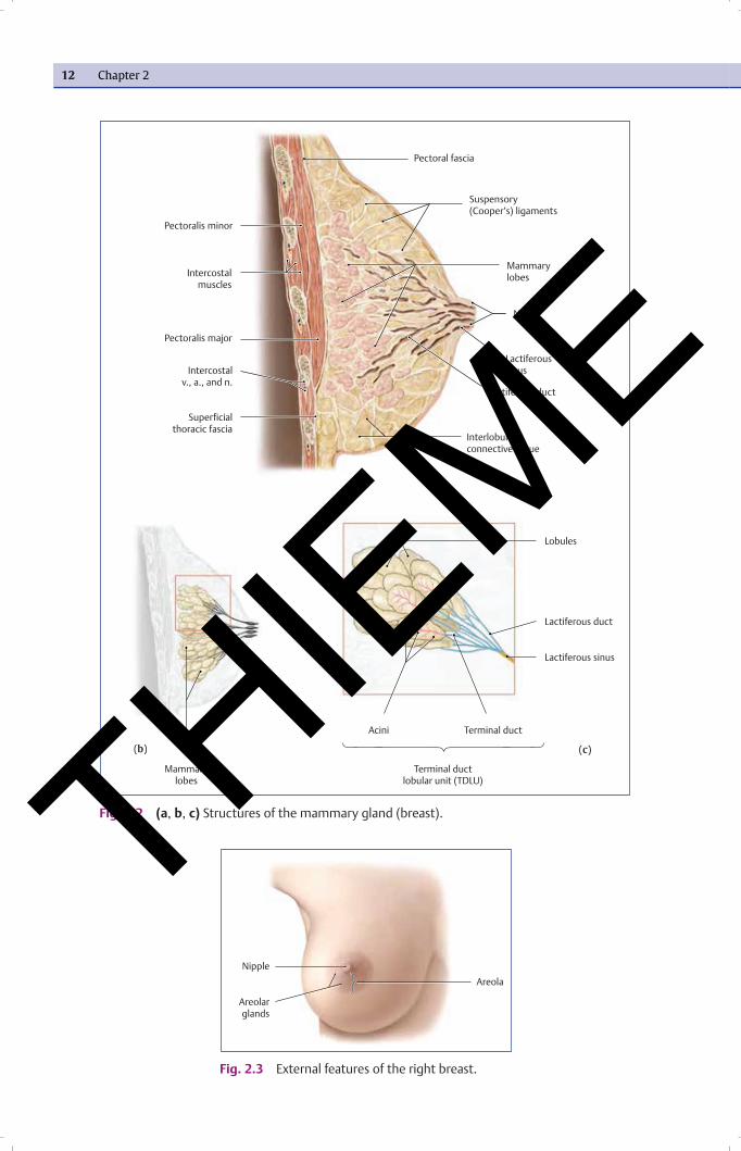
Mammary GlandThe mammary gland is a modified sweat gland extending vertically between the second and the sixth rib in the midclavicular line and sideways from the lateral border of the sternum to the midaxillary line. It lies within the superficial fascia anterior to the pectoralis major muscle. The pectoral fascia covers the anterior surface of the pectoralis major and is connected to the overlying skin by the sus-pensory ligaments of the breast (ligaments of Cooper), which pass between the lobes of the mammary gland (Fig. 2.2).
The mammary gland consists of 15 to 20 lobes; each lobe has a lactiferous duct which is dilated to form the lactiferous sinus at the base of the nipple and opens separately at the apex. Axillary tail (tail of Spence) of the gland passes superolaterally into the axilla to the level of the third rib. The gland is sur-mounted by the nipple lying at the level of the fourth intercostal space. It is surrounded by the areola, which is pink in colour at young age. In the early stages of pregnancy and shortly thereafter owing to pigmentation, both nipple and areola become dark permanently (Fig. 2.3).
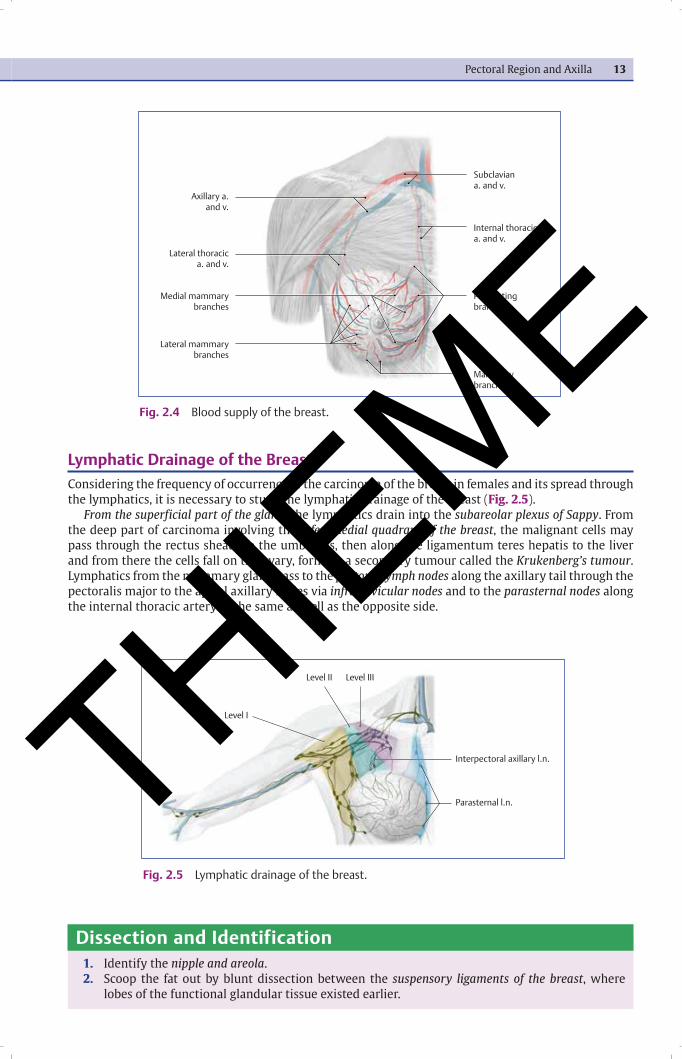
Blood SupplyBlood supply to this region occurs by the perforating branches of the intercostal and internal thoracic arteries medially and the lateral thoracic artery laterally (Fig. 2.4).
THIEME
12 Chapter 2
Pectoralis major
Pectoralis minor
Intercostalmuscles
Intercostalv., a., and n.
Superficialthoracic fascia
Interlobular connective tissue
Lactiferous duct
Lactiferous sinus
Nipple
Mammary lobes
Suspensory (Cooper’s) ligaments
Pectoral fascia
Mammarylobes
Terminal ductlobular unit (TDLU)
Acini Terminal duct
Lactiferous sinus
Lactiferous duct
Lobules
(a)
(b) (c)
Fig. 2.2 (a, b, c) Structures of the mammary gland (breast).
Nipple
Areolarglands
Areola
Fig. 2.3 External features of the right breast.
THIEME
Pectoral Region and Axilla 13
Lymphatic Drainage of the BreastConsidering the frequency of occurrence of the carcinoma of the breast in females and its spread through the lymphatics, it is necessary to study the lymphatic drainage of the breast (Fig. 2.5).
From the superficial part of the gland, the lymphatics drain into the subareolar plexus of Sappy. From the deep part of carcinoma involving the inferomedial quadrant of the breast, the malignant cells may pass through the rectus sheath to the umbilicus, then along the ligamentum teres hepatis to the liver and from there the cells fall on the ovary, forming a secondary tumour called the Krukenberg’s tumour. Lymphatics from the mammary gland pass to the pectoral lymph nodes along the axillary tail through the pectoralis major to the apical axillary nodes via infraclavicular nodes and to the parasternal nodes along the internal thoracic artery of the same as well as the opposite side.
Axillary a.and v.
Lateral thoracica. and v.
Lateral mammarybranches
Medial mammarybranches
Perforating branches
Internal thoracic a. and v.
Subclavian a. and v.
Mammary branches
Fig. 2.4 Blood supply of the breast.
Level IIILevel II
Level I
Interpectoral axillary l.n.
Parasternal l.n.
Fig. 2.5 Lymphatic drainage of the breast.
Dissection and Identification 1. Identify the nipple and areola. 2. Scoop the fat out by blunt dissection between the suspensory ligaments of the breast, where
lobes of the functional glandular tissue existed earlier.
THIEME
14 Chapter 2
3. About 15 to 20 lactiferous ducts, each arising from a lobe of the gland, converge on the nipple and expand into the lactiferous sinus deep to the nipple.
4. Attempt to pass a bristle through one of the ducts of the nipple and try to identify a lobe of the gland. The attempt may not be very successful in elderly females.
5. At the anterior ends of the intercostal spaces deep to the breast, small neurovascular bundles exist. Trace the branches of these nerves medially and laterally.
At this stage, it would be pertinent to have short descriptions of the cutaneous nerves, vessels and deep fascia in the region before we explore the deeper structures.
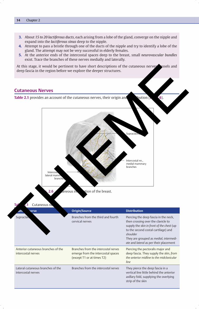
Cutaneous NervesTable 2.1 provides an account of the cutaneous nerves, their origin and distribution (Fig. 2.6).
Intercostal nn.,lateral mammary
branches
Intercostal nn., medial mammary branches
Supraclavicular nn.
Fig. 2.6 Cutaneous innervation of the breast.
Table 2.1 Cutaneous nerves
Name of nerve Origin/Source Distribution
Supraclavicular Branches from the third and fourth cervical nerves
Piercing the deep fascia in the neck, then crossing over the clavicle to supply the skin in front of the chest (up to the second costal cartilage) and shoulderThey are grouped as medial, intermedi-ate and lateral as per their placement
Anterior cutaneous branches of the intercostal nerves
Branches from the intercostal nerves emerge from the intercostal spaces (except T1 or at times T2)
Piercing the pectoralis major and deep fascia. They supply the skin, from the anterior midline to the midclavicular line
Lateral cutaneous branches of the intercostal nerves
Branches from the intercostal nerves They pierce the deep fascia in a vertical line little behind the anterior axillary fold, supplying the overlying strip of the skin
THIEME

Pectoral Region and Axilla 15
Deep FasciaThe deep fascia covering the pectoralis major (pectoral fascia) blends with the periosteum of the clavicle and sternum, and then passes over the deltopectoral groove to be continuous with the fascia over the deltoid. At the lower border of the pectoralis major, it is continuous along the axillary floor as axillary fascia.
Clavipectoral FasciaIt is a strong sheet of fascia, lying deep to the pectoralis major muscle (Fig. 2.7). It is attached to the clavi-cle above and splits to enclose the subclavius and pectoralis minor muscles. Lower down, it continues as a suspensory ligament of the axilla which blends with the axillary fascia. Between the subclavius and pectoralis minor, it is pierced by the following structures:
1. Lateral pectoral nerve 2. Thoracoacromial trunk/artery 3. Cephalic vein 4. Lymphatics from the deep part of the breast
Muscles of the Pectoral Region Muscles of the pectoral region (Fig. 2.8) are described in Table 2.2.
Table 2.2 Muscles of the pectoral region Name of muscle
Origin Insertion Nerve supply Action/Function
Pectoralis major (Fig. 2.8)
Clavicular head: Anterior surface of the medial half of the clavicleSternocostal head: Anterior surface of the sternum, upper six costal cartilages, aponeurosis of the exter-nal oblique muscle
Lateral lip of the in-tertubercular sulcus of the humerus
Lateral and medial pectoral nerves,clavicular head (C5, C6),sternocostal head (C7, C8 and T1)
1. Adducts and medially rotates the humerus; draws the scapula anteriorly and inferiorly2. Acting alone, the clavicular head flexes the humerus and the sternocostal head extends it from the flexed position
Clavicle
SubclaviusPectoralis major
Pectoral fasciaClavipectoral fascia
Axillary artery
Pectoralis minor
Axillary fascia
Fig. 2.7 Clavipectoral fascia as seen in oblique section parallel to the axillary artery.
(continued)
THIEME

16 Chapter 2
Name of muscle
Origin Insertion Nerve supply Action/Function
Pectoralis minor (Fig. 2.9)
Third to fifth ribs near their costal cartilages
Medial border and superior surface of the coracoid pro-cess of the scapula
Medial pectoral nerve (C8, T1)
Stabilizes the scapula by drawing it inferiorly and an-teriorly against the thoracic wall
Subclavius(Fig. 2.9)
Junction of the first rib and its costal cartilage
Inferior surface of the middle third of the clavicle
Nerve to the sub-clavius (C5, C6)
Anchors and depresses the clavicle
Serratus anterior (Fig. 2.10)
External surface of the upper eight ribs
Anterior surface of the medial border of the scapula
Long thoracic nerve (C5, C6, C7)
Protracts the scapula and holds it against the thoracic wall; rotates the scapula
Pectorails major,sternocostal part
Sternum
Pectoralis major,clavicular part ClavicleAcromion
Coracoidprocess
Greater tuberosity
Lessertuberosity
Intertuberculargroove
Crest of greatertuberosity
Coraco-brachialis
Pectoralis major,abdominal part
Humerus
Fig. 2.8 Pectoralis major.
Table 2.2 (continued) Muscles of the pectoral region
THIEME

Pectoral Region and Axilla 17
First ribClavicle
Acromion
Coracoidprocess
Subclavius
Pectoralisminor
Thirdthroughfifth ribs
Fig. 2.9 Pectoralis minor and subclavius.
Coracoidprocess
Acromion
Glenoidcavity
Medialborder
Scapula
Serratusanterior
Inferiorangle
First throughninth ribs
Fig. 2.10 Serratus anterior muscle.
THIEME

18 Chapter 2
Dissection and Identification 1. Remove the fascia from the anterior surfaces of the pectoralis major and deltoid to define their
attachments. 2. Identify the clavicular and sternocostal head of the pectoralis major and trace the muscle up to
its insertion on the humerus. 3. Divide the deep fascia by blunt dissection along the deltopectoral groove to reveal the cephalic
vein (Fig. 2.11). 4. Identify the deltopectoral triangle (Fig. 2.11) bounded by the deltoid, pectoralis major and
clavicle.
https://www.winkingskull.com/dissector/V1/video.aspx?vid=242
Transverse nerve of neck
External jugular vein
Accessory nerve
Sternocleidomastoid
Subclavian vein
Clavicle
Anterior jugular vein
Infraclavicular fossa
Sternum
Pectoralis major
Great auricularnerve
Lateral cervicaltriangle
Trapezius
Transversecervical vein
Supraclavicularnerves
Deltoid
Cephalic vein indeltopectoral
groove
Fig. 2.11 Superficial veins and nerves of the right shoulder region.
5. Cut through the clavicular head of the pectoralis major (Fig. 2.12) just below the clavicle and lift it laterally. This exposes the lateral pectoral nerve and thoracoacromial artery piercing the clavipectoral fascia.
https://www.winkingskull.com/dissector/V1/video.aspx?vid=81
6. Cut through the sternocostal head of the pectoralis major (Fig. 2.12) and lift it carefully so that the underlying structures are not damaged. Reflect the cut parts to expose the medial pectoral nerve that pierces the pectoralis minor and then the pectoralis major thus innervating both muscles.
7. Find under the reflected pectoralis major muscle the clavipectoral fascia, pectoralis minor muscle and subclavius muscle.
8. Trace the cephalic vein and lateral pectoral nerve through the clavipectoral fascia. Trace the ce-phalic vein to its termination into the axillary vein.
9. Trace the pectoralis minor muscle to its attachments. 10. Before you reflect the pectoralis minor, note that it delineates the three parts of the axillary artery. 11. Trace the branches of the thoracoacromial artery. 12. First, trace the pectoral branch which passes downwards and medially between the pectoralis
major and minor and supplies them, then trace it proximally and identify the acromial branch
THIEME

Pectoral Region and Axilla 19
which passes upwards and laterally deep to the deltoid, crossing the tip of coracoid process to the acromion.
13. Find out the clavicular branch ascending medially to supply the sternoclavicular joint and deltoid branch in the deltopectoral groove accompanying the cephalic vein.
14. Trace the lateral thoracic artery, a branch of the second part of the axillary artery, along the lat-eral border of the pectoralis minor muscle.
https://www.winkingskull.com/dissector/V1/video.aspx?vid=243
Fig. 2.12 Clavicular and sternocostal heads of pectoralis major muscle are cut to explore the underlying structures.
Internal jugular vein
Interscalenespace
Brachialplexus
Suprascapularartery
Omohyoid
Trapezius
Axillary artery
Thoracoacromialartery
Deltoid
Cephalic vein
Musculo-cutaneous nerve
Pectoralis major(cut edge)
Median nerve
Ulnar nerve
Axillary arteryand vein
Common carotid artery
Scalene muscles
Phrenic nerve
Inferior thyroid artery
Ascending cervical artery
Transverse cervical artery
External jugular vein
Thyrocervical trunk
Subclavian vein
Clavicle
Subclavius
Superior thoracic artery
Long thoracic nerve
Pectoralis major
Pectoralis minor
Medial and lateralpectoral nerves
Lateralthoracic artery
Subscapularartery
Thoraco-dorsal artery
Circumflexscapular artery
AxillaThe axilla (armpit) is a pyramidal-shaped space between the upper part of the arm and the lateral tho-racic wall.
Boundaries and Contents
Anterior wall: Pectoralis major, pectoralis minor and subclavius with enclosing investing fascia.Posterior wall: Subscapularis, teres major and latissimus dorsi (below).Medial wall: Serratus anterior.Lateral wall: Anterior and posterior walls approximate at the bicipital groove of the humerus.Apex of the axilla: It is bounded by the clavicle, first rib and superior border of the scapula and is continu-ous with the root of the neck through which pass the axillary vessels and nerves of the brachial plexus. The contents of axilla include axillary vessels, axillary lymph nodes, nerves of brachial plexus and loose connective tissue (Fig. 2.13).
THIEME

20 Chapter 2
Dissection and Identification
Fig. 2.13 Boundaries and contents of the axilla.
Longthoracic nerve
Superiorthoracic artery
Pectoralismajor
Medial andlateralpectoral nerves
Pectoralisminor
Subscapular nerve
Circumflexscapular artery
Thoracodorsalartery
Thoracodorsalnerve
Longthoracic nerve
Externaloblique
Serratusanterior
Latissimusdorsi
Posteriorcord of
brachial plexus
Tricepsbrachii
Posterior brachialcutaneous nerve
Brachial fascia
Ulnar nerve
Median nerve
Radial nerve
Biceps brachii
Anterior and posteriorcircumflex humeral arteries
Pectoralis major(cut edge)
Tendon of long headof biceps brachii
Cephalic vein
Deltoid
Musculo-cutaneous nerve
Brachial veinAxillary artery
and vein
Lateral thoracic artery Subclavian artery Subclavian vein
Clavicle SubclaviusThoraco-acromial artery
1. Re-identify the previously dissected pectoralis major, pectoralis minor, clavipectoral fascia, the medial and lateral pectoral nerves and thoracoacromial artery.
https://www.winkingskull.com/dissector/V1/video.aspx?vid=247
2. With the help of fingers, separate the pectoralis minor from the underlying thoracic wall. 3. Cut its medial attachments to the thoracic wall; reflect the muscle laterally retaining its attach-
ment to the coracoid process and this exposes the contents of the axilla. 4. Abduct the arm to gain better access to the axilla and to appreciate the linear pathway of the
brachial plexus into the upper limb. 5. Remove the fat from the axilla to expose the axillary sheath. 6. Subsequently, expose the vessels and nerves in the axilla, superior to the pectoralis minor
(Fig. 2.14). 7. Cut through the anterior layer of the clavipectoral fascia just inferior to the clavicle to expose the
subclavius muscle. 8. Expose the contents of the axilla by removing the loose connective tissue, fat and lymph nodes. 9. The lymph nodes cannot be seen unless enlarged by disease. 10. Identify the extensive axillary venous plexus. 11. Remove the smaller tributaries of the vein in order to get better visualization of the brachial
plexus and the axillary artery with its branches.
https://www.winkingskull.com/dissector/V1/video.aspx?vid=131
12. Trace the branches of all three parts of the axillary artery.
https://www.winkingskull.com/dissector/V1/video.aspx?vid=246
THIEME

Pectoral Region and Axilla 21
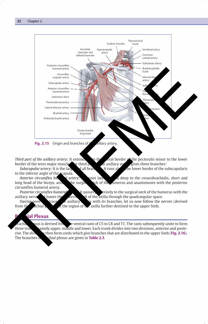
Axillary Artery The axillary artery begins as a continuation of the third part of the subclavian artery at the outer bor-der of the first rib and ends at the lower border of the teres major to continue as the brachial artery (Fig 2.15).
The proximal part of the axillary artery, axillary vein and brachial plexus and their branches are enclosed in the axillary sheath, derived from the prevertebral layer of the deep cervical fascia.
The axillary artery is surrounded by the brachial plexus and inferomedially it is related with the axil-lary vein throughout its course.
The axillary artery is divided into three parts by the pectoralis minor muscle.First part of the axillary artery: It lies proximal to the pectoralis minor muscle and extends from the outer border of the first rib to the medial border of the pectoralis minor muscle.Branches from the first part: The superior thoracic artery is the only branch given by the first part of the axillary artery. It arises near the apex of the axilla and supplies blood to the first and second intercostal spaces. Second part of the axillary artery: It lies behind the pectoralis minor muscle. It gives two branches, the thoracoacromial artery and lateral thoracic artery. The thoracoacromial artery is divided into four branches:
1. Pectoral branch 2. Acromial branch 3. Deltoid branch 4. Clavicular branch
The lateral thoracic artery arises at the lateral border of the pectoralis minor muscle and descends along the lateral border of the pectoralis minor, and provides, in females, large lateral mammary branches to supply the breast.
Medial and lateralcord branches
Radial n.
Median n.
Ulnar n.
Brachial v.
Brachial a.
Circumflexscapular a.
Radial n.,motor branches
Thoracodorsal a. and n.
Lateral thoracic a.
Upper subscapular n.
Long thoracic n.,superior thoracic a.
Axillary a.
Medial cord
Axillary v.
Lower subscapular n.
Axillary n.
Subscapular a.Thoracoacromial a.
Lateral cord
Fig. 2.14 Removed: Anterior wall (pectoral major and minor, and clavipectoral fascia). Retracted: Medial and lateral cords of the brachial plexus.
THIEME
22 Chapter 2
Third part of the axillary artery: It extends from the lateral border of the pectoralis minor to the lower border of the teres major muscle. The third part of the axillary artery gives three branches:
Subscapular artery: It is the largest of all branches. It runs along the lower border of the subscapularis to the inferior angle of the scapula.
Anterior circumflex humeral artery: It passes laterally and deep to the coracobrachialis, short and long head of the biceps, around the surgical neck of the humerus and anastomoses with the posterior circumflex humeral artery.
Posterior circumflex humeral artery: It passes posteriorly to the surgical neck of the humerus with the axillary nerve and leaves the posterior wall of the axilla through the quadrangular space.
Having seen and traced the axillary artery with its branches, let us now follow the nerves (derived from the brachial plexus) in the region of the axilla further destined to the upper limb.
Brachial PlexusBrachial plexus is derived from the ventral rami of C5 to C8 and T1. The rami subsequently unite to form three trunks, namely upper, middle and lower. Each trunk divides into two divisions, anterior and poste-rior. The divisions then form cords which give branches that are distributed to the upper limb (Fig. 2.16). The branches of brachial plexus are given in Table 2.3.
Vertebral artery
Commoncarotid artery
Subclavian artery
Brachiocephalictrunk
Internal thoracicartery
Axillary artery
Superiorthoracic artery
Thoracoacromialartery
Pectoral branch
Pectoralisminor
ThyrocervicaltrunkScalene muscles
Suprascapularartery
Acromial,clavicular and
deltoid branches
Posterior circumflexhumeral artery
Circumflexscapular artery
Subscapular artery
Anterior circumflexhumeral artery
Latissimus dorsi
Thoracodorsal artery
Lateral thoracic artery
Brachial artery
Profunda brachii artery
Triceps brachii,long head
Fig. 2.15 Origin and branches of the axillary artery.
THIEME

Pectoral Region and Axilla 23
Table 2.3 Brachial plexus
Name of nerve Source with root value
Course Distribution
Lateral pectoral Lateral cord, C5, C6 and C7
Pierces the clavipectoral fascia to reach the deep surface of the pectoralis major
Pectoralis major
Musculocutaneous Terminal branch of the lateral cord (C5–C7)
Exits the axilla by piercing the coraco-brachialis; descending between the bi-ceps and brachialis, it continues as the lateral cutaneous nerve of the forearm
Coracobrachialis, biceps brachii and brachialis muscle and lateral forearm skin
Median Lateral root of the median nerve from the lateral cord (C5–C7)and medial root from medial cord (C8, T1)
Lateral and medial roots unite to form the median nerve lateral to the axillary artery, then descends through the arm adjacent to the brachial artery
Muscles of the flexor compart-ment of the forearm (except for the flexor carpi ulnaris, medial half of the flexor digitorum profundus), intrinsic muscles of the thenar part and palmar skin
Medial pectoral
Medial cutaneous nerve of the arm
Medial cutane-ous nerve of the forearm
Medial cord(C8, T1)
Passes between the axillary artery and vein, pierces the pectoralis minor and then pierces the deep surface of the pectoralis major muscle
Runs by the medial side of the axillary and brachial veins and communicates with the intercostobrachial nerve
In close relation with the ulnar nerve, pierces the deep fascia along with the basilic vein and then divides into ante-rior and posterior branches
Pectoralis major (sternocostal part) and pectoralis minor
Skin of the medial side of the arm up to the medial epi-condyle of the humerus and olecranon process of the ulnaSkin of the medial side of the forearm up to the wrist
Ulnar Medial cord (C7, C8 and T1)
Terminal branch of the medial cord of the brachial plexus descends on the medial side of the arm; running behind the medial epicondyle of the humerus, it supplies the medial aspect of the forearm and hand
Flexor carpi ulnaris and medial half of the flexor digitorum profundus, intrinsic muscles of the hand, medial half of the skin of the hand
Upper subscapular Posterior cord (C5) Passes posteriorly and enters the sub-scapularis muscle directly
Upper part of the subscapularis muscle
Lower subscapular Posterior cord (C6) Runs inferolaterally, deep to the sub-scapular artery and vein
Lower part of the subscapularis and teres major muscles
Thoracodorsal Posterior cord (C6, C7 and C8)
Arises between the upper and lower subscapular nerves, passes along the posterior axillary wall to the latissimus dorsi muscle
Latissimus dorsi muscle
Axillary Terminal branch of the posterior cord (C5, C6)
Exits the axilla through the quadran-gular space. Winds around the surgical neck of the humerus
Shoulder joint, deltoid, teres minor muscles and skin overly-ing the inferior part of the deltoid
Radial Terminal branch of the posterior cord (C5–T1)
Exits the axilla lying posterior to the axillary artery, passes into the radial groove, perforates the lateral intermus-cular septum and enters the cubital fossa
All the muscles of the extensor compartment of the forearm, skin of the posterior and infero-lateral arm
THIEME

24 Chapter 2
Dissection and IdentificationLet us now trace the branches of the brachial plexus.
https://www.winkingskull.com/dissector/V1/video.aspx?vid=245
Lateral cord of the brachial plexus:
1. Trace the coracobrachialis and short head of the biceps springing from the tip of the coracoid process.
Fig. 2.16 Brachial plexus. (a and b) Formation and structure. (c) Course in relation to axillary artery stretched for clarity.
First
(a)
(c)
(b) THIEME
Pectoral Region and Axilla 25
2. Find the musculocutaneous nerve entering the coracobrachialis, and trace this nerve upwards to find the lateral cord of the brachial plexus.
3. Lateral root of the median nerve also arises from the lateral cord of the brachial plexus. To find out the median nerve, trace the lateral root distally.
4. Find the axillary artery and median nerve medial to the coracobrachialis muscle.Medial cord of the brachial plexus:
1. Trace the medial root of the median nerve proximally to find out the medial cord of the brachial plexus.
2. Identify the ulnar nerve from the medial cord of the brachial plexus. 3. Trace the previously identified medial and lateral pectoral nerves to the medial and lateral cords
of the brachial plexus, respectively. 4. Identify the medial cutaneous nerve of the forearm, medial cutaneous nerve of the arm and
branches from the medial cord of the brachial plexus by tracing them to a short distance into the arm.
Posterior cord of the brachial plexus:
1. To get a better view of the posterior cord of the brachial plexus, retract the axillary artery, the medial and lateral cords of the brachial plexus superiorly.
2. With the help of blunt dissection, clean the axillary nerve passing posterior to the humerus ac-companied by the posterior circumflex humeral artery.
3. Identify the radial nerve, which passes behind the humerus but anterior to the latissimus dorsi and teres major muscles.
4. Identify the subscapular nerves arising from the posterior cord of the brachial plexus and supply-ing the subscapularis muscle.
5. At this stage, one can also see the teres major and latissimus dorsi along with the subscapularis, forming the post axillary wall.
6. Please note that the medial, lateral and posterior cords of the brachial plexus are disposed around the second part of the axillary artery in their respective positions behind the pectoralis minor.
1. Carcinoma of the breast is a common cancer in females. It can cause dimpling of the skin from the malignant infiltration and contraction of the suspensory ligaments of the breast (ligaments of Cooper).
2. Peau d’orange sign: An orange peel appearance of the skin because of cancer cells interfering with the lymph drainage leading to lymph edema with puffy skin between the dimpled pores.
3. Radical mastectomy: In radical mastectomy, excision of the breast tumour is done with pectoralis major and axillary node clearance. The clavicular head of the pectoralis major is usually spared for cosmetic reasons.
4. Breast abscess is drained by radial incisions to avoid cutting through a number of lactiferous ducts. 5. Krukenberg’s tumour is a secondary ovarian tumour and may occur in case of carcinoma of the
breast, involving the inferomedial quadrant of the breast. 6. Erb–Duchenne palsy: Injury to the upper part of the brachial plexus (C5, C6) results in the paralysis
of the shoulder and arm muscles. The upper limb is adducted in the shoulder, arm is medially ro-tated and the elbow extended (Porter’s tip hand/Policeman’s tip hand).
7. Klumpke’s paralysis: Injury to the lower part of the brachial plexus, damaging the lower trunk (C8, T1), results in claw hand due to paralysis of the short muscles of the hand.
Clinical Notes
THIEME

THIEME