leaf anatomy of deschampsia antarctica (poaceae) from the
TRANSCRIPT

Revista Chilena de Historia Natural 72: 411-425,1999
Leaf anatomy of Deschampsia antarctica (Poaceae) from the Maritime Antarctic and its plastic response to
changes in the growth conditions Anatomfa foliar de Deschampsia antarctica (Poaceae) de la Antartida Maritima y su
respuesta plastica a variaciones de las condiciones de crecimiento
MAGDALENA ROMERO1, ANGELICA CASANOVA!, GRICELDA ITURRA, AURELIO REYES, 1 GLORIA MONTENEGR02 and MIREN ALBERDI
1Instituto de Botanica, Facultad de Ciencias, Universidad Austral de Chile, Casilla 567, Valdivia, Chile E-mail: 1 malberdi@ .uach.cl
2Facultad de Ciencias Biol6gicas Pontificia Universidad Cat6lica de Chile, Casilla 114-D, Santiago, Chile
ABSTRACT
The leaf blade anatomical features of Deschampsia antarctica Desv. growing in Robert Island, South Shetland Islands, Maritime Antarctic (62°22'S 59°43'W) and in clones cultivated in the laboratory for two years, at 2 ± 1.5 and 13 ± 1.5 oc and 180 J.UllOI m·2 s 1 of irradiance were studied by light and scanning electron microcospy. Since D.antarctica is growing under the harsh environmental conditions of the Maritime Antarctic for at least five millennia, it is postulated, that their leaf anatomy may show genotypic adaptations to this environment, which should be maintained when clones of this plant are cultivated under different conditions. In this Antarctic habitat, mean air temperature of January was ea. 2.8 oc ( < 8 to -2.5 °C)and the maximal irradiance was ea. 2000 mmol m·2 s·1• A strong variation was found in the anatomical characteristics of the leaf surface and in the leaf cross section, between plants growing in the field and their clones growing at the highest temperature in the laboratory ( 13 °C). The leaf surface of the Antarctic samples showed more xerophytic characteristics (smaller leaf surface and epidermal cells, higher leaf thickness, higher stomata density and number of cells per area) than the leaves of plants cultivated at 13 oc. Additionally, Antarctic samples presented stomata in both surfaces and epidermal cells with turgid papillae. Therefore, the taxonomic value of epidermal characteristics for identification of Poaceae could be questioned. In the leaf transverse section the vascular bundles of the Antarctic samples appeared surrounded with two bundle sheaths: an outer, with parenchymatous cells without chloroplasts, and an inner or mestome with thick walls. The outer bundle sheath was absent in leafs of plants growing at 13 °C. Xylem of leaf Antarctic samples did not present lacuna and their vessel lumens were smaller than at 13 °C. Leaf anatomical characteristics of plants growing at 2 oc correspond to an intermediate state between the two mentioned conditions. The results suggest that the leaf anatomical features of D. antarctica do not correspond to a genotypic adaptation to the harsh environmental Antarctic conditions, but rather to a plastic response of the phenotype to ameliorated growth conditions in the laboratory.
Key words: Deschampsia antarctica, Poaceae, Antarctic, leaf anatomy, phenotypical anatomical changes and temperature.
RESUMEN
Se estudia !as caracteristicas anat6micas de la lamina foliar en plantas de Deschampsia antarctica Desv. creciendo en Isla Robert, Islas Shetland del Sur, Antartida Maritima (62°22'S 59°43'W) yen clones cultivados en ellaboratorio a 2± 1,5 oc y 13 ± 1,5 ocy 180mmol m·2 s-1 deirradiancia, utilizando microscopia6pticay de barrido. YaqueD. antarctica crece, desde aproximadamente cinco milenios, bajo !as condiciones ambientales desfavorables de la Antartida Maritima, se postula que su anatomia foliar presentaria adaptaciones genotipicas a este ambiente, !as que deberian mantenerse cuando clones de esta planta son cultivados bajo diferentes condiciones. En el habitat antartico, !as temperaturas promedio de enero fueron de aproximadamente 2,8 oc (< 8 a -2,5 °C) y la irradiancia maxima de 2000 J.UllOI m-2 s-1• Muestras del habitat y de Ios cultivos en ellaboratorio (13 °C) presentaron una marcada variaci6n en !as caracterfsticas anat6micas, de la superficie foliar y del corte transversal de la hoja, con el incremento de la temperatura. La superficie foliar de las plantas creciendo en la Antartida evidenci6 caracteristicas más xeroffticas (men or superficie foliar y tamafio de !as celulas epidermicas, mayor grosor foliar y densidad de estomas y mimero de celulas por área) que !as hojas de plantas cultivadas a 13 oc. Ademas, !as muestras de la Antartida presentaron estomas en ambas superficies y celulas epidermicas con papilas turgentes. En cortes transversales de !as hojas, Ios haces vasculares de !as muestras antárticas aparecen rodeados de dos vainas vasculares: una externa, con celulas parenquimatosas sin cloroplastos y una interna o mestoma con paredes engrosadas. La vaina externa está ausente en !as hojas de plantas creciendo a 13 oc. El xilema de !as muestras foliares de la Antártida no present6lagunas, y ellumen de sus vasos fue
(Received 29 November, 1998, accepted 28 May, 1999; managed by L. Corcuera)
412 ROMERO ET AL.
menor que a 13 °C. Las caracterfsticas anat6micas foliares de !as plantas creciendo a 2 oc ocuparon un lugar intermedio entre !as dos condiciones de crecimiento mencionadas. Los resultados sugieren que !as caracterfsticas anat6micas foliares de D. antarctica no corresponderfan a una adaptaci6n genotfpica a! clima desfavorable de la Antartida, sino más bien a una respuesta plastica del fenotipo a !as condiciones de crecimiento menos adversas dellaboratorio.
Palabras clave: Deschampsia antarctica, Poaceae, Antartida, cambios fenotfpicos de la anatomfa foliar y temperatura.
INTRODUCTION
Controversies exist on the relationships be-tween the anatomical and morphological characteristics of plants and the environ-mental conditions (Palta & Li 1979, Levitt 1980). Small cell sizes, thick cell walls, high leaf thickness, and high stomatal den-sity of leaves, among others features, are associated with extreme environmental con-ditions, such as low temperature (Palta & Li 1979, Korner & Larcher 1988). In tuber-bearing Solanum species, Palta & Li ( 1979) reported that the number and thickness of palisade parenchyma layers and the stomatal index on the upper leaf surface were directly related to their cold resistance and the low temperatures of their habitats. Thus, the anatomical leaf structure of cold resistant species results from their constitutive adap-tation to low temperatures, as a consequence of a selection pressure (Tieszen & Helgager 1968, Palta & Li 1979).
In this paper, we will study the anatomi-cal leaf blade characteristics of
A B
1 F G
Deschampsia antarctica Desv. (Poaceae), one of two unique native angiosperms that inhabits the Antarctic regions, one of the harshest ecosystems of the world (Edwards & Lewis-Smith 1988, Zufiiga et al. 1996). Since D. antarctica has been present in the Maritime Antarctic for at least five millen-nia (Birkenmeyer et al. 1985, Lewis-Smith 1994 ), we hypothesized that in the course of the evolution the harsh environmental conditions of the Antarctic have determined the anatomical leaf adaptations that are maintained, when clones of this plant spe-cies are cultivated under different condi-tions.
MATERIALS AND METHODS
Sampling and collection site
The study area was located in Robert Island (South Shetland Islands, Maritime Antarc-tic 62° 22'S 59° 43'W ), where D. antarctica grows as isolated small tussocks less than
CD E
XX H I J
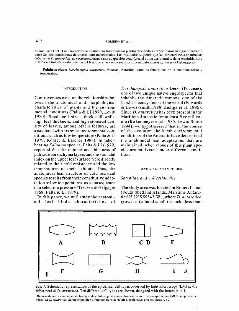
Fig. 1: Schematic representation of the epidermal cell types observed by light microscopy (LM) in the foliar peel of D. antarctica. Ten different cell types are shown, designed with the letters A to J. Representaci6n esquematica de Ios tipos de celulas epidermicas observados por microscopfa 6ptica (MO) en epidermis
foliar de D. antarctica. Se muestran diez diferentes tipos de celulas, designadas.con !as letras A a J.

LEAF ANATOMY OF Deschampsia antartica 413
10 cm high (Casaretto et al. 1994 ). In this place, the mean air temperature of January was 2.8 °C, being the maximal always be-low 8 oc and the minimum around -2.5 °C (Zufiiga et al. 1996). The maximum photo-synthetic active radiation was approxi-mately 2000 jlmol m-2s-: on clear days. More information about the microclimatic conditions of the Maritime Antarctic habi-tats are reported by Edwards & Lewis-Smith (1988) and Casanova (1997).
Thirty plants of the Poaceae D. antarctica were randomly collected from tussocks in the above mentioned site in January 1995. Since these plants have vegetative repro-duction (Casaretto et al. 1994) they can be
considered as clones. Leaves from the sec-ond node of ten different plants were col-lected and fixed in FAA (Sass 1958) for anatomical studies. Fresh plants were placed in plastic bags and transported by air to the laboratory in Valdivia (Chile). These plants were grown for two years in climatic chambers under two different tem-peratures: a) 2 ± 1.5 oc DIN (similar to the mean temperature of the Antarctic sum-mer), and b) 13 ± 1.5 °C DIN (optimal photosynthetic temperature for this spe-cies; Edwards & Lewis-Smith 1988). The photoperiod was 16 h, and light intensities ea. 180 jlmol m-2s-'; the substrate was a mixture of organic soil and turf (3:2 wlw).
TABLE 1
LM measuraments of different epidermal cell types located in the edge, internervic and epinervic zone in leaves of D.antarctica growing in the Antartic and in climatic chamber at 2 ±
1.5 oc and 13 ± 1.5 °C. Dimensions of stomata (guard cells) are also given. Data represent mean of 30 measurements in three leaves of different plants. Standard deviation was lower
than 10%. Not present(-), length (L), greater width (wl), smaller width (w2). Letters
represent the cells types us in Fig.l
Mediciones a! MO en diferentes tipos de celulas epidermicas localizadas en el horde, zona internervica y epinervica de hojas de D.antarctica creciendo en la Antartida y en camaras climaticas a 2 ± I ,5 oc y 13 ± 1,5 °C. Se entregan tambien de !as dimensiones del estoma (celulas.guardianas). Los datos representan el promedio de 30 mediciones en
tres hojas de diferentes plantas. Desviaci6n estandar fue inferior a! 10%. No presente (-) . Largo (L), ancho maximo (w'), ancho minimo (w2). Las letras representan Ios diferentes tipos celulares mostrados en Fig.1
Plants growing at: Describer Cell Antarctic Climatic chamber at:
Type 2 ± 1.5 oc 13 ± 1.5 oc
L wl w2 L wl w2 L wl w2
Adaxial epidermal (!1JI1) Edge I 65.7 16.9 9.0 70.5 13.9 7.5 217.5 14.0 14.0
J 22.4 16.9 16.9 27.1 16.1 16.1 Internervic A 145.2 19.6 12.1 104.7 18.9 12.8 295.3 18.8 6.9
E 229.0 16.2 14.2 H 38.9 8.0 6.0
Epinervic A 101.6 15.9 8.4 84.5 15.3 10.7 220.2 25.4 15.0 B 113.2 17.0 12.9 c 16.5 11.7 11.7 D 8.9 10.3 10.3
Stomata 24.3 16.2 16.2 33.8 20.3 20.3 Abaxial epidermal (!lJil)
Edge 70.9 18.0 10.0 86.1 16.0 9.0 215.9 14.8 7.0 J 21.7 17.2 17.2 26.1 12.8 12.8
Internevic A 100.5 15.9 9.3 188.1 21.9 13.2 344.9 16.8 8.1 F 80.1 16.0 26.8 213.1 17.3 26.9 296.4 13.6 23.5 G 38.9 8.0 8.0
Epinervic A 122.0 28.8 16.0 202.5 36.9 18.7 294.7 28.1 14.1 Stomata 35.5 8.3 8.3 40.3 10.9 10.9 44.7 8.3 8.3
414 ROMERO ET AL.
Ten plants in each treatment were used. Plants were irrigated every three days at 80% field capacity with water or nutrient solution. The leaves from the second node, collected directly in the field and those grown in the laboratory and taken from the middle of the leaf blade were used for light (LM) and scanning electron microscopy (SEM). Each sample was estimated as an experimental entity for the observations and measurements. Means of particular measurements (described below) were cal-culated from the individual values. The statistical differences between the means were established by an ANOV A (P < 0.05),
and a Tukey test was performed when the distance between means was significant (Little & Hills 1976).
Epidermal isolation for light microscopy (LM)
Adaxial and abaxial epidermal were iso-lated and cleared according to Aiken & Lefkovitch ( 1984 ). Peeled leaf epidermal preparation montage was made in glycerin-ated-gelatin and observed by LM (Zeiss). Measurements of length and width (w 1 = widest part and w2 = narrowest part) were
~·J ,;,/··(i
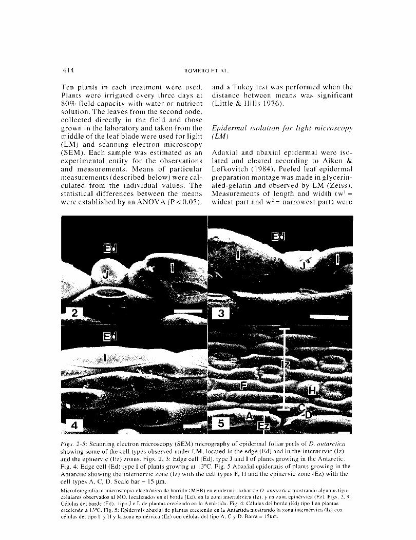
Fif?s. 2-5: Scanning electron microscopy (SEM) micrography of epidermal foliar peels of D. antarctica showing some of the cell types observed under LM. located in the edge (Ed) and in the internervic (Iz) and the epinervic (Ez) zones. Figs. 2, 3: Edge cell (Ed), type J and I of plants growing in the Antarctic. Fig. 4: Edge cell (Ed) type I of plants growing at l3°C. Fig. 5 Abaxial epidermis of plants growing in the Antarctic showing the internervic zone (Iz) with the cell types F, H and the epinervic zone (Ez) with the cell types A, C, D. Scale bar = I 5 jlm. Microfotograffa al rnicroscopio electr6nico de barrido (MEB) en epidermis foliar de D. antarctica mostrando algunos tipos celulares observados al MO. localizados en el borde (Ed), en la zona internervica (lz). yen wna epinervica (Ez). Figs. 2, 3: Celulas del borde (Ed), tipo J e I, de plantas creciendo en la Antartida. Fig. 4: Celulas del borde (Ed) tipo I en plantas creciendo a I3°C. Fig. 5: Epidermis abaxial de plantas creciendo en la Antartida mostrando la zona internervica (lz) con celulas del tipo F y H y la zona epinervica (Ez) con celulas del tipo A, C y D. Barra = l5f.Lm.
LEAF ANATOMY OF Deschampsia antartica 415
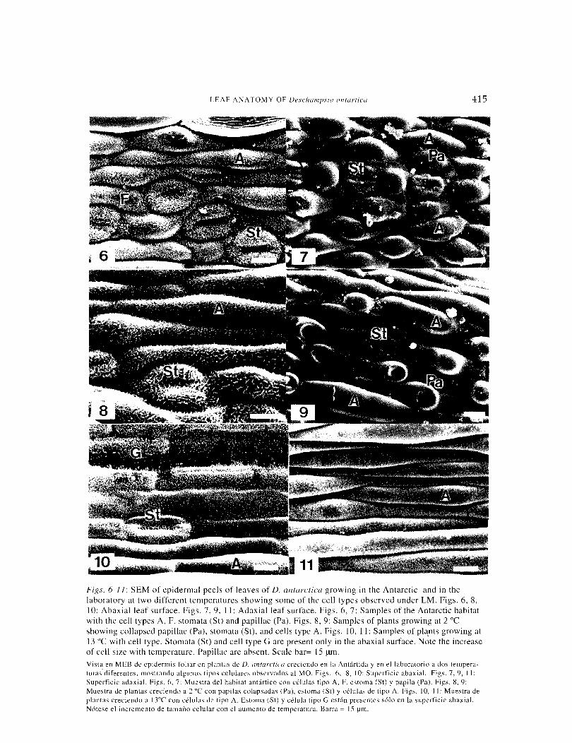
Figs. 6-11: SEM of epidermal peels of leaves of D. antarctica growing in the Antarctic and in the laboratory at two different temperatures showing some of the cell types observed under LM. Figs. 6, 8, I 0: Abaxial leaf surface. Figs. 7, 9, I I: Adaxial leaf surface. Figs. 6, 7: Samples of the Antarctic habitat with the cell types A, F, stomata (St) and papillae (Pa). Figs. 8, 9: Samples of plants growing at 2 oc showing collapsed papillae (Pa), stomata (St), and cells type A. Figs. I 0, !I: Samples of pla_nts growing at 13 oc with cell type. Stomata (St) and cell type G are present only in the abaxial surface. Note the increase of cell size with temperature. Papillae are absent. Scale bar= 15 f..liD. Vista en MEB de epidermis foliar en plantas de D. antarctica creciendo en la Antartida yen el laboratorio a dos tempera-turas.diferentes. mostrando algunos tipos celulares observados al MO. Figs. 6, 8, 10: Superficie abaxial. Figs. 7, 9, 11: Superficie adaxial. Figs. 6, 7: Muestra del habitat antartico con celulas tipo A. F. estoma (St) y papila (Pa). Figs. 8, 9: Mucstra de plantas creciendo a 2 oc con papilas colapsadas (Pa). estoma (St) y celulas de tipo A. Figs. 10, 11: Muestra de plantas creciendo a 13"C con celulas de tipo A. Estoma (St) y celula tipo G estan presentes solo en la superficie abaxial. N6tese el incremento de tamano celular con el aumento de temperatura. Barra = 15 !Jm.
416 ROMERO ET AL.
made from thirty cells of each different cell type, located at the edge, internervic and epinervic zones. Only some of these cell types are shown in the SEM micrographs. Additionally, cell and stomatal densities were determined.
Scanning electron microscopy (SEM)
Samples of leaves were vacuum infiltrated with Karnovsky and glutaraldehyde 25% in sodium phosphate buffer 0.2 M (pH 7.4) for two hours. The samples were rinsed with phosphate buffer 0.1 M for three-five minutes three times and dehydrated on ice in a graded acetone series, followed by critical point (Hitachi mod HCP-2) drying with liquid carbon dioxide. The samples were mounted on aluminum stubs and coated with gold in an Edwards S 150 B sputter-coater. Samples were observed in a Bausch & Lomb Nanolab 2000 scanning electron microscope. Images were photo-graphed on Polaroid 665 in Forte pan 50 film.
Leaf cross sections (LM)
Cross sections {lJ.Lm thick) of leaves fixed in Karnovsky, embedded in epon-araldite,
and stained with toluidine blue (1%) were made. Free hand cross sections of leaf samples ( 16 J.Lm thick) were also made. Direct LM measurements of the length and the width of cells, the thickness of the epidermis, the thickness of palisade and spongy-parenchyma layers, the total leaf thickness as well the vessel lumen area, were performed in several cross sections using an eye-piece micrometer. In addi-tion, leaf dry weight and leaf. area were determined.
RESULTS
Epidermal characteristics
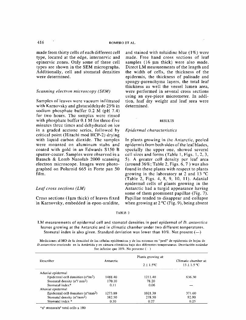
In plants growing in the Antarctic, peeled epidermis from both sides of the leaf blades, specially the upper one, showed several cell sizes and forms (Table 1, Figs. 1, 2, 3, 5). A greater cell density per leaf area (around 36%; Table 2, Figs. 6, 7) was also found in these plants with respect to plants growing in the laboratory at 2 and 13 oc (Table 2, Figs. 4, 8, 9, 10, 11). Adaxial epidermal cells of plants growing in the Antarctic had a turgid appearance having some of them prominent papillae (Fig. 7). Papillae tended to disappear and collapse when growing at 2 oc (Fig. 9), being absent
TABLE 2
LM measurements of epidermal cell and stomatal densities in peel epidermal of D. antarctica leaves growing at the Antarctic and in climatic chamber under two different temperatures.
Stomatal index is also given. Standard deviation was lower than 10%. Not present(-)
Mediciones a! MO de la densidad de !as celulas epidermicas y de Ios estomas en "peel" de epidermis de hojas de D.antarctica creciendo en la Antartida y en camara climatica bajo dos diferentes temperaturas. Desviaci6n estandar
fue inferior que 10%. No presente (-)
Describer
Adaxial epidermal Epidermal cell densities (n°/m2) Stomatal density (n°/ mm2) Stomatal index*
Abaxial epidermal Epidermal cell densities (n°/mm2) Stomatal density (n°/mm2) Stomatal index *
*n° stomata/no total cells x 100
Antarctic
1488.40 170.10
0.11
1273.80 382.50
0.30
Plants growing at:
2 ± l.SOC
1211.40 78.20
0.06
1021.30 278.50
0.27
Climatic chamber at: 13 ± 1.5 oc
636.30
371.60 92.90
0.25
LEAF ANATOMY OF Deschampsia antartica 417
12
Ad
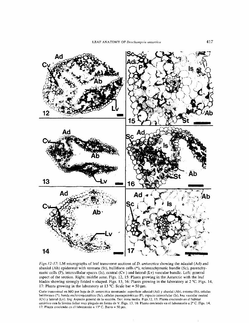
lit Figs.l2-17: LM micrographs of leaf transverse sections of D. antarctica showing the adaxial (Ad) and abaxial (Ab) epidermal with stomata (St), bulliform cells(*), sclerenchymatic bundle (Se), parenchy-matic cells (P), intercellular spaces (Is), central (Cv ) and lateral (Lv) vascular bundle. Left: general aspect of the section. Right: middle zone. Figs. 12, 15: Plants growing in the Antarctic with the leaf blades showing strongly folded v-shaped. Figs. 13, 16: Plants growing in the laboratory at 2 oc. Figs. 14, 17: Plants growing in the laboratory at 13 °C. Scale bar = 50 J.Uil. Corte transversal en MO por hoja de D. antarctica mostrando: superficie adaxial (Ad) y abaxial (Ab), estoma (St), celulas buliformes (*), banda esclerenquimatica (Se), celulas parenquimaticas (P), espacio intercelular (Is), haz vascular central (Cv) y lateral (Lv). Izq: Aspecto general de la secci6n. Der: zona media. Figs.12, 15: P1anta creciendo en el habitat antartico con la lamina foliar muy plegada en forma de V. Figs. 13, 16: Planta creciendo en ellaboratorio a 2" C. Figs. 14, 17: Planta creciendo en ellaboratorio a 13" C. Barra =50 J.I!Il.
418 ROMERO ET AL.
in leaves cultivated at 13 oc (Fig. 11). The complexity of cell types of the adaxial epi-dermal of the Antarctic samples, disap-peared at 13 °C (maintaining only the cell type I and A) (Table 1). Some of these cell types are shown also in figures 7 and 11. At 13 °C the length of cells (type A) was increased by 224% in comparison to the Antarctic plants (Table 1, Figs. 7, 11 ). In the abaxial epidermal, the cell complexity of Antarctic samples was maintained at 13 °C with exception of the cell type J, located in the edge zone, that disappeared (Table 1, Figs. 2, 3, 4). The length of the epidermal cells of samples growing at 13 oc was increased by ea. 200% in comparison to the Antarctic samples (P < 0.05) (Table 1, Figs. 6, 10). Epidermal cells of plants growing at 2 °C showed intermediate sizes between the other two groups of plants (Table 1, Figs. 8, 9). Stomata were present on both epidermis, in Antarctic samples (Figs. 6, 7) and in plants growing at 2 oc (Figs. 8, 9), being more frequent in the abaxial surface. Stomata density was higher in leaves of plants growing in the Antarctic than in plants culti-vated in the laboratory (P < 0.05) (Table 2, Figs. 6, 8, 10). In plants growing at 13 °C stomata were present only in the abaxial epi-dermis, being its density significantly lower
18
than in plants growing in the Antarctic (P < 0.05) (Table 2, Figs. 10, 11).
Transverse sections of leaf blades
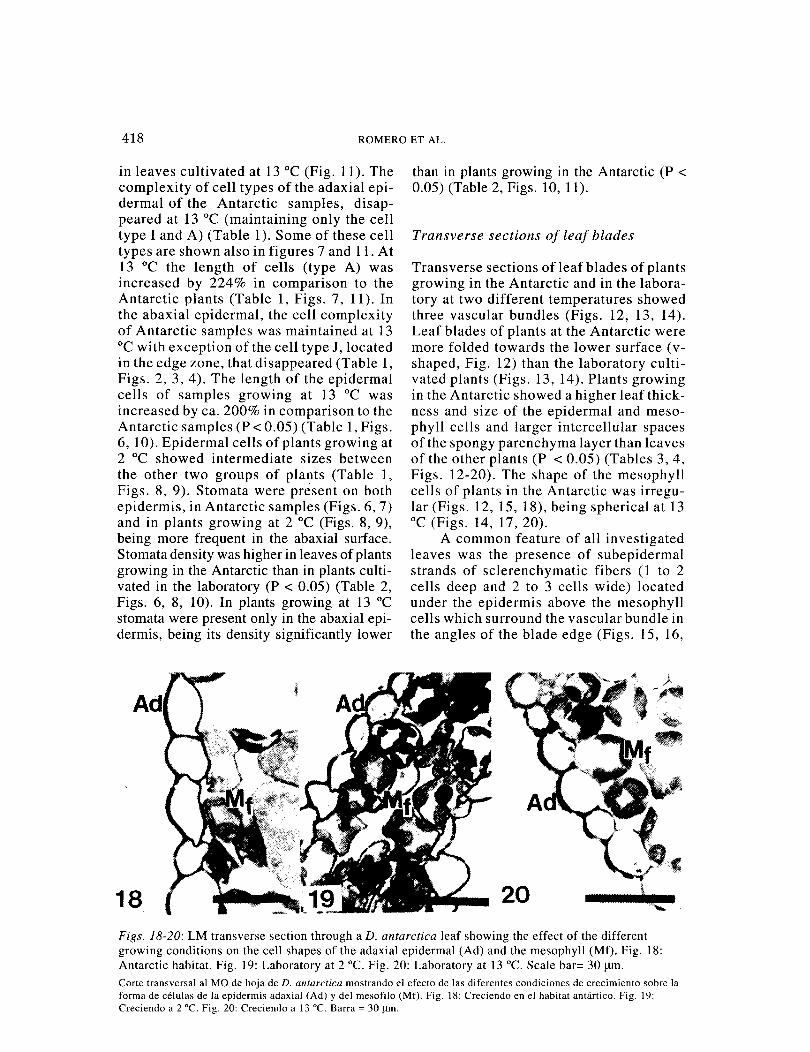
Transverse sections of leaf blades of plants growing in the Antarctic and in the labora-tory at two different temperatures showed three vascular bundles (Figs. 12, 13, 14). Leaf blades of plants at the Antarctic were more folded towards the lower surface (v-shaped, Fig. 12) than the laboratory culti-vated plants (Figs. 13, 14). Plants growing in the Antarctic showed a higher leaf thick-ness and size of the epidermal and meso-phyll cells and larger intercellular spaces of the spongy parenchyma layer than leaves of the other plants (P < 0.05) (Tables 3, 4, Figs. 12-20). The shape of the mesophyll cells of plants in the Antarctic was irregu-lar (Figs. 12, 15, 18), being spherical at 13 oc (Figs. 14, 17, 20).
A common feature of all investigated leaves was the presence of subepidermal strands of sclerenchymatic fibers (1 to 2 cells deep and 2 to 3 cells wide) located under the epidermis above the mesophyll cells which surround the vascular bundle in the angles of the blade edge (Figs. 15, 16,
20 Figs. 18-20: LM transverse section through a D. antarctica leaf showing the effect of the different growing conditions on the cell shapes of the adaxial epidermal (Ad) and the mesophyll (Mf). Fig. 18: Antarctic habitat. Fig. 19: Laboratory at 2 °C. Fig. 20: Laboratory at 13 °C. Scale bar= 30 !liD· Corte transversal al MO de hoja de D. antarctica mostrando el efecto de !as diferentes condiciones de crecimiento sobre la forma de celulas de la epidermis adaxial (Ad) y del mesofilo (Mf). Fig. 18: Creciendo en el habitat antartico. Fig. 19: Creciendo a 2 °C. Fig. 20: Creciendo a 13 °C. Barra = 30 J.ll11.

LEAF ANATOMY OF Deschampsia antartica 419
17). The number of cells of these strands increased concomitantly with the tempera-ture. Bulliform cells appeared in the angles of the blade of the abaxial epidermis (Figs. 12, 13, 14). These cells are often much larger and more vacuolated than neighbor-ing epidermal cells. They may be enabling the rolling movements of the blades (Esau 1985).
Vascular bundles.
The central and lateral vascular bundles of the Antarctic leaf samples (Figs. 15, 21, 22) showed the two differentiated bundle sheaths (an outer parenchymatous and an inner or mestome) characteristic of the Pooideae type (Esau 1985), while those of the plants growing in the laboratory at 13 oc presented only mestome (Figs. 17, 25, 26). Plants growing in the laboratory at 2 oc showed a transition state (Figs. 16, 19, 23, 24).
The outer bundle sheath of Antarctic leaves is incomplete and formed by thin-walled parenchymatous cells without vis-ible chloroplasts (Figs. 12, 15, 21, 22). The cells of the outer and inner sheath of the central vascular bundle are spherical (Fig. 21) while those of the lateral bundles are very irregular in form (Fig. 22). The inner bundle sheath or mestome of leaf blades at the Antarctic is continuous and constituted by small cells with U-shape thickened walls of endodermoid type (Esau 1985). These cells reacted positively for lignin with the phloroglucin test (Sass 1958). Mestome of plants growing at 13 oc showed scarcely turgid lumen and thicker walls than those of plants growing at lower temperature (Figs. 15, 17, 21-26). Lumen vessels of vascular bundles of plants growing in the Antarctic were smaller (Fig. 22) than those of plants growing at 2 and 13 oc (P < 0.05) (Table 3, Figs. 23-25). The vascular bundle of the Antarctic samples did not present lacuna as the other samples (Figs. 21, 23,
TABLE3
LM morphometric parameters in leaf transverse section of D. antarctica growing in the Antarctic and in climatic chamber at two differents temperatures (2 ± 1.5 oc and 13 ± 1.5 °C).
Additionally, other leaf parameters are given. Not present.(-). Standard deviation (±)
Parametros morfometricos medidos a! MO en corte transversal de hojas de D. antarctica creciendo en la Antartida y
en camaras climaticas a dos diferentes temperaturas (2 ± 1.5 "C y 13 ± 1.5 "C). Adicionalmente se entregan otros parametros. No presente (-). Desviaci6n estandar (±)
Plants growing at: Describer Antarctic Climatic chamber at:
2 ± 1.5 "C 13 ± 1.5 "C
Adaxial epidermal Thickness (J.Ull) 20.0 ± 0.4 16.3 ± 0.6 18.3 ± 1.0 Abaxial epidermal Thickness (J.Ull) 21.1 ± 0.3 17.2 ± 0.2 17.9± 1.0
Mesophyll Palisade layer thickness (J.Ull) 28.4 ± 1.4 36.8 ± 1.2 48.4 ± 1.4 Ratio palisade/palisade + spongy 0.1 0.3 0.4 Spongy layer thickness (J.Ull) 180.6 ± 2.6 100.0 ± 2.4 60.5 ± 3.0 Number of vascular bundles 3.0 3.0 3.0
Vessel elements Lumen area (J.Ull2) 80.0 ± 4.2 99.4 ± 2.4 157.0~6.1
Lumen area increase (%) 24.3 96.3 Other characteristics
Leaf thickness (!lm) 250.3 ± 3.5 188.4 ± 2.5 164.2 ± 2.0 Leaf dry weight (% PF) 25.6 22.0 19.4 Total leaf area (mm2) 30.1 ± 4.8 145.3 ± 11.6 184.1 ± 16.4 Leaf area increase(%) 382.7 511.6

420 ROMERO ET AL.
Figs. 21-26: LM leaf transverse section of D. antarctica showing the central (left side) and lateral (right side) vascular bundle. Fig. 21: Samples of plants growing in the Antarctic showing the central vascular bundle surrounded by two sheaths: an outer of parenchymatous cells without chloroplasts (Os) and an inner sheath or mestome with thickened internal cell walls (Ms), small lumen vessels of the metaxylem (V). Fig. 22: Antartic samples. Lateral vascular bundle with very irregular cell forms in the outer sheat (Os) and inner sheath or mestome (Ms). Fig. 23: Central vascular bundle of plants growing at 2 oc without outer sheaths showing the mestome (Ms), vessels (V), tracheary elements of protoxylem (par-tially destroyed) with an anular tracheid (T) and a small lacuna (L). Fig. 24: Samples at 2 oc. Lateral vascular bundle showing a transition state of the outer sheath (Os) with chloroplasts, and a reduced mestome (Ms). Adittionally the adaxial (Ad) and abaxial (Ab) epidermal, intercellular space (Is), stomata (St) and sclerenchymatic bundle (Se) are shown. Fig. 25: Samples at 13 oc showing the central vascular bundle without outer sheaht and a mestome (Ms) with some collapsed cells, two very differentiated tracheary elements (V) and a great lacuna (L). Fig. 26: Samples at 13 oc showing the mestome (Ms) of lateral vascular bundle with cell walls thinner than those of Antarctic samples. Scale bar=l5 J.llll. Note the increase of the lumen vessels and lacuna with the temperature increase. Corte tranversal en MO por hoja de D. antarctica, mostrando el haz vascular central (!ado izquierdo) y lateral (!ado derecho). Fig. 21: Muestra foliar de la Antartida con el haz vascular central (Cv) rodeado por dos vainas: una externa de celulas parenquimatosas sin cloroplastos (Os) y una interna o mestoma (Ms) con la pared celular interna engrosada, vasos (V) del metaxilema de lumen pequefio. Fig. 22: Muestra Antartida. Haz vascular lateral (Lv) con celulas de forma irregular en ambas vainas: la externa (Os) y la interna o mestoma (Ms). Fig. 23: Haz vascular central (Cv) de plantas creciendo a 2 °C, sin vaina externa, mostrando mestoma (Ms), vasos (V), elementos traqueidales del protoxilema (parcialmente destrufdos) con traqueida (T) anular y una pequefia laguna (L). Fig. 24: Muestra foliar a 2 °C. Haz vascular lateral (Lv) mostrando la vaina externa (Os) con cloroplastos (estado de transici6n) , y un reducido mestoma (Ms). Ademas, se observa: la superficie adaxial (Ad) y abaxial (Ab), espacio intercelular (Is), estoma (St) y banda esclerenquimatica (Se). Fig. 25: Muestra a 13 °C mostrando el haz vascular central (Cv) sin vaina externa y un mestoma (Ms) con algunas celulas colapsadas, dos elementos traqueales muy diferenciados (V) y una gran laguna (L). Fig. 26: Muestra a l3°C mostrando el mestoma (Ms) del haz vascular lateral (Lv) con la pared celular mas delgadas que las de la muestras antartica. Barra=15 J.IIIl. N6tese el incre-mento de tamafio del lumen de la laguna y vasos con el incremento de temperatura.

LEAF ANATOMY OF Deschampsia antartica 421
25). The annular tracheids, only appears at 2 oc (Fig. 23). The dimensions of the la-cuna in samples growing in the laboratory increased with the growth temperature (Figs. 23, 25).
DISCUSSION
Our results showed that leaf anatomical characteristics of specimens of D. antarctica growing in the Antarctic, in com-parison with those growing in the labora-tory (at 2 and 13 °C), were strongly xero-phytic (e.g. smaller epidermal cell sizes, higher cell density and complexity of cell forms, thicker cuticle, higher stomata den-sity, greater leaf thickness, smaller lumen vessels). In these habitats, as well in other cold regions, low temperature can induce drought stress and thus xerophytic leaf
structures (Korner & Larcher 1988). The changes of the majority of these character-istics when plants grew in the laboratory reflected modifications not consistent with the reduced intraspecific morphologic vari-ability of the epidermis of the Poaceae (Esau 1985). These results suggest that the taxonomic value of epidermal characteris-tics as proposed by Upadhyaya & Fumes ( 1994) may not be applicable to all plant groups, specially when members of the Poaceae are considered. The presence of papillae in D. antarctica growing in their natural habitat could be interpreted as a storage mechanism of carbohydrates in-volved in freezing point depression, as re-ported for other plants (Levitt 1980, Sakai & Larch er 1987, Larch er 1995). That is consistent with the high contents of su-crose and fructans found in D. antarctica growing in the Antarctic (~ufiiga et al.
TABLE4
LM cytological leaf parameters (high, width, area and ratio width/high) of D. antarctica growing in the Antartic and in climatic chambers at 2 ± 1.5 and 13 oc ± 1.5. Measurements
were made under LM in leaf transverse sections. Data represent means of 10 leaves of different plants. Standard deviation (±)
Panimetros citol6gicos (alto, ancho, area y radio ancho/alto) en hojas de D.antarctica creciendo en la Antartida yen camara climatica a 2 ± 1,5 oc y 13 ± 1,5 oc. Mediciones realizadas a! MO en cortes transversales de hoja. Los datos
representan a! promedio de 10 hojas de plantas diferentes. Desviaci6n estandar (±)
Describer
Adaxial epidermal cells (Jlll1) High Width Area (Jlll12) Ratio width/high
Abaxial epidermal cells (Jlll1) High Width Area (Jlll12) Ratio width/high
Palisade parenchyma cells (Jlll1) High Width Area (Jlll12) Ratio width/high
Spongy parenchyma cells (Jlll1) High Width Area (Jlll12) Ratio width/high
Antarctic
16.8 ± 0.5 14.8 ± 0.6
248.6 0.9 ± 0.5
20.8 ± 0.9 11.2 ± 0.4
233.0 0.5 ± 0.5
24.0 ± 1.1 20.0 ± 1.9
480.0 0.8 ± 1.5
32.6 ± 2.1 22.9 ± 1.0
883.9 0.7 ± 1.6
Plants growing at:
2 ± 1.5 oc
12.4 ± 0.4 11.6 ± 0.3
143.8 0.9 ± 0.4
13.6 ± 0.5 9.6 ± 0.3
130.6 0.7 ± 0.4
15.0 ± 0.3 14.8 ± 0.2
222.0 1.0 ± 0.1
21.2 ± 2.0 19.6 ± 1.0
415.5 0.9 ± 1.5
Climatic chamber at: 13 ± 1.5 oc
18.4 ± 0.3 15.6 ± 0.5
287.0 0.8 ± 0.4
18.8 ± 0.6 15.2 ± 0.4
285.8 0.8 ± 0.5
16.8 ± 0.2 16.0 ± 0.3
268.8 1.0 ± 0.1
25.1 ± 1.3 21.1 ± 0.8
529.6 0.8 ± 1.1
422 ROMERO ET AL.
1996), although their localization in the papillae remains to be established. Fructans are often related with cold hardiness of plants (Pontis & Del Campillo 1985, Livingston Ill & Henson 1998). The high frost resistance (LT 50 around -27 °C) of D. antarctica (Casanova 1997) could be re-lated, at least partially, with these sub-stances. Fructans participate also in the osmoregulation of cells in periods of lim-ited water availability (Virgona & Barlow 1991, Pollock & Cairns 1991, Hendry 1993).
The v-shaped position of leaf blades of plants at the Antarctic habitat are similar to the typical graminoid leaves of both, high mountains and polar tundra ecosystems (Kdrner & Larcher 1988). They could serve as an isolation leaf strategy, conferring independence with respect to the environ-ment, maintaining the stomata of the abaxial surface under a high relative humidity, thus decreasing the transpiration rates and main-taining the gas exchange (Larcher 1995, Hardy et al. 1995). Zuiiiga et al. (1996) found increased leaf temperatures with re-spect to the air temperatures in D. antarctica growing in the Antarctic which could be produced by low transpiration rates in this species. It is known, that in non transpiring plants the temperature of leaves increased in various grades with respect to air tem-peratures (Larcher 1995). Under these con-ditions the photosynthesis could be main-tained by C02 accumulation in the abun-dant intercellular spaces of the mesophyll. The irregular mesophy ll cell forms of plants growing in the Antarctic are not an artifact of the fixation method because we observed the same mesophyll cell forms in fresh preparations (not shown). It is reported that the irregular mesophyll cell forms may provide an important interface favoring a more intensive C02 gas exchange (No bel & Walker 1985, Kdrner & Larcher 1988, Upadhyaya & Furness 1994). The maxi-mum stomatal conductance for C02 uptake and loss of water vapor is greater in leaves with stomata present on both surfaces than on the lower surface only (Beer ling & Kelly 1996). This morphologic characteristic could improve the water absorption under water stress induced by low temperature
(Larcher 1995). Our results showed a rela-tionship between the leaf thickness, the occurrence of amphistomaty (stomata present on the upper and lower surfaces) and Antarctic habitat with high irradiance. These results support the suggestions by Mott et al. ( 1982) about the occurrence of amphistomatic, bifacial and thicker leaves in plants exposed to full sun. Indeed, thick and amphistomatic leaves can facilitate C02 diffusion to the mesophyll cells on each side of a leaf (Nobel & Walker 1985).
Sclerenchymatic bands may provide an advantageous characteristic to the leaves of plants growing in unfavorable habitats with regard to a possible disturbance of water conduction, conferring strength to the mesophyll and preventing collapse of leaf tissues under water stress (Pyykkd 1966). With respect to the high xerophytic characteristics of the Antarctic samples, we had expected a high development of their sclerenchymatic tissues. Unexpect-edly, they were scarcely developed with respect to plants growing in the laboratory at 13 oc.
The presence of two bundle sheaths (an outer and an inner) in the vascular bundles of leaves of some plants has been associ-ated with an adaptation to a high radiation intensity (Brown 1958, Pyykkd 1966). This structural feature was also present in leaves of D. antarctica growing in the Antarctic where also high photon flux densities are expected (Casanova, 1997). Recent ultra-structural studies in this plant material re-vealed the presence of inconspicuous chlo-roplasts in this sheath (own unpublished results). This feature resembles the "Kranz syndrome" of some Gramineae, where re-duced grana are present (Smith & Brown 1973). These results could suggest a func-tional specialization to optimize photosyn-thetic success under the harsh Antarctic environmental conditions as reported for other plants by Hall & Langdale (1996). These authors suggest, that the optimiza-tion of the photosynthesis to different en-vironmental stress conditions, requires the development of one or more photosynthetic pathways as occurs in the genus Flaveria where a progression of photosynthetic types from C3 to C 4 occurs (Dai et al. 1996).

LEAF ANATOMY OF Deschampsia antartica 423
Measurements with carbon isotope (ratio of stable isotopes 13C to 12C in dry matter) according Smith & Brown (1973) have re-vealed, that D. antartica, as expected, does not correspond to a C4 plant. The 013C value was -28.3% (own unpublished results) typi-cal for a C3 plant (refs. in Larcher 1995). Thus, according to the structural features of vascular bundles of D. antarctica could present, depending on the environmental conditions, some of the different C4 photo-synthetic subtypes mentioned by Hall & Langdale ( 1996) and Dai et al. ( 1996). The absence of the outer bundle sheath in plants growing in the laboratory at 13 oc and 180 !Jmol m-2 s- 1 supports the assumption that this structure and function could be more associated to the high irradiance of the Antarctic habitat, than to low temperature. More studies with respect to the photosyn-thetic enzyme types and their compartmen-talization in this plant growing under dif-ferent environmental conditions are need.
The survival capacity to freezing condi-tions of D. antarctica growing in the Ant-arctic could be ensured by a more devel-oped mestome. Mestome may provide, as in other plants, resistance to plasmolysis, because of the intimate contact of cell con-tent with the thick walls (Esau 1985). It favoured, also the depression of the freez-ing water point (Kaku 1971, Huner 1985). The small area of the lumen of vessels can also confer resistance to low temperature in the Antarctic because water freezes slowly in smaller than in greater area ves-sels (Ash worth & Abeles 1984 ).
Most of the leaf characteristics found in plants growing in the Antarctic have been described for plants from other cold areas (Korner & Larcher 1988, Consaul & Aiken 1993). Our results suggest a plastic modifi-cation (sense Pyykko 1966) of leaves of D. antarctica to the harsh Antarctic habitat. In addition, the strong tussock graminoid growth form of D. antarctica in the Antarc-tic (Casaretto et al. 1994) disappeared and the leaf surface increased in plants grow-ing in the laboratory, although their inner surface decreased. All these changes showed that D. antarctica can loose some of the anatomical characteristics acquired in the harsh Antarctic habitat when brought
to milder conditions. Therefore, our as-sumption about a genotypic adaptation of anatomical leaf features of D. antarctica to colder temperatures is not proved, specially with the disappearance of the outer sheath of vascular bundle and the increase in the vessel lumens at increasing temperatures ( 13 °C). Additional factors, not reproduced in the climatic chamber, but present in the natural habitat, may be responsible for the anatomical variations between plants grow-ing in the Antarctic and in the laboratory. For example, the quality and quantity of the light in the growth chamber and in the natural habitat were different. While total incoming radiation under the full midday sun can reach around 2000 !Jmol m-2 s- 1 in the Antarctic (Edwards & Lewis-Smith 1988), in our growth chamber the light intensities were ea. 180 !Jmol m-2 s- 1• The light quality was also different in the Ant-arctic compared to the light in the climatic chamber (fluorescent light source).The possible effects of increased UV -Blight in the Antarctic on D. antarctica are yet to be studied. Becwar et al. ( 1982) reported that the leaf structure of plants growing at high altitude was modified when compared to leaves growing in chambers, with a loss of radiation of about 38 to 50%, with respect to natural habitats. High radiation also brings secondary effects (heat and water deficit) that may change the response of the plant to local light intensity (Larcher 1995). These effects, specially water deficit, could also be expected as consequence of low temperatures in the Antarctic habitat dur-ing the growing season. The experiments in the laboratory were made under uniform environmental conditions (light intensities, relative humidity, irrigation) varying only the growth temperature. They showed that the xerophytic leaf features of plants of_ D. antarctica decreased when growing at a higher temperature.
With respect to regional warming in the Antarctic, Lewis-Smith (1994) pointed out that the increase in populations of D. antarctica and Colobanthus quitensis (Kunth) Bartl. in this region, could be used as a valuable bioindicator. Our study pro-vides a complement for future evaluations of Antarctic climatic warming. It provides
424 ROMERO ET AL.
information about the present leaf anatomi-cal characteristics of D. antarctica and the variation possibilities under more moder-ate conditions. Finally, these results indi-cate that the leaf anatomical characteristics of D. antarctica are not an adaptation produced by natural selection under harsh Antarctic conditions, but rather a plastic response of the phenotype to these condi-tions.
ACKNOWLEDGMENTS
We thank Prof. Dr. Peter W einberger for reviewing the manuscript and to Instituto de Histologfa, Universidad Austral de Chile, Valdivia, Chile, for the use of the scanning electron microscope and for the training and the advising on the SEM. This work was supported by DID-UACH S- 95-39, FONDECYT 1970637 and 1980967 and Instituto Antartico Chileno (INACH~ 148).
LITERATURE CITED
AIKEN SG & LP LEFKOVITCH (1984) The taxonomic value of using epidermal characteristics in the Cana-dian rough fescue complex (Festuca altaica, F. campestris, F. hallii, F. scabrella). Canadian Journal of Botany 62: 1864-1870.
ASHWORTH EN & FB ABELES (1984) Freezing behav-ior of water in small pores and the possible role in the freezing of plant tissues. Plant Physiology 76: 201-204.
BECW AR MR, FD MOO RE, Ill & MJ BURKE (1982) Effects of deletion and enhancement of ultraviolet-B (280-31 5nm) radiation on plants grown at 3000m elevation. Journal American Society Horticulture Sciences 107: 771-774.
BEERLING DJ & CK KELL Y (1996) Evolutionary com-parative analyses of the relationship between leaf structure and function. New Phytologist 134: 35-5 I.
BIRKENMA YER K, R OCHYRA, IU OLSON & L STUCHLIK ( 1985) Mid- Holocene radiocarbon- dated peat at Admiralty Bay, King George Island (South Shetland Islands. West Antarctica). Bulletin of the Polish Academy of Sciences Earth Sciences 33: 7-13.
BROWN W (1958) Leaf anatomy in grass systematics. Botanical Gazette 119: 170-202.
CASANOVA MA ( 1997) Eficiel!cia fotoqufmica del PSII en Deschampsia antarctica (Desv.): una gramfnea tolerante a la congelaci6n. Tesis de Magister, Facultad de Ciencias, Universidad Austral de Chile, Chile. 123 pp.
CASARETTO JA, LJ CORCUERA, I SEREY & GE ZUNIGA ( 1994) Size structure of tussocks of a popu-lation of Deschampsia antarctica in Robert Island, Maritime Antarctica. Serie Cientffica INACH 44: 61-66.
CONSAUL LL & SG AIKEN (1993) Limited taxonomic value of palea intercostal characteristics in North American Festuca (Poaceae). Canadian Journal of Botany 71: 1654-1659.
DAI Z, M SB KU & GE EDW ARDS (1996) Oxygen sensitivity of photosynthesis and photorespiration in different photosynthetic types in the genus Flaveria. Planta 198: 563-571.
EDW ARDS JA & RI LEWIS-SMITH (1988) Photosyn-thesis and respiration of Colobanthus quitensis and Deschampsia antarctica from the maritime Antarc-tic, British Antarctic. Survey Bulletin 81: 43-63.
ESAU K (1985) Anatomfa Vegetal. Third edition. Omega, Barcelona. 740 pp.
HALL LN & JA LANGDALE ( 1996) Molecular genetic of cellular differentiation in leaves. New Phytologist 132: 533-553.
HARDY JP, VJ ANDERSON & JS GARDNER (1995) Stomatal characteristics, conductance ratios and drought induced leaf modifications of semi arid grass-land species. American Journal of Botany 82: 1-7.
HENDRY GAF (1993) Evolutionary origins and natural functions of fructans: a climatological, biogeographic and mechanistic appraisal. New Phytologist 123: 3-14.
HUNER NPA (1985) Morphological, anatomical, and molecular consequences of growth and development at low temperature in Secale cereale L. cv Puma. American Journal of Botany 72: 1290-1306.
KAKU S (1971) A possible role of the endodermis as a barrier for air propagation in the freezing of pine needles. Plant and Cell Physiology 12: 941-948.
KORNER CH & W LARCHER (1988) Plant life in cold climates. In: Long SF & FY Woodward (eds) Plants and temperatures: 25-57. Symposium Society Experi-mental Biology vol. 42. The Company of Biologists Limited, Cambridge.
LARCHER W (1995) Physiological Plant Ecology. Third edition, Springer-Verlag, New York. 504 pp.
LEVITT J (1980) Responses of plants to environmental stresses. Chilling, freezing and high temperature stresses. Second edition, Academic Press, London. 607 pp.
LEWIS-SMITH RI ( 1994) Vascular plants as bioindicators of regional warming in Antarctica. Oecologia 99: 322-328.
LITTLE T & J HILLS (1976) Metodos estadfsticos para la investigaci6n en Agricultura. Pergamon Press, Mexico.
LIVINGSTON Ill DP & HENSON CA (1998) Apoplastic sugars, fructans, fructan exohydrolase, and invertase in winter oat: Responses to second-phase cold hard-ening. Plant Phisiology 116: 403-408.
MOTT KA, AC GIBSON & JW 0' LEARY (1982) The adaptive significance of amphistomatic leaves. Plant, Cell and Environment 5: 455-460.
NOB EL PS & DB WALKER (1985) Structure of leaf photosynthetic tissue. In: Barber J & NPR Baker (eds) Photosynthetic mechanisms and the environ-ment: 502-536. Elsevier, London.
LEAF ANATOMY OF Deschampsia antartica 425
PAL TA JP & PH LI (1979) Frost hardiness in relation to leaf anatomy and natural distribution of several Solanum species. Crop Science 19: 665-671.
POLLOCK CJ & AJ CAIRNS (1991) Fructan metabolism in grasses and cereals. Annual. Review of Plant Physi-ology 42: 77-101.
PYYKKO M ( 1966) The leaf anatomy of East Patagonian xeromorphic plants. Annales Botanici Fennici 3: 453-622.
PONTIS HG & E DEL CAMPILLO (1985) Fructans. In: Dey PM & R Dixon (eds) Biochemistry of storage carbohydrates in green plants: 205 -227. Academic Press, London.
SAKAI A & W LARCHER ( 1987) Frost survival of plants. In: Billings WD, F Golley, OL Lange, JS Olson & H Remmert (eds) Ecol Studies 62. Springer Press, Berlin.
SASS J (1958) Botanical microtechnique. Third edition, The Iowa State College Press, Ames, Iowa. 228 pp.
SMITH BN & WV BROWN (1973) The Kranz syndrome in the Gramineae as indicated by carbon isotopic ratios. American Journal of Botany 60: 505-513.
TIES ZEN LL & J A HELGAGER ( 1968) Genetic and physi-ological adaptation in the Hill reaction of Deschampsia caespitosa. Nature 219: 1066-1067.
UPADHY AY A MK & NH FURNES (1994) Influence of light intensity and water stress on leaf surface charac-teristics of Cynoglossum officinale, Centaurea spp., and Tragopogon spp. Canadian Journal of Botany 72: 1379-1386.
VIRGONA JM & EWR BARLOW (1991) Drought stress and carbohydrate changes. Australian Journal Plant Physiology 18: 227-232.
ZUNIGA GE, M ALBERDI & LJ CORCUERA ( 1996) Non-estructural carbohydrates in Deschampsia antartica Desv. from South Shetland Islands, Mari-time Antarctic. Environmental and Experimental Botany 36: 393-398.