leader in digital cd earn womens health healthcare
TRANSCRIPT

JANUARY 2021 I 1
Leader in digital CPD for Southern African healthcare professionals
Women’s healthEarn 3 free CEUs
© 2021 deNovo Medica
This report was made possible by an unrestricted educational grant from Astrazeneca. The content of the report is independent of the sponsor.
Click here – you need to watch the video in order to complete the CPD questionnaire.
Dr Ronwyn van EedenSpecialist Physician and Medical OncologistMedical Oncology Centre of Rosebank and Park Lane Breast Care UnitJohannesburg
Warning signs of breast cancer
Guidance for primary care
Learning objectivesYou will learn:
• Why early diagnosis of breast cancer is important
• The risk factors associated with breast cancer
• The tools and recommendations for breast cancer screening
• What is required to make a breast cancer diagnosis.
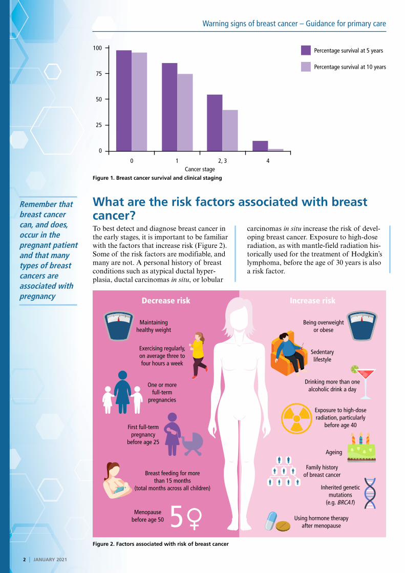
IntroductionThe National Cancer Registry1 shows that breast cancer is the most common cancer in women in South Africa. Women face an approximate 12% overall lifetime risk for developing breast cancer, which accounts for the most frequent cause of cancer deaths in women throughout the world. Early detection of breast cancer is important, as the early stages can be cured and are more easily managed; much poorer outcomes are evident in those with locally advanced breast cancer or in the metastatic setting (Figure 1). In recent decades, extraordinary progress has been made in the understanding of breast cancer, which has given rise to precision medicine for targeted therapies that are less toxic as compared to older treatments.
General practitioners are not only well placed to empower both women and men with the knowledge to lower their cancer risk, but to also encourage monthly breast self-examinations. Furthermore, screening of the general population for breast cancer is suited to general practice and so it is imperative to recognise the early warning signs.
2 I JANUARY 2021
Warning signs of breast cancer – Guidance for primary care
Remember that breast cancer can, and does, occur in the pregnant patient and that many types of breast cancers are associated with pregnancy
What are the risk factors associated with breast cancer?To best detect and diagnose breast cancer in the early stages, it is important to be familiar with the factors that increase risk (Figure 2). Some of the risk factors are modifiable, and many are not. A personal history of breast conditions such as atypical ductal hyper-plasia, ductal carcinomas in situ, or lobular
carcinomas in situ increase the risk of devel-oping breast cancer. Exposure to high-dose radiation, as with mantle-field radiation his-torically used for the treatment of Hodgkin’s lymphoma, before the age of 30 years is also a risk factor.
Figure 1. Breast cancer survival and clinical staging
Figure 2. Factors associated with risk of breast cancer
100
75
50
25
0
Percentage survival at 5 years
Percentage survival at 10 years
0 1 2, 3 4Cancer stage
Decrease risk Increase risk
Being overweight or obese
Sedentary lifestyle
Drinking more than one alcoholic drink a day
Exposure to high-dose radiation, particularly
before age 40
Ageing
Family history of breast cancer
Inherited genetic mutations
(e.g. BRCA1)
Using hormone therapy after menopause
Maintaining healthy weight
Exercising regularly, on average three to four hours a week
One or more full-term
pregnancies
First full-term pregnancy
before age 25
Breast feeding for more than 15 months
(total months across all children)
Menopause before age 50 5♀

JANUARY 2021 I 3
Warning signs of breast cancer – Guidance for primary care
EARN FREECPD POINTS
Join our CPD community at
and start to earn today!
www.denovomedica.com
A new lump, or a lump that does not ‘look right’ or is increasing in size should always be imaged or biopsied, irrespective of the age of the patient
Modifiable risk factors
Modifiable risk factors include an active life-style with regular exercise, a healthy diet, and not smoking or abusing alcohol. Patients with a high body mass index (BMI) >30kg/m2 not only have a higher risk of developing breast
cancer than patients who are smaller or thin-ner, but there are also many restrictions to managing the breast cancer in a patient with a high BMI in terms of imaging and distribu-tion of chemotherapeutic agents, as examples.
Personal history of pregnancy and lactationPatients who have never had children are at increased risk for breast cancer. Also at increased risk are patients with full-term pregnancies when over the age of 30 years, and those that did not breast feed. Breast feeding for more than 15 months across all children has been found to be protective against the development of breast cancer.
Remember that breast cancer can, and does, occur in the pregnant patient and that many types of breast cancers are associated with pregnancy. After a patient has given birth, some manifestations of breast cancer may be confused with a blocked duct or inflamma-tion, it is important that any patient with new symptoms or a lump that is increasing in size, for example, is referred for imaging or biopsy.
What does age have to do with it? Ageing is a risk factor for the development of breast cancer, although it must be noted that women are increasingly presenting at younger ages with most of Dr van Eeden’s patients being diagnosed aged younger than 40 years. A new lump, or a lump that does not ‘look
right’ or is increasing in size should always be imaged or biopsied, irrespective of the age of the patient. Some young patients have fibroadenomas which, upon imaging, may resemble some breast cancers.
Hormone replacement therapyMenopause before the age of 50 years decreases the risk for development of breast cancer. However, patients using hormone replacement therapy (HRT) for extended periods of time are at higher risk for the
development of breast cancer and so it is important to consider all risk factors for breast cancer when considering HRT for a menopausal patient. Risk usually dissipates with time after discontinuing HRT.
Family historyRisk of breast cancer increases in those with a first-degree relative (mother or sister) who has had breast cancer, risk further increases if the family member was diagnosed at a young age (<50-years-old). A male relative who has been diagnosed with breast cancer is often
indicative of a hereditary condition such as BRCA mutations. Certain hereditary can-cers occur more commonly in certain South African populations, such as Ashkenazi Jewish and Afrikaans patients.
Genetic mutations associated with breast cancers Of patients that develop breast cancer, 60-70% have no obvious risk factors. Of breast cancer patients with known risk fac-tors, approximately 4% are associated with hereditary breast cancers such as BRCA or other mutations (Figure 3). Dr van Eeden strongly recommends that patients are referred to a genetic counsellor prior to genetic testing, as this can often be a source of anxiety to patients.
Figure 3. Genome mutations that increase the risk for breast cancer
BRCA1BRCA2
TP53PTEN
ATM CASP8CHEK2,BRIP1,PALB2
Alleles identified through GWAS
4 I JANUARY 2021
Warning signs of breast cancer – Guidance for primary care
Dr van Eeden strongly recommends that patients are referred to a genetic counsellor prior to genetic testing, as this can often be a source of anxiety to patients
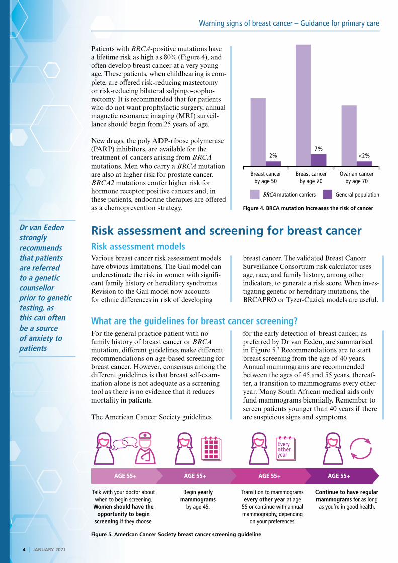
Patients with BRCA-positive mutations have a lifetime risk as high as 80% (Figure 4), and often develop breast cancer at a very young age. These patients, when childbearing is com-plete, are offered risk-reducing mastectomy or risk-reducing bilateral salpingo-oopho-rectomy. It is recommended that for patients who do not want prophylactic surgery, annual magnetic resonance imaging (MRI) surveil-lance should begin from 25 years of age.
New drugs, the poly ADP-ribose polymerase (PARP) inhibitors, are available for the treatment of cancers arising from BRCA mutations. Men who carry a BRCA mutation are also at higher risk for prostate cancer. BRCA2 mutations confer higher risk for hormone receptor positive cancers and, in these patients, endocrine therapies are offered as a chemoprevention strategy.
Risk assessment and screening for breast cancerRisk assessment modelsVarious breast cancer risk assessment models have obvious limitations. The Gail model can underestimate the risk in women with signifi-cant family history or hereditary syndromes. Revision to the Gail model now accounts for ethnic differences in risk of developing
breast cancer. The validated Breast Cancer Surveillance Consortium risk calculator uses age, race, and family history, among other indicators, to generate a risk score. When inves-tigating genetic or hereditary mutations, the BRCAPRO or Tyzer-Cuzick models are useful.
What are the guidelines for breast cancer screening?For the general practice patient with no family history of breast cancer or BRCA mutation, different guidelines make different recommendations on age-based screening for breast cancer. However, consensus among the different guidelines is that breast self-exam-ination alone is not adequate as a screening tool as there is no evidence that it reduces mortality in patients.
The American Cancer Society guidelines
for the early detection of breast cancer, as preferred by Dr van Eeden, are summarised in Figure 5.2 Recommendations are to start breast screening from the age of 40 years. Annual mammograms are recommended between the ages of 45 and 55 years, thereaf-ter, a transition to mammograms every other year. Many South African medical aids only fund mammograms biennially. Remember to screen patients younger than 40 years if there are suspicious signs and symptoms.
Figure 5. American Cancer Society breast cancer screening guideline
BRCA mutation carriers General population
Breast cancer by age 50
Breast cancer by age 70
Ovarian cancer by age 70
Figure 4. BRCA mutation increases the risk of cancer
2% <2%7%
Talk with your doctor about when to begin screening.
Women should have the opportunity to begin
screening if they choose.
Begin yearly mammograms
by age 45.
Transition to mammograms every other year at age
55 or continue with annual mammography, depending
on your preferences.
Continue to have regular mammograms for as long as you’re in good health.
AGE 55+AGE 55+AGE 55+AGE 55+
Every other year

JANUARY 2021 I 5
Warning signs of breast cancer – Guidance for primary care
EARN FREECPD POINTS
Join our CPD community at
and start to earn today!
www.denovomedica.com
Consensus among the different guidelines is that breast self-examination alone is not adequate as a screening tool as there is no evidence that it reduces mortality in patients
Appropriate use of screening modalities
Breast screening modalities that are available to patients include mammography, ultra-sonography, MRI, and clinical or self-exam-ination of the breasts. Mammography will usually reveal a lesion before it is clinically palpable. Mammography is now standardised by the breast imaging reporting and data system (BIRADS) tool that indicates risk of malignancy and recommends a manage-ment strategy (Table 1). Only 2% of screening mammograms detect an abnormality, 80% of these will be benign. Radiation exposure is so
low that the benefits far outweigh the risks.
It is important to remember that sensitivity and specificity of mammography decreases with the younger age of the patient and that the mammogram can be incorrectly interpreted; benign calcifications and breast implants can lead to radiological conundrums, giving rise to false-positive and false-negative results. In general, patients respond negatively to mammography because it is a painful, uncomfortable, and impersonal process.
Table 1. BIRADS tool
Category Management Likelihood of cancer
0 Need additional imaging or prior examinations
Recall for additional imaging and/or await prior examinations
–
1 Negative Routine screening Essentially 0%
2 Benign Routine screening Essentially 0%
3 Probably benign Short interval follow-up (6 month) or continued
>0% but ≤2%
4 Suspicious Tissue diagnosis 4a: low suspicion for malignancy (>2% to ≤10%)4b: moderate suspicion for malignancy (>10% to ≤50%)4c: high suspicion for malignancy (>50% to <95%)
5 Highly suggestive of malignancy
Tissue diagnosis ≥95%
6 Known biopsy-proven Surgical excision when clinically appropriate
–
As a stand-alone, ultrasound is not good for screening but it is useful for looking at lymph nodes when used as an adjunct to mammog-raphy. Ultrasound is also useful for localising the area for biopsy and is used to monitor the
reduction in lesion size in patients undergoing neoadjuvant chemotherapy prior to surgery. MRI is the modality of choice in high-risk BRCA patients who have very dense breasts.
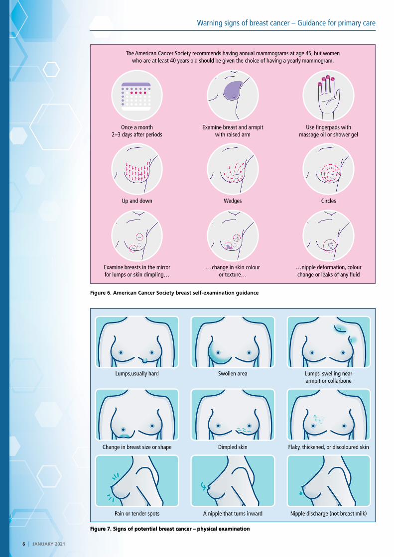
What are the clinical signs of breast cancer? Patients should be encouraged to regularly perform a breast self-examination (Figure 6). It is important to note if there is any swell-ing under the arms or in the supraclavicular areas, as well as any changes in the skin (flaky or dimpled), tender or painful breasts (not always associated with breast cancer), changes in the nipple, or discharge that is not breast milk or looks bloody (Figure 7).
Dr van Eeden brings special attention to the red breast. This is often thought to be an
inflammation such as mastitis, or an abscess or infection. These patients may be treated with antibiotics again and again, an inflam-matory cancer is regularly detected upon mammography. Inflammatory, or hormone receptor-negative, breast cancers are often very aggressive and require chemotherapy as the inflammation needs to be treated prior to considering surgery in these patients. Inflammatory breast cancers have a far worse prognosis than other types of breast cancer.
6 I JANUARY 2021
Warning signs of breast cancer – Guidance for primary care
Figure 6. American Cancer Society breast self-examination guidance
Figure 7. Signs of potential breast cancer – physical examinationFigure 7. Signs of potential breast cancer – physical examination
Once a month 2–3 days after periods
Examine breast and armpit with raised arm
Use fingerpads with massage oil or shower gel
Lumps,usually hard Swollen area Lumps, swelling near armpit or collarbone
Examine breasts in the mirror for lumps or skin dimpling…
…change in skin colour or texture…
…nipple deformation, colour change or leaks of any fluid
Pain or tender spots A nipple that turns inward Nipple discharge (not breast milk)
Up and down Wedges Circles
Change in breast size or shape Dimpled skin Flaky, thickened, or discoloured skin
The American Cancer Society recommends having annual mammograms at age 45, but women who are at least 40 years old should be given the choice of having a yearly mammogram.

JANUARY 2021 I 7
Warning signs of breast cancer – Guidance for primary care
EARN FREECPD POINTS
Join our CPD community at
and start to earn today!
www.denovomedica.com
Fine needle aspiration biopsy is never adequate for breast cancer diagnosis; image-guided core needle biopsy, performed in a mammography unit, is preferred
Depending on the type of breast cancer, metastases may be present in the lungs, liver, bones and brain. Signs and symptoms of
metastases (Table 2) will be determined by the site of disease.
Table 2. Signs and symptoms of metastases
• Breathing difficulties• Cough• Bone pain• Symptoms of hypercalcaemia• Abdominal distension• Jaundice• Hepatomegaly
• Bone pain or pathological fracture• Localising neurological signs• Altered cognitive function• Headache• General symptoms of weight loss, loss of appetite
and fatigue.
What is needed to make a diagnosis? Dr van Eeden maintains that once a suspi-cious lesion is found, “it’s the tissue that’s the issue.” Fine needle aspiration biopsy is never adequate for breast cancer diagnosis; image-guided core needle biopsy, performed in a mammography unit, is preferred. Biopsy is usually sonar-guided but can also be MRI-guided if there is a non-palpable and
indistinct mass. Without proper biopsy of a breast mass, cancer cannot be correctly diagnosed, sub-typed and classified. Dr van Eeden emphasises that there is no such thing as an emergency mastectomy and that breast cancers are always treated by a multidiscipli-nary team.
Breast cancer subtypesThe many different subtypes of breast cancer (Figure 8) have distinct molecular and cellular origins. Luminal A, a hormone receptor-posi-tive breast cancer, is the most common sub-type usually seen in older patients and has a good prognosis, responding well to endocrine therapy without the need for chemotherapy. HER2-negative breast cancers, accounting for
approximately 15% of luminal A cancers, are treated with trastuzumab. New anti-HER2 therapies have recently become available in South Africa. Triple negative cancers require chemotherapy as they are generally very aggressive; they are often seen in younger patients with BRCA-associated cancers that can often mimic fibroadenomas.
Figure 8. Subtypes of breast cancerFigure 8. Subtypes of breast cancer
44%
2%
11%
19%
24%
Luminal ALuminal BBasal-likeHER2- enrichedNormal-like
Luminal A(ER+&/or PR+, HER2-)
• Most common subtype• Less aggressive• Lower histological grade• Good prognosis• Hormone responsive• Associated with increasing age
Luminal B(ER+ &/or PR+, HER2+)
• Similar to Luminal A• More frequently ER+/PR-• Worse outcome than Luminal A
Basal-like(Triple negative, cytokeratin:
5/6+ &/or EGFR+)• Aggressive subtype• High grade histology, and high mitotic rate• Risk at younger age (<40)• More likely premenopausal African American
woman
HER2+ (ER-)• Less common, highly aggressive subtype• High grade histology• Risk at young age (<40) greater than luminal
subtypes• African American ethnicity may be a risk factor• Outcome improved with HER2
In young patients – Triple negative
breast cancers can often mimic a
fibroadenoma on radiological findings
and clinally
Differentiated byKi67 >/<14%

DisclaimerThe views and opinions expressed in the article are those of the presenters and do not necessarily reflect those of the publisher or its sponsor. In all clinical instances, medical practitioners are referred to the product insert documentation as approved by relevant control authorities.
8 I JANUARY 2021
Warning signs of breast cancer – Guidance for primary care
EARN FREECPD POINTS
Are you a member of Southern Africa’s leading
digital Continuing Professional Development
website earning FREE CPD points with access to
best practice content?
Only a few clicks and you can register to start
earning today
Visit
For all Southern African healthcare professionals
www.denovomedica.com
DeNovo Medica
@deNovoMedica
deNovo Medica
Find us at
Published by
70 Arlington Street, Everglen, Cape Town, 7550Tel: (021) 976 0485 I [email protected]
© 2021 deNovo MedicaReg: 2012/216456/07
This CPD accredited programme was written for deNovo Medica byGlenda HardyBSc(Hons) Medical Cell Biology
Key learnings
• Most early breast cancers are asymptomatic
• Breast cancer is often detected as an abnormality on a mammogram before it is palpable
• Clinical breast exam, imaging, and needle biopsy form the basis of a general approach to assessment for and diagnosis of breast cancer
• Improvement in screenings and different therapies have led to an increased survival rate
• For most patients with low-risk early-stage breast cancer, treatment is curative.
NOW EARN FREE CPD POINTS
Click here to access and submit deNovo Medica’s CPD modules
ReferencesClick on reference to access the scientific article1. Singh E, Motsuku L, Khoali L, et al. Ekurhuleni population-
based cancer registry 2018 annual report.
2. American Cancer Society recommendations for the early
detection of breast cancer.