lateral wall of nose
DESCRIPTION
PPT PRESENTATION DESCRIBING ABOUT LATERAL WALL OF NOSETRANSCRIPT

ANATOMY OF LATERAL WALL OF NOSE
EMBRYOLOGY IN BRIEF

The development of NOSE starts at about 4th week of gestational age.
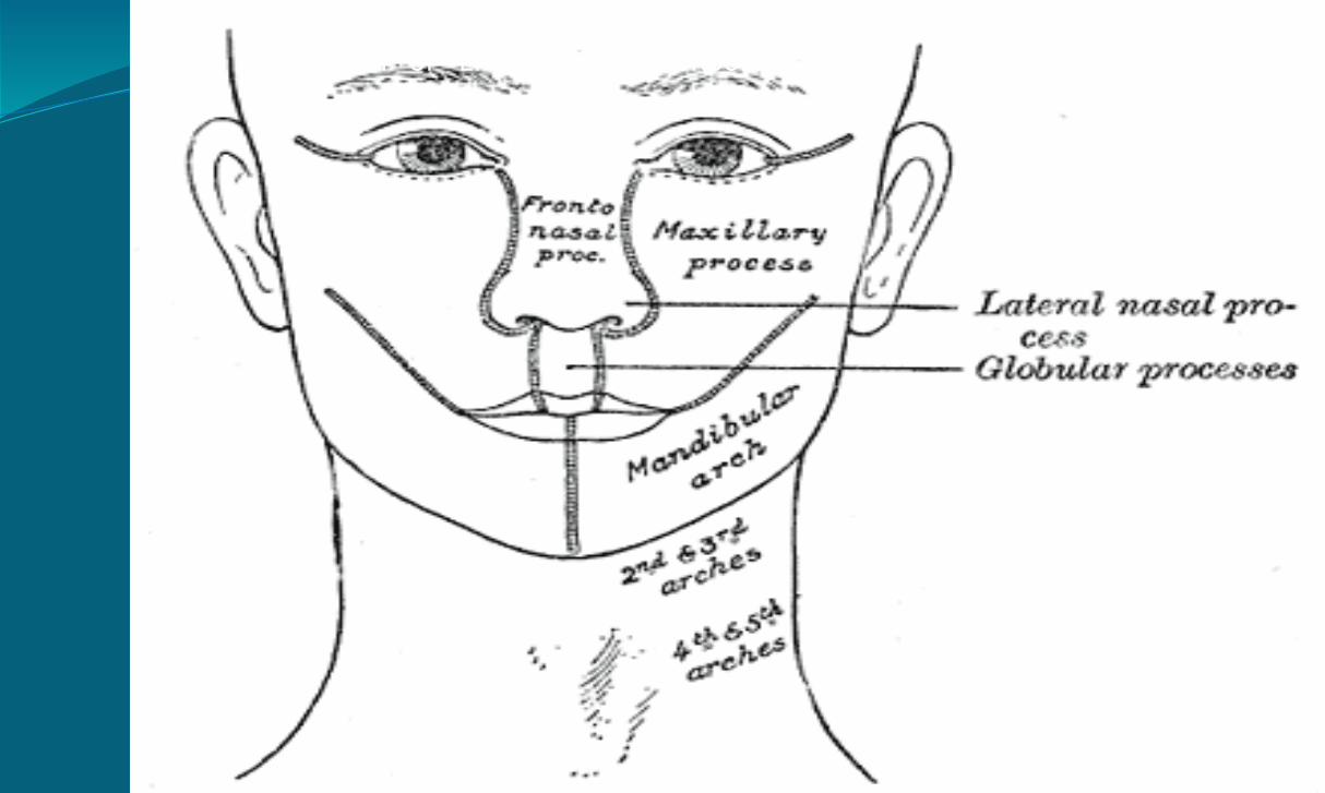
Three prominences appear around stomatodaeum(future mouth)….
FRONTONASAL PROCESS…mesoderm covering the developing forebrain Proliferates,& forms downward projection that overlaps upper part of stomatodaeum.
MANDIBULAR ARCHES( Rt & Lt)….arising from 1st pharyngeal arch.
Mandibular arch divides into maxillary and mandibular process.
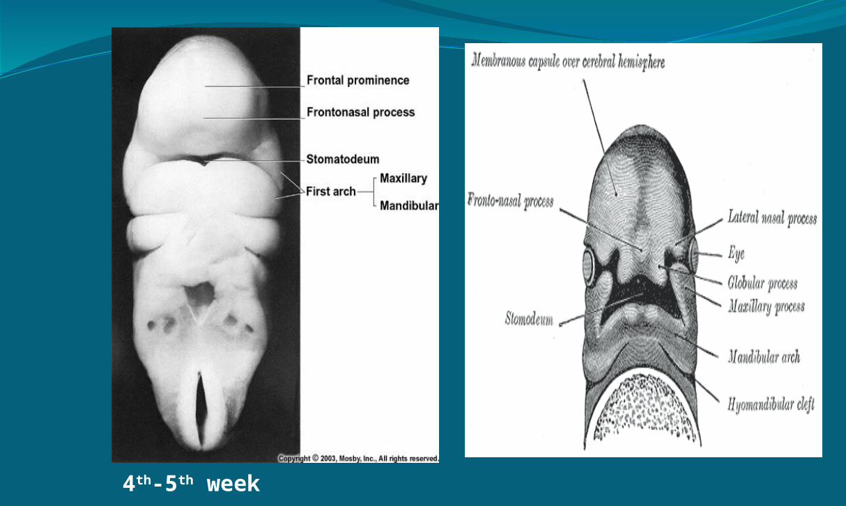
4th-5th week

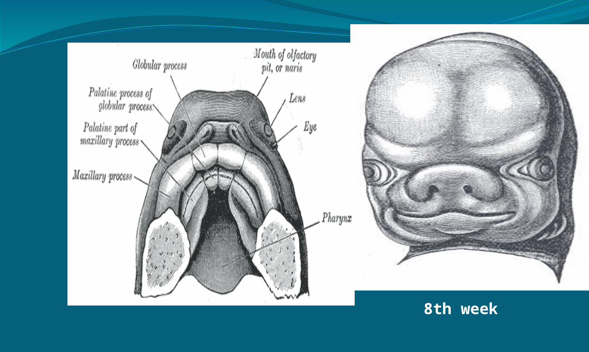
The Nose is derived from frontonasal process in 4th fetal week.
5th fetal week ectodermal plaques develop on lateral aspect of FNP & become paired NASAL PLACODES,early precursors of nares.
End of 5th week these convex placodes develop into concave nasal grooves, the medial & lateral sides of placodes protrudes forwards to become Medial & Lateral Nasal Process.
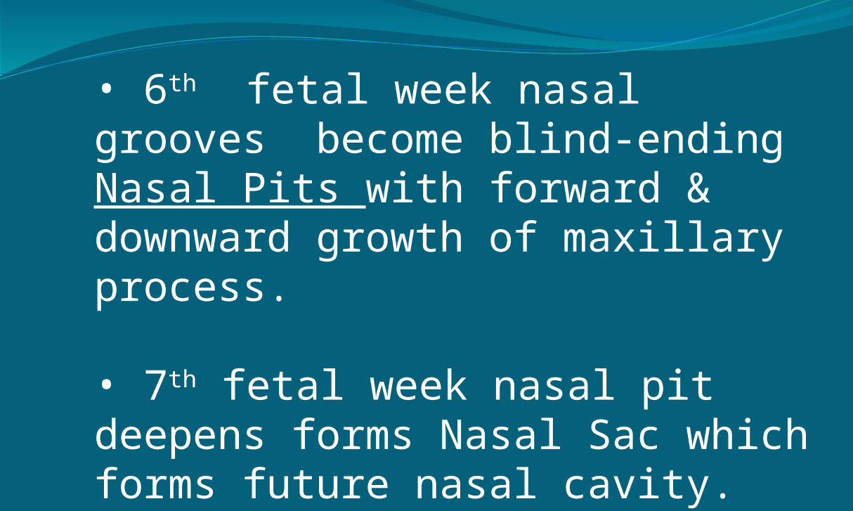
• 6th fetal week nasal grooves become blind-ending Nasal Pits with forward & downward growth of maxillary process.
• 7th fetal week nasal pit deepens forms Nasal Sac which forms future nasal cavity.

6th -7th week

LATERAL WALL OF NOSE devolopment….
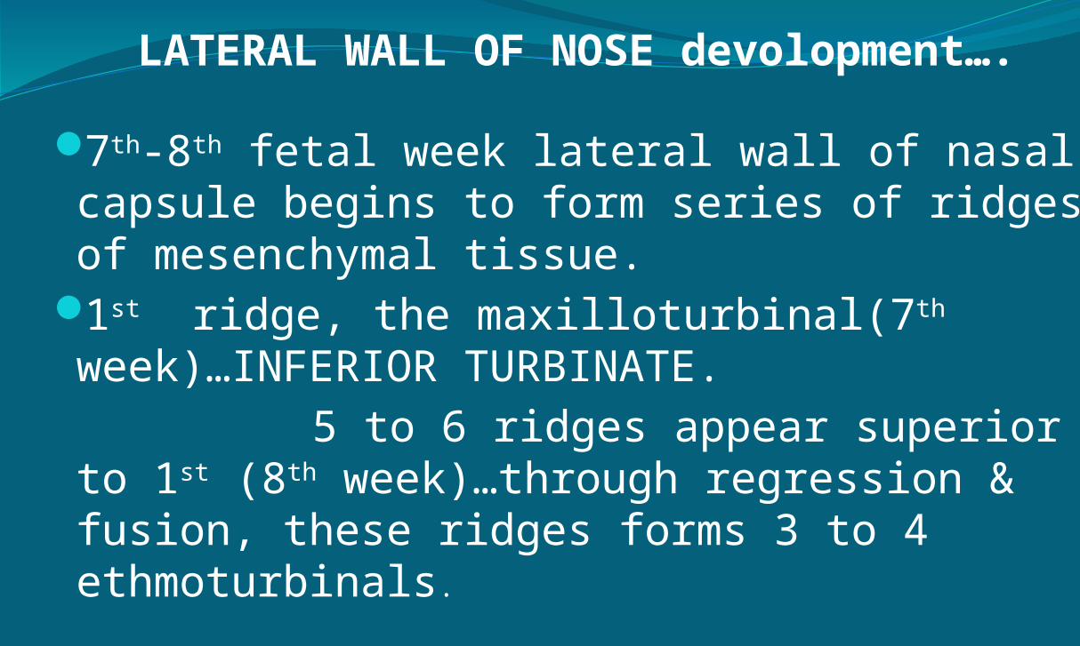
7th-8th fetal week lateral wall of nasal capsule begins to form series of ridges of mesenchymal tissue.
1st ridge, the maxilloturbinal(7th week)…INFERIOR TURBINATE.
5 to 6 ridges appear superior to 1st (8th week)…through regression & fusion, these ridges forms 3 to 4 ethmoturbinals.
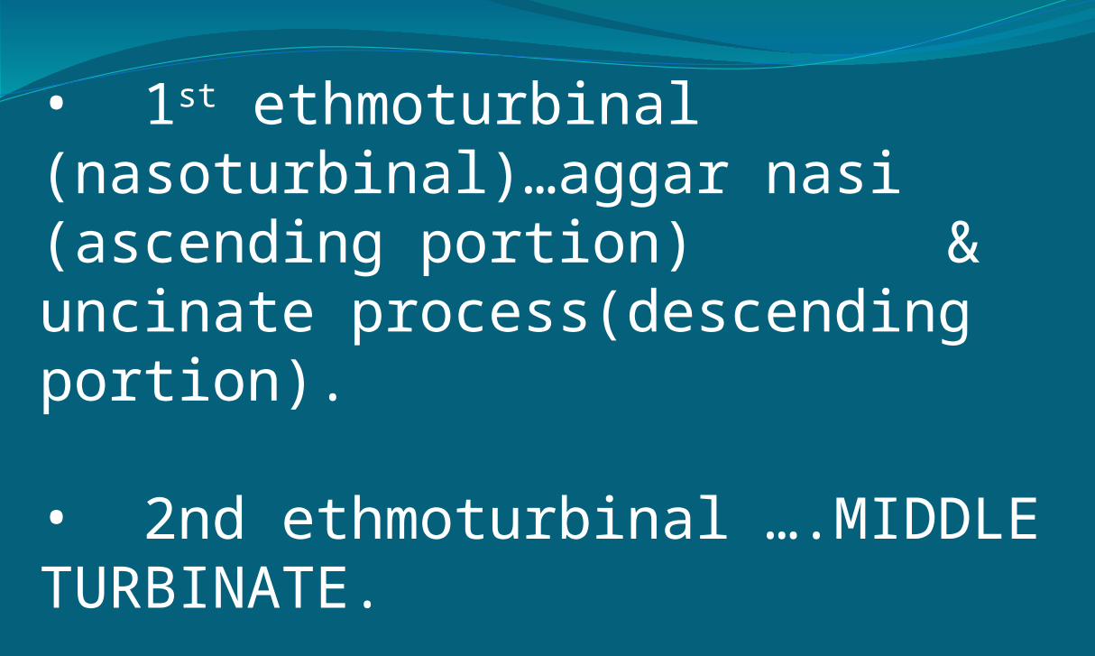
• 1st ethmoturbinal (nasoturbinal)…aggar nasi (ascending portion) & uncinate process(descending portion).
• 2nd ethmoturbinal ….MIDDLE TURBINATE.
• 3rd forms …SUPERIOR TURBINATE
• rest regress or join supreme turbinate.
8th week
SUMMARY OF EMRYONIC PRECURSORS
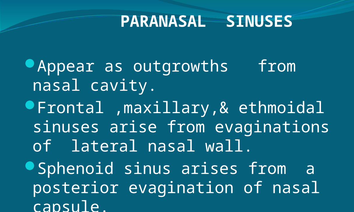
PARANASAL SINUSES
Appear as outgrowths from nasal cavity.
Frontal ,maxillary,& ethmoidal sinuses arise from evaginations of lateral nasal wall.
Sphenoid sinus arises from a posterior evagination of nasal capsule.
PARANASAL SINUSES
The sinuses begin to develop in 3rd fetal month & only ethmoidal & maxillary sinus, are present at birth.
Maxillary sinus begins as an outpounching of lateral nasal wall at 10th fetal week.
Ethmoidal sinus begins at 3rd month of fetal life.

Frontal sinus develops during 4th fetal month as an outpounching medial the most superior aspect of uncinate process.
Sphenoid sinus are unique in that they arise from within the nasal capsule of embryonic nose.
undeveloped until 3 yrs by 7 yrs pneumatization reaches sella tursica
by age 9 to 12 it is generally complete

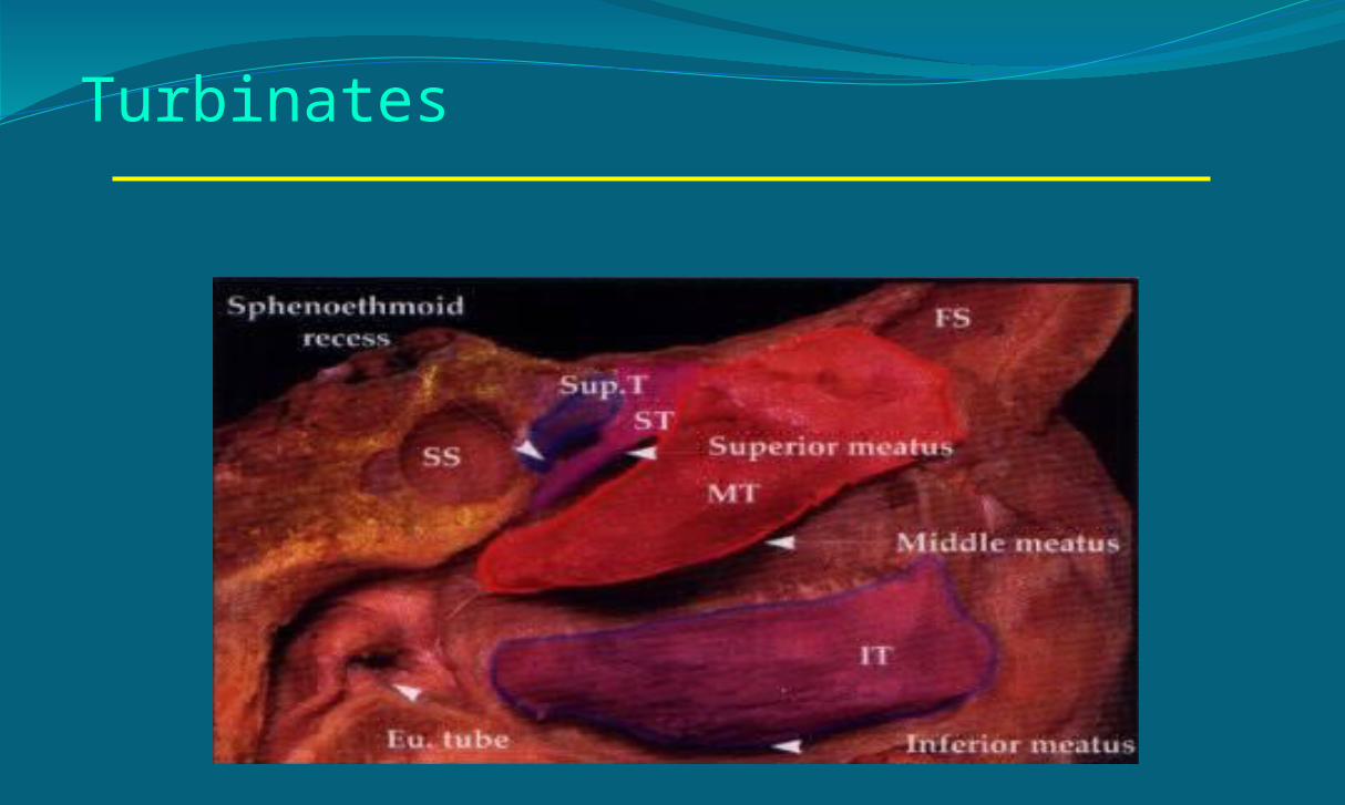
Lateral nasal wallFormed by 3 or 4 conchae(or turbinates)
Named from below inferior, middle, superior, supreme conchae.
Meatus refers to air spaces located beneath conchae
Remaining nasal cavity posterior to turbinates is nasopharyngeal meatus.


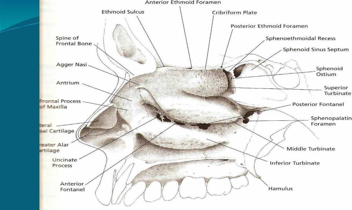
The lateral wall of nose:Maxillary boneEthmoid bonesphenoid boneInferior turbinateLacrimal bonePalatine bone
Turbinates

Lamella

Lateral nasal wallInferior turbinate/meatus Largest turbinate and largest meatus Highest at the jnctn of ant and middle 3 rd (1.6-2.3 cm)
Separate bone covered by thick mucous membrane
Nasolacrimal opening in anterior portion of lateral wall of inferior meatus
Slit like opening is protected by fold of mucous membrane, the plica lacrimalis or valve of Hasner
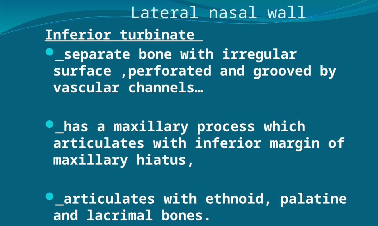
Lateral nasal wallInferior turbinate separate bone with irregular surface ,perforated and grooved by vascular channels…
has a maxillary process which articulates with inferior margin of maxillary hiatus,
articulates with ethnoid, palatine and lacrimal bones.

Middle turbinate/meatus Portion of ethmoid bone It recieves drainage from the frontal, maxillary and ant ethmoidal cells
hiatus semilunaris and ethmoid infundibulum.
maxillary hiatus. ant and post fontanelles recesses terminalis

The agger nasi remnant of the nasoturbinal in lower mammals
most anterior part of the ethmoid.
Represented by small crest or mound on the lateral wall just ant to attachment of middle turbinate.
it may be pnuematised ( 5 – 80 % )

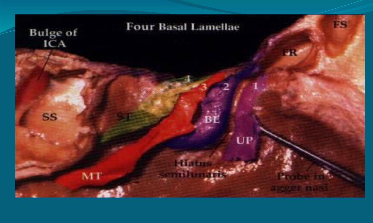
Middle turbinate, basal lamella
Part Site Direction Attachment
1st part (anterior) Vertical Skull base2nd part (middle) Oblique Lamina
papyracea3rd part (posterior) Transverse Perpendicular
plate
of palatine bone


Basal lamella and ethmoids
• Basal lamella:– Anterior ethmoids– Posterior ethmoids

Uncinate process
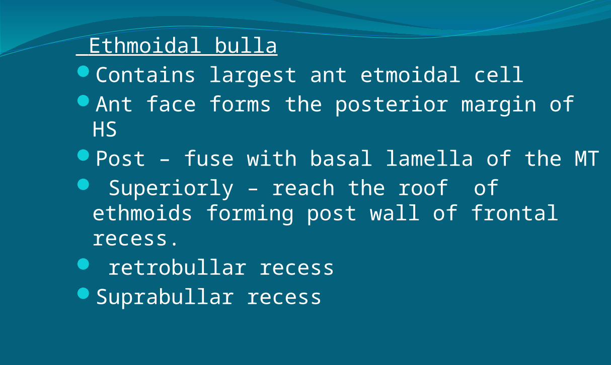
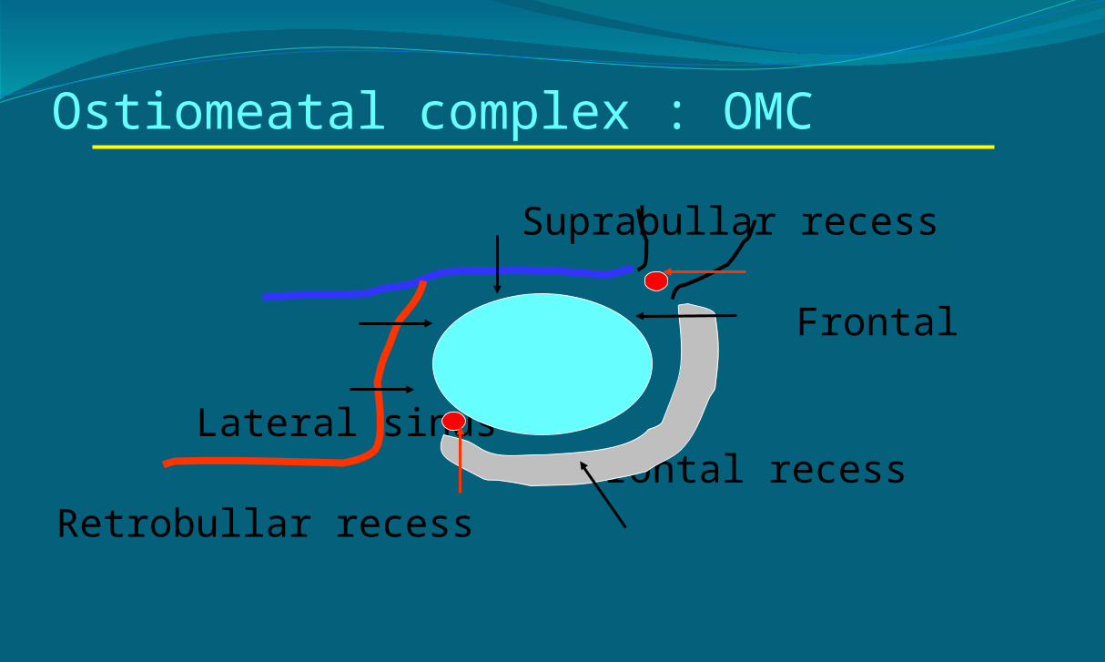
Ethmoidal bullaContains largest ant etmoidal cellAnt face forms the posterior margin of HSPost – fuse with basal lamella of the MT Superiorly – reach the roof of ethmoids
forming post wall of frontal recess. retrobullar recessSuprabullar recess

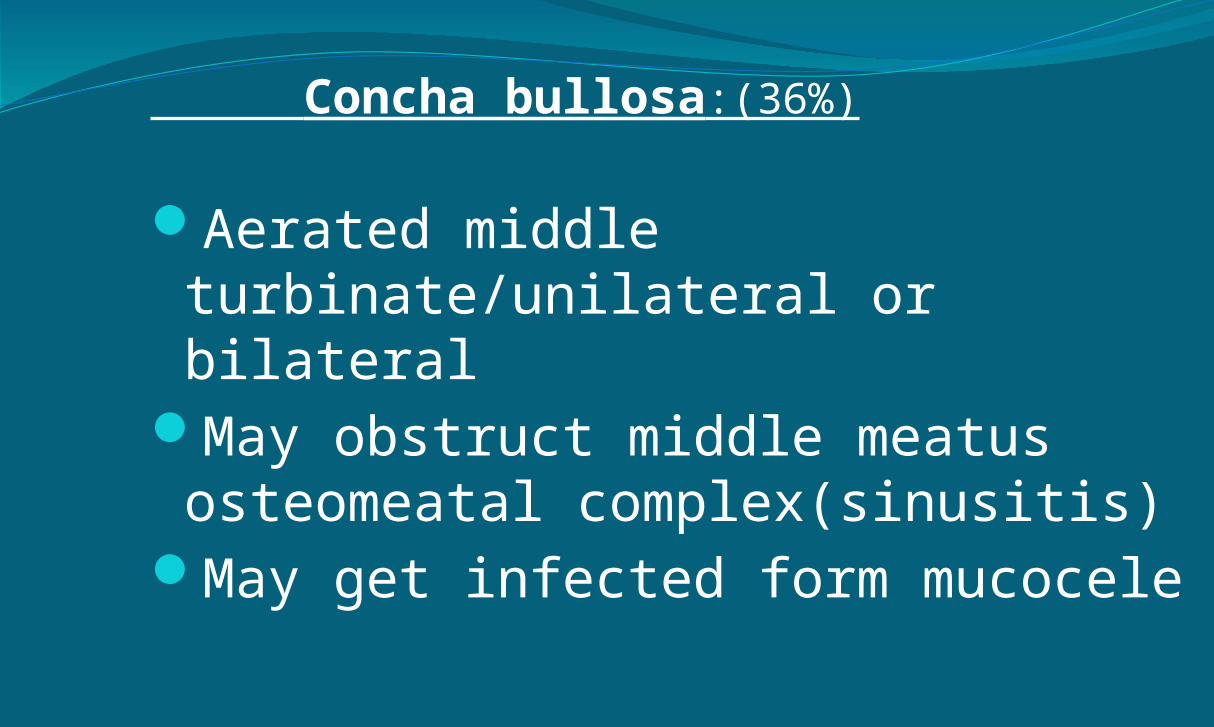
Concha bullosa:(36%)
Aerated middle turbinate/unilateral or bilateral
May obstruct middle meatus osteomeatal complex(sinusitis)
May get infected form mucocele


Paradoxical middle turbinate:
Greater curvature of middle turbinate is concave to middle meatus
Double middle turbinate:
Anteriorly bent UP may come in contact with middle turbinate
Narrows middle meatus & appear as additional turbinate

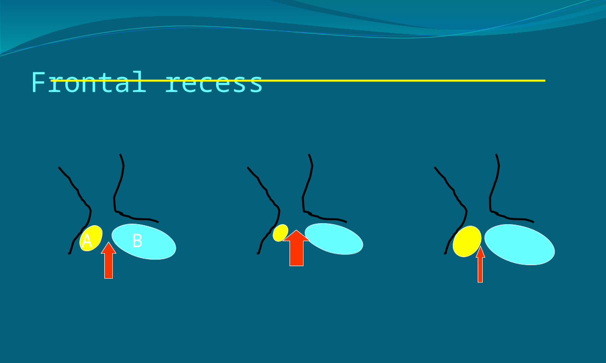
Frontal recess
A B

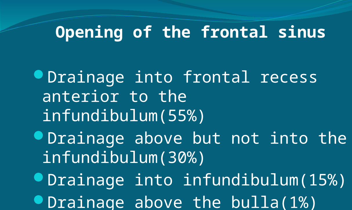
Opening of the frontal sinus
Drainage into frontal recess anterior to the infundibulum(55%)
Drainage above but not into the infundibulum(30%)
Drainage into infundibulum(15%)Drainage above the bulla(1%)

Uncinate process
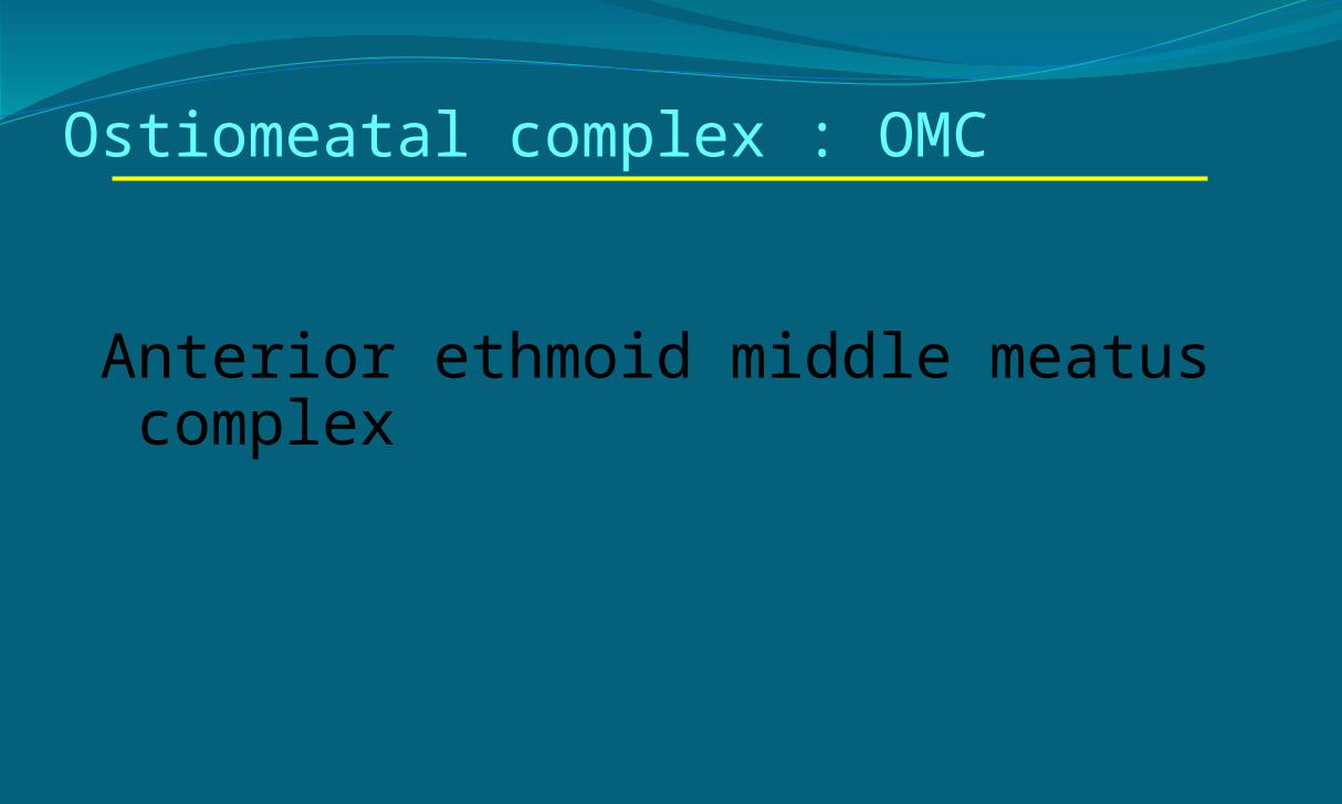
Ostiomeatal complex : OMC
Anterior ethmoid middle meatus complex
Ostiomeatal complex : OMC
Suprabullar recess
Frontal ostium Lateral sinus
Frontal recessRetrobullar recess
Maxillary ostium Infundibulum

Ostiomeatal complex : OMC
Bulla ethmoidalisHiatus semilunaris inferiorInfundibulum
Frontal recess & Frontal ostiumMaxillary ostium


Maxillary ostium
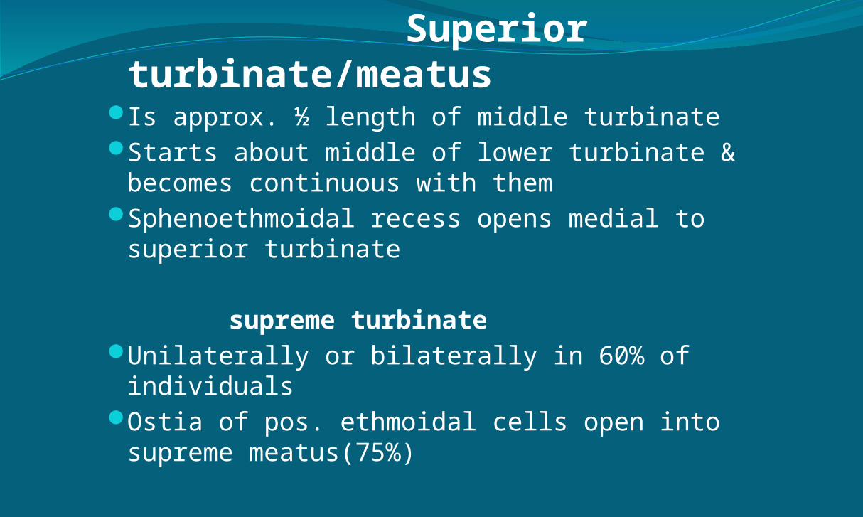
Superior turbinate/meatus
Is approx. ½ length of middle turbinateStarts about middle of lower turbinate &
becomes continuous with themSphenoethmoidal recess opens medial to
superior turbinate
supreme turbinateUnilaterally or bilaterally in 60% of individualsOstia of pos. ethmoidal cells open into supreme
meatus(75%)
APPLIED ANATOMY OF LATERAL WALL OF NOSE ……..mainly during
endoscopic surgery

Skull base: anterior ethmoids & AEA
Crista galliSeptumCribriform plateMiddle turbinateLateral lamellaFovea ethmoidalis

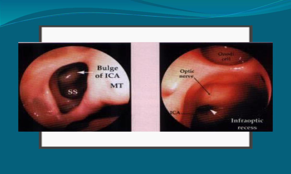
Sphenoid sinus
Optic nerveCarotid arterySella tursicaMaxillary nerveVidian nerve


Blood Supply The arterial supply is from external and internal
carotid.
Majority from branches of the maxillary artery, one of the terminal branches of the external carotid artery
The most important branch is the sphenopalatine artery

Blood Supply Sphenopalatine enters via sphenopalatine
foramen which lies just inf to the horizontal attachment of the middle turbinate
An area anteriorly is supplied by the branch from the facial and part of the lateral wall adjacent to the palate recieves blood from greater palatine.
The internal carotid art via ethmoidals supply superior lateral walls


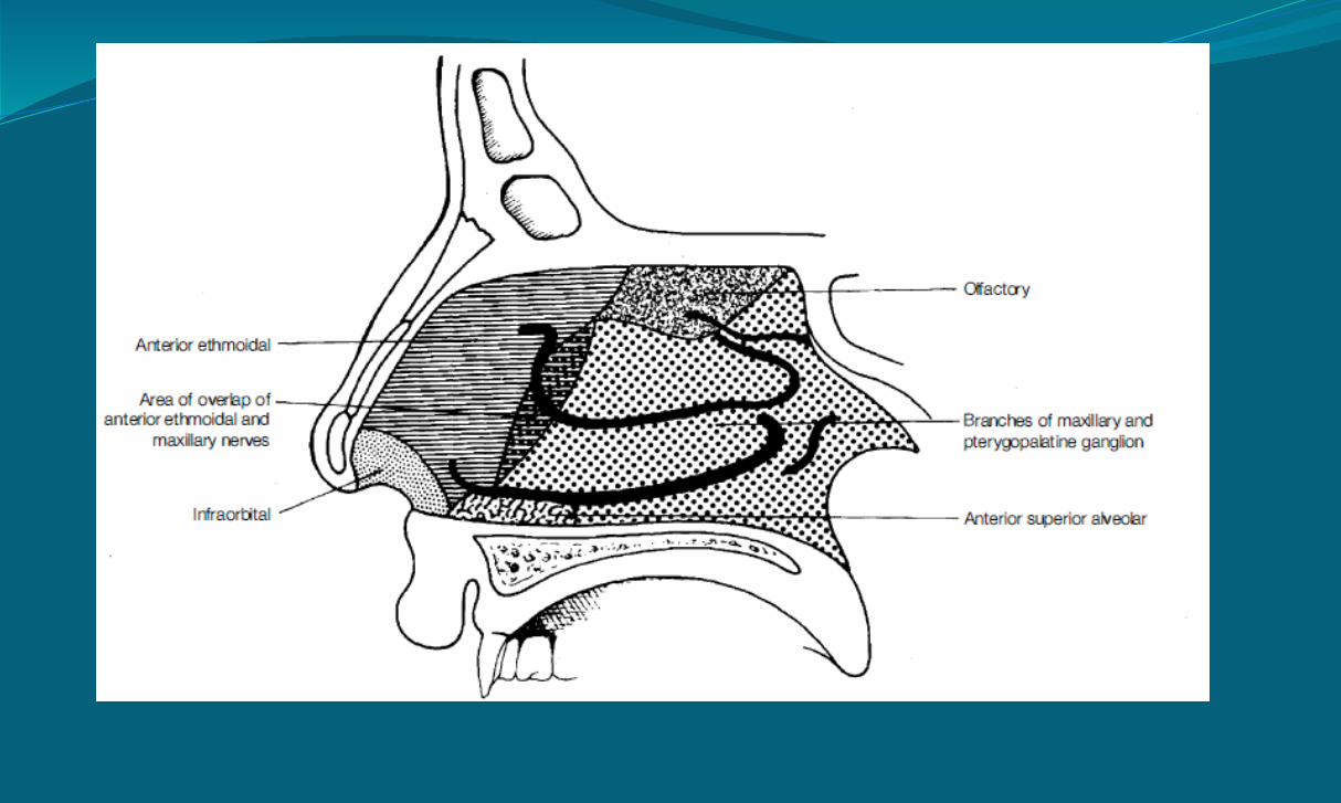
Nerve Supply Superior part – olfactory nerve.
Anterosuperiorly – ant ethm nerve
Post – branches of pterygopalatine ganglion ant pal nerves
Infraorbital nerve
Ant sup alveolar n


Lymph DrainageThe lymph vessels draining the vestibule end in
the submandibular nodes
The remainder of the nasal cavity is drained by vessels that pass to the upper deep cervical nodes

..