late onset foreign body reaction due to poly-l-lactic acid
TRANSCRIPT

Late Onset Foreign Body Reaction due to PLLA
Vol. 32, N o. 6, 2020 519
Received February 6, 2020, Revised July 30, 2020, Accepted for publication August 4, 2020
Corresponding author: Joong Sun Lee, Department of Dermatology, Eulji University Hospital, Eulji University School of Medicine, 95 Dunsanseo- ro, Seo-gu, Daejeon 35233, Korea. Tel: 82-42-611-3037, Fax: 82-42-259-1111, E-mail: [email protected]: https://orcid.org/0000-0003-2562-4090
This is an Open Access article distributed under the terms of the Creative Commons Attribution Non-Commercial License (http://creativecommons.org/licenses/by-nc/4.0) which permits unrestricted non-commercial use, distribution, and reproduction in any medium, provided the original work is properly cited.
Copyright © The Korean Dermatological Association and The Korean Society for Investigative Dermatology
pISSN 1013-9087ㆍeISSN 2005-3894Ann Dermatol Vol. 32, No. 6, 2020 https://doi.org/10.5021/ad.2020.32.6.519
CASE REPORT
Late Onset Foreign Body Reaction due to Poly-L-Lactic Acid Facial Injections for Cosmetic Purpose
Yu Jin Jeon, Dae Won Koo, Joong Sun Lee
Department of Dermatology, Eulji University Hospital, Eulji University School of Medicine, Daejeon, Korea
The safety and efficacy of Poly-L-lactic acid (PLLA) as an in-jectable facial volumizer for the treatment of lipoatrophy and facial rejuvenation has been widely proven. We experienced a remarkable case of deep-seated nodules on both the cheeks of 57-year-old female 18-months after administration of PLLA filler injection for cosmetic purpose and performed a skin biopsy. With hematoxylin and eosin stain, the nodule showed non-caseating granulomas consisting of histiocytes with central foreign bodies in the dermis. This case report represents the late-onset foreign body reaction due to PLLA facial injections. (Ann Dermatol 32(6) 519∼522, 2020)
-Keywords-Foreign-body reaction, Granuloma, Poly-L-lactic acid
INTRODUCTION
Correction of aging changes and contour deformities has been a general issue for decades. Various materials have been used as tissue augmentation agents by administering into the tissues. Cosmetic fillers are mainly classified into four categories; the calcium hydroxyapatite-based agent,
the poly-L-lactic acid (PLLA) agent (SculptraⓇ; Dermik La-boratories, Paris, France), the collagen-based filler with pol-ymethyl methacrylate crystals, and hyaluronic acid-based fillers1.Considering the stability of PLLA and ease of procedure, PLLA filler injection is often performed for the manage-ment of facial aging. PLLA filler has been approved by the United States Food and Drug Administration as the first in-jectable facial volumizer for the treatment of lipoatrophy in 2004. During the last decade, PLLA filler has been uti-lized for various purposes such as restoration of lipoa-trophy in patients infected with human immunodeficiency virus or correction of facial lines or wrinkles and augment-ing depressed parts of the face. The mechanism of action involves the stimulation of collagen and the synthesis of other connective tissues. As the PLLA filler is biodegrad-able in nature, the duration of correction remains for 1 to 2 years2. However, despite its noted efficacy, many side effects have also been reported2. Herein, we describe a case of formation of nodules due to foreign body reaction followed by injection of PLLA filler.
CASE REPORT
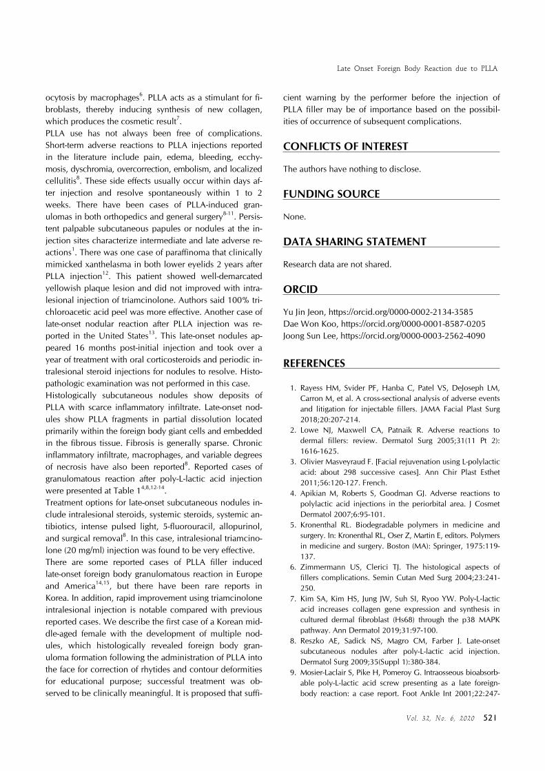
A 57-year-old female patient presented with asymptomatic skin-colored firm nodules on both the cheeks, which had persisted for one month. She had a history administration of PLLA filler (SculptraⓇ) injection for restoration of facial volume at these sites 18 months back.Physical examination revealed multiple, bilaterally distri-buted, well-marginated, round-shaped, and firm skin-col-ored nodules on both the cheeks (Fig. 1).Histopathological examination of the cheek revealed non- caseating granulomas consisting of histiocytes with central foreign bodies in the dermis. The translucent, long, and spiky particles were considered as filler constituents lo-

YJ Jeon, et al
520 Ann D erm atol
Fig. 1. (A) Skin colored nodules symmetrically distributed on both the infraorbital areas of the face. (B) Close-up view of ill- demarcated, oval-shaped, and firm nodules on the right infraor-bital area. The right nodule is slightly erythematous to orange colored and larger than the left one. (C) Side view of the lesions which are showing protrusion. Lesions are indicated as red arrowheads.
Fig. 2. (A) Histopathologic findings show multiple and scattered polymorphous foreign body reactive to fibrotic change in the dermis (H&E, ×40). (B) At higher power, non-caseating granulo-mas consisting of histiocytes, lymphocytes, and multinucleated giant cells with a central foreign body in the deep dermis can be visualized (H&E, ×400).
Table 1. Summary of reported cases of granulomatous reaction after poly-L-lactic acid injection for cosmetic purpose
No StudyAge(yr)/sex, nationality
Onset duration after injection
Treatment Site of skin lesion
1 Apikian et al.4 (2007) 49/Female, Australian
6 wk Surgical excision Infraorbital area
2 56/Female, Australian
7 d No treatment Infraorbital area
3 57/Female, Australian
6 wk No treatment Intraorbital area
4 Reszko et al.8 (2009) 62/Female, Caucasian
12 mo Intralesional triamcinolone injection, minocycline
Intraorbital area
5 Dijkema et al.14 (2005) 64/Female, Netherlander
14 mo surgical excision Upper lip
6 44/Female, Caucasian
6 mo Surgical excision Zygoma region
7 Kim et al.12 (2016) 47/Female, Korean
16 mo 100% trichloroacetic acid peel Lower eyelids
8 O’Daniel13 (2017) 50/Female, American
16 mo Oral corticosteroids, intralesional triamcinolone injection
Jawline
9 Present case 57/Female, Korean
18 mo Intralesional triamcinolone injection Infraorbital area
cated in the foreign body-type multinucleated giant cells. Granulomas were surrounded by chronic inflammatory cells, which were predominantly lymphocytes accompa-nied by reactive dermal fibrosis. The histopathological fea-tures were supportive of foreign body reactions (Fig. 2). The patient was treated with two courses of 20 mg/ml tri-amcinolone intralesional injections in the lesions at a 2-weeks interval. Subsequent to treatment, complete dis-appearance of the palpable nodules was observed. No re-currence of the lesions was observed for 2 years. We re-ceived the patient’s consent form about publishing all photographic materials.
DISCUSSION
The PLLA filler is still a commonly used important agent, and it has known to facilitate correction of aging with min-imal adverse events3,4. The action of the mechanism of PLLA is as follows. After intradermal injection of PLLA, mannitol, carmellose, and xylocaine are reabsorbed rap-idly between 24 hours to 3 days, and the PLLA spheres are left behind. Subsequently, the PLLA spheres undergo a process of biodegradation. PLLA is progressively hydro-lyzed into monomers or oligomers5. Later, the remains of the hydrolyzed products of PLLA are subjected to phag-
Late Onset Foreign Body Reaction due to PLLA
Vol. 32, N o. 6, 2020 521
ocytosis by macrophages6. PLLA acts as a stimulant for fi-broblasts, thereby inducing synthesis of new collagen, which produces the cosmetic result7.PLLA use has not always been free of complications. Short-term adverse reactions to PLLA injections reported in the literature include pain, edema, bleeding, ecchy-mosis, dyschromia, overcorrection, embolism, and localized cellulitis8. These side effects usually occur within days af-ter injection and resolve spontaneously within 1 to 2 weeks. There have been cases of PLLA-induced gran-ulomas in both orthopedics and general surgery8-11. Persis-tent palpable subcutaneous papules or nodules at the in-jection sites characterize intermediate and late adverse re-actions1. There was one case of paraffinoma that clinically mimicked xanthelasma in both lower eyelids 2 years after PLLA injection12. This patient showed well-demarcated yellowish plaque lesion and did not improved with intra-lesional injection of triamcinolone. Authors said 100% tri-chloroacetic acid peel was more effective. Another case of late-onset nodular reaction after PLLA injection was re-ported in the United States13. This late-onset nodules ap-peared 16 months post-initial injection and took over a year of treatment with oral corticosteroids and periodic in-tralesional steroid injections for nodules to resolve. Histo-pathologic examination was not performed in this case.Histologically subcutaneous nodules show deposits of PLLA with scarce inflammatory infiltrate. Late-onset nod-ules show PLLA fragments in partial dissolution located primarily within the foreign body giant cells and embedded in the fibrous tissue. Fibrosis is generally sparse. Chronic inflammatory infiltrate, macrophages, and variable degrees of necrosis have also been reported8. Reported cases of granulomatous reaction after poly-L-lactic acid injection were presented at Table 14,8,12-14.Treatment options for late-onset subcutaneous nodules in-clude intralesional steroids, systemic steroids, systemic an-tibiotics, intense pulsed light, 5-fluorouracil, allopurinol, and surgical removal8. In this case, intralesional triamcino-lone (20 mg/ml) injection was found to be very effective.There are some reported cases of PLLA filler induced late-onset foreign body granulomatous reaction in Europe and America14,15, but there have been rare reports in Korea. In addition, rapid improvement using triamcinolone intralesional injection is notable compared with previous reported cases. We describe the first case of a Korean mid-dle-aged female with the development of multiple nod-ules, which histologically revealed foreign body gran-uloma formation following the administration of PLLA into the face for correction of rhytides and contour deformities for educational purpose; successful treatment was ob-served to be clinically meaningful. It is proposed that suffi-
cient warning by the performer before the injection of PLLA filler may be of importance based on the possibil-ities of occurrence of subsequent complications.
CONFLICTS OF INTEREST
The authors have nothing to disclose.
FUNDING SOURCE
None.
DATA SHARING STATEMENT
Research data are not shared.
ORCID
Yu Jin Jeon, https://orcid.org/0000-0002-2134-3585 Dae Won Koo, https://orcid.org/0000-0001-8587-0205 Joong Sun Lee, https://orcid.org/0000-0003-2562-4090
REFERENCES
1. Rayess HM, Svider PF, Hanba C, Patel VS, DeJoseph LM, Carron M, et al. A cross-sectional analysis of adverse events and litigation for injectable fillers. JAMA Facial Plast Surg 2018;20:207-214.
2. Lowe NJ, Maxwell CA, Patnaik R. Adverse reactions to dermal fillers: review. Dermatol Surg 2005;31(11 Pt 2): 1616-1625.
3. Olivier Masveyraud F. [Facial rejuvenation using L-polylactic acid: about 298 successive cases]. Ann Chir Plast Esthet 2011;56:120-127. French.
4. Apikian M, Roberts S, Goodman GJ. Adverse reactions to polylactic acid injections in the periorbital area. J Cosmet Dermatol 2007;6:95-101.
5. Kronenthal RL. Biodegradable polymers in medicine and surgery. In: Kronenthal RL, Oser Z, Martin E, editors. Polymers in medicine and surgery. Boston (MA): Springer, 1975:119- 137.
6. Zimmermann US, Clerici TJ. The histological aspects of fillers complications. Semin Cutan Med Surg 2004;23:241- 250.
7. Kim SA, Kim HS, Jung JW, Suh SI, Ryoo YW. Poly-L-lactic acid increases collagen gene expression and synthesis in cultured dermal fibroblast (Hs68) through the p38 MAPK pathway. Ann Dermatol 2019;31:97-100.
8. Reszko AE, Sadick NS, Magro CM, Farber J. Late-onset subcutaneous nodules after poly-L-lactic acid injection. Dermatol Surg 2009;35(Suppl 1):380-384.
9. Mosier-Laclair S, Pike H, Pomeroy G. Intraosseous bioabsorb-able poly-L-lactic acid screw presenting as a late foreign- body reaction: a case report. Foot Ankle Int 2001;22:247-

YJ Jeon, et al
522 Ann D erm atol
251.10. Tannirandorn Y, Tuchinda K. Vaginal vault granulations
after total abdominal hysterectomy using polyglactin for vault closure. J Med Assoc Thai 2001;84:693-696.
11. Gammelgaard N, Jensen J. Wound complications after closure of abdominal incisions with Dexon or Vicryl. A randomized double-blind study. Acta Chir Scand 1983;149: 505-508.
12. Kim MW, Park HS, Yoon HS, Cho S. Late-onset complica-tion of fillers: paraffinoma of the lower eyelids clinically
mimicking xanthelasma. Ann Dermatol 2016;28:753-756.13. O’Daniel G. Management of late-onset, recurrent facial
nodular reaction after poly-L-lactic (PLLA) injections. J Drugs Dermatol 2017;16:1297-1299.
14. Dijkema SJ, van der Lei B, Kibbelaar RE. New-fill injections may induce late-onset foreign body granulomatous reaction. Plast Reconstr Surg 2005;115:76e-78e.
15. Daines SM, Williams EF. Complications associated with injectable soft-tissue fillers: a 5-year retrospective review. JAMA Facial Plast Surg 2013;15:226-231.