last class: 1. transportation at the plasma membrane a. carrier protein, b. channel protein 2....
TRANSCRIPT

• Last Class:
• 1. transportation at the plasma membrane
• A. carrier protein, B. channel protein
• 2. intracellular compartments
• A. nucleus, B. Endoplasmic Reticulum

• Intracellular Vesicular Traffic
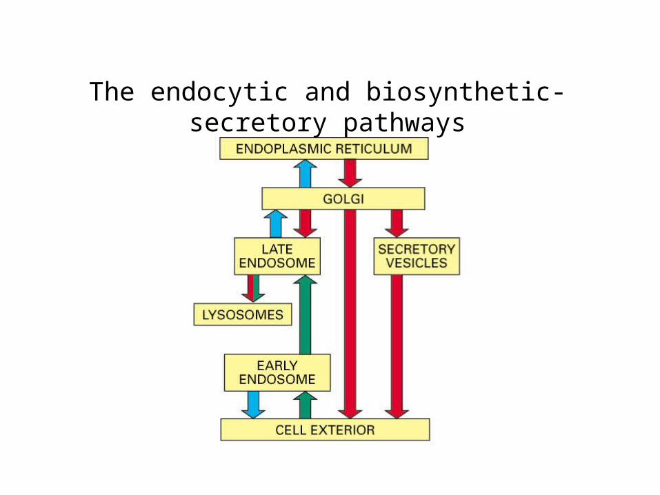
The endocytic and biosynthetic-secretory pathways

Vesicular transport

The intracellular compartments of the eucaryotic cell in the biosynthetic-secretory and endocytic
pathways
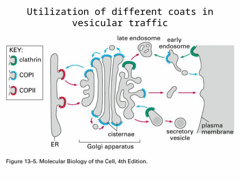
Utilization of different coats in vesicular traffic

Clathrin-coated pits and vesicles
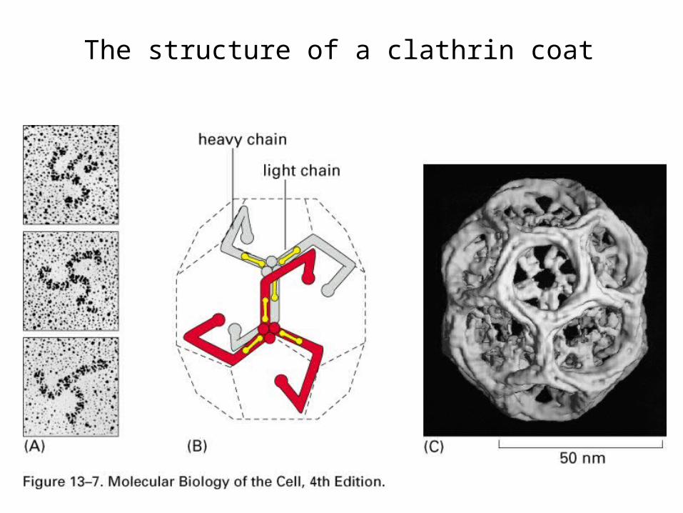
The structure of a clathrin coat
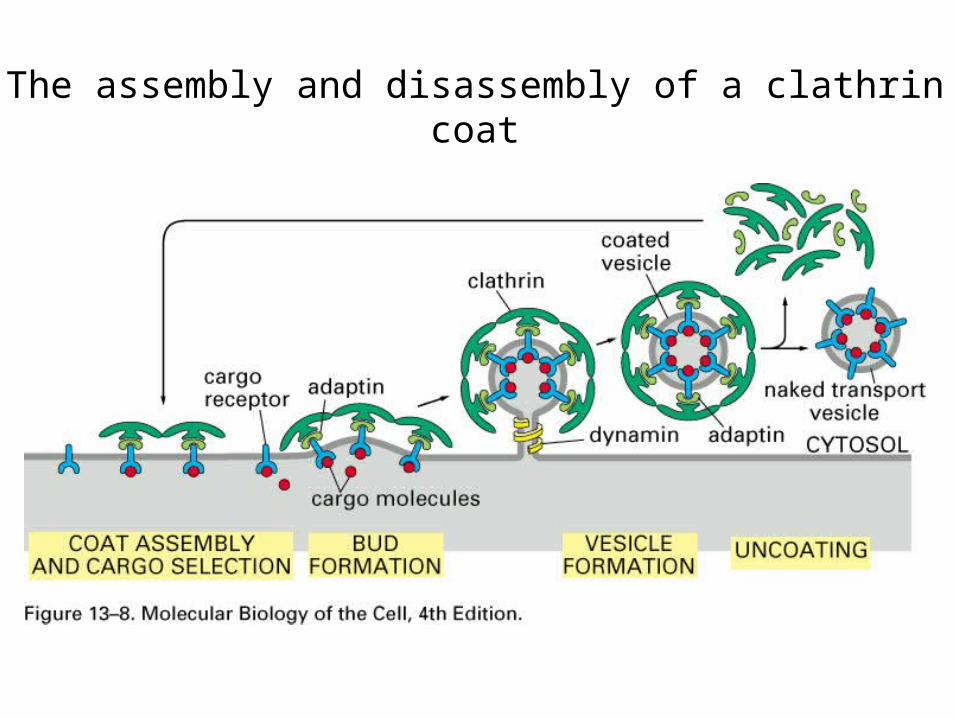
The assembly and disassembly of a clathrin coat

The role of dynamin in pinching off clathrin-coated vesicles from the membrane
Shibire mutant in drosophila

A current model of COPII-coated vesicle formation

The postulated role of SNAREs in guiding vesicular transport

The structure of paired snare
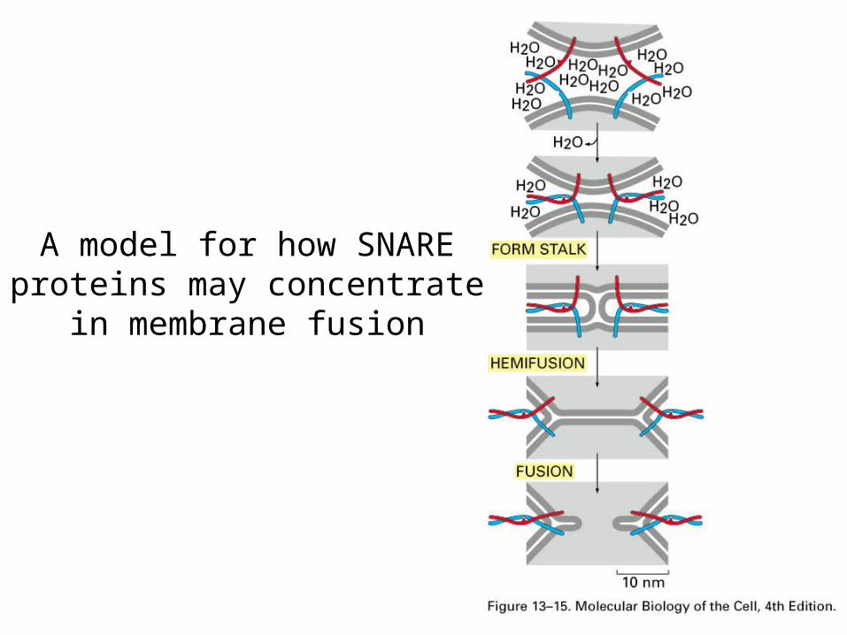
A model for how SNARE proteins may concentrate in
membrane fusion

Dissociation of SNARE pairs by NSF after a membrane fusion cycle is completed

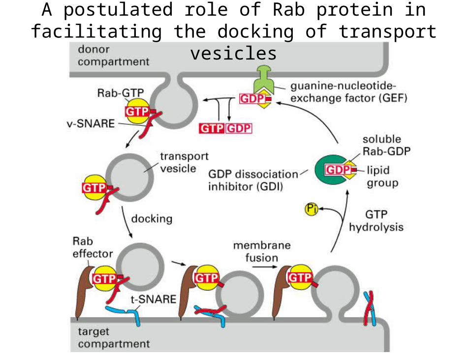
A postulated role of Rab protein in facilitating the docking of transport vesicles

The entry of enveloped viruses into cells

• Transport from the ER through the Golgi apparatus
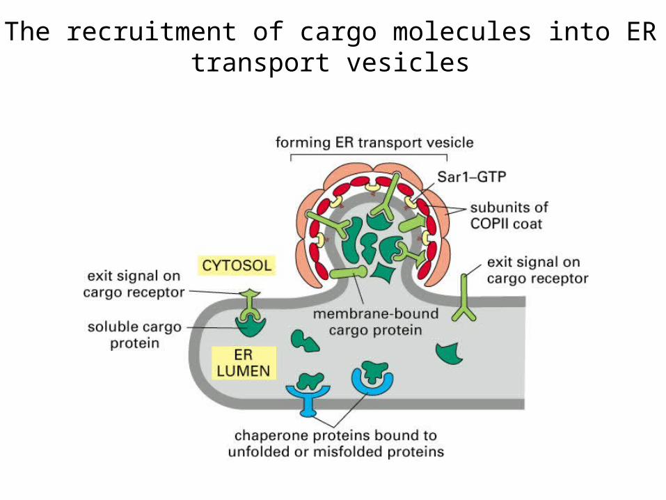
The recruitment of cargo molecules into ER transport vesicles

Retention of incompletely assembled antibody molecules in the ER

Vesicular tubular clusters

A model for the retrieval of ER resident proteins
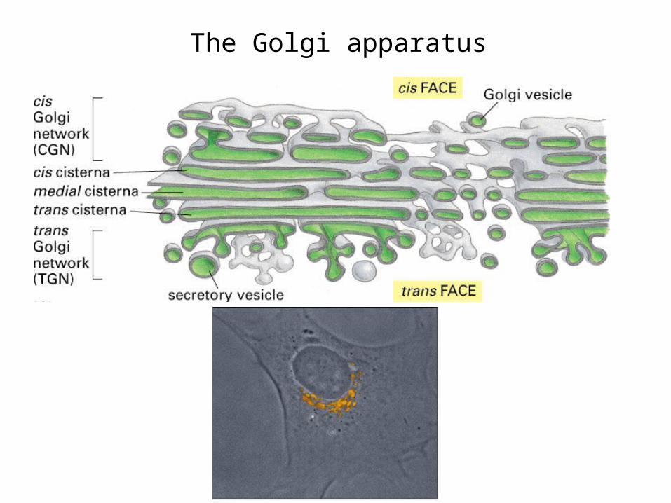
The Golgi apparatus
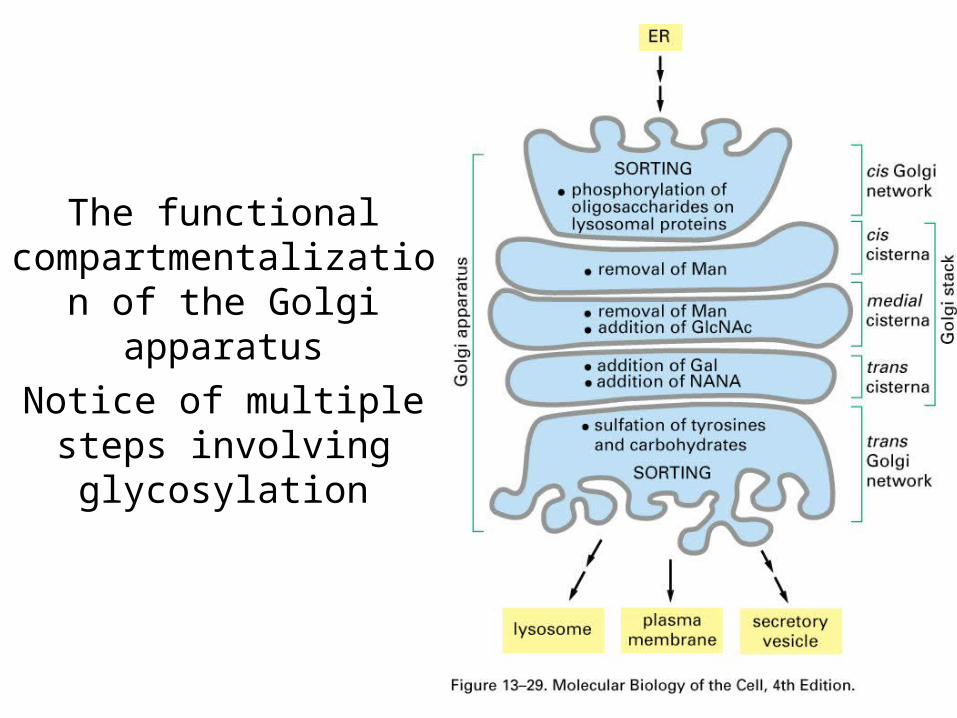
The functional compartmentalization of the Golgi apparatus
Notice of multiple steps involving glycosylation

Two possible models explaining the
organization of the Golgi apparatus and the
transport of proteins from one cisterna to the next

• Transport from the trans Golgi nextwork to Lysosomes

LysosomesLow pH
Full of Acid hydrolases

The structure of mannose 6-phosphate on a lysosome enzyme

The transport of newly synthesized lysosomal hydrolases to lysosomes

• Transport into the cell from the plasma membrane endocytosis
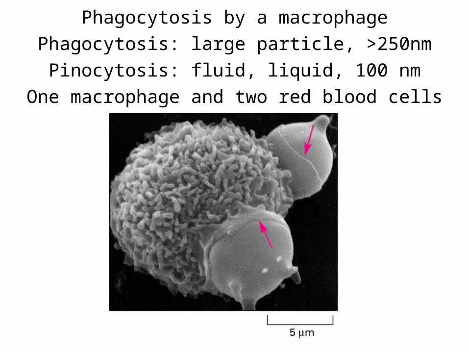
Phagocytosis by a macrophagePhagocytosis: large particle, >250nm
Pinocytosis: fluid, liquid, 100 nmOne macrophage and two red blood cells

The formation of clathrin-coated vesicles
from the plasma membrane

Caveolae in the plasma membrane of a fibroblast

A low-density lipoprotein (LDL)
particle

Normal and mutant LDL receptor

The receptor-mediated endocytosis of LDL

Possible fates for transmembrane receptor proteins that have been endocytosed

Storage of plasma membrane proteins in recycling endosomes

Transcytosis

Sorting of membrane
proteins in the endocytic pathwayGreen: EGF-EGFRRed: transferrin and its receptor

The sequestration of endocytosed
proteins into internal
membranes of multivesicular
bodies

• Transport from the trans Golgi network to the cell exterior: exocytosis
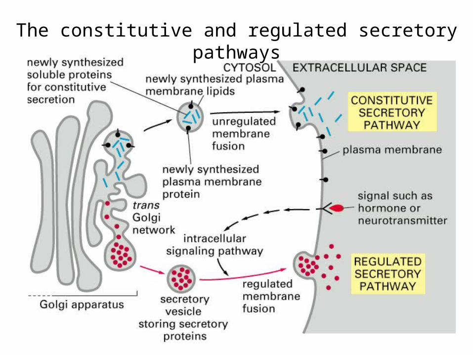
The constitutive and regulated secretory pathways
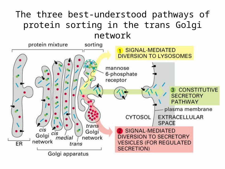
The three best-understood pathways of protein sorting in the trans Golgi network

Exocytosis of secretory vesicles

Electron micrographs of exocytosis in rat mast cells
Release of histamine

Exocytosis as a localized responsesBeads attachment
localized the release

Model of lipid rafts in the trans Golgi network
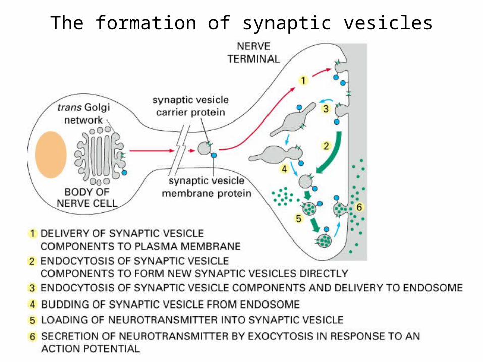
The formation of synaptic vesicles
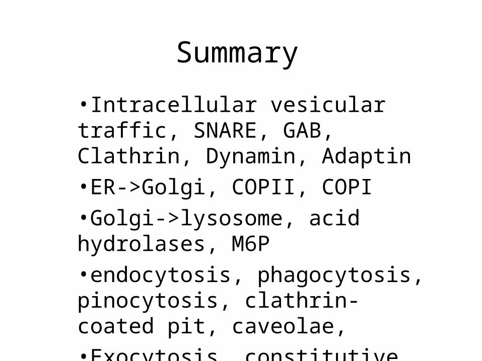
Summary
•Intracellular vesicular traffic, SNARE, GAB, Clathrin, Dynamin, Adaptin
•ER->Golgi, COPII, COPI
•Golgi->lysosome, acid hydrolases, M6P
•endocytosis, phagocytosis, pinocytosis, clathrin-coated pit, caveolae,
•Exocytosis, constitutive and regulated mechanisms

•Cell Signaling 1: General Concepts

A simple intracellular signaling pathway

Extracellular signaling molecules bind to receptors

Signals can be tranmitted either short or long distances (I)

Signals can be tranmitted either short or long distances (II)

For Long distance, two typical ways
Endocrine signalingDifferent cells need specific ligands and receptors

Synaptic signalingMore efficient, same set of ligands and receptors

Signaling via GAP JunctionsNo ligand-receptor system needed

Combinatory effect of multiple inputs
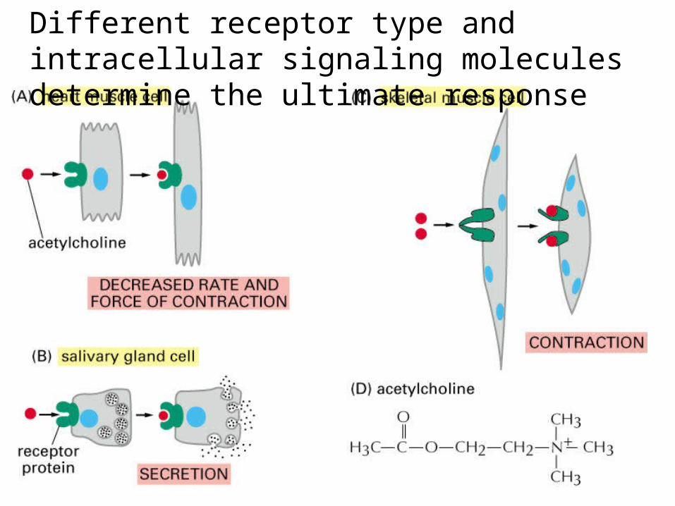
Different receptor type and intracellular signaling molecules determine the ultimate response

Many signaling molecules have short lifetime

NO (nitric oxide) induces the relaxation of SMCThe function of viagra is to inhibit cyclic GMP phosphodiesterase, hence elongate the lifetime of cyclic GMP and relaxation
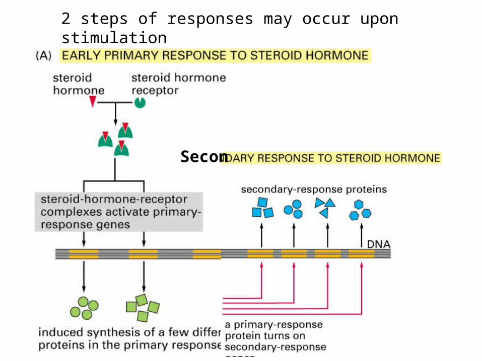
2 steps of responses may occur upon stimulation
Secon
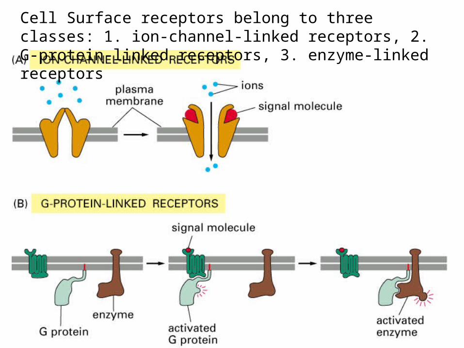
Cell Surface receptors belong to three classes: 1. ion-channel-linked receptors, 2. G-protein-linked receptors, 3. enzyme-linked receptors
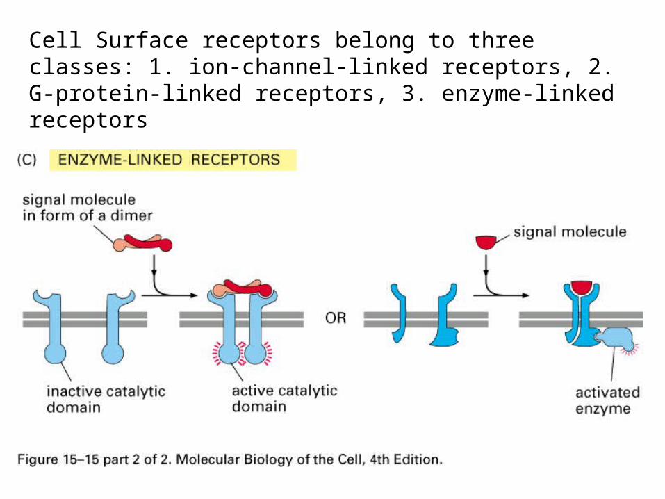
Cell Surface receptors belong to three classes: 1. ion-channel-linked receptors, 2. G-protein-linked receptors, 3. enzyme-linked receptors

Different Kinds of intracellular proteins serving as signaling molecules
1. Relay proteins2. Messenger proteins3. Adaptor proteins4. Amplifier proteins5. Transducer proteins6. Bifurcation proteins7. Integrator proteins8. Latent gene regulatory proteins

Two kinds of molecule switch eventsPhosphorylation and GTP binding
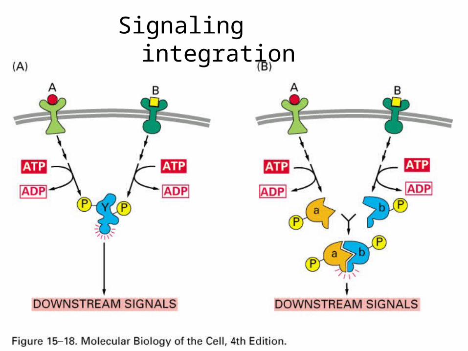
Signaling integration

Intracellular signaling complexes enhance the speed, efficiency, and specificity
2 types: Preassembled vs. Assembled after stimulation
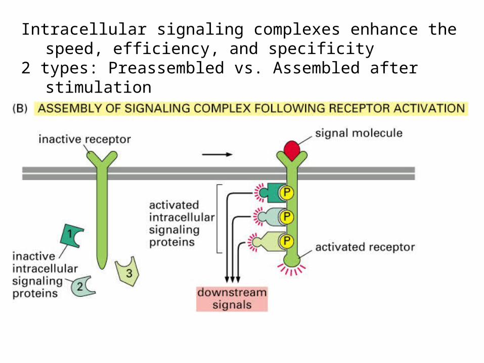
Intracellular signaling complexes enhance the speed, efficiency, and specificity
2 types: Preassembled vs. Assembled after stimulation

Binding domains for interactions between proteins and complex assembly
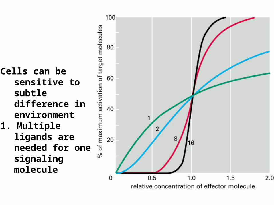
Cells can be sensitive to subtle difference in environment
1. Multiple ligands are needed for one signaling molecule
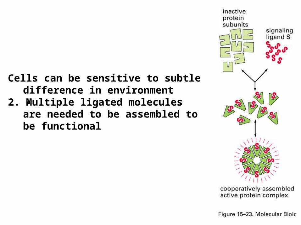
Cells can be sensitive to subtle difference in environment
2. Multiple ligated molecules are needed to be assembled to be functional
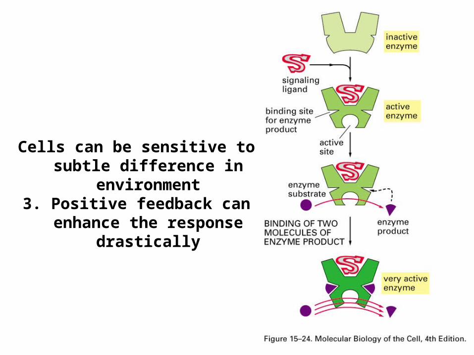
Cells can be sensitive to subtle difference in environment
3. Positive feedback can enhance the response drastically
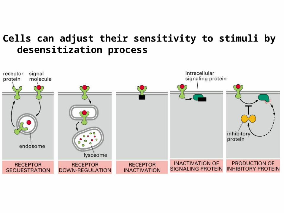
Cells can adjust their sensitivity to stimuli by desensitization process
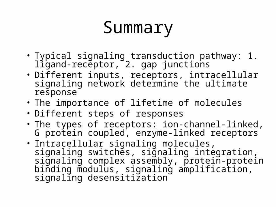
Summary
• Typical signaling transduction pathway: 1. ligand-receptor, 2. gap junctions
• Different inputs, receptors, intracellular signaling network determine the ultimate response
• The importance of lifetime of molecules• Different steps of responses• The types of receptors: ion-channel-linked, G
protein coupled, enzyme-linked receptors• Intracellular signaling molecules, signaling switches,
signaling integration, signaling complex assembly, protein-protein binding modulus, signaling amplification, signaling desensitization